Copyright 0 1996 by the Genetics Society of America
Drosophila syntaxin
Is
Required
for Cell
Viability and May Function
in
Membrane Formation
and Stabilization
Karen
L. Schulze*”
and
Hugo
J. Bellen*,+
*Division of Neuroscience and +Department of Molecular and H u m a n Genetics, +Department of Cell Biology, Howard Hughes Medical Institute, Baylor College of Medicine, Houston, Texas 77030
Manuscript received June 25, 1996 Accepted for publication September 17, 1996
ABSTRACT
The role of the Drosophila homologue of syntaxin-lA (syx) in neurotransmission has been extensively studied. However, developmental Northern analyses and in situ hybridization experiments show that SYX mRNA is expressed during all stages and in many tissues. We have isolated new mutations in syx
that reveal roles for syx outside the nervous system. In the ovary, SYX is present in the germarium, but it is predominantly localized to nurse cell membranes. Mitotic recombination experiments in the germ- line show SYX is essential for oogenesis and may participate in membrane biogenesis in the nurse cells. In the early embryo, a large contribution of maternally deposited RNA is present, and the protein is localized at cell membranes during cellularization. After the maternal contribution is depleted, zygotically produced SYX assists secretion events occurring late in embryogenesis, such as cuticle deposition and neurotransmitter release. However, SYX is also required in larval imaginal discs, as certain hypomorphic mutant combinations exhibit rough eyes and wing notch defects indicative of cell death. Furthermore, recombinant clones that lack syx cause cell lethality in the developing eye. We propose that, similar to its roles in cuticle secretion and neurotransmitter release, SYX may mediate membrane assembly events throughout Drosophila development.
T
HE study of vesicle-mediated transport in a number of systems has greatly enhanced our understand- ing of the process by which membranes and their pro- tein constituents are delivered to the proper target site during secretion and membrane addition. The observa- tion that the molecular mechanisms that underlie vesi- cle trafficking in two distinct systems, constitutive secre- tion in yeast and regulated neurotransmitter release in higher eukaryotes, are conserved has led to the hypoth- esis that an ubiquitous machinery exists to execute the fusion event (reviewed in BENNETT and SCHELLER 1993; FERRO-NOVICK andJmN 1994). Extensive studies of the molecules that are proposed to mediate vesicle docking and fusion events in both systems have generated a number of theories regarding how these events occur. The model that has enjoyed the most popularity is the “SNARE hypothesis.” The basic premise relies upon the interaction of putative membrane-anchored recep- tors, termed v- (vesicle) and t- (target) SNARES, depen- dent on the membrane within which they reside. The SNAREs (SNAP receptors) are so named for their ability to attract the soluble NSF attachment proteins (SNAPS) and the ATPase NSF (Nethylmaleimide-sensitive fusion protein) to the appropriate position to effect vesicleCorresponding uuthor: Hugo J. Bellen, Howard Hughes Medical Insti- tute, Baylor College of Medicine, Room T630, One Baylor Plaza, Houston, TX 77030. E-mail: [email protected]
’
Present address: Howard Hughes Medical Institute, University of California, 5-748 MRL, P.O. Box 951662, Los Angeles, CA 90095- 1662.Genetics 144: 1713-1724 (December, 1996)
fusion with its target membrane (SOLLNER et al. 1993a,b). In brain, the SNAREs have been identified as synaptobrevin on the synaptic vesicle and syntaxin and SNAP-25 within the presynaptic membrane. These pro- teins have homologues in yeast that have independently been identified as essential genes that lie along the secretory pathway. It has therefore been speculated that the association of tissue- or cell-specific vesicle and tar- get receptors with each other and/or with the NSF- SNAP complex may mediate the docking and fusion event (SOLLNER et al. 1993b; PEVSNER et al. 1994; SCHI-
The phenotypes of yeast SNARE mutants (secretion blockade and post-Golgi vesicle accumulation) con- firms the absolute necessity for these proteins in exo- cytosis (NOVICK et al. 1980). Furthermore, the brain SNARE homologues have also been shown to be essen- tial to neurotransmitter release, as they are the targets of proteolytic cleavage by the clostridial and botulinum neurotoxins that block neurotransmission (SCHIAVO et al. 1992; HAYASHI et al. 1994; reviewed in JAHN and
NIEMANN 1994). Though these data certainly do not contradict the premises of the theory, recent evidence has challenged the fundamental tenets of the SNARE hypothesis. Mutational analyses of two proteins, the pu- tative v-SNARE n-synaptobrevin and the potential t- SNARE syntaxin, have been performed in Drosophila (SWEENEY et al. 1994; BROADIE et al. 1995; SCHULZE et
al. 1995). These experiments have established that sy- naptobrevin and syntaxin both function downstream
1714 K. L. Schulze and H. J. Bellen
of the vesicle docking event, in contrast to what was originally proposed in the SNARE hypothesis. More importantly, genetic manipulations such as these that removed N-SYB or SYX function revealed that although synaptobrevin may assist in Cay’-regulated neurotrans- mitter release, its function is not essential to the fusion event
per
se as spontaneous fusions still occur (SWEENEYet al. 1994; BROADIE et al. 1995). O n the other hand, the role of syntaxin was shown to be absolutely essential to vesicle fusion in neurotransmitter release, as all forms of exocytosis, both spontaneous and evoked, are absent in syx null mutants (BROADIE et al. 1995; SCHULZE et al. 1995). The precise function of syntaxin, whether in a prefusion or “priming” step, or as a catalyst of the fusion event itself, has yet to be elucidated.
Clues to syntaxin’s function have been revealed by rigorous analysis of its protein structure and its interac- tions with other proteins. Syntaxin has been shown to bind in vitro to all the components that may form the SNARE complex (synaptobrevin, SNAP-25, NSF and a- SNAP), and amino acid domains within the protein that are crucial for these interactions have been delineated
(CMOS et al. 1994; HAYASHI et al. 1994; PEVSNER et al.
1994; HANSON et al. 1995; K E E et al. 1995). Syntaxin was originally isolated due to its interaction with the synap- tic vesicle membrane protein synaptotagmin, and this interaction has been shown to occur in a Ca‘+-depen- dent manner (BENNEM et al. 1992; CHAPMAN et al. 1995). Finaily, syntaxin also interacts with presynaptic N-type calcium channels (SHENG et aE. 1994) in a man- ner that may effect their function (BEZPROZVANNY et al. 1995). The association of syntaxin with synaptotagmin and Ca2+ channels in the neuron strongly supports the placement of syntaxin’s function late in the cascade of events that culminate in vesicle fusion upon Ca2+ influx. We initially reported that the expression of the Dro- sophila s y n t a x i n - l A homologue is not restricted to the nervous system as is thought to be the case for its verte- brate counterpart. syxfunctions in cuticle secretion and possibly in the garland cells where it may assist in the clearance of waste from the hemolymph via endo- and exocytosis (SCHULZE et al. 1995). To gain further insight into SYNTAXIN’S ( S Y X ) potential role in vesicle fusion, we have examined its function in a variety of secretory processes occurring throughout Drosophila develop ment. Herein we present preliminary evidence that SYX
may be required for many exocytotic events during fu- sion or prefusion steps that may occur in a similar man- ner as has been suggested for neurotransmission. Cu- multatively, these data point to a critical and essential role for SYX in membrane fusion and biogenesis.
MATERIALS AND METHODS
Drosophila stocks and mutagenesis: All Drosophila strains were maintained on standard cornmeal-molasses medium at 25” unless otherwise noted. Canton-Swas used as the wild-type strain. A deficiency that uncovers all g x alleles (Df?R)ml~’~~~, breakpoints 95D7- 11; 95F15) was provided by Dr. EIJZABETH
KNUST (TEPASS et al. 1990). Some syx alleles were rebalanced over TM6B and placed in a y w background using either the balancer strain y w/y w; D3/TM6B, T6 P ( w + , AbdA-lacZJ or y
w/y w; L/CyO; P{ry’, 1acZJslp; @/TMGB, Tb P[w’, A b d - l a c Z ) . The P-element insertion strain P(ly+ tsyx” t y / TM3, SI, ry (ab- breviated syx”) was obtained from the Drosophila Stock Cen- ter in Bloomington, Indiana (P[syx], SCHULZE et uL. 1995). Excision of the syx‘insertion was achieved by mating individ- ual P[ry+]syx~/TTM? males with virgin females of the genotype y w/y w; Ki
p”
A2-3/Kip‘
A2-3. Individual y w; P(ly+)syx” y/Ki
6’
A2-3 males (or females) were backcrossed to females (males) of the original insertion strain to balance the excision chromosome. The progeny of these crosses were scored for individuals with rosy eye color, indicative of an excision event, from each of which Asyx‘ ry/ TM?, Sb ly balanced stocks were established. Complementation tests with the original insertion strain were subsequently performed to define the nature of the excision events as precise or imprecise.To isolate point mutations, isogenized ry emales were fed 25 mM EMS suspended in 1% sucrose using a standard protocol (LEWIS and BACHER 1968). The mutagenized chromosomes (10,500) were balanced over TMGB, Tband assessed for failure to complement the original Pelement insertion syx” at 28”.
Induction of mosaic animals and germline clones: Genetic mosaics were generated using the FLP/FRT recombinase sys- tem (CHOU and PERRIMON 1992) essentially as outlined by
XU and RUBIN (1993). The alleles ~yx’’~~’, syx‘ (Df(?R)A229 and Df(?R)A6, SCHULZE et al. 1995) and syx’ (each in a y 711 background) were recombined onto a chromosome con- taining Ply+ hs-neo FRT}82B using selection for resistance to G418 (Geneticin, GIBCO) and verification of failure to complement the original J-yxallele. Males containing the heat- inducible FLP recomhinase (y w P/ly+ hsFLPI/Y) and the chromosomes Plry* hs-neo FRT/82B P{w’ ly’}9OE (permitting detection of mosaic clones in the eye) or Plry’ hs-neo FRTjr82B
P{w+ a v d ” / ~ ~ P{U+ OVO”‘/?RZ (for assaying germline clone development) were constructed and crossed to y 7u; P(y+ hs- neo FRT)82B syx females. First instar larvae (24-48 hr AEL) were incubated in a 38” water bath (in the vials in which they were laid) for 1 hr to induce mitotic recombination. Adult females of the appropriate genotype were examined for clones ( w + ) or assayed for egg-laying ability after several days of mating to y w; syxnZz9/TM6B, Tb males. In some cases, the ovaries of females that failed to lay eggs were dissected (see below). P(FRT} and P(rnarker] strains were obtained from the Bloomington, Indiana Stock Center.
Construction of genomic rescue transgenes: An 11 .O-kb XbaI fragment from genomic phage A10 and a 6.0-kb NotI fragment from genomic phage A6 (see Figure 3) were sub- cloned into the vector pCaSpeR3, which contains the 5’ and
3’ P-element integration sequences flanking the white gene (PIRROTTA 1988). The constructs were injected into y w; Ki ppA2-3/+ embryos (BELLEN et al. 1992), and surviving adults were backcrossed to y w. Progeny bearing w+ eyes (indicating stable integration of the construct at a chromosomal locus) were selected and served as founders of stocks. Seven second chromosome insertion strains were generated that contained the A10 X6aI construct, and six second chromosome insertion strains were created that bore the A6 NotI construct.
To determine if either of the two P[w+lsyx’ constructs could rescue the lethality of syx‘ and ~ y x ” ~ ~ ~ , y w; P[w’lsyx+/CyO; D/ TM6B, Tb males were crossed to y w; s y x - / T M 6 3 , TI, females. Of the progeny of this cross, brothers and sisters of the geno- type y w; P(w+)syx+/+; syx-/TMGB, Tb were mated. Recovery of non-Tubby progeny was indicative of rescue.
Role of q x in Membrane Biogenesis 1715
pared as described in the Genius RNA nonradioactive labeling detection kit (Boehringer Mannheim)
.
In situ hybridization to whole mount embryos was performed as previously de- scribed by INGHAM et al. (1991) and TAUTZ and PFEIFLEImmunobistochemis~ Appropriately staged wild-type
and syx mutant embryos were collected and processed for immunocytochemical staining according to standard tech- niques outlined in SALZBERG et al. (1994). MAb 8C3, which recognizes Drosophila Syntaxin-1A and was given by Dr. SEY-
MOUR BENZER, was used at a final dilution of 1:10 (FUJITA et al. 1982). MAb 1D4, which recognizes the cytoplasmic domain of Drosophila Fasciclin I1 (VAN VACTOR et al. 1993), was o b tained from Dr. COREY S. GOODMAN and was used at a final dilution of 1:50 to determine if motorneuron growth cone guidance and fasciculation occurred normally. MAb 22C10, a neuronal marker that recognizes a membrane antigen on all cells of the Drosophila peripheral nervous system (FUJITA et al. 1982; GOODMAN et al. 1984) was utilized at a 1:200 dilu- tion to analyze overall neuronal morphology. Nuclear staining of embryos was achieved by treating fixed embryos with 125 units ribonuclease A for 30 min at 37". Propidium iodide (50 pg/mL) was applied concurrently with the primary antibody. Ovaries were dissected and fixed as described in XUE and COOLEY (1993). Fixed ovaries were processed for immunohis- tochemistry using the procedure for embryos outlined in SALZBERG et al. (1994). T o visualize SYX protein distribution, MAb 8C3 was utilized at a dilution of 1:10. Biotinylated goat anti-mouse secondary antibody (Vector) was used at a 1:200 dilution. The Vecta-Stain ABGHRP kit (Vector) was used to augment the DAB-peroxidase signal. Fluorescein isothiocya- nate-conjugated anti-mouse IgG was utilized at a 1:200 dilu- tion.
Paraffin sections of adult Canton-S heads were prepared for mass histology using a modification of the protocol ofJAGER and FISCHBACH (1987). The primary antibody (anti-rat Syntaxin polyclonal 1378, HATA et al. 1993) was diluted 1:200.
Molecular techniques: Total RNA for Northern analysis was isolated by LiCl precipitation of phenol-chloroform extracted Drosophila homogenates, and poly-A+ RNA was purified with oligodT cellulose ( SAMBROOK et al. 1989). Approximately 5 pg poly-A+ RNA was loaded per lane. The probe was gener- ated by PCR of the 3' untranslated region of the syx cDNA, including 430 bp between the stop codon and the polyadenyl- ation signal. A 3.0-kb EcoRI genomic fragment served as the template for the reaction. The primers used were 5'-ClTACA- CGCCACATTCACTCC-3' and 5"CTTTGTTTGCAATTA- CGTGTGJ' with the following conditions: 1 cycle at 95" (5 min); 35 cycles at 94" (1.5 rnin), 51" (2 min) and 72" (3 min); 1 cycle at 72" (3 min). Other probes tested were the 3' half of the cDNA from the internal EcoRI site to the end (encodes last 174 amino acids) and a 330-bp HindIII/SacII fragment from the 5' half of the cDNA (includes codons of the first 44 amino acids).
To determine the molecular defects of EMSinduced point mutants, crude genomic DNA was prepared from individual
~yx'/syx""~ or syx'/syx' embryos or single male s y ~ ' ~ ~ ~ / s y ~ ~ ~ ~ ~ flies to serve as a template for PCR amplification of the open
reading frame (ORF) using complementary primers with overhangs containing BamHI and Hind111 restriction sites to facilitate subcloning. The conditions for the PCR were as fol- lows: 1 cycle at 95" (5 min); 35 cycles at 94" (1.5 min), 57" (2 min), 72" (3 min); 1 cycle at 72" (3 min). The product of the reactions were subcloned into Bluescript I1 SK+ (Stratagene), and the universal and reverse primers as well as a panel of internal primers were utilized to prime sequencing reactions by the dideoxy chain-termination method using the auto- mated fluorescent procedure (Applied Biosystems). Two clones independently isolated in this manner were sequenced
(1989).
for each mutant. The Genetics Computer Group Sequence Analysis Package Version 7.3.1-UNIX was utilized for all subse- quent nucleotide and amino acid sequence analyses.
The molecular defects of excision alleles were determined by performing Southern analyses of genomic DNA prepared from balanced strains carried out essentially as described in SAMBROOK et al. (1989). PCR analysis was performed on crude genomic DNA of single flies using the 31-bp inverted P-ele-
ment repeat (5'-CGACGGGACCACClTATGTTATTTCAT-
CATG-3') as well as two primers designed from syx sequence (5'GTTATTTGTGACTCACACTGGTACC-3' and 5'-GTT- CACACTCGAAAATTGTITGCCC-3') to determine if the P element had been completely removed or internally deleted (see SCHULZE et al. 1995). The conditions for the PCR were as follows: 1 cycle at 95" (5 min); 35 cycles at 94" (1.5 min), 51-53" (2 min) and 72" (3 min); 1 cycle at 72" (3 min).
RESULTS
y x expression is widespread throughout Drosophila development: We initially reported the expression and distribution of SYX transcripts and SYX protein during the latter half of embryogenesis (SCHULZE et al. 1995). The extent of expression of SEX during Drosophila de- velopment was further investigated by Northern blot- ting of RNA representing all stages of embryogenesis, larval life and adulthood. We tested three probes: the 3' end of the cDNA containing coding region that would be most conserved (C-terminal portion of the protein), a probe containing 5' untranslated sequence and sequence coding for the first 44 amino acids (not well conserved among syntaxins), and a probe con- taining only 3' untranslated sequence. All three probes recognized the same complex pattern composed of up to six transcripts, ranging in size from 3.5 to 12 kb (see Figure 1). SYX messages are present during every stage of Drosophila development. Although the pattern of transcripts is very complex considering that the small (1.4 kb) cDNA we and others (CEREZO et al. 1995) have recovered is contained within a single exon, it is likely that alternative splicing, various promoters, and differ- ent polyadenyltion signals are used to generate this het- erogeneity. We feel confident that each of these tran- scripts represent SYXmessages and not transcripts from other related and possibly highly homologous Drosoph- ila syntaxins that may exist, since three different probes generated identical patterns. Further, these transcripts are likely to encode a single protein as developmental Westerns display only a single band (SCHULZE et al. 1995; data not shown). In addition, low stringency hy- bridization of genomic Southern blots failed to reveal any other bands than those corresponding to SYX (data not shown).
1716 K. I,. Schulze and H. J. Bcllcn
kb
9.4.
7.4.
4 1 2 + 9.0
*
8.0*
7.04.4
2.4
4- 4.2 4 3.5
L
RP49FI(:L‘RI.: l.--Dc\c~lop~~~c~lt“1 csprcwiion o f .syx. Six principal
s ~ x transcripts (ranging in size from 3.5 to 12 kb) are present rlrlring Drosophila development. The Northern blot was hy- bridized w i t h a specific . yprobe amplified by PCR from the 3‘ untl-anslatetl region. All the lanes were equally loaded based o n hyhridization of RP49 probe, w i t h the exception of second instars, pupae and adults that are slightly under- loaded.
respectively. During pupariation, all five transcripts re- appear, and the adult expression pattern resembles that of the
pupa.
MTe observed that three of the adult tran- scripts (3..5,4.2 and 8.0 kb) are also present in 0- to 9 h r - old embryos, suggesting some or all of these messages represent the maternal contribution to the embryo.We continued the developmental analysis by per- forming in situ hybridization and immunohistochemis- ty to more closely examine syx’s expression in young embryos. Digoxigenin-labeled antisense RNA probes were utilized to visualize the distribrltion of SYXmRNA in embryos of a11 stages. In agreement with the North- ern analysis, which indicated three prevalent transcripts in embryos aged 0-3 h r AEL (Figure l ) , very young embryos contain high levels of syntaxin RNA and pro- tein (Figure 2).
Precellalarized embryos exhibit ubiquitously distrik uted syx mRNA, although it accumulates most densely in the germ plasm at the extreme posterior tip of the embryo where the pole cells will form (Figure 2B). As the future pole cell membranes pinch off from the pos-
terior tip, SJ’X RNA is located within each bud and re- mains concentrated at the posterior of the embryo (data not shown). Upon the completion of somatic cel- lularization, syx RNA is uniformly distributed within all cells of the embryo and seems concentrated near the cell membrane (data not shown). As gastrulation pro- ceeds, s y ~ RNA is abundant in all cells. At stage 9, the message is present in most ectodermal cells, the ante- rior and posterior midgut invaginations, and the central nervous system (CNS) precursor cells (Figure 2F). Ex- pression of syxduring later stages of embryogenesis was previously characterized and includes the garland cells, midgut, nervous system and ectoderm (SCHULZE rt nl. 199.5).
To determine the distribution of SYX protein during oogenesis and embryogenesis, we immunocytochemi- cally stained ovaries and embryos with either a polyclonal antibody raised against rat syntaxin-lA (HATA rt 01. 1993) o r monoclonal antibody 8C3 (a gift from KOSRXI) ZINS Mr\IE:R and SI?Y%IOL!R BENZER). The former antibody spe- cifically recognizes a protein of the appropriate molecu- lar weight in extracts prepared from Drosophila heads and identifies the protein encoded by the .yx gene
(SCHUI.7.E rt nl. 1995), whereas the latter was prepared against the native Drosophila SYX protein ( F ~ ) ~ I T A rt nl. 1982). In wild-type ovaries stained with MAb 8C3, we detected SYX protein in regions 2 and 3 of the germar- ium, outlining the membranes of germline cyst cells.
SYX continues to be abundantly expressed in the nurse cell membranes of egg chambers during stages 1-8, and
SYX levels fade during stages 8 and 9 (Figure 2A). In young embryos during mitotic cycles 9-13, the plasma membrane partially envelops each dividing nn- cleus, forming “cytoplasmic buds”. Beginning in mi- totic cycle 9, “pole buds” form at the posterior tip of the embryo, marking the formation and segregation of the germ cells (FOE P t nl. 1993). As is the case for syx mRNA, SYX is present ubiquitously in precellularized embryos but is concentrated at the posterior tip, and accumulates beneath these pole buds as they undergo cytokinesis (Figure 2, C and D). Similarly during cycle 11 “somatic buds” form around dividing somatic nuclei but undergo several rounds of formation and collapse as the nuclei beneath the buds divide. At metaphase of each mitotic division, the membrane of the bud invagi- nates markedly, and “pseudocleavage furrows” form between the buds (FOE rt nl. 1993;
SCHF..TER
andM‘IESCHAUS 1993). SYX expression appears to follow the outline of the buds as they partially enwrap the dividing nuclei during mitotic cycles 10-14 (Figure 2C). Finally, during cycle 14, the somatic nuclei become completely surrounded by the elongating membranes of the cyto- plasmic buds, and cellularization is completed. SYX Itr
calization outlines these membranes as they form (Fig- ure 2D). SYX continues to be expressed in the membranes of each cell of the fully cellularized e m h r y (Figure 2E).
Syntaxin protein expression does not become re- stricted to any tissue o r cell type until stage 9 of em- bryogenesis. During germ band extension, the protein is present in the mesoderm, ectoderm and invaginating neurectoderm but is excluded from the amnioserosa (data not shown). During later stages of embryogenesis, the protein is most prominent in the CNS and the gar- land cells and is weakly expressed in the midgut and ectoderm (SCHULLE rt 01. 1995).
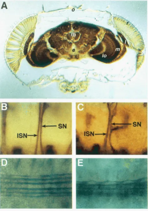
SYX protein is also present in the adult brain and the synaptic substations of the visual system (Figure 5A). Staining of paraffin sections through adult wild-type heads with an anti-rat syntaxin polyclonal antibody re- veals SYX is concentrated in the neuropil regions of
Role of syx in Membrane Biogenesis 1717
FIGURE 2.-Distribution of SYX mRNA and SYX protein in ovarioles and young embryos. Wild-type ovariole and embryos stained for SYX with MAb 8C3 or hybridized with an antisense SYXRNA probe. Embryos are oriented anterior to the left, dorsal up. Staging is according to CAMPOS-~RTEGA and HARTENSTEIN (1985). (A) Syntaxin is present in the nurse cell membranes of the ovary. The protein is abundant in the nurse cell membranes of the germarium (g) and the early stages of oocyte development but dissipates beginning at stage 8 (S8). (B) SYXRNA is present ubiquitously in embryos before cellularization but is concentrated at the posterior pole. (C) SYX distribution in an embryo that has not yet completed mitotic cycle 13 is observed in large, roughly hexagonal-shaped outlines that correspond to the pseudocleavage furrows of the cytoplasmic buds. (D) An embryo that nears the completion of somatic cellularization (cycle 14) demonstrates SYX staining along the membranes of each cell (green). SYX is also concentrated beneath the pole cells. Nuclei are labeled with propidium iodide (red). (E) A completely cellularized embryo is beginning to undergo gastrulation (stage 6), as the cephalic and ventral furrows are forming. SYX is present in all the membranes of all cells at this stage. (F) A stage 9 embryo exhibits SYXRNA localization in the anterior (am) and posterior
(pm) midgut invaginations, the ventral neuroblasts (n) and the ectoderm.
such as in the lamina and medulla of the optic lobes where photoreceptor axons form synaptic contacts with second order neurons. However, SYX distribution in the brain differs from that of synaptic vesicle-specific proteins such as synaptotagmin (LITTLETON et al. 1993), as SYX is also present in axons and cell bodies whereas synaptotagmin is restricted to synaptic terminals.
Generation
and
molecular characterization of muta-tions in syx: To gain a better insight into syx's function, and to identify possible new phenotypes associated with partial loss of function alleles of syx, we performed two mutagenesis screens. In the first, the P element in ? x p
1718 K. L. Schulze and H. J. Bellen
muscle contraction waves and emerge from the egg case. In addition, two of the embryonic lethal mutations recov- ered in this screen were phenotypically characterized in the previous work. qXnZz9, a null allele, exhibits complete blockade of all forms of neurotransmitter release. In addition, nonneuronal forms of secretion are impaired, as yolk digestion in the gut is abnormal and several layers of the cuticle are missing (SCHULZE et al. 1995; A. PRO-
KOP, personal communication). syx6 also exhibits secre- tory defects that are intermediate in severity between syxn229 and syx'.
In the second mutagenesis, -10,500 chromosomes were screened for EMS-induced mutations. The molec- ular defects borne by the excision mutations were ana- lyzed by genomic Southern and PCR analyses, and the EMS-induced mutations were sequenced. The data is described below and pictorially in Figure 3.
syxnZz9 is a null allele that almost entirely deletes the ORF and flanking regulatory regions
(1.7
kb),
whereas all other excision events produced alleles of varying severity. syxRh and syxZx4 appear to be severe hypo- morphs or null alleles on the basis of their molecular deficiencies and the fact that, like syxnZ2', they fail to secrete most cuticle and are embryonic lethal (Table 1). We have previously shown that syx6 is a severe hypo- morph but is not a null allele, as some residual protein is present (SCHULZE et al. 1995). syx', syx4, S J X ' ~ , sydl', s ~ x " ~ , syxZa4, syxZs7 and syx294 bear incomplete internal deletions of the Pelement (Figure 3 ) . syx', syxi7, syx"' and syxz94 have an intermediate phenotype similar to that caused by the original Pelement insertion syx'; these alleles are embryonic lethal, display wild-type cu- ticular structures, but fail to exhibit muscle contraction waves. Finally, on the basis of their viability, syx4, SYX"~, syxZo4 and syxZR7 are weak hypomorphs.Only two point mutations were recovered from the EMS mutagenesis (1/5250 chromosomes screened). syx' carries a premature termination signal (QlOl to
stop) and behaves as a null allele (see Table 1). s ~ x ~ ~ " contains no detectable aberrations in the coding region
and probably carries a temperature-sensitive mutation in a regulatory domain (see below and DISCUSSION).
Table 1 summarizes the complementation data among all the syx alleles recovered, as well as the lethality of each individual allele when tested in trans to itself and the two null alleles, syxnzZy and sy'. Almost all the al- leles are embryonic lethal as homozygotes and as trans- heterozygotes with the null alleles. Their phenotypes range in severity from that of the null allele itself, which lacks cuticle and bears other secretory defects, to that of the original P-element allele, which exhibits a normal cuticle but no spontaneous rnuscie contractions. These two phenotypic categories seem to correspond well to the molecular defects, with the null-like phenotype re- stricted to alleles with an imprecise deletion of the P element that removed surrounding genomic DNA (syx', syx14, syxx6 and syxZR4). The hypomorphic phenotype is displayed by alleles bearing small internal deletions
within the Pelement (syx', syx", syx ' I y and syxZy4). How-
ever, a third group, represented by alleles with <1 kb of the Pelement insertion remaining (syx4, syxii7, SYX'"~ and syxZR7), are very weak hypomorphs and often pro- duce viable progeny in trans to other syxalleles. Further- more, syx4 homozygotes are completely normal and fer- tile adults, whereas males homozygous for 53~''~ (an
allele with a molecular defect indistinguishable from syx4) are sterile (data not shown).
Rescue of the lethality associated with syxp and syxdZ9:
To determine if syx' and the deletions generated from its excision were bonajde mutations in syx, we rescued the lethality associated with the original Pelement insertion allele and the most severe loss of function (null) allele, sy~"'~'. Two rescue constructs (shown in Figure 3) were comprised of the genomic region encompassing the ORF and u p and downstream sequences. This genomic DNA was engineered into the pCaSpeR3 vector and injected into embryos, and from those that had been stably inte- grated, 12 second chromosome transformant lines were generated. The second chromosomes bearing these con- structs were crossed into the syxp and q~~~~~ genetic back- grounds and assessed for their ability to rescue the lethal- ity associated with these q x alleles. Two of the 12 transformants completely rescued the phenotypes associ- ated with both q x p and syxnZz9, as homozygotes carrylng the rescue transgene were viable and fertile and overtly morphologically normal (data not shown). These results demonstrate that the P element and the deletion associ- ated with .yxnz2' both disrupt the syx gene and further, that a number of the transcripts may not be essential for viability and fertility, as their length exceeds that of the rescue construct.
Syniaxin is required in the female gennline for oo-
cyte development: Since Northern analysis and in situ
hybridizations to very young embryos (0-3 hr AEL) indicated an abundance of SYX transcripts, we wished to determine the effect on early embryonic develop- ment of the removal of all maternally derived SYX activ- ity. Mitotic recombination was induced by activating FLP recombinase expression in first instar larvae bear- ing the syx null allele, s ~(located at 95E1-2), distal x ~ ~ ~ ~ to a flippase recognition target site (FRT) on one chro- mosome arm
3R
and two copies of P(w' ovd") distal to the FRT on the homologous chromosome arm. These animals were analyzed for egg-laying ability upon eclo- sion. ovo"' on the third chromosome causes dominant female sterility leading to rudimentary ovaries that fail to develop beyond stage 8 or 9. Hence, females hetero- zygous for this P-element insertion carlying ovo'" can- not lay eggs. Nevertheless, individual ovarioles that have developed from a progenitor germline cell in which a mitotic recombination event has taken piace, exchang- ing ovo"' for syx, produce viable oocytes. In control ex- periments of this type, 69% of heat-induced females examined exhibited restoration of fertility (see TableRole of .S?.Y in Membrane Biogenesis 1719
I
A10 Xbal 11.0 kb rescue fragmentI
16 Not1 6.0 kb rescue fragment
H
l k bcDNA
e
syx
position of Pelement
I I I I t I I I
**
I t I I I I I composite mapI I I I I I I I I I I I I I I I
x
E EHE E H S N NS S IS XH H1.7 kb deletion
-
1.3 kb deletion
-
229
6
3.4 kb deletion
-
14
1.8 kb deletion
-
86
1.9 kb deletion
-
284
P
-
2.1 kb deletion118
-
500 bp deletion2
I 300 bp deletion
17,294
800 bp remaining
4
177
500 bp remaining I
287
-50 bp remaining I
204
FIGI‘RE 3.-Molecular defects exhibited hv .vx alleles. Schematic representation of genomic region encompassing the .SJ.Y
locus. T h e 8 7 6 h p ORF (black box) is shown flanked 9’ and 3’ untranslated regions comprising the 1.4-kh partial cDNA. T h e direcion of transcription is indicated hv the horizontal arrow.
*
marks a genomic ,Vo/I fragment that recognizes a transcipt o na Northern hlot and may thus indicate a nearhy or nested gene (data not shown). Thc position of the original Pelement insertion is shown hv the vertical arrow. Hatched hars within the P element indicate ?+ and InrZ genes. The molecular delict of each .qx allele is diagrammed and the name of the affected allele is shown to the right of rach map. E, EroRI; H, HintlIlI; S ;
A’d; S , Sicn; X, X M ; O R I , origin of replication; Km”, kanamycin-resistance gene.
the 918 FRT . ~ ~ x ” ” ~ / F R T 7u+ 0710’” females tested for egg-laying ability, 212 were dissected and their ovaries examined. All were rudimentary in their appearance and indistinguishable from that of ovo”‘ heterozygotes.
M’e repeated these experiments using hvo weaker syx alleles, syx”and syx‘’. We have previously shown by “est- ern blotting that some residual
SYX
activity is present in syx” mutants, whereas syx” homozygotes display even more reduced.m
levels than q x ” mutants (S<:HUI.%EP/ n/. 1995). Although heat-shocked FRT syx”/FRT 711’
ozd” females are sterile, as shown in Table 2, 84% of the heat-shocked FRT .syx”/FRT 70+
ova'"
females tested demonstrated egg-laving ability. \%en these females were mated to 7ct; . s y ~ ” ~ ~ ” / TM(iB, Th males, 54% of theirprogeny sunived because the zygotic syx+ provided by the balancer chromosome of the male was sufficient to rescue the maternal syx”effect. The remainder (zygotic genotvpe .y~x”/~yx”~’”, maternal .syx/.syx) displayed a phenotype similar to that of syx zygotic null embryos
(lack of cuticle structures and accumulation of yolk in the gut). Therefore, although SYX is essential for oo-
genesis to proceed to completion, the requirements for
S Z X in the development of the germline and for mater- nally derived
SYX
during zygotic development can be provided sufficiently by the weak syx” allele. Further- more, any maternal effects of the syx”allele npon early embryonic development can be fully rescued by a wild- type zygotic contribution from the male.A temperature-sensitive mutation reveals Syxs role in eye and wing development: The isolation of a series of partial loss of function alleles of syx immediately re- vealed that syx plays a role in imaginal tissues. T h e most intriguing adult phenotype was observed among allelic combinations including thc EMSinduced s ~ x ’ ~ ‘ ’ . In ev- ery combination (except in / r m s t o SJK’, sec I W X : ~ ~ .
1720 K. L. Schulze and H. J. Bellen
TABLE 1
syx complementation and lethality
229 6 14 86 284 P 2 17 118 294 287 204 4 177 I 15ts
Complementation
229 6
14
86 284 P
294 28 7 204 4 177 1
15ts
-
-/+
-/+
+/-+
+*
-
-
-/+
-/+
+
+
+*
-
-
-
-/+
--/+
-/+
+
+/-
+
+
+/-
+/- +/-+/-
-
-/+
+/-
-/+
+/-
+/-
+
+/-
+/- +/-
-/+
- -
+
+
-+*
+*
+*
+*
+
-
+/-
-
+
+*
-
-
+
+/-
+*
-
+
+/-
-
c*
+
+/-
-
+*
+*
+*
Lethality
20 23
22 24
20 23
+*
Homozygous Over 229 Over 1
26 26 20
25 25 26
22 25 26
27 22 25
25 24 32
26 33 31
19 12 27
26 28 21
n.d. V V V 20 1L-2L
3L-P 3L-P
v
v
20v
n.d. n.d. n.d. n.d. 20 V
Complementation of given syx allele (name of allele indicated in left column) with other syx allele (name of allele indicated in top row), performed at 25°C and scored for viability. Lethality expressed as percentage of total fertile progeny that fail to emerge. -, no adults survive;
-/+,
510% of adults are viable (fair number of escapees); +/-, 11-22% transheterozygous viable (slightly less than 30% expected);+,
226% transheterozygous viable progeny (30% expected if complements);+*,
transheterozygous viable progeny with eye and wing phenotypes; V, viable; 1L-2L, first or second instar lethal; 3L-P, third instar or pupal lethal; n.d., not determined.are sometimes fused and often are improperly rotated and misaligned with respect to one another (Figure 4B). These defects are often observed near an invagi- nation of the photoreceptor array that occurs at the edge of the cuticle near the antenna. At times, this region of cuticle is enlarged and protrudes into the eye proper (compare arrows in wild type and mutant, Fig- ure 4C). This rough eye phenotype was exacerbated by raising these progeny at 28", as roughened eyes were observed in 100% of the transheterozygotes. However,
when the cross is performed at 18" the effect is abol- ished, and all transheterozygotes expected emerge with- out defects (data not shown).
Another defect present in many of the ~ y x ' ~ ' ' transhet- erozygous combinations is notching along the posterior margin of the wing (Figure 4D). This effect is observed more prevalently in the transheterozygous syx progeny of a cross performed at 25" than in a similar cross raised at 28". Interestingly, the number of viable progeny that emerge from crosses at 28" is reduced compared to the
TABLE 2
TX- clone frequency
p {w+l P {w+ o v 8 ' )
Eye color Eye color Egg-laying ability
Allele Red (%) White (%I 'n Red (%) White (%) n Sterile (%) Fertile (%) n
229 18 0 355 20 0 1,315 100 0 918
6 ND ND 0 29 0 280 100 0 212
P ND ND 0 31 0 961 16 84 187
+
31 31 36 36 36 492 31 69 111Frequency of recombination between an FRT syx chromosome (syx allele tested shown in left column) with an FRT P(marker1 chromosome (qmarker) tested shown in top row) is expressed as a percentage. In the case of eye color, the percentage of FRT syx/FRT 4wi] female progeny that exhibited one or more mosaic patches (either red or white) is listed. The total number of females of this genotype scored is given (n). Egg-laying ability was determined as described in MATERIALS AND METHODS and is expressed as the percent of the total number ( n ) of FRT syx/FRT P(wf O V ~ ' ] females examined for this phenotype. ND, not
Role of syx in Membrane Biogenesis 1'721
4
w
P
FIGURE 4 . - - ~ y x ' ~ ~ transheterozygotes display eye and wing defects. (A) Wild-type eye, lateral view. (B) s y ~ ' ~ ~ / s y x ~ @ ' eye, lateral view. The ommatidial array is disorganized leading to a "roughened" appearance of the eye. A protrusion of cuticle (arrow) invades the ommatidial array. (C) Left: frontal view of a wild-type head. Right: frontal view of - ~ y x ' ~ ~ / s y x ' ~ head. Note the excess cuticle (arrow) present on either side of the antennae in the mutant. (D) Wing of s y ~ " ~ / s y x ~ ~ individual displays notching along the posterior margin.
same cross at 25" (e.g., syx14, S Y X ' ~ ~ , syx17, data not
shown), suggesting that a more severe wing phenotype or, a complete penetrance of the wing phenotype among the progeny cannot be observed due to the con- current effect on viability. Both the notched wing and reduced viability phenotypes are completely nullified by raising S J X ' ~ " transheterozygotes at 18". The rough-
ened eye and notched wing defects exhibited by these flies suggest that SYX functions during eye and wing development.
Syntaxin is required for cell viability
in
the eye: Al- though the defects in syx15ts transheterozygotes are mild, we proceeded to further investigate the role SYXplays during eye development. Since the complete a b sence of syx results in embryonic lethality, mitotic re- combination experiments were again performed to ana- lyze the effects of lack of SYX function in individual cells of the developing eye. Recombination experi- ments were performed using the FRT/FLP recombi- nase system as described above for the construction of
w' ov8' mosaic animals. In this case, a second P{ marker] chromosome was utilized that carried a Pelement inser- tion at 90E containing only the eye color marker w+.
As
shown in Table 2, 18% of the adult FRT S J X " ' ~ ~ /FRT w+ females examined displayed w" clones. Similar results were obtained when FRT s~x""~/FRT w+ ov8'
females were scored for eye clones (20%). However, none of these females exhibited the concomitant syxnZz9
and thus w- clone, indicating that the syx- sister cell clone born of the recombination event failed to survive in the absence of
SYX.
In control experiments using an FRT on a wild-type syx chromosome, 36% of the female FRT syx+/FRT w+ progeny displayed both w' and w-sister clones. Hence, lack of SYX activity in cells of the developing eye causes lethality.
The weaker syx alleles syx6 and syxp both produce a cell lethal effect in the eye.
As
shown in Table 2, of the heat shocked FRT syx6/FRT w" orFRT
syxp/FRT wtfemales examined, none displayed w- clones. Hence, when SYX is either abolished ( syxnZz9) or reduced (syxp
or syx6), the w- clone bearing two copies of either of these alleles fails to develop alongside its sister w+/w'
clone. No trace of the syx- clone is visible, such as a scar or a change in the overall size or pattern of the eye, suggesting that the clone dies soon after the recom- bination event. These results show that the requisites for SYX in the germline and the eye are different, with the eye requiring more SYX to complete its develop- ment.
Syntaxin
plays a rolein
membranes of the nervous system:As
we have previously shown, SYX's principal role in the nervous system of Drosophila is its essential function in neurotransmitter release (SCHULZE et al. 1995). However, in syxnZz9 homozygotes, incomplete compaction of the ventral nerve cord is observed(SCHULZE et al. 1995). The effects of zygotic loss of SYX
were closely examined by immunohistochemistry to
syxnZ9 and syx6 embryos using MAbs 1D4 and 22C10. These antibodies respectively recognize fasciclin 11 and a neuronal membrane glycoprotein, and are excellent markers to observe defects in axonal structure. Late- stage homozygous S ~ X " ' ~ ~ or syx6 embryos (stage 16 and
17)
display incomplete fasciculation of the interseg- mental (ISN) and segmental (SN) nerve bundles, which contain motor and sensory nerve fibers exiting and en- tering the CNS to and from the periphery (Figure 5C).A wild-type embryo stained with MAb 22C10 (Figure 5B) displays properly fasciculated axons in its ISN and SN bundles; the axons are tightly apposed and no gaps can be distinguished. The staining of the ISN and SN of a syx6 homozygote is quite diffuse, suggesting that the individual axons cannot adhere to one another perhaps due to inconsistencies in their membrane constituents (Figure 5C). Similar defects are observed in the longitu- dinal tracts of the CNS when observed with anti-fasciclin
II
antibody (Figure 5E). In the wild-type CNS, three longitudinal tracts are observed on either side of the midline, and each set of axon fibers exhibits a tight arrangement (Figure 5D). In the CNS of a syxnZz9 homo- zygote, the axon bundles are irregular and slightly de- fasciculated (Figure 5E). These data indicate that SYXaffects properties of neuronal membranes. In addition, these defects worsen in aging embryos, suggesting that
SYX plays a critical role in maintaining membrane com- position and integrity.
DISCUSSION
1722 K. L. Schulze and H. J. Bellen
FIGURE 5.-Absence of SYX causes membrane defects in the mature embryonic nervous system. (A) Frontal section through adult brain and visual system stained with a poly- clonal antibody prepared against rat syntaxin-1A. SYX is abun- dant in synaptic regions although it is also present along axo-
nal tracts and in cell bodies. Identified regions of visual system: r, photoreceptor array of retina; 1, lamina; m, medulla; lo, lobula. fb, fan-shaped body; e, esophagus; 0, ocelli. (B and C) Embryos are oriented anterior to the left, dorsal up. (B) Lateral view of a stage 16 wild-type embryo stained with MAb 22C10, which recognizes a membrane glycoprotein present in all neurons of the PNS and CNS. The intersegmental (ISN) and segmental (SN) nerve bundles of one abdominal segment are indicated; note the axons are tightly fasciculated. (C) A similar view of a stage 16 ?x6 embryo stained with MAb 22C10. The ISN and SN fibers are defasciculated. (D and E) Embryos are oriented anterior to the left. (D) Ventral view of a stage 16 wild-type embryo stained with MAb 1D4, which recognizes the cell adhesion molecule fasciclin 11. Three tightly fascicu- lated longitudinal tracts are visible on either side of the mid- line of the CNS. (E) Ventrolateral view of a stage 16 syxnZZ9
embryo; only the three longitudinal tracts of one side of the midline are in focus. Note these tracts (particularly the most peripheral of the three) are irregularly fasciculated.
essential for cell viability in the developing eye, may play a role in membrane stabilization in the nervous system, and is required for the completion of oogenesis. Furthermore, syntaxin’s expression in the cellularizing embryo suggests it may perform an important role in membrane biogenesis at this stage. All of these events may involve membrane formation or addition and may
thus rely upon syntaxin’s essential function in the fu- sion of vesicles with target membrane.
Developmental Northern analysis and in situ hybrid- ization to wild-type embryos clearly indicate a large amount of maternally derived RNA is deposited into the oocyte. Furthermore, the localization of syntaxin protein during the early stages of embryogenesis sug- gests that SYX’s function may contribute to the tremen- dous amount of membrane biogenesis that occurs at this time. It is estimated that 23 times the amount of membrane overlying the syncytium of nuclei at mitotic cycle 14 is needed to completely cellularize the 6000 blastoderm cells of the Drosophila embryo (FOE et al.
1993; SCHEJTER and WIESCHAUS 1993). Although the mechanism by which membrane addition occurs during cellularization is unknown, investigators have divided the process into two phases: a slow phase involving salta- tory transport along astral microtubules and a fast phase in which the apical microvilli disappear. The contribu- tion to the membranes of the developing blastoderm cells during the fast phase is believed to involve a flat- tening of the microvilli and hence may not require the synthesis and addition of new membrane (FULLILOVE
and JACOBSON 1971). However, during the earlier slow phase, particles have been observed to move along the microtubules with anterograde (toward the yolk) and retrograde (toward the nucleus at the apex) speeds re- sembling that observed in extruded squid axoplasm (FOE a n d h E R T s 1983; VALE 1987). Hence, in a similar way that vesicles are transported to the presynaptic ter- minals of neurons, phospholipid vesicles may be trans- ported further away from the periphery of the cellulariz- ing embryo to sites of membrane addition. SYX may play a role in the fusion of the vesicles at these sites, as it is enriched within the cytoplasmic buds that envelop the dividing blastodenn nuclei. This localization corre- sponds to that of the cytoskeletal elements and products of such genes as nullo and serendipitya, which are be- lieved to form the structural foundation for somatic membrane invagination (SCHEJTER and WIESCHAUS
1993). Its localization to the apical and lateral walls at cycle 14 appropriately places SYX where it may perform this putative function in the fusion of membranous vesi- cles to achieve cellularization.
Role of syx in Membrane Biogenesis 1723
of their cytoplasmic contents into the oocyte. However, a weak syx allele estimated to produce only 30% of wild- type levels of
SYX
was sufficient to rescue the defect in oogenesis such that viable eggs could be produced. In fact, when these eggs are fertilized by sperm carrying a syx+ chromosome, the zygotes are capable of devel- oping normally. Hence, these data suggest the maternal and zygoticSYX
components overlap in embryonic de- velopment.A role for
SYX
in eye development or function was suggested by the observation that a temperature-sensi- tive allele, S Y X ~ ~ " , produces roughened eye phenotypes in trans to other syx alleles. The rough eye phenotype could be the result of improper secretion of factors that influence cell-cell communication between developing ommatidia or direct decision-making events that nor- mally guarantee the precision and regularity of the om- matidial array. The defect could also arise from anoma- lies in membrane biogenesis within cells comprising each ommatidium. Further, the presence of nodules of cuticle at the edge of the ommatidial array indicates that secretion of cuticle is aberrant in these mutants. However, no molecular defects were detected in the ORF of the s y ~ ' ~ ' ' chromosome. The observation that the phenotype is observed only in trans to alleles exhib- iting a loss of part of the syx regulatory region suggests that the defect of this allele lies within the syx promoter. In addition,sy~'~"/Df(3R)
crbF8p4 progeny are inviable, confirming that the mutation is cytologically near the syx locus. The fact that s y ~ ' ~ " / s y x ' embryos are viable and exhibit no defects suggests that, in the presence of at least one functional syx promoter, transcription is directed via transvection (LEWIS 1954; MULLER and SCHAFFNER 1990). Although the precise functional anomaly caused by the SYX'~'' mutation cannot be pre-dicted from these data, the phenotype does suggest a role for
SYX
in eye development and prompted further investigation.When SYX production is abolished or diminished by the presence of either homozygous null or hypomor- phic syx alleles in single cells of the developing eye imaginal disc, these cells or the clonal descendants de- rived from them are never observed in the adult. If a cell were to lose
SYX
function due to a mitotic recombi- nation event, it is conceivable that it or its descendants may not be able to complete its division due to an inability to fuse and pinch off the membranes of the resultant sister cells. Such a cytokinesis defect has been suggested to underlie the phenotype of knolle mutants. The KNOLLE gene encodes an Arabidopsis thaliana syx homologue, and in these mutants the concentric pat- tern of tissue primordia is absent due to a failure of the highly ordered cell divisions that produce this array to progress to completion (LUKOWITZ et al. 1996). The authors suggest that the Knolle protein may function in the addition of membrane at the cell plate during cytokinesis. Similarly, in dividing cells of the Drosophila eye imaginal disc as well as at cellular blastoderm andduring pole cell formation,
SYX
may be required to assist in cytokinesis and membrane biogenesis by di- rectly affecting the fusion of vesicles containing newly synthesized membranes. It is interesting to note that mutations inrap,
a nSecl/Muncl8/UNC-18 homo- logue (SALZBERG et al. 1994), a gene encoding a protein thought to interact with SYX in vivo (SCHULZE et al. 1994), cause cell lethality in the eye (HARRISON et al. 1994). In addition, mutations in rop cause very similar defects (HARRISON et al. 1994) to those observed in syx mutants throughout development (this work), sug- gesting that both genes may function in general secre- tion and membrane biogenesis.In conclusion these lines of evidence suggest but do not demonstrate that
SYX
may function in membrane assembly events throughout Drosophila development. All of the tissues in whichSYX
is highly expressed or in which mutations in syx have a dramatic effect on development or function share the common character- istic that they have a high requirement for fusion of vesicles to form membranes or secrete or release vesicle contents. We feel this evidence is highly suggestive of a role forSYX
in membrane biogenesis in these tissues.To this end, additional experiments to define
SYX
function in these tissues can now be performed using the panel of mutations described in this study. Electron microscopic analysis can be utilized to ascertain the severity of membrane instability in the nervous system of syx null and hypomorphic alleles. Immunolocaliza- tion of
SYX
in the cellularizing embryo can also be ascertained with electron microscopy. Since it was not possible to obtain embryos lacking the maternal syx con- tribution, the function ofSYX
during cellularization could be disrupted by the application of anti-syntaxin antibodies or the production of antisense SYXRNA. syx clones derived from mitotic recombination in the eye imaginal disc could be followed microscopically to de- termine if and when division by cells in the clone goes awry. These experiments should provide more conclu- sive answers as to the role ofSYX
in the development of these tissues.We acknowledge MANZOOR A. BHAT for assistance with confocal microscopy and YUCHUN HE for microinjection of rescue constructs. We thank R r c m C. ATKINSON, J. TROY LITTLETON, and MARK N. WU for critical reading of the manuscript. This work was supported in part by an National Institutes of Health grant to MARK S. PERIN and H.J.B. H.J.B. is an Associate Investigator for the Howard Hughes Medical Institute.
LITERATURE CITED
BELLEN, H. J., D. D'EVELYN, M. H m w a n d S. J. ELLEDGE, 1992 Isc- lation of temperaturesensitive diphtheria toxins in yeast and their effects o n Drosophila cells. Development 114: 787-796. BENNETT, M. K , and R. H. SCHELLER, 1993 The molecular machin-
Acad. Sci. USA 90: 2559-2563.
ery for secretion is conserved from yeast to neurons. Proc. Natl.
BENNETT, M. K, N. CALAKOS and R. H. SCHELLER, 1992 Syntaxin: a synaptic protein implicated in docking of synaptic vesicles at presynaptic active zones. Science 257: 255-259.