International Journal of Nanomedicine
Dove
press
O r I g I N a l r e s e a r c h
open access to scientific and medical research
Open access Full Text article
sequential optimization of methotrexate
encapsulation in micellar nano-networks
of polyethyleneimine ionomer containing
redox-sensitive cross-links
samira sadat abolmaali1 ali Tamaddon1,2
gholamhossein Yousefi1,2 Katayoun Javidnia3 rasoul Dinarvand4
1Department of Pharmaceutics, shiraz school of Pharmacy, 2center for Nanotechnology in Drug Delivery, 3Medicinal and Natural Products chemistry research center, shiraz University of Medical sciences, shiraz, Iran; 4Nanotechnology research centre, Faculty of Pharmacy, Tehran University of Medical sciences, Tehran, Iran
correspondence: ali Tamaddon center for Nanotechnology in Drug Delivery, Department of Pharmaceutical Nanotechnology, school of Pharmacy, shiraz University of Medical sciences, PO Box 71345-1583, shiraz, Iran
Tel +98 711 242 4127 Fax +98 711 242 4126 email [email protected]
Abstract: A functional polycation nanonetwork was developed for delivery of water soluble chemotherapeutic agents. The complexes of polyethyleneimine grafted methoxy polyethylene glycol (PEI-g-mPEG) and Zn2+ were utilized as the micellar template for cross-linking with dithiodipropionic acid, followed by an acidic pH dialysis to remove the metal ion from the micellar template. The synthesis method was optimized according to pH, the molar ratio of Zn2+, and the cross-link ratio. The atomic force microscopy showed soft, discrete, and uniform nano-networks. They were sensitive to the simulated reductive environment as determined by Ellman’s assay. They showed few positive ζ potential and an average hydrodynamic diameter of 162±10 nm, which decreased to 49±11 nm upon dehydration. The ionic character of the nano-networks allowed the achievement of a higher-loading capacity of methotrexate (MTX), approximately 57% weight per weight, depending on the cross-link and the drug feed ratios. The nano-networks actively loaded with MTX presented some suitable properties, such as the hydrodynamic size of 117±16 nm, polydispersity index of 0.22, and a prolonged swelling-controlled release profile over 24 hours that boosted following reductive activation of the nanonetwork biodegradation. Unlike the PEI ionomer, the nano-networks provided an acceptable cytotoxicity profile. The drug-loaded nano-networks exhibited more specific cytotoxicity against human hepatocellular carcinoma cells if compared to free MTX at concentrations above 1 µM. The enhanced antitumor activity in vitro might be attributed to endocytic entry of MTX-loaded nano-networks that was found in the epifluorescence microscopy experiment for the fluorophore-labeled nano-networks. Keywords: nanonetwork, polyethyleneimine, ionomer complex, template assisted synthesis, methotrexate, cytotoxicity
Introduction
Self-assembled polymeric nanoparticles based on amphiphilic copolymers represent a relatively new class of nanostructures built on the bottom-up technological approach that has attracted considerable attention as a potential delivery vehicle, especially
for poorly soluble drugs.1 Bottom-up techniques simply arrange building blocks into
desirable nanostructures, usually spontaneously, through self-assembly.2 Their benefits
include particle sizes in the range of 10–200 nm, with a fairly narrow size distribution, simplicity of fabrication as a consequence of the bottom-up approach, and superior
loading of active pharmaceutical agents.3
A rather new class of the polymer micelles could be prepared through the for-mation of lyophobic complexes with the polyelectrolyte segment of an ionomer.
International Journal of Nanomedicine downloaded from https://www.dovepress.com/ by 118.70.13.36 on 23-Aug-2020
For personal use only.
Number of times this article has been viewed
This article was published in the following Dove Press journal: International Journal of Nanomedicine
Dovepress
abolmaali et al
Ionomers are composed of covalently linked polyelectrolyte
and nonionic polymers.4 Depending on the topology of
linking, they can be classified as block copolymers, graft copolymers, and nano-networks. They behave as unim-ers like classical polymunim-ers or polyelectrolytes in aqueous solution; whereas, the micellization of these ionomers can be induced by external stimuli, such as temperature, pH, ionic strength, or by the complex formation of a polyelectrolyte segment with oppositely charged molecules, such as synthetic
linear polyelectrolytes,5,6 block ionomers,7 surfactants,8,9
proteins,10,11 deoxyribonucleic acid (DNA),12,13 or metal
ions.14,15
The poly ion metal complexation of ionomers has been used to induce the formation of polymer micelles, as addressed by different authors. The electrostatic neu-tralization of carboxylate-based polyanionic segments of
polyethylene oxide (PEO)-b-polymethacrylic acid,16
PEO-b-polyacrylic acid (PAA)17,18 or pluronic-b-PAA,9,19 and alkaline
earth metal ions, such as Ca2+ and Ba2+, results in spontaneous
micellization of the copolymers.
Alternatively, highly stable, metal-based nanoparticles are prepared through the formation of coordination bonds with transition metal ions. Complexes of hydrophilic block
ionomers, such as PAA-b-polyhydroxyethyl acrylate,20
PAA-b-polyacrylamide,21 or PEO-b-PAA,22 and Al3+, La3+,
or Cu2+ lead to the formation of spherical assemblies with
core-corona morphology and hydrodynamic diameters in the range of 25–150 nm.
The colloidal stability of ionomer complexes is highly dependent on the environmental parameters, such as pH
and ionic strength.23 The low structural stability of ionomer
micelles in biological media could be problematic for their biomedical applications. Thus, there is an interest in the stabilization of ionomer micelles by introducing cross-links between the polymer chains, either within the core domain
or throughout the corona layer. Bronich et al14,19 developed
a procedure in which ionomer micelles were initially pre-pared by the self-assembly of the ionic blocks of PEO-b-polymethacrylic acid (PMA) or pluronic-PAA copolymers
with an oppositely charged condensing agent (Ca2+) that was
followed by the chemical cross-linking of the ionic blocks in the core by ethylenediamine. Similarly, reversible
core-cross-linked micelles were synthesized with cystamine.15
Branched polyethyleneimine (PEI) is a cationic polyelectrolyte that contains primary, secondary and ter-tiary amino groups with the average pKa value between
8.5–9.24 The polymer has a high content of amine
nitro-gen able to donate electrons and coordinate with
transi-tion metal ions.25,26 This property may be applied for the
immobilization of proteins and enzymes,27–29 anchoring
metal nanoparticles,30 or drug delivery.31,32 Different
authors have reported ionomer complex micelles of PEI grafted with methoxypolyethylene glycol (mPEG) and
anionic surfactants,9,33 metal ions (AuCl
3 or PtCl62–),26,34
oligonucleotides,35 or plasmid DNA.36 The covalent
attach-ment of mPEG is commonly used to improve aqueous solubility and to reduce immunogenicity and cytotoxicity
of polymeric systems.37,38 The different copolymers of PEI
and mPEG form PEI ionomers with a hydrophilic steric shield, which improves the stability of the PEI complexes
and prevents aggregation.39 The mPEG-grafted PEI has
been shown to display decreased interactions with proteins, a reduced activation of the complement system, and an
enhanced circulation time in the bloodstream.40 Moreover,
less safety problems are associated with mPEG grafted than
unmodified polymers in vitro and in vivo.41
Methotrexate (MTX) is one of the most widely studied and effective therapeutic agents available to treat many solid tumors, hematologic malignancies, and autoimmune
diseases;42 however, the poor pharmacokinetics, the
nar-row safety margin, and the impaired cellular uptake of the drug have limited the therapeutic outcomes of conventional delivery systems. Large amounts of the administrated MTX are eliminated by the kidneys within a short period of time, resulting in a short plasma half-life of 5–8 hours and a low
drug concentration in target tissues.43 Increasing the dose of
MTX may result in a higher therapeutic efficacy, but it also
leads to a higher risk of side effects;44,45 moreover, MTX
resistance appears due to impaired cellular uptake.46
For an improved MTX delivery, several unique pathophysiological features of the diseases can be used either as targets or as tools for drug delivery, such as
angiogenesis,47,48 enhanced permeability and retention
(EPR) effect,49 acidosis, and the expression of specific
antigens and receptors.50,51 Some delivery systems have
been developed to improve the pitfalls of MTX therapy that ranged from polymeric conjugates, such as human serum albumin, liposomes, microspheres, solid lipid nano-particles, polymeric nanonano-particles, dendrimers, polymeric micelles, in situ forming hydrogels, carrier erythrocyte, and nanotechnology-based materials, including carbon nanotubes, magnetic nanoparticles, and gold nanopar-ticles. They are successful in providing a prolonged and nonfluctuating plasma profile or an enhanced activity in vitro and in vivo; nevertheless, many of them still need different complementary studies to fine-tune the carrier in terms of pharmaceutical properties, as well as safety
and efficacy.52
International Journal of Nanomedicine downloaded from https://www.dovepress.com/ by 118.70.13.36 on 23-Aug-2020
Dovepress sequential optimization of methotrexate encapsulation in micellar nano-networks
Following our recent study on the synthesis of homo-geneous nano-networks via the core cross-linking of the
complexes of Zn2+ and the PEI ionomer and the subsequent
removal of Zn2+ from the micellar template,53 we aimed to
study the application of the developed carrier for the active encapsulation and delivery of MTX in vitro. The effect of different physicochemical and formulation parameters was investigated on colloidal (particle size distribution and morphology) and pharmaceutical (drug-loading and release kinetic) properties of the MTX-loaded nano-networks. It was hypothesized that the cross-linked polyamines core of the nano-networks can encapsulate the negatively charged chemotherapeutic agents, such as MTX, efficiently.
Moreover, the redox-sensitive cross-links can provide the required stability of the nano-networks if they are diluted in the simulated extracellular medium; whereas, the break-down of the network in the simulated reductive medium can promote the release of the encapsulated MTX. Finally, the cellular internalization of the fluorophore-labeled nanonet-work and the in vitro cellular activity of the MTX-loaded nanonetwork were noticed.
Materials and methods
chemicals
Branched PEI 10 kDa (corresponding to Mw/Mn of 1.4) and
mPEG 2 kDa (mPEG2000–COOH) were purchased from
Polysciences, Inc., (Warrington, PA, USA) and JenKem Technology USA (Allen, TX, USA), respectively.
MTX (United States Pharmacopeia grade) was obtained from Heumann Pharma GmbH and Co Generica KG (Nürnberg, Germany). Dithiodipropionic acid (DTDP), 1-(3-dimethylaminopropyl)-3-ethylcarbodiimide hydro-chloride (EDC), and N-hydroxysuccinimide (NHS), 2,4,6-trinitrobenzene sulfonic acid (TNBS), disodium ethylenediaminetetraacetic acid (EDTA), sodium
boro-hydride (NaBH4), 5,5′-dithiobis-(2-nitrobenzoic acid)
(Ellman’s reagent, DTNB), and 2-yl)-2,5-diphenyltetrazolium bromide (MTT) were
sup-plied by Sigma-Aldrich, St Louis, MO, USA. ZnSO4⋅7H2O,
dichloromethane (DCM), dimethylsulfoxide (DMSO), diethyl ether, triethylamine, 2-morpholinoethanesulfonic acid (MES), and potassium bromide (KBr) were purchased from Merck KGaA (Darmstadt, Germany). Deionized water (Direct Q UV3; EMD Millipore, Billerica, MA, USA) was used for all experiments.
Template-assisted synthesis
of nano-networks
The synthesis method includes the following consecutive steps: PEGylation of PEI; formation of the micellar template
of Zn2+ and PEI ionomer; the cross-linking reaction; and
the subsequent removal of Zn2+ from the micellar template
(Figure 1).
The ionomers composed of branched PEI and grafted mPEG–COOH were synthesized by carbodiimide reaction in the presence of triethylamine as a proton quencher, as
reported before.54 Accordingly, the required volumes of the
NHS-activated mPEG2000−COOH in methanol reacted with
5% weight/volume PEI solution in DCM at different nominal
ethylene oxide weight ratios (fETO) of 0.1, 0.3, and 0.5. The
reaction media were supplemented with 1% triethylamine and the required volumes of methanol to obtain the constant volume ratio of methanol/DCM of 2:1. Then, the vessels were
incubated for 3 hours while mixing at 25°C. The products
were concentrated using rotational speed-vacuum (RVC 2-18; Martin Christ GmbH, Osterode am Harz, Germany) until a yellowish white wax was obtained. The reconstituted products in deionized water were dialyzed using the Float-A-Lyzer 6–8 kDa (Spectrum Laboratories Inc., Rancho Dominguez, CA, USA), according to the manufacturer’s instructions. Then, the product solutions were lyophilized (Alpha 1-2 LD; Martin Christ GmbH). Proton nuclear magnetic resonance
1
2
+ DTDP/EDC/NHS
Empty nanonetwork Methotrexate-loaded nanonetwork
+ Acidic dialysis
O O
O
mHN N
NH
NH HN
HN N
O
O
O OO
m HN N COO−
COO−
+NH2
H2N+
NH
NH
+NH2 H2N+
N NH
n OO m
O COO−
COO− NH N O
O SS HN
N
n MTX
MTX
S S NH
NH
NH
+MTX solution pH 6
NH
O O
O
n m
HN
N N
n
Figure 1 schematic presentation of synthesis of empty nano-networks and after loading of MTX.
International Journal of Nanomedicine downloaded from https://www.dovepress.com/ by 118.70.13.36 on 23-Aug-2020
Dovepress
abolmaali et al
(1H-NMR) spectroscopy was performed for the
characteriza-tion of the PEI ionomer. It showed multiple peaks at 2.3–3.1 ppm attributed to the ethyleneimine proton and the peak of the ethylene glycol proton at 3.6 ppm in deuterium oxide
(D2O) (data not shown). Relative yields of the reactions were
about 86%–100%.
To prepare complexes of Zn2+ and the PEI ionomers used
as a template for the cross-linking reaction, the stock solu-tions of mPEG-g-branched PEI copolymers (PEI ionomers) were prepared by reconstituting 30 mg of the lyophilized copolymers with the required volumes of deionized water cor-responding to the final concentration of 2.3 M total polymer
nitrogen. The ZnSO4 was added at different molar ratios of
metal ion to the polymer nitrogen (M2+/N=0.1, 0.4, and 0.7)
at different 100 mM MES buffer pH (5, 6.5, and 8) and NaCl concentrations (0, 50, 150 mM). The mixtures were vigorously micropipetted several times, incubated at ambient temperature for 30 minutes, and diluted ten times with deionized water.
The L9-type Taguchi orthogonal array design was adopted to
establish the optimum condition for the preparation of the small and uniform ionomer complexes (Table 1).
The cross-linking reaction was achieved via condensa-tion occurring between the reactive amine groups of the copolymers and the carboxylic functional groups of 100 mM DTDP solution in DMSO using EDC and NHS, at the molar ratio 2:2:1 NHS/EDC/COOH. The degree of cross-linking was controlled by the cross-linking ratio (C), defined as the molar ratio of DTDP to primary amines that were studied in three levels (0.3, 0.6, and 1.2). The reaction mixture was allowed to stir overnight at room temperature. Then, the cross-linked ionomer complexes were dialyzed against the
acidic deionized water (pH ,3) using the Float-A-Lyzer
(6–8 kDa) to remove the Zn2+ template and to prepare the
nano-networks.
Dynamic light scattering and
ζ
potential
The intensity-average diameter (Z-average) and
polydisper-sity index (PDI) of the Zn2+-templated ionomer dispersions
or the nano-networks were measured at the concentration of 1–2 mg/mL, using a Zetasizer 3000 HSA from Malvern Instruments (Malvern Instruments, Malvern, UK) at a
fixed scattering angle of 90°, using a helium–neon laser
(λ=633 nm). The PDI is a dimensionless measure of the
heterogeneity of particle sizes calculated from a cumulants analysis of the measured intensity autocorrelation function.
The viscosity and refractive index of pure water at 25°C
were used for data analysis. Zeta-potential measurements were performed for 1/10 dilutions of the dispersions in
phosphate-buffered saline (pH =7.4), based on the
elec-trophoretic mobility of the particles in a clear, disposable capillary electrophoresis cell. Values were given as the mean of triplicate samples.
remote loading of MTX
The aqueous dispersions of the PEI ionomer (fETO=0.3, C=0)
and the corresponding nano-networks at different cross-link
ratios (C=0.3, C=0.6) were simply mixed by pipetting with an
aqueous solution of MTX in the deionized water at different ratios of MTX to molar amine moieties of the PEI ionomer
(R=0.15, 0.3, and 0.6) at different pH (5, 6, and 7).
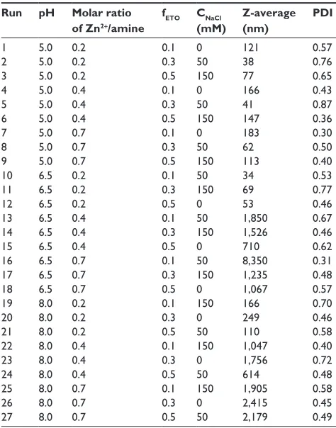
The mixtures were incubated for 72 hours at room tem-perature while shaking at 400 rpm. The unbound MTX was removed by ultrafiltration using Amicon YM-30 centrifugal filter devices (EMD Millipore). The concentration of MTX in filtrates was determined using a Knauer high-performance liquid chromatography (HPLC) system with P1000 pump (Herbert Knauer GmbH, Berlin, Germany) equipped with a multiple wavelength ultraviolet (UV) detector (UV 2600 detector). Separation of MTX was achieved at ambient Table 1 Taguchi orthogonal array design
Run pH Molar ratio
of Zn2+/amine
fETO CNaCl
(mM)
Z-average (nm)
PDI
1 5.0 0.2 0.1 0 121 0.57
2 5.0 0.2 0.3 50 38 0.76
3 5.0 0.2 0.5 150 77 0.65
4 5.0 0.4 0.1 0 166 0.43
5 5.0 0.4 0.3 50 41 0.87
6 5.0 0.4 0.5 150 147 0.36
7 5.0 0.7 0.1 0 183 0.30
8 5.0 0.7 0.3 50 62 0.50
9 5.0 0.7 0.5 150 113 0.40
10 6.5 0.2 0.1 50 34 0.53
11 6.5 0.2 0.3 150 69 0.77
12 6.5 0.2 0.5 0 53 0.46
13 6.5 0.4 0.1 50 1,850 0.67
14 6.5 0.4 0.3 150 1,526 0.46
15 6.5 0.4 0.5 0 710 0.62
16 6.5 0.7 0.1 50 8,350 0.31
17 6.5 0.7 0.3 150 1,235 0.48
18 6.5 0.7 0.5 0 1,067 0.57
19 8.0 0.2 0.1 150 166 0.70
20 8.0 0.2 0.3 0 249 0.46
21 8.0 0.2 0.5 50 110 0.58
22 8.0 0.4 0.1 150 1,047 0.40
23 8.0 0.4 0.3 0 1,756 0.72
24 8.0 0.4 0.5 50 614 0.48
25 8.0 0.7 0.1 150 1,905 0.58
26 8.0 0.7 0.3 0 2,415 0.45
27 8.0 0.7 0.5 50 2,179 0.49
Notes: Taguchi orthogonal array design for optimization of Z-average and PDI as functions of ph, the molar ratio of Zn2+/amine of PeI ionomer, the weight fraction of
PeO (feTO), and the concentration of Nacl (mM).
Abbreviations: PeO, polyethylene oxide; PDI, polydispersity index; PeI, polyethy-leneimine; feTO, weight fraction of PeO; c, concentration.
International Journal of Nanomedicine downloaded from https://www.dovepress.com/ by 118.70.13.36 on 23-Aug-2020
Dovepress sequential optimization of methotrexate encapsulation in micellar nano-networks
temperature, using monolithic reversed phase column
(Chromolith® Guard column, RP-18e, 5–4.6 mm; Merck).
The mobile phase was composed of 100 mM sodium acetate (pH 4) acetonitrile (volume ratio of 88:12) and was delivered isocratically at a flow rate of 2 mL/minute.
The HPLC method was validated in terms of specificity, linearity, accuracy, precision, and limit of quantification for an in vitro assay. Accordingly, the entrapment efficiency (the percentage of MTX entrapped to the total amount added) and the entrapment capacity (percentage of the entrapped MTX to the total amount of loaded system) were calculated.
The effects of the different factors (pH, feeding ratios, and the cross-link ratios) on the loading parameters were investigated by full factorial design in three levels (Table 2).
The factors were analyzed using Design-Expert® Software
version 6.02 (Stat-Ease, Inc., Minneapolis, MN, USA).
The particle sizes and ζ potentials of MTX-loaded
nano-networks were determined at different cross-link ratios (0.0, 0.3, and 0.6) and a constant MTX feed ratio of 0.3, according to the section “Remote loading of MTX.”
atomic force microscopy
To explore the morphology of the particles, samples were prepared for atomic force microscopy (AFM) imaging by
depositing 10 µL of 0.2 mg/mL aqueous dispersion of empty
and MTX-loaded nano-networks (R=0.3) at different cross-link
ratios (0.3 and 0.6) in comparison with the complexes of the MTX and the PEI ionomer, onto a prewashed glass slide. The AFM imaging was performed in air using a vibration-damped
NanoWizard® II (JPK Instruments AG, Berlin, Germany).
Commercial pyramidal tips (MicroMasch Inc., Tartu, Estonia)
attached to I-type cantilevers with a length of about 230 µm,
a resonance frequency of about 380 kHz, and a nominal force
constant of 40 N/m were used. Images were recorded in an intermittent contact mode at a scan speed of approximately 1 Hz to avoid damage of the sample surface. The acquired
images had a resolution of 512×512 pixels. The topological
characteristics, including projected diameter (d), surface area
(A), perimeter (P), and maximum height (Zmax), were measured
randomly by the software for at least 300 particles.
Subsequently, the shape factors (circularity and aspect ratio) were calculated according to Equation 1 and Equation 2 to express the morphology of the particles:
Circularity A
P
= 4π2 (1)
Aspect ratio d
Z =
max
(2)
In vitro release study
The release of the MTX from the nano-networks prepared at the cross-link ratio of 0.3 was evaluated by the dialysis method
at R=0.3 for 24 hours in phosphate buffered saline (PBS;
pH 7.4; 0.14 M NaCl) and acetate buffered saline (pH 5.5; 0.14 M NaCl) without or with 1 mM tris(2-carboxyethyl) phosphine (TCEP) as a reducing agent.
Briefly, 4 mL MTX-loaded nano-networks containing 2.5 mg of the PEI ionomer and 1.4 mg of MTX were placed in the Float-A-Lyzer 6–8 kDa dialysis tube. The dialysis was carried out against 400 mL of the release media in a shaking
incubator at 37°C and 100 rpm.
At scheduled times, 1 mL of the medium was withdrawn, and the amount of MTX released was estimated by the HPLC method as described in the section “Remote loading of MTX.” The cumulative release percentage of MTX was determined at each point and plotted as a function of time.
Table 2 Z-average, PDI, and zeta-potential of PeI ionomer
Formulation Cross-link
ratio
Condition Z-average
(nm)
PDI Zeta potential
(mV)*
1 0 + Zn2+ 33.7±6.4 0.22±0.07 –
2 0.3 + Zn2+ 112.8±42.6 0.14±0.11 –
3 0.6 + Zn2+ 139.6±11.5 0.24±0.06 –
4 1.2 + Zn2+ 227.3±12.5 0.30±0.01 –
5 0.3 after acid dialysis 254.0±46.6 0.36±0.06 +9.7±3.7
6 0.6 after acid dialysis 162.0±10.0 0.27±0.04 +1.7±4.1
7 1.2 after acid dialysis 175.0±0.0 0.47±0.02 −0.1±3.4
8 0.00 + MTX 109.0±16.8 0.18±0.06 +5.5±9.9
9 0.3 + MTX 117.0±15.9 0.22±0.01 −1.5±4.8
10 0.6 + MTX 133.0±21.4 0.19±0.02 −0.1±4.5
Notes: The Z-average, PDI, and zeta-potential of the PeI ionomer at different cross-link ratios before and after the removal of Zn2+ from the micellar templates and following
the encapsulation of the MTX in the nano-networks at the feeding ratio of 0.3. *In PBs (10 mM phosphate salts +140 mM Nacl; ph 7.4). Abbreviations: PDI, polydispersity index; PeI, polyethyleneimine; MTX, methotrexate; PBs, phosphate buffered saline.
International Journal of Nanomedicine downloaded from https://www.dovepress.com/ by 118.70.13.36 on 23-Aug-2020
Dovepress
abolmaali et al
The control experiment was done to study free MTX transport through the dialysis membrane at the simulated media. To find out the mechanism of drug release from the nano-networks, the Korsmeyer–Peppas equation (Equation 3) was best fitted for the first 60% drug release data in com-parison to other mathematical equations, including zero order,
first order, and the Higuchi models.55 The release rate (k), the
release exponent (n), and the lag time (t0) were determined
from the fraction of MTX released (M/M∞) versus time (t) by
a nonlinear regression method, using SPSS Statistics version 16.0 (IBM Corporation, Armonk, NY, USA).
M
M k t t
n
∞= ⋅ −( 0)
(3)
redox-sensitive degradation
To study the redox biodegradability of the nano-networks, the thiol content of the cross-linked nanoparticles was deter-mined prior to and after reducing the disulfide bond present in
the DTDP molecule, according to the published method.56
Briefly, 25 µL of the 0.2 M NaBH4 solution in NaOH
0.2% was added to 45 µL of the sample solutions, which
contained 4 mM nitrogen, and incubated for 1 hour at room temperature while shaking at 300 rpm. Then, pH was adjusted to 4 using HCl 1 N and further incubated for 10 minutes to
remove the NaBH4 from the reaction medium. The medium
was supplemented with NaOH 1 N to adjust the pH again
to 8. The volume reached 90 µL with the addition of
(4-(2-hydroxyethyl)-1-piperazineethanesulfonic acid) (HEPES)
buffer (300 mM; pH 8.0). Then, 10 µL (4 mg/mL) DTNB
in the HEPES buffer (50 mM; pH 8.0) was added, and the mixture was incubated for 15 minutes at room temperature. Absorbance values were obtained with a PowerWave HT
microplate reader (BioTek, Winooski, VT, USA) at λ=412
nm, and the thiol content was calculated using a standard curve of the reduced glutathione and expressed as mmol/L free thiol.
In vitro cellular studies
The MTT-based cytotoxicity assay was carried out based
on the protocol described previously.57 Accordingly, the
HepG2 cells were plated into 96-well microtiter plates
(Orange Scientific, Braine-l′Alleud, Belgium) at a density
of 25,000 cells/cm2. After 24 hours, the cells were treated in
serum-supplemented culture medium. First, the assay was performed for the nano-networks at the cross-link ratios of 0.3 and 0.6, in comparison to the PEI ionomer and the
branched PEI (Mw=10 kDa). In another experiment, the
cytotoxicity of MTX was determined in HepG2 cells incu-bated with the free drug for 24, 48, and 72 hours. Finally, the cytotoxicity of the MTX-loaded nano-networks was compared to the free MTX after 24 hours. Each treatment was similarly prepared by the primary dilution of the stock solutions in phosphate buffered saline (pH 7.4) and the final
10× dilution in RPMI 1640 medium (PAA Laboratories
GmbH, Germany) supplemented with 10% fetal bovine serum (Gibco BRL).
Following incubation, the medium was aspirated and
replaced by 100 µL of 1/10 diluted MTT stock solution
in the culture medium to obtain the final concentration of 0.5 mg/mL. After 3 hours, the medium was aspirated again, and the insoluble formazan crystals were dissolved in
100 µL/well DMSO and measured spectrophotometrically
in a microplate reader at λ=570 nm, and corrected for
back-ground absorption at λ=650 nm. Cell viability was calculated
for blank-corrected data relative to untreated control cells. To determine cellular uptake potential of the nano-networks, the cells were grown on Lab-Tek II 4-well cham-ber slides with covers (Nunc, USA Scientific, Ocala, FL,
USA) at a density of 2×10⁴ cells per chamber. The cells
were treated with 50 µg/mL of nano-networks labeled with
fluorescein isothiocyanate (FITC), washed three times with cold PBS, fixed with 3.7% paraformaldehyde solution in
PBS at 4°C for 15 minutes, followed by washing with PBS.
Cellular internalization of the nanohydrogels was studied by epifluorescence microscope (Nikon Eclipse E400; Nikon Corporation, Tokyo, Japan).
statistics
The statistical analysis was performed by GraphPad Software Inc., version 5.0 (GraphPad Software, Inc., La Jolla,
CA, USA). P-values 0.05 were considered statistically
significant. Data were expressed as mean ± standard
deviation.
Results and discussion
Template-assisted synthesis
of nano-networks
The preparation method requires a low-energy mixing pro-cess, and it does not require the use of any organic solvent.
The donor–acceptor complexes of Zn2+ and the amines of the
PEI ionomers can be considered as a special type of copolymer with lyophobic segments formed from the phase separation
of the ethyleneimine moieties with Zn2+. The water-soluble
mPEG segments stabilize the coacervates and prevent the aggregation or the macroscopic phase separation.
International Journal of Nanomedicine downloaded from https://www.dovepress.com/ by 118.70.13.36 on 23-Aug-2020
Dovepress sequential optimization of methotrexate encapsulation in micellar nano-networks
Similarly, the formation of metal colloids has been shown in aqueous medium by the reduction of the lyophobic
com-plexes of diblock or graft PEO-PEI with Pd2+ or Pt2+ at low
pH.25,34 The preparation requires a precise tuning of
differ-ent physicochemical parameters, such as fETO, pH, the ionic
strength of the solution, and the type and the concentration
of the ionic species, inducing the gelation of ionomers.23,58
Therefore, it was hypothesized that the colloid property of
the complexes of PEI ionomers with Zn2+ depends on some
factors such as pH, the molar ratio of Zn2+, f
ETO, and salt
concentration. The factors were considered in the Taguchi orthogonal array design for optimization of the size distribu-tion. The Taguchi method is a fractional factorial design pro-posed by Genichi Taguchi, a Japanese engineer, to improve the quality of products or processes.
Accordingly, the particle size data (Z-average and PDI) were analyzed statistically by changing the experimental factors simultaneously, according to the designed array in three levels (Table 1). The data were successfully fitted in
individual two-factor interaction models (P,0.0001 and
P,0.05, respectively).
If the Z-average was considered as a response variable,
pH (P,0.0001), the molar ratio of Zn2+ (P,0.0001), and
their interaction (P approximately 0.001) were determined significant. Figure 2A shows the sizes increased abruptly if
the molar ratio of Zn2+ increased from 0.2, possibly due to
an insufficiency of mPEG chains to solubilize the lyophobic
PEI-Zn2+ domains. Similarly, it was shown that the
aggrega-tion occurs for the block ionomer of polyacrylic acid at a
relatively high concentration of Ba2+ and Gd3+.59 The particles
enlarged by increasing pH from 5–6.5, especially at the molar
ratios of Zn2+$0.4 (Figure 2B). This could be explained by
the reduced protonation of the PEI backbone at a more basic
pH, and the very low solubility of the Zn2+ complexes, due
to the binding of the hydroxyl ion ligands.
The main effect of the molar ratio of Zn2+ (P,0.05) and
the related interaction effect with salt concentration (P,0.05)
were statistically significant terms influencing PDI. A
rela-tively high level of the molar ratio of Zn2+ resulted in more
homogeneous particles that might occur due to the
cross-link action of Zn2+, though the PDI values were still high.
An increase of the salt concentration brought about a higher
PDI at the low level of Zn2+; however, the lowest PDI was
attained at the high level of salt concentration (150 mM) if the
molar ratio of Zn2+ was 0.4. This controversial effect of salt
concentration might be due to displacement of Zn2+ from their
complexes at the high NaCl concentration during the phase-separation step. This phenomenon was reported previously
for PEO-g-PEI/surfactant complexes.60,61 Nevertheless, the
salt might stabilize the lyophobic complexes of Zn2+ at the
molar ratio of 0.4, possibly through the formation of the salt bridges on the interface of the lyophobic complexes.
The optimum condition defined by the minimum
Z-average × PDI comprises the molar ratio of Zn2+ =0.2,
pH =5.7, fETO=0.3, and without additional salt. The resulted
self-assembly exhibited Z-average of 33.7 nm and PDI of 0.22 and served as a micellar template for synthesis of cross-linked polymer micelles in the next step.
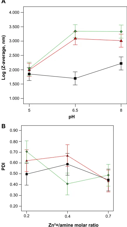
Cross-linking of the polyamine core of the micellar template was achieved via condensation reaction with the carboxylic functional groups of DTDP, a homobifunctional carboxylic acid based electrophilic cross-linking reagent with a length of 1.2 nm and a cleavable disulfide linkage. Following
B
PDI
Zn2+/amine molar ratio
A
4.000
3.500
3.000
2.500
2.000
1.500
Log (Z-average, nm)
1.000
5
0.90
0.80
0.70
0.40
0.30
0.20
0.2 0.4 0.7
0.50 0.60
6.5
pH
8
Figure 2 Interaction effect of ph and the molar ratio of Zn2+ on Z-average (A) and
PDI (B) of the PeI ionomer micellar template , , and denote various molar ratios of Zn2+/amines of the PeI ionomer (0.2, 0.4, and 0.7, respectively).
Abbreviations: PDI,polydispersity index; PeI, polyethyleneimine.
International Journal of Nanomedicine downloaded from https://www.dovepress.com/ by 118.70.13.36 on 23-Aug-2020
Dovepress
abolmaali et al
the reaction, acid dialysis was performed to remove Zn2+. The
1H-NMR spectra of the nano-networks showed the total
inte-gral intensity of the signals due to −S−CH2CH2−C=O protons
of DTDP (m, δ: 2.4–2.9) increased by the cross-link ratio in
comparison to the ethylene glycol peak of the PEI ionomers in the swollen state (data not shown). The estimated degree of cross-links per primary amines with the nominal cross-link ratio of 0.3 was calculated as 7.36%. This value gradually increased and reached 15.72% for the nominal cross-linking ratio of 1.2. It is noteworthy to mention that DTDP can also form a loop within a single chain; therefore, the calculated degrees of cross-linking can be overestimated.
Figure 3A shows the sizes increased after removing
Zn2+ to about 150–250 nm at the relatively lower cross-link
ratios (0.3 and 0.6). These observations are consistent with
the swelling nature of the nano-networks. It was understood from Table 2 that the average volumes of the swollen nano-networks changed more than ten times upon the removal
of Zn2+ at C=0.3, while the volumes exhibited only a
mod-est increase at the higher ratios compared to those of the
precursor complexes of Zn2+ and PEI ionomer. The
aver-age sizes of the nano-networks decreased by increasing C from 0.3–1.2.
This is in line with other studies that showed that if the cross-link ratio increased, the smaller particles of
PEO-b-PAA were prepared.15,16 Nevertheless, the average sizes of
the cross-linked complexes of the Zn2+ and the PEI ionomer
increased markedly at the cross-link ratio of 1.2 before
removing Zn2+ from the micellar templates. This could
be explained by an increase of the hydrophobicity of PEI
ionomer/Zn2+ complexes, due to the amide bond formation
mediated by DTDP and the failure of the mPEG chains to prevent aggregation.
Figure 3B shows that an increase of PDI happened upon
the removal of Zn2+ that was less pronounced at C=0.6. The
changes of PDI could be explained by the positive physical
cross-link action of Zn2+, which disappeared after acid
dialysis. Taking into consideration these results, the synthe-sis method produces a hydrophilic network with a relatively more uniform population of small particles if compared to other methods, such as polyionic complexation, desolvation,
emulsion droplet coalescence, and direct cross-linking.62
remote loading of MTX
MTX, a weak bicarboxylic acid with pKa values of 3.8 and 4.8, could be immobilized into the PEI nano-networks (appar-ent pKa approximately 8.5) by the simple mixing of the MTX solution with the aqueous dispersions of the nano-networks. Although it seems that the MTX loading was mainly governed through electrostatic charge complexation, we cannot exclude the possibility that the drug molecules can be physically entrapped and retained in the networks, due to nonspecific hydrophobic interactions.
Table 3 shows full-factorial design optimization of the MTX loading into the nano-networks. The drug loading was expressed as a function of pH, C and the R in terms of the loading efficiency (LE%) and the loading capacity (LC%). The results of analysis of variance revealed that MTX loading follows the two-factor interaction
statisti-cal model (P,0.0001 similarly for LE% and LC%). The
main effects of C and R and their interaction (C × R) were
determined as the significant terms included in the cor-responding models.
300
A
B
250
Z-average (nm)
PD
I
200
150
100
50
0
0.3
0.6
0.5
0.4
0.3
0.2
0.1
0
0.3 0.6 1.2
Cross-link ratio (C)
Cross-link ratio (C)
0.6 1.2
Figure 3 effect of the cross-link ratio.
Notes: Theeffect of the cross-link ratio on Z-average (A) and PDI (B) of the Zn2+
micellar template of the PeI ionomer, prior to () and after () removal of Zn2+.
Abbreviations: PDI,polydispersity index; PeI, polyethyleneimine.
International Journal of Nanomedicine downloaded from https://www.dovepress.com/ by 118.70.13.36 on 23-Aug-2020
Dovepress sequential optimization of methotrexate encapsulation in micellar nano-networks
Figure 4A shows that an increase of R led to a significant
decline of LE% of the PEI ionomer (P=0.001), while LE%
was little changed by R for the nano-networks; however, Figure 4B shows that LC% was almost constant for the PEI
ionomer (P0.05), but increased for the nano-networks
(P=0.001). The increase was more pronounced for R in the
range of 0.15 and 0.30, but less at R=0.6, possibly due to
reduced binding sites for MTX loading.
Remarkably, the LC% in the PEI ionomer was lower than
the nano-networks at R=0.6. It could be explained by the
formation of the hydrophobic MTX-bound domains in the polyionic segment of the PEI ionomer stabilized by the mPEG chains that could lead to a self-assembly, preventing the further loading of MTX molecules. Unlike the PEI ionomer, hollow spaces of the nano-networks provide the free diffusion of MTX molecules, so the interaction of MTX with the outer layer amines does not restrict the subsequent loading.
Higher degrees of cross-linking may lead to a decreased LC% (Figure 4B and C) as reported before for the
cross-linked PEO-b-PMA micelles.16 The pH changes provide
an opportunity to tune the electrostatic binding between the MTX and the ionomer chains. LC% did not change by pH in the range of 5–7, since the MTX molecule has a similar negative-charge density in that pH range, and the polymer
chain is protonated in the same way at pH =5–7. Both the
optimum LE% and LC% were achieved at the average pH
of 6 and R=0.3 for the nano-networks, especially at C=0.3.
A
Loading efficiency (%)
Feeding ratio (R) 100
90
80
70
60
50
40
30
20
10
0.15 0.3 0.6
B
Loading capacity (%)
Feeding ratio (R) 70
60
50
40
30
20
10
0.15 0.3 0.6
C
Loading capacity (%)
pH 70
60
50
40
30
20
10
5 6 7
Figure 4 Loading efficiency and loading capacity.
Notes: Theloading efficiency % (A) and loading capacity % (B and C) of the MTX-loaded nano-networks at different cross-link ratios: (0), (0.3), and (0.6). The loading parameters were shown as a function of MTX-feeding ratio (A and B) or ph (C).
Table 3 Full factorial design optimization of MTX loading
Run Cross-link ratio
pH Feeding
ratio
Loading efficiency (%)
Loading capacity (%)
1 0.0 5.0 0.15 57.67 37.20
2 0.0 5.0 0.30 59.81 55.13
3 0.0 5.0 0.60 40.22 62.28
4 0.0 6.0 0.15 91.83 48.54
5 0.0 6.0 0.30 55.85 53.43
6 0.0 6.0 0.60 27.27 52.82
7 0.0 7.0 0.15 87.03 47.20
8 0.0 7.0 0.30 48.22 49.76
9 0.0 7.0 0.60 21.94 47.39
10 0.3 5.0 0.15 59.87 38.08
11 0.3 5.0 0.30 57.13 53.99
12 0.3 5.0 0.60 52.73 68.40
13 0.3 6.0 0.15 58.10 37.37
14 0.3 6.0 0.30 57.25 54.04
15 0.3 6.0 0.60 39.96 62.13
16 0.3 7.0 0.15 70.49 42.00
17 0.3 7.0 0.30 67.53 58.11
18 0.3 7.0 0.60 39.93 62.11
19 0.6 5.0 0.15 29.04 22.97
20 0.6 5.0 0.30 36.41 42.79
21 0.6 5.0 0.60 20.83 46.10
22 0.6 6.0 0.15 46.59 32.37
23 0.6 6.0 0.30 54.42 52.78
24 0.6 6.0 0.60 31.17 56.13
25 0.6 7.0 0.15 47.98 33.01
26 0.6 7.0 0.30 48.38 49.85
27 0.6 7.0 0.60 33.12 57.62
Notes: Full factorial design optimization of methotrexate loading, according to the cross-link ratio, ph, and the feeding ratio in three levels.
Abbreviation: MTX, methotrexate.
International Journal of Nanomedicine downloaded from https://www.dovepress.com/ by 118.70.13.36 on 23-Aug-2020
Dovepress
abolmaali et al
The optimum LC% of 57% was noticeably higher than the
previous report of 12%,63 so the nano-networks satisfactorily
loaded MTX.
Table 2 shows that MTX loading was accompanied by reduction of the Z-average of the nano-networks especially at the low cross-link ratio (C=0.3) (P<0.05), suggesting that the drug loading attenuated swelling of the nano-networks through formation of charge-neutralized hydrophobic domains; moreover, the drug loading did not cause any adverse effect on the particle stability.
Notably, the MTX-loaded nano-networks exhibited almost zero value of zeta potential, suggesting that MTX was effectively neutralized by the core amines; nevertheless, nano-networks showed only few positive charges that reduced by increasing the cross-link ratio. The reduced zeta potentials are possibly due to an arrangement of the mPEG chains around the cross-linked polyamine core that shifts the shear
plane away from the positive core, as indicated before.64
aFM
An AFM experiment was performed to investigate topo-logical properties of the empty and the MTX-loaded
nano-networks (R=0.3) at C=0.3 (cross-link 1) and 0.6
(cross-link 2) compared to the complex of the PEI iono-mer and the MTX. As shown in Figure 5, discrete, small, and round particles were observed for the nano-networks
at C=0.6 that were smaller and more uniform than the
complexes of the ionomer and the MTX. Table 4 shows that the average projected diameters of dehydrated empty nano-networks decreased to about 50 nm, if compared to the hydrodynamic diameters as determined by Zetasizer. After loading the MTX into the nano-networks, the diameters and
the Z-values increased significantly (P,0.05), as compared
to the empty particles. The average circularity of the empty
nanonetwork (C=0.3; R=0) increased significantly (P,0.05)
A B
PEI ionomer – MTX complex MTX-loaded nanonetwork
4 µm 4 µm
Figure 5 aFM images of the methotrexate-loaded PeI ionomer complex (A) and the nano-networks loaded with MTX at the nominal cross-link ratio of 0.6 (B). Abbreviations: aFM, atomic force microscopy; PeI, polyethyleneimine; MTX,
methotrexate. Table
4
a
FM determination of topological and shape factors
Polymer
Condition
Surface area (m
2)
Perimeter (m)
Projected diameter (m)
Z value (height, m)
Circularity
Aspect ratio
Nanonetwork (cross-linked
1 ) empty 23.3 × 10 − 16± 17.9 × 10 − 16 22.6 × 10 − 8± 10.7 × 10 − 8 55.0 × 10 − 9± 9.2 × 10 − 9 14.6 × 10 − 9± 2.4 × 10 − 9 0.59 ± 0.17 3.87 ± 1.08
Nanonetwork (cross-linked
2 ) empty 22.0 × 10 − 16± 6.3 × 10 − 16 17.9 × 10 − 8± 5.7 × 10 − 8 48.9 × 10 − 9± 11.2 × 10 − 9 7.8 × 10 − 9± 1.8 × 10 − 9 0.81 ± 0 .09 6.15 ± 1.60 Pe I ionomer MTX-loaded 37.6 × 10 − 16± 11.4 × 10 − 16 29.3 × 10 − 8± 14.3 × 10 − 8 85.0 × 10 − 9± 18.6 × 10 − 9 31.3 × 10 − 9± 2.9 × 10 − 9 0.71 ± 0.04 2.71 ± 0.22
Nanonetwork (cross-linked
1 ) MTX-loaded 32.4 × 10 − 16± 0.8 × 10 − 16 24.6 × 10 − 8± 0.4 × 10 − 8 70.9 × 10 − 9± 0.7 × 10 − 9 19.5 × 10 − 9± 0.2 × 10 − 9 0.78 ± 0.09 3.57 ± 0.52
Nanonetwork (cross-linked
2 ) MTX-loaded 27.7 × 10 − 16± 0.4 × 10 − 16 23.9 × 0 − 8± 0.3 × 10 − 8 73.1 × 10 − 9± 0.6 × 10 − 9 30.7 × 10 − 9± 0.2 × 10 − 9 0.78 ± 0.01 2.41 ± 0.02 Notes: The a
FM determination of topological and shape factors of the P
eI ionomer and the nano-networks at the cross-link ratio of
0 .3 (cross-linked 1 ) or 0 .6 (cross-linked 2
), prior to and after methotrexate loading.
Abbreviations:
a
FM, atomic force microscopy; P
eI, polyethyleneimine.
International Journal of Nanomedicine downloaded from https://www.dovepress.com/ by 118.70.13.36 on 23-Aug-2020
Dovepress sequential optimization of methotrexate encapsulation in micellar nano-networks
24
Free MTX
MTX-loaded nanonetwork MTX-loaded nanonetwork (plus TCEP)
20 16 12
Time (h)
8 4 0 0 20 40 60 80 100
M/M
∞
(%)
A
24
Free MTX
MTX-loaded nanonetwork MTX-loaded nanonetwork (plus TCEP)
20 16 12
Time (h)
8 4 0 0 20 40 60 80 100
M/M
∞
(%)
B
Figure 6 In vitro cumulative release of MTX from the nano-networks.
Notes: Invitro cumulative release of MTX from the nano-networks at the nominal cross-link ratio of 0.6 in PBs (ph =7.4), or acetate buffered saline (ph =5.5) without or with 1 mM TceP.
Abbreviations: MTX,methotrexate; PBs, phosphate buffered saline; TceP, tris(2-carboxyethyl)phosphine; M/M∞, drug released.
from 0.59–0.78 (C=0.6; R=0) and 0.81 (C=0.3; R=0.3) in a
similar pattern that could be explained by the formation of more homogeneous and spherical particles. Upon adsorption of the nano-networks on a glass surface, they exhibited a high aspect ratio, suggesting that the particles were flattened on
the substrate as previously addressed.59
In vitro release study
Figure 6 shows the release profiles of the MTX loaded
nano-networks (C=0.6; R=0.3) in isotonic-balanced release
media (pH 5.5 or 7.5 + 140 mM NaCl). A prolonged release
of MTX achieved over 24 hours is consistent with the
previ-ous reports of a sustained MTX release for 8–24 hours.65,66
No burst release or very significant lag-time was recognized
(P0.05) for the MTX-loaded nano-networks if compared
to free MTX, although the initial burst behavior has been reported in several studies, due to the fast escape of the free
MTX out of the nanoparticles.67,68 The better control of the
initial burst release might be explained by the formation of a core-enriched model of an active MTX loading within the closely packed amine moieties. The release data were analyzed, according to the mathematical power function (Korsmeyer–Peppas model) to understand the mechanisms by which the drug release happened and to calculate the corresponding constants (Table 5). The MTX release was determined swelling-controlled, that means the reduced drug concentration gradient over the time period was compensated by an increased swelling of the nano-network which happens
International Journal of Nanomedicine downloaded from https://www.dovepress.com/ by 118.70.13.36 on 23-Aug-2020
Dovepress
abolmaali et al
following MTX release. It was found that the nano-networks show a control over the MTX release, since n was calculated to be near the value of one, especially at the physiologic pH of 7.5. It could be explained by the reservoir action of the nanonetwork for the negatively charged molecules, such as MTX. The effect of pH and the cross-linking reaction on the
rate constant (K) and n were not found significant (P0.05).
MTX is a weak acidic drug that exhibits more solubility at pH 7.5 than at 5.5. The lower solubility of MTX at the acidic environment of inflamed tissues can be considered as a hurdle for the delivery of MTX. The negative solubility behavior of MTX was improved most likely by a pronounced swelling of the nano-networks that compensates the reduced solubility of the drug in the relatively acidic pH of 5.5.
To evaluate whether reducing the disulfide containing cross-link (DTDP) can trigger MTX release from the nano-networks, the study was repeated in the release media supplemented with a reducing agent, such as TCEP, which is active in a broad range of pH and simulates the reducing action of glutathione. The TCEP treatment increased K, irrespective of the medium pH
(P,0.05), that – in turn – indicates that the MTX release can
be accelerated in the intracellular reducing environment.
redox-sensitive degradation
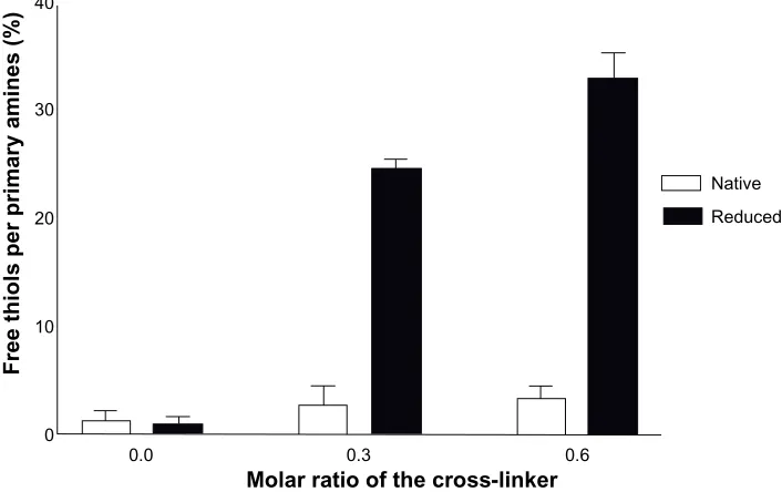
To fabricate a stabilized redox-sensitive system for intra-venous administration, bioreversible disulfide containing cross-links (DTDP) were used for the preparation of the nano-networks.
To study whether the disulfide bonds in the core of the nano-networks undergo cleavage, the empty particles were incubated in a simulated medium containing
reduc-ing agents, such as NaBH4. Figure 7 shows the ratio of
free thiol to total primary amines of the nano-networks increased in the presence of the reducing agent, as the cross-link density increased. The cross-links were stable during synthesis of the nano-networks since no significant free thiol was found at the end of the reaction; nevertheless, the stable disulfide containing cross-links could be prone to glutathione mediated reduction, which predominantly
occurs in the intracellular medium.69
Table 5 rate constants, release exponents for MTX release from nano-networks at nominal cross-link ratio
Release condition Rate constant (K) Release exponent (n) Lag time (hours) R2
ph =7.5 0.099±0.022 0.92±0.09 0.65±0.36 0.998
ph =7.5 + TceP 0.199±0.028 0.66±0.01 0.30±0.02 0.999
ph =5.5 0.082±0.027 0.88±0.19 0.57±0.37 0.986
ph =5.5 + TceP 0.217±0.004 0.51±0.01 0.53±0.03 0.999
Notes: The rate constants (K) and release exponents (n) for MTX release from the nano-networks at the nominal cross-link ratio of 0.6 (cross-linked 2), in PBs (ph =7.4), or acetate buffered saline (ph =5.5) without or with 1 mM TceP.
Abbreviations: MTX, methotrexate; PBs, phosphate buffered saline; TceP, tris(2-carboxyethyl)phosphine.
Free thiols per primary amines (%)
Molar ratio of the cross-linker
Native
0.0 0
10 20 30 40
0.3 0.6
Reduced
Figure 7 redox-sensitive degradation of the nano-networks demonstrated by ellman’s assay of free thiols prior to and after addition of 0.2 M NaBh4.
International Journal of Nanomedicine downloaded from https://www.dovepress.com/ by 118.70.13.36 on 23-Aug-2020
Dovepress sequential optimization of methotrexate encapsulation in micellar nano-networks
In vitro cellular studies
MTT assay is a sensitive method for determination of the cytotoxicity of nanostructures, since they primarily exert toxicity on the mitochondrial compartment
follow-ing cellular internalization.70 The assay was performed to
compare cytotoxicity of native PEI, the PEI ionomer, or the
nano-networks at C=0.3 (cross-linked 1) or C=0.6
(cross-linked 2) in the HepG2 cell line after incubation for 24 hours (Figure 8A). PEI demonstrated a concentration-dependent cytotoxicity while the PEI ionomer showed a reduced
0
0
0.1 20
0.01 10
Polymer concentration (µg/mL)
Methotrexate concentration (µM)
20 40
Viability (%)
Viability (%)
60 80 100
100 1,000
24 hours 48 hours 72 hours Branched PEI (Mw =10 kDa) PEI ionomer
Nanonetwork (cross-linked 1) Nanonetwork (cross-linked 2)
A
B
40 60 80 100
1 10 100
0.1 20
0.01
Methotrexate concentration (µM)
Viability (%)
C
40 60 80 100
1 10 100
Free MTX
MTX-loaded (cross-linked 1) MTX-loaded (cross-linked 2)
Figure 8 MTT-based cytotoxicity.
Notes: TheMTT-based cytotoxicity of: (A) empty nano-networks in comparison with PeI ionomer and branched PeI after exposure for 24 hours; (B) free MTX incubated for 24, 48, and 72 hours; (C) MTX-loaded in nanonetwork versus free MTX after 24 hours of exposure at 37°c in hepg2 cell line. cross-linked 1 and cross-linked 2 denote the theoretical cross-link ratios of 0.3 and 0.6, respectively. Data are expressed as the mean ± SD for five replicates.
Abbreviations: MTX,methotrexate; MTT, 3-(4,5-dimethylthiazol-2-yl)-2,5-diphenyltetrazolium bromide; PeI, polyethyleneimine; sD, standard deviation; Mw, weight-average molecular weight.
International Journal of Nanomedicine downloaded from https://www.dovepress.com/ by 118.70.13.36 on 23-Aug-2020
Dovepress
abolmaali et al
cytotoxic effect. In contrast, the nano-networks showed practically no cytotoxic effect at concentrations below
100 µg/mL. This interesting result could be explained by
the substantially reduced ζ-potentials of the nano-networks
and the shielding effect of the mPEG shell, as discussed by
different authors.71,72
Prior to evaluating the activity of the MTX-loaded nano-networks, the cytotoxicity of free MTX was studied as a function of the drug concentration after 24, 48, and 72 hours (Figure 8B). The incubation of HepG2 cells with increasing
concentrations of MTX in the range of 0.1–100 µM did not
result in significant changes in cell viability at each time interval; however, a time-dependent cytotoxic effect was
observed (P,0.001), as previously reported,73 since MTX is
a cytostatic chemotherapeutic agent that mainly inhibits cell proliferation. The toxicity on HepG2 cells was augmented
for the MTX loaded in the nano-networks (P,0.05),
espe-cially at the concentrations above 1 µM (Figure 8C), though
the empty nano-networks did not demonstrate cytotoxicity. The finding might be related to the mechanisms by which free MTX transports into cells. The cellular entry of MTX is restricted to saturable mechanisms, such as the reduced
folate transmembrane carrier and the folate receptor.74 Unlike
free MTX, the MTX-loaded nano-networks can enter the cells via a parallel endocytic pathway. Moreover, the drug molecules that escaped from the endosomal compartment are less accessible to efflux pumps; therefore, a higher cellular accumulation of MTX and an enhanced cytotoxicity can be achieved. The issue was similarly addressed for different
nanoparticulate systems.75,76
Conclusion
The nano-networks synthesized successfully through the cross-linking of the micellar template of the PEI ionomer complex, whose size distribution is controlled by pH and the
molar ratio of a complexing metal ion, such as Zn2+. They
comprise a polycation swollen core structure stabilized by a hydrophilic mPEG shell, avoiding unfavorable interactions with biological interfaces. The nano-networks exhibited some appropriate pharmaceutical properties, such as small sizes, uniformity, globular morphology, and flexibility without using high shear forces or organic solvents for manipulation. They were able to accommodate negatively charged molecules, such as MTX, efficiently featured with a higher encapsula-tion capacity than the corresponding polyionic micelles and a prolonged MTX release in a swelling-controlled manner; moreover, the strategy of reductive activation of nanonetwork biodegradation boosted the release rate in the simulated
intracellular medium. Taken together, these observations and the enhanced antitumor activity in vitro might imply the potential application of the nano-networks for the successful delivery of MTX that should be tested in vivo.
Acknowledgment
The authors gratefully acknowledge the use of the facilities at the Center for Nanotechnology in Drug Delivery at Shiraz University of Medical Sciences and at the Nanotechnology Research Center, Tehran University of Medical Sciences. This study was funded by a Shiraz University of Medical Sciences grant for the accomplishment of the PhD thesis of Dr Samira Abolmaali.
Disclosure
The authors declare there is no conflict of interest.
References
1. Honglawan A, Ni H, Weissman D, Yang S. Synthesis of random copolymer based pH-responsive nanoparticles as drug carriers for cancer therapeutics. Polym Chem. 2013;4(13):3667–3675.
2. Whitesides GM, Kriebel JK, Mayers BT. Self-Assembly and Nanostructured Materials. In: Huck WTS, editor. Nanoscale Assembly:
Chemical Techniques. New York: Springer Science + Business Media; 2005:217–239.
3. Kamimura M, Kim JO, Kabanov AV, Bronich TK, Nagasaki Y. Block ionomer complexes of PEG-block-poly(4-vinylbenzylphosphonate) and cationic surfactants as highly stable, pH responsive drug delivery system. J Control Release. 2012;160(3):486–494.
4. Oh KT, Bronich TK, Bromberg L, Hatton TA, Kabanov AV. Block ionomer complexes as prospective nanocontainers for drug delivery.
J Control Release. 2006;115(1):9–17.
5. Gohy JF, Varshney SK, Antoun S, Jérôme R. Water-soluble complexes formed by sodium poly(4-styrenesulfonate) and a poly(2-vinylpy ridinium)-block-poly(ethylenoxide) copolymer. Macromolecules. 2000;33(25):9298–9305.
6. Kabanov A, Bronich TK, Kabanov VA, Eisenberg A. Soluble stoichio-metric complexes from poly(N-ethyl-4-vinylpyridinium) cations and poly(ethylen oxide)-block-polymethacrylate anions. Macromolecules. Jul 1996;29(4):6797–6802.
7. Harada A, Kataoka K. Formation of polyion complex micelles in an aqueous milieu from a pair of oppositely-charged block copolymers with poly(ethylene glycol) segments. Macromolecules. 1995;28(15): 5294–5299.
8. Bronich TK, Popov AM, Eisenberg A, Kabanov VA, Kabanov KV. Effects of block length structure of surfactant on self-assembly and solution behavior of block ionomer complexes. Langmuir. 2000;16(2):481–489.
9. Bronich TK, Nehls A, Eisenberg A, Kabanov VA, Kabanov AV. Novel drug delivery systems based on the complexes of block ionomers and surfactants of opposite charge. Colloid Surf B: Biointerfaces. 1999;16(1–4):243–251.
10. Harada A, Kataoka K. Switching by pulse electric field of the elevated enzymatic reaction in the core of polyion complex micelles. J Am Chem
Soc. 2003;125(50):15306–15307.
11. Kishimura A, Koide A, Osada K, Yamasaki Y, Kataoka K. Encapsulation of myoglobin in PEGylated polyion complex vesicles made from a pair of oppositely charged block ionomers: a physiologi-cally available oxygen carrier. Angew Chem Int Ed Engl. 2007;46(32): 6085–6088.
International Journal of Nanomedicine downloaded from https://www.dovepress.com/ by 118.70.13.36 on 23-Aug-2020
Dovepress sequential optimization of methotrexate encapsulation in micellar nano-networks
12. Kabanov VA, Kabanov AV. Interpolyelectrolyte and block ionomer complexes for gene delivery: physico-chemical aspects. Adv Drug Deliv
Rev. 1998;30(1–3):49–60.
13. Kakizawa Y, Kataoka K. Block copolymer micelles for delivery of gene and related compounds. Adv Drug Deliv Rev. 2002;54(2):203–222. 14. Bronich TK, Bontha S, Shlyakhtenko LS, Bromberg L, Hatton TA,
Kabanov AV. Template-assisted synthesis of nanogels from Pluronic-modified poly(acrylic acid). J Drug Target. 2006;14(6):357–366. 15. Kim JO, Sahay G, Kabanov AV, Bronich TK. Polymeric micelles
with ionic cores containing biodegradable cross-links for delivery of chemotherapeutic agents. Biomacromolecules. 2010;11(4):919–926. 16. Kim JO, Kabanov AV, Bronich TK. Polymer micelles with cross-linked
polyanion core for delivery of a cationic drug doxorubicin. J Control
Release. 2009;138(3):197–204.
17. Oh KT, Bronich TK, Kabanov VA, Kabanov AV. Block polyelectrolyte networks from poly(acrylic acid) and poly(ethylene oxide): sorption and release of cytochrome C. Biomacromolecules. 2007;8(2):490–497. 18. Sondjaja HR, Hatton TA, Tam KC. Self-assembly of poly(ethylene
oxide)-block-poly(acrylic acid) induced by CaCl2: mechanistic study.
Langmuir. 2008;24(16):8501–8506.
19. Bronich TK, Ouyang M, Kabanov VA, Eisenberg A, Szoka FC Jr, Kabanov AV. Synthesis of vesicles on polymer template. J Am Chem
Soc. 2002;124(40):11872–11873.
20. Sanson N, Bouyer F, Destarac M, In M, Gérardin C. Hybrid polyion complex micelles formed from double hydrophilic block copolymers and multivalent metal ions: size control and nanostructure. Langmuir. 2012;28(8):3773–3782.
21. Sanson N, Bouyer F, Gérardin C, In M. Nanoassemblies formed from hydrophilic block copolymers and multivalent ions. Phys Chem Chem
Phys. 2004;6(7):1463–1466.
22. An L, Wang Y, Liu X, et al. Block ionomer complex micelles based on the self-assembly of poly(ethylene glycol)-block-poly(acrylic acid)
and CdCl2 for anti-tumor drug delivery. Chem Pharm Bull (Tokyo).
2011;59(5):559–563.
23. Solomatin SV, Bronich TK, Bargar TW, Eisenberg A, Kabanov VA, Kabanov AV. Environmentally responsive nanoparticles from block ionomer complexes: effects of pH and ionic strength. Langmuir. 2003; 19(19):8069–8076.
24. Kunath K, von Harpe A, Fischer D, et al. Low-molecular-weight polyethylenimine as a non-viral vector for DNA delivery: comparison of physicochemical properties, transfection efficiency and in vivo distribution with high-molecular-weight polyethylenimine. J Control
Release. 2003;89(1):113–125.
25. Bronstein LM, Sidorov SN, Gourkova AY, et al. Interaction of metal compounds with ‘double-hydrophilic’ block copolymers in aque-ous medium and metal colloid formation. Inorganica Chimica Acta. 1998;280(1–2):348–354.
26. Sidorov SN, Bronstein LM, Valetsky PM, et al. Stabilization of metal nanoparticles in aqueous medium by polyethyleneoxide-polyethyleneimine block copolymers. J Colloid Interface Sci. 1999;212(2):197–211.
27. Bhattacharjee S, Dong J, Ma Y, et al. Formation of high-capacity protein-adsorbing membranes through simple adsorption of poly(acrylic acid)-containing films at low pH. Langmuir. 2012;28(17):6885–6892. 28. Vignesh G, Arunachalam S, Vignesh S, James RA. BSA binding
and antimicrobial studies of branched polyethyleneimine-copper(II) bipyridine/phenanthroline complexes. Spectrochim Acta A Mol Biomol
Spectrosc. 2012;96:108–116.
29. Wu S, Zhang L, Yang K, Liang Z, Zhang L, Zhang Y. Preparing a metal-ion chelated immobilized enzyme reactor based on the polyacrylamide monolith grafted with polyethylenimine for a facile regeneration and high throughput tryptic digestion in proteomics. Anal Bioanal Chem. 2012;402(2):703–710.
30. Dacarro G, Cucca L, Grisoli P, Pallavicini P, Patrini M, Taglietti A. Monolayers of polyethilenimine on flat glass: a versatile platform for cations coordination and nanoparticles grafting in the preparation of antibacterial surfaces. Dalton Trans. 2012;41(8):2456–2463.
31. Chertok B, David AE, Yang VC. Polyethyleneimine-modified iron oxide nanoparticles for brain tumor drug delivery using magnetic targeting and intra-carotid administration. Biomaterials. 2010;31(24): 6317–6324.
32. Müller M, Keßler B, Fröhlich J, Poeschla S, Torger B. Polyelectrolyte complex nanoparticles of poly(ethyleneimine) and poly(acrylic acid): preparation and applications. Polymers (Basel). 2011;3(2): 762–778.
33. Bontha S, Kabanov AV, Bronich TK. Polymer micelles with cross-linked ionic cores for delivery of anticancer drugs. J Control Release. 2006;114(2):163–174.
34. Bronstein LM, Kostylev M, Shtykova E, et al. Mixed Co/Fe oxide nanoparticles in block copolymer micelles. Langmuir. 2008;24(21): 12618–12626.
35. Vinogradov SV, Bronich TK, Kabanov AV. Self-assembly of polyamine-poly(ethylene glycol) copolymers with phosphorothioate oligonucleotides. Bioconjug Chem. 1998;9(6):805–812.
36. Nguyen HK, Lemieux P, Vinogradov SV, et al. Evaluation of polyether-polyethyleneimine graft copolymers as gene transfer agents.
Gene Ther. 2000;7(2):126–138.
37. Frankel AE. Reducing the immune response to immunotoxin: commentary re R Hassan et al, pretreatment with rituximab does not inhibit the human immune response against the immunogenic protein LMB-1. Clin Cancer Res. 2004;10:16–18. Clinical Cancer Research. 2004;10(1):13–15.
38. Ghiamkazemi S, Amanzadeh A, Dinarvand R, Rafiee-Tehrani M, Amini M. Synthesis, and characterization, and evaluation of cellular effects of the FOL-PEG-g-PEI-GAL nanoparticles as a potential non-viral vector for gene delivery. J Nanomater. 2010;50.
39. Merdan T, Kunath K, Petersen H, et al. PEGylation of poly(ethylene imine) affects stability of complexes with plasmid DNA under in vivo conditions in a dose-dependent manner after intravenous injection into mice. Bioconjug Chem. 2005;16(4):785–792.
40. Mao S, Neu M, Germershaus O, et al. Influence of polyethylene glycol chain length on the physicochemical and biological properties of poly(ethylene imine)-graft-poly(ethylene glycol) block copolymer/ SiRNA polyplexes. Bioconjug Chem. 2006;17(5):1209–1218. 41. Fischer D, Osburg B, Petersen H, Kissel T, Bickel U. Effect of
poly(ethylene imine) molecular weight and pegylation on organ distribution and pharmacokinetics of polyplexes with oligodeoxynucle-otides in mice. Drug Metab Dispos. 2004;32(9):983–992.
42. Purcell WT, Ettinger DS. Novel antifolate drugs. Curr Oncol Rep. 2003;5(2):114–125.
43. Grim J, Chládek J, Martínková J. Pharmacokinetics and pharmacody-namics of methotrexate in non-neoplastic diseases. Clin Pharmacokinet. 2003;42(2):139–151.
44. Widemann BC, Adamson PC. Understanding and managing methotrex-ate nephrotoxicity. Oncologist. 2006;11(6):694–703.
45. Abelson HT, Fosburg MT, Beardsley GP, et al. Methotrexate-induced renal impairment: clinical studies and rescue from systemic toxicity with high-dose leucovorin and thymidine. J Clin Oncol. 1983;1(3): 208–216.
46. Rodenhuis S, McGuire JJ, Narayanan R, Bertino JR. Development of an assay system for the detection and classification of methotrexate resistance in fresh human leukemic cells. Cancer Res. 1986;46(12 Part 1):6513–6519. 47. Koch AE, Distler O. Vasculopathy and disordered angiogenesis in
selected rheumatic diseases: rheumatoid arthritis and systemic sclerosis.
Arthritis Res Ther. 2007;9 Suppl 2:S3.
48. Koning GA, Schiffelers RM, Wauben MH, et al. Targeting of angio-genic endothelial cells at sites of inflammation by dexamethasone phosphate-containing RGD peptide liposomes inhibits experimental arthritis. Arthritis Rheum. 2006;54(4):1198–1208.
49. Gaffo A, Saag KG, Curtis JR. Treatment of rheumatoid arthritis. Am J
Health Syst Pharm. 2006;63(24):2451–2465.
50. Levick JR. Hypoxia and acidosis in chronic inflammatory arthritis; relation to vascular supply and dynamic effusion pressure. J Rheumatol. 1990;17(5):579–582.
International Journal of Nanomedicine downloaded from https://www.dovepress.com/ by 118.70.13.36 on 23-Aug-2020