(Received on 14.12.2009. Accepted after revision on 9.3.2010)
[Indian J Tuberc 2010; 57:75-79]
P.D. Hinduja National Hospital & Medical Research Centre, Mahim, Mumbai
Correspondence:Dr. Camilla Rodrigues, Consultant Microbiologist, P. D. Hinduja National Hospital & Medical Research Centre, Veer Savarkar Marg (West), Mumbai – 400 016 (Maharashtra), India; Phone: +91- 22 – 24447794/95; Fax: +91 - 22 - 2444 91 51 2318 ; Email: [email protected]
CAN CORD FORMATION IN BACTEC MGIT 960 MEDIUM BE USED AS A PRESUMPTIVE METHOD FOR IDENTIFICATION OF M. TUBERCULOSIS
COMPLEX?
Mugdha Kadam, Anupama Govekar, Shubhada Shenai, Meeta Sadani, Asmita Salvi, Anjali Shetty and Camilla Rodrigues
Summary
Background: Serpentine cord formation in BACTEC MGIT 960 medium was evaluated as a rapid method for the presumptive identification of M. tuberculosiscomplex (MTBC).
Material & Methods: Total 2527 samples were processed for AFB culture using MGIT 960 TB system over a period of three months. AFB smears were prepared from 1000 MGIT tubes flagged positive by the MGIT instrument and stained by ZN method to examine presence or absence of serpentine cording. The cord formation was compared with PNBA [p-nitro benzoic acid] test on MGIT system and all controversial cases were further evaluated by NAP [p- [p-nitro-a-acetylamino-phydroxypropiophenone] test on BACTEC 460 TB system.
Results & Discussion: Of the 1000 culture positives, 904 (90.4%) were identified as mycobacteria, of which 869 (96%) showed cording by smear microscopy. One (0.1%) was identified as nocardia. In the remaining 95 (9.5%) cases, primary smear made from MGIT vial was negative. Of 869 cultures showing serpentine cord formation, 842 were confirmed as MTBC and 27 as NTM by PNBA assay on MGIT 960 TB system. The sensitivity, specificity, positive and negative predictive values are found to be 99.6%, 54%, 96% and 91% respectively. An average detection time for PNBA assay was found to be eight days whereas cording results were available on the same day of culture positivity.
Conclusion: Though highly sensitive it is not very specific and hence cannot be the only test for presumptive diagnosis of MTBC.
Key words: Cord formation, Presumptive Identification, Mycobacterium tuberculosis.
INTRODUCTION
Isolation of mycobacteria by Acid Fast Bacilli (AFB) culture represents the corner stone on which definitive diagnosis of tuberculosis (TB) and other Non-Tuberculous Mycobacteria (NTM) disease relies. Most of the laboratories in the developing world rely on conventional Lowenstein and Jensen (L.J) media for culture followed by use of different biochemical tests for identification of mycobacteria, limitations of which are well known. Use of automated liquid culture systems like BACTEC MGIT 960, MB/Bact, Versa Tech is slowly increasing in disease endemic countries as India. These automated liquid culture systems,when combined with commercial molecular techniques like probe hybridization for species identification, are capable of producing positive results in two weeks
or less forthe vast majority of sputum smear-positive specimens, and withinthree weeks for smear-negative specimens.1 However, such techniques are expensive, technically demanding and limited to a few clinically relevant species. Immuno chromatographic techniques such as CAPILIA are expensive and are still not available in India. Therefore in low-resource countries, many laboratories report a presumptive identification of
Mycobacterium tuberculosis complex (MTBC) to physicians on the basis of a simple, rapid and cost-effective method i.e cord formation in liquid culture media.
Virulent strains of the MTBC, when grown in a liquid medium, often display characteristic serpentine cord formation.2-4 Avirulent variants of MTBC grow in liquid media in a non-oriented,
dispersed fashion.3 NTM can form true cords in liquid culture but do so rarely, despite the fact that many species contain the cell wall glycolipid that mediates cord formation.2,3 The interpretation of cording morphology, particularly in NTM such as
M. kansasii, M. avium complex, M. marinum, M. szulgai, M. chelonae, M. gordonae M. terrae, and
M. phlei that can frequently form looser aggregates or “pseudocords”, is also subject to intero-observer differences.2,3
Cord formation has been advocated as a guide for the cost-effective utilization of DNA probes for the identification of Mycobacterium species5, but to date only a few studies have evaluated the utility of cord formation for the presumptive identification of MTBC.2,3,5-7 The present study was undertaken to determine the reliability of serpentine cording in BACTEC MGIT 960 medium as a rapid method to report the presumptive identification of MTBC. MATERIAL AND METHODS
A total of 2527 consecutive clinical specimens were processed for AFB culture using MGIT 960 TB system over a period of three months (May 2009 to August 2009). All contaminated clinical specimens were digested and decontaminated by the standard N-acetyl-L-cysteine-NaOHmethod.8 The sedimentwas suspended in 1 ml of sterile phosphate-buffered saline (pH6.8). 0.5 ml of the processed specimen was then inoculated into MGIT 960 vials supplemented as described by the manufacturer, and 0.2 ml onto L.J medium slants. CSF and specimens collected from sterile sites were inoculated directly to MGIT vials. All inoculated MGIT vials were incubated in the MGIT 960 instrument either till they were flagged positive by the instrument or for a maximum of six weeks. L.J medium slants were examined daily for the first one week and thereafter, biweekly, for twelve weeks, for the visible appearance of colonies. Of the total 2527, 1000 MGIT vials were flagged positive by MGIT 960 TB system and checked for cording by ZNCF staining by two different observers. All controversial results were further rechecked by an experienced microbiologist. All positive cultures were further subjected to identification by p-nitro benzoic acid
(PNBA) assay on MGIT 960 TB system.9-13All the MGIT vials flagged positive by machine but AFB negative by smear microscopy were further incubated at 370C and smear was repeated periodically after every three days.Obvious turbidity in the MGIT vial was confirmed by Gram staining of the smear as well as subculture on blood agar medium. In addition, 0.2 ml of positive broth was subcultured on an additional L.J. slant. Growth on this L.J subculture was used to rule out mixed infection, of MTB and NTM strains.
Serpentine cords were defined as ropelike aggregates of AFB in which the long axes of the bacteria paralleled the long axis of the cord. Identification using PNBA
It has been reported that the growth of MTB isolates is inhibited by PNB 500 g/ml whereas NTM are resistant to this concentration. The PNB stock solution was prepared to ensure a final concentration of 500 mg/ml in the MGIT vial.9-13 This stock solution was aliquoted and stored at -200. The PNBA test
was performed by inoculating the positive culture into two MGIT tubes with and without PNBA and incubated in the MGIT 960 system. The growth Control (GC) was flagged positive by the MGIT system when Growth Unit reached 400. Cut off values of less than or equal to 100 was taken as sensitive indicating the growth of M. tuberculosis
complex. Any value more than 100 was considered resistant indicating NTM. All cultures showing growth of NTM were further confirmed by r -nitro-a-acetylamino-b-hydroxypropiophenone (NAP) test in BACTEC 460 TB system.14
RESULTS
Of the total 2527 clinical specimens processed, 1000 (39.57%) were positive by MGIT 960 TB system (Table 1) which were further analysed for cording by AFB smear, and further processed for identification using PNBA test. These specimens included 742 respiratory specimens (650 Sputum, 62 Brochoalveolar lavage or BAL, 06 tracheal secretions, 24 pleural fluid) and 258 non-respiratory specimens (28 lymphnode, 25 tissue, 84
(n=845) whereas by observer 2 in 96.66% (n = 840). Final rechecking of controversial results by experienced microbiologist confirmed cord formation in 869/904 (96%) of MGIT positive cultures. Of 869 cultures showing serpentine cord formation, 842 were confirmed as MTBC and 27 as NTM by PNBA assay on MGIT 960 TB system. Of the 35 cultures negative for cording by ZNCF microscopy, three were found to be MTBC and 32 NTM by PNBA Test. Confirmation of all NTM by NAP test did not show any change in the results. Overall sensitivity, specificity, positive and negative predictive values are 99.6%, 54%, 96% and 91%
Table 1:
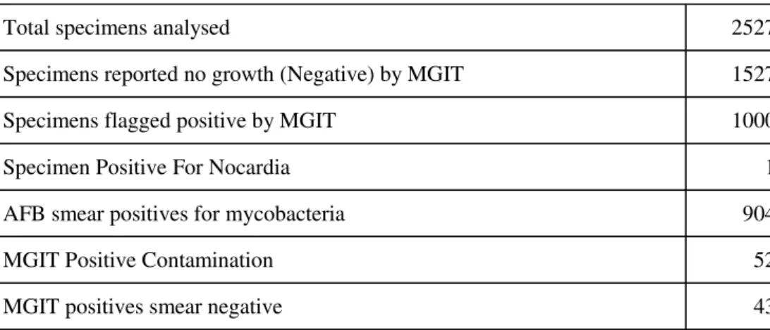
Distribution of the total clinical specimens analysed
Table 2:
Comparison of PNBA and Cord Formation
Total specimens analysed 2527
Specimens reported no growth (Negative) by MGIT 1527
Specimens flagged positive by MGIT 1000
Specimen Positive For Nocardia 1
AFB smear positives for mycobacteria 904
MGIT Positive Contamination 52
MGIT positives smear negative 43
N = 904
NO. of Samples showing Cording
(869)
No. of Samples Absent for Cording
(35) Final Result PNBA Test Positive for MTBC (845) 842 (99.6%) 3 (0.4%) MTB
PNBA Test Negative for MTBC
(59)
27 (46%) 32 (54%) NTM
Sensitivity = 99.6% Specificity = 54%
pus and aspirates, 07 body fluids, 20 CSF, 16 urine, 29 Abcess , 33 biposy , 16 Others). Of these 1000 specimens flagged positive by MGIT, 904 were positive for mycobacteria, one was showing Nocardia by smear microscopy. In the remaining 95 cases, primary smear made from MGIT vial was negative.
As shown in Tables 2 & 3, all 904 MGIT positive cultures were checked for cording by ZNCF method by two different observers. 845 were positive by observer 1 and 840 were positive by observer 2. Cords were recorded by observer 1 in 97.23%
respectively. An average detection time for PNBA assay was found to be eight days whereas cording results were available on the same day of culture positivity.
Of the 95, MGIT positive and AFB smear negative cases, 52 were identified as contamination. All re-inoculated MGIT vials after decontamination showed no growth. In the remaining 43 cases, organisms were not apparent by smear microscopy even after incubation till six weeks so all these were finally reported as no growth. All these were considered as MGIT false positives.
DISCUSSION
Rapid diagnosis of TB is critical to control of the disease; therefore, use of the most rapid methods available for culture and identification of MTBC is advocated. Cord formation has been reported as a simple, cost effective method for rapid presumptive identification of mycobacteria cultivated in liquid medium.2,3,5,6 We evaluated the characteristics of cord formation of MTB complex in the liquid MGIT medium and results were compared with PNBA identification assay on MGIT. All NTM identified by PNBA method were further rechecked by NAP assay on BACTEC 460 TB system.
Of the 1000 MGIT tubes flagged positive by the MGIT 960 instrument, only 904 (90.4%) were identified as mycobacteria of which 869 (96%) showed cording by smear microscopy. One (0.1%) was identified as nocardia. Of the remaining 95 (9.5%) MGIT positive AFB smear negative cases, 52 were contaminated and 43 were AFB smear and culture negative for mycobacteria or other bacteria
(at the end of the 42-day protocol). Reason for false positives can be depletion of oxygen due to other live cells present in the samples e.g. pus cells or cells present in tissues.
Gram positive cocci were the prevalent organisms responsible for contamination of MGIT 960 tubes in 52/2517 (2%) cases. Being highly enriched media contaminants like gram positive cocci can easily grow and utilized the oxygen in the medium giving false positive fluorescence signal. Hence smear preparation of all MGIT tubes flagged positive by MGIT will be helpful before processing it for further identification.
Studies conducted on the utility of cord formation for presumptive identification of MTBC, yield discordant data with sensitivity ranging from 22.9% to 90%.2,3,5,6 Cording was found to be very specific for MTBC by Yagupsky et al and Morris et al.3,6 However, in the present study, 27/59 (46%) NTM showed cording were misinterpreted as MTBC, decreasing the specificity of this test to 54%.
Cord formation is strictly an in vitro
phenomenon and the proportion of isolates that demonstrate this phenomenon has been shown to vary greatly between clinical labs. The factor responsible for cord formation has been identified as trehalose 6,6’-dimyolate (TDM), a glycolipid with two long chain b-hydroxyl-a-branched fatty acids of variable length. TDM is a virulence factor and has been detected in NTM including MAC which rarely forms true cords, suggesting that the glycolipid is not sufficient for this property. Alternatively, the specific length of the fatty acid chains in TDM or species specific interaction with other cell wall components may determine its tendency to promote cord formation.
In the present study, the basis of microscopic morphology is available, on average, eight days earlier than the presumptive identification provided by the PNBA or and five days earlier than NAP differentiation test.
It should be noted that recognition of true cording sometimes would be difficult for the
N = 869 Cording Present Cording Absent Observer 1 845 24 Observer 2 840 29
Table 3:
Comparison between two observers’
microscopist. The loose, incomplete pseudocords produced by NTM may be misinterpreted as true cording. This is reflected by the differences in observer one and two’s results (Table 3). In the present study, cord formation was not seen in three MTBC culture isolates by both the observers.
In a study carried out by McCarter et al, 54% MTBC from specimens incubated in liquid media for less than seven days did not show cording indicating a sufficient length of time is required to permit cord formation to develop.2 Various other factors that can attribute to false negative results are strain differences, culture composition, growth conditions, differences in the handling of culture prior to ZNCF staining, number of fields observed and experience of the microscopist. The interpretation of cording morphology, particularly in NTM, is also subject to inter-observer differences. CONCLUSION
The evaluation of cording provides rapid preliminary information before the results of other identification methods are available. Though highly sensitive, it is not very specific and hence cannot be the only test for presumptive diagnosis of MTBC. Before cord formation is used for presumptive identification, laboratory workers must be aware of the criterion. This method can be used in deciding how to progress with identification method but should not be used to generate preliminary reports to physicians.
REFERENCES
1. Sharp SE, Lemes M, Sierra SG, Poniecka A, Poppiti RJ Jr. Lowenstein-Jensen media. No longer necessary for mycobacterial isolation. Am J Clin Pathol 2000; 113: 770-73.
2. McCarter, Y. S., I. N. Rarkiewicz, and A. Robinson. Cord formation in BACTEC medium is a reliable, rapid method for presumptive identification of
Mycobacterium tuberculosis complex. J Clin Microbiol
1998; 36: 2769–71.
3. Yagupsky, P. V., D. A. Kaminski, K. M. Palmer, and F. S. Nolte. Cord formation in BACTEC 7H12 medium for rapid, presumptive identification of Mycobacterium tuberculosis complex. J Clin Microbiol 1990; 28:1451– 53.
4. Middlebrook, G., R. J. Dubos, and C. Pierce. Virulence and morphological characteristics of mammalian tubercule bacilli. J Exp Med 1947; 86:175–84 5. Kaminski, D. A., and D. J. Hardy. Selective utilization
of DNA probes for identification of Mycobacterium
species on the basis of cord formation in primary BACTEC 12B cultures. J Clin Microbiol 1995; 33:1548– 50.
6. Morris, A. J., and L. B. Reller. Reliability of cord formation in BACTEC media for presumptive identification of mycobacteria. J Clin Microbiol 1993;
31:2533–34.
7. Badak FZ, Goksel S, Sertoz R, Guzelant A, Kizirgil A and Bilgic A. Cord formation in MB/Bactec TB medium is a Reliable Criterion for Presumptive Identification of
Mycobacterium tuberculosis complex in Laboratories with High Prevalence of M. tuberculosis. J Clin Microbiol 1999; 37:4189-91.
8. Kent and Kubica GP. Public health mycobacteriology: a guide for level III lab. Atlanta, GA: U.S. Department of health and human services, Public health services. Centre for disease control. 1985, 64-8.
9. C. Rodrigues, S Shenai, M Sadani, N Sukhadia, M Jani, K Ajbani, A Sodha, A Mehta. Evaluation of The BACTEC MGIT 960 TB system for recovery and identification of M. tuberculosis complex in a high through put tertiary care centre. Indian Journal of Medical Microbiology
2009; 27(3): 317-21.
10. Salman H. Siddiqi, Ph.D. (BD Fellow, Sparks, Maryland, USA ) , Sabine Rüsch-Gerdes, Ph.D. (Director, National Reference Center for Mycobacteria, Borstel, Germany) 2006. BACTEC™ MGIT 960™ TB System Manual.
11. Barrie JD. Use of thiosemicarbazone and
para-nitrobenzoic acid in screening tests for anonymous mycobacteria. J Clin Path 1967; 20: 86-8.
12. Giampaglia CMS, Martins MC, Inumaru VTG, Butuem
IV and Telles MAS. Evaluation of a rapid differentiation test for the M. tuberculosis complex by selective inhibition with p-nitrobenzioc acid and thiophene-2-carboxylic acid hydrazide. Int J Tuberc & Lung Dis
2005; 9(2): 206-09.
13. Giampaglia CMS, Martins MC, Chimara E. et al.
Differentiation of Mycobacterium tuberculosis from other mycobacteria with p-nitrobenzoic acid using MGIT 960. Int J of TuberC & Lung Dis 2007; 11(7): 803-07.
14. Rodrigues C, Shenai S, Almeida D, Sadani M, Vadher C and Mehta A. Use of BACTEC 460 TB system in the diagnosis of tuberculosis. Indian Journal of Medical Microbiology 2007; 25(1): 32-5.