Abstract: In current decades, an automatic teeth segmentation is an emergent research area in the field of diagnosing dental anomalies and periapical radiographs and also increases the usage of digital records in orthodontics. In digital dental models, the first step is to segment the models or finding the boundaries of each tooth. Earlier, the segmentation of models are done by human operator manually to draw the boundaries, which divides the teeth from each other. Then, the fully automated or ideal segmentation methodologies are developed to interact with the digital dental models. The purpose of this review is to increase the penetration of machine learning methodologies in healthcare; specifically in dental medical images are quiet slow compared to the other research fields. This review also discussed about the advantage of machine learning methodologies for segmenting and classifying the dental medical images and also described about the issues of traditional methods over machine learning based methods in medical imaging.
KEYWORDS:DENTAL IMAGE,MACHINE LEARNING,IMAGE SEGMENTATION, IMAGE CLASSIFICATION,DEEP NEURAL NETWORK,SVM,KNN,RANDOM FOREST CLASSIFIER.
I.INTRODUCTION
In recent years, the digital atlases of human anatomy have become more attention and also reviewed a lot in the area of medical image analysis research and also much effort has been put in developing medical imaging systems for clinical procedures [1]. Actual interpretation of the obtained images is essential for successful achievement of the treatments [2], [3]. However, the human body’s internal organs are naturally complex and hard to diagnose [4]. In medical image analysis research, the teeth segmentation is the most successive strategy that involves the co-ordination of several applications in dental technology [5], [6]. In computer aided systems, the tooth segmentation on dental model is a necessary step for orthodontic virtual treatment planning [7]. A lot of dental methodologies are implemented for segmenting the tooth images such as, back ground subtraction, fuzzy c-means, k-means clustering, region growing methodologies, dual clustering methodologies, histogram based methodologies, etc. [8]. Also, the segmentation methodologies are generally divided into three classes: structural approaches, stochastic approaches and hybrid approaches [9]. Structural
Revised Manuscript Received on December 22, 2018.
Vivek K Verma, Computer science, SCIT, Manipal University Jaipur, INDIA
Tarun Jain, Computer science, SCIT, Manipal University Jaipur,
INDIA
Horesh Kumar, Computer science, IEC College of Engineering & Technology, INDIA.
methodologies are utilized to find out the structural properties of the images, deformable approaches like contour level sets are comes under the category of structural methodology [10].
The stochastic methodologies are used to identify the local information of the medical images, whether the image pixels are belongs to the desired region or not [11]. Methodologies like thresholding approaches, segmentation using covariance, distance measure are comes under the category of stochastic methodology [12]. The hybrid methodology is the combination of both structural and stochastic approaches [13]. However, these methodologies are not accurate and fast in cutting boundaries for separating the teeth from dental models. Still, it remains a challenging task to over these issues the penetration of machine learning methodologies (support vector machine, decision tree, deep neural networks, convolution neural networks, super pixel segmentation, semantic segmentation, etc.) in healthcare, specifically in dental medical images should be increased [15], [16]. The main aim of this review is to provide the comprehensive review of machine learning based algorithms in teeth segmentation and classification to analysis the issues by comparing the traditional methods and machine learning based methods. This review also provides the fundamental knowledge and the state of the art approaches about machine learning in the domain of teeth segmentation and classification.
II. LITERATURE REVIEW
O. Nomir, and M. Abdel-Mottaleb [18] evaluated a new methodology, integral projection and signature vector for separating and matching the teeth contours using salient points. At last, the matching scores were calculated using the distance between the signature vectors. The experimental outcome confirmed that the proposed methodology was more significant than existing approaches by means of classification rate. By considering the both inter and intra modal variations in a united frame work, increases the complexity of frame work.
M.K. Alsmadi, [19] developed an fully automatic segmentation methodology named as hybrid fuzzy c means along with neutrosophic methodology for segmenting and detecting the jaw region in panoramic X-ray images. The experimental research was performed on publicly available dental panoramic images to validate its classification rate in terms of similarity index, specificity band sensitivity. The proposed approach was
only applicable for datasets with noiseless teeth images. Otherwise, it
Machine Learning Perspectives for Dental
Imaging
leads to inconsistency with realistic applications. Some of the conventional methodologies result of teeth segmentation and classification is stated in the table 1.
Table 1. Performance comparison of existing methodologies
Authors Models Dataset Performance measure L. Wang, et al.
[17]
Gaussian filtering regularized level
set and Kernel means
Micro-CT datasets
Classification accuracy = 81.5%
Precision = 85.90% Recall = 87.86%
F- measure = 0.868 O. Nomir, and
M. Abdel-Mottaleb
[18]
Iterative and adaptive thresholding
-
Segmentation accuracy = 82.5%
M.K. Alsmadi,
[19] Fuzzy C-Means
Dental panoramic
images
Similarity index = 80% Specificity = 77% Sensitivity = 83%
To overcome the above mentioned drawbacks, the machine learning based methodologies are implemented for enhancing the performance of dental segmentation and classification. Some of the recent machine learning based segmentation and classification methods are detailed below.
III.ML APPROACHES FOR DENTEL ANALYSIS Several techniques are suggested by researchers in the both teeth segmentation and classification for identifying dental injuries, tooth decay, etc. In this section, a brief evaluation of some important contributions to the existing literatures are presented for both segmentation and classification.
A.Segmentation methodologies Super pixel segmentation
Super pixel segmentation is extensively utilized in several applications such as, pre-processing step in classical segmentation, as mid-level cues utilized for tracking, pre-processing step for semantic segmentation and graph based stereo matching etc. The sample image of super pixel segmentation is represented in the figure 1. In recent decades, several super pixel methodologies are proposed, some of the methodologies are determined below,
1. Super pixels from normalized cuts 2. Quick-shift
3. Turbo-pixels
4. Simple linear iterative clustering
5. Compact super-pixels and constant intensity super-pixels
6. Entropy rate super pixels
[image:2.595.319.555.216.356.2]7. Super-pixels energy driven sampling, etc.
Fig. 1 image of super pixel segmentation
Semantic segmentation
Semantic segmentation is the process of clustering the image parts, which belongs to the similar object class. This segmentation algorithms are extensively utilized in several applications such as, detecting road signs, tumor detection, colon crypts segmentation, land use and land cover classification etc. The sample image of semantic segmentation is represented in the figure 2. Currently, several semantic segmentation methodologies are proposed, some of the methodologies are determined below,
1. Conditional random fields 2. Texton-Boost
3. Texton-Forest etc.
Fig 2. Sample image of semantic segmentation
B.Classification methodologies Neural network
Usually, most of the traditional methods for teeth segmentation are iterative procedure or it is based on the particular image features. The major advantage of Neural Network (NN) method in teeth segmentation is that once the network is trained, it has the capacity to deliver accurate transformation parameters, even if the original image is altered. Also, the input pattern is delivered to get an extreme fast output. The term network indicates inter-connection between the neurons in the diverse framework. NN framework has three layers, primary layer consists of input neurons, which transfer data in terms of neurotransmitters to the second layer and the third layer is output layer. The neurotransmitter parameters called “weights” that control the information in the computations. The graphical structure of three layer NN is characterized in the figure 3. The major types of NN are Convolutional Neural Network (CNN) and Deep Neural Network (ANN).
[image:2.595.55.268.617.770.2]Convolution neural network
CNN is a standard state-of-the-art NN classifier, which performs classification procedures based on vectors, without the knowledge of input topology. The CNN approach extracts the modest features at a higher resolution and then transform the modest features into more composite features at a coarser resolution. CNN is a computational model stimulated by animal central sensory systems (specifically the brain) that is capable for machine learning. CNN is displayed as frameworks of interrelated “neurons” that can register values from inputs by feeding data through the system.
Like other machine learning techniques, NNs have been utilized to resolve an extensive range of jobs that are difficult to solve by utilizing normal rule-based programming. CNN is normally characterized by three sorts of parameters, for example, interconnection design between various layers of neurons, learning procedure for updating the weights between inter-connections and activation function that transforms neuron’s input weight to output activation. While updating weight, each input and the following result of the inputs are summed. A bias is also being included in the result of the sums and the respective sum is delivered through an activation function. Most habitually employed activation function is a sigmoid function.
Deep neural network
[image:3.595.338.564.160.317.2]The Deep Neural Network (DNN) typically work as feed forward networks and it is an unsupervised pre-training technique with greedy layer wise training. Here, the data flows from input layer to the output layer without looping function. The major advantage of DNN classifier is, during classification the possibilities of missing value is very low. The DNN technique executes only one layer in unsupervised pre-training stage. The first sparse auto encoder (1st hidden layer) is trained on the raw inputs to learn primary features in the inputs. The structure of an auto encoder is represented in the figure 4.
Fig 4. Structure of an auto encoder
In the pre-training stage, all the weights and bias parameters are learned to reduce the cost function. The
figure 5 shows the soft-max classifier using auto encoder. The input data use the forward propagation to train sparse auto encoder to attain the basic features. In the next hidden layer of pre training data, the auto encoder technique calculates its features using the same method from the preceding hidden layers.
Fig 5. Stacked auto encoder with soft-max classifier
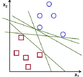
Support Vector Machine
[image:3.595.56.273.525.700.2]Support Vector Machine (SVM) enables an efficient way of extracting the features and a set of rules to perform classification. SVM is a discriminative classification approach represented by a separate hyperplane. The goal of SVM is to train the classifier utilizing training data and generate an optimal model. The SVM classifier is popularly used in several applications like bioinformatics, teeth segmentation, computer vision fields, etc., due its high performance in accuracy, ability of processing the high dimensional data problems such as, gene expression and modelling of diverse data source. General representation of SVM classification scheme is mentioned in figure 6.
[image:3.595.343.515.529.688.2]Kernel nearest neighbor
Kernel Nearest Neighbor (KNN) is one of the emerging classification method with the advantage of limited usage of resources, while training the large dataset. KNN is a supervised learning classification algorithm and also one of the most widely used non-parametric pattern classification method, which reduce the concern of complexity of probability density. The KNN rule classifies the attributes by allocating the label frequently in -nearest samples. Initially, KNN methodology trains the record and then identifies the new records, which are similar to the trained records. It searches the space for training records that are nearest to the new record as the new record neighbors. In this algorithm, the nearest distance is defined by means of a distance metric named as Euclidean distance.
Random forest
Random forest classifier is one of the emerging classification method with the advantage of limited usage of resources, while training the large dataset. Random forest is a supervised learning classification algorithm and also one of the most widely used non-parametric pattern classification method, which reduces the concern of complexity of probability density. In this classifier, every tree is regarded as an individual classifier and the classification output is voted by all the decision trees. To build a random forest classifier, the growth rules of each tree are summarized as follow.
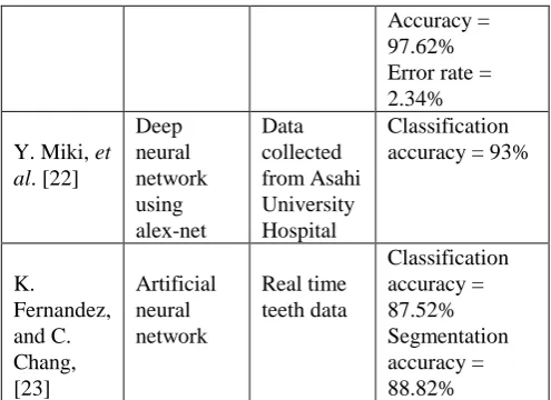
If, is the number of training set, then sample the data’s randomly from the training set. is the dimension of input features, suppose is the specified dimension of the randomly selected sub-features from the original feature vectors. Then feature variables are selected randomly from the features and the best split on these dimensional features are used to split the node. Each tree keeps growing until all these training samples are totally separated without pruning. Some of the recent machine learning methodologies result of teeth segmentation is represented in the table 2.
[image:4.595.309.557.48.228.2]
Table 2. Performance comparison of machine learning methodologies
Authors Models Dataset Performance
measure
Y.E. Mahmoud, et al. [20]
Kernel Nearest Neighbor
Data collected from modern science and arts university
Accuracy = 95.65%
P. Mortaheb, and M. Rezaeian, [21]
Support vector machine
Dental CT images
Sensitivity = 83.24% Specificity = 98.35% Precision = 72.77%
Accuracy = 97.62% Error rate = 2.34%
Y. Miki, et al. [22]
Deep neural network using alex-net
Data collected from Asahi University Hospital
Classification accuracy = 93%
K.
Fernandez, and C. Chang, [23]
Artificial neural network
Real time teeth data
Classification accuracy = 87.52% Segmentation accuracy = 88.82%
In this reviw, the traditional methodologies delivers 81.5%, 82.5% and 80% of classification and segmentation accuracy. From the table 1 and table 2, the machine learning methodologies performs effectively compared to the traditional methods on digital dental database. Also, it shows 4-10% of improvement in the both segmentation and classification accuracy.
IV.Conclusion
This review illustrates the efficiency of machine learning methodologies over traditional approaches for segmenting and classifying the dental parts. In recent decades, machine learning methodologies gained more attention in automation and also achieved good performance in several fields (medical, computer vision, biometrics, etc.) compared to the traditional methodologies. Whereas, the penetration of machine learning methodologies in healthcare, especially in dental medical images are quiet slow compared to the other research areas. This review emphasized the barriers that are reducing the growth of machine learning methodologies in health care and also emphasized the research issues in traditional methodologies.
REFERENCES
1. Z. Xia, Y. Gan, L. Chang, J. Xiong, and Q. Zhao, “Individual tooth segmentation from CT images scanned with contacts of maxillary and mandible teeth,” Computer methods and programs in biomedicine, vol.138, pp.1-12, 2017.
2. T.M. Tuan, “Dental segmentation from X-ray images using semi-supervised fuzzy clustering with spatial constraints,”
Engineering Applications of Artificial Intelligence, vol.59, pp.186-195, 2017.
3. P.L. Lin, P.Y. Huang, P.W. Huang, H.C. Hsu, and C.C. Chen, “Teeth segmentation of dental periapical radiographs based on local singularity analysis,” Computer methods and programs in biomedicine, vol.113, no.2, pp.433-445, 2014.
6. E.H. Said, D.E.M. Nassar, G. Fahmy, and H.H. Ammar, “Teeth segmentation in digitized dental X-ray films using mathematical morphology,” IEEE transactions on information forensics and security, vol.1, no.2, pp.178-189, 2006.
7. E.I. Sela, S. Hartati, A. Harjoko, R. Wardoyo, and M.S. Munakhir, “Segmentation on the dental periapical x-ray images for osteoporosis screening,” International Journal of Advanced Computer Science and Applications, vol.4, no.7, pp.147-51, 2013. 8. I.B. Pavaloiu, N. Goga, A. Vasilateanu, I. Marin, A. Ungar, I. Patrascu, and C. Ilie, “Neural network based edge detection for CBCT segmentation,” In E-Health and Bioengineering Conference (EHB), IEEE, pp. 1-4, 2015.
9. S.V. Tikhe, A.M. Naik, S.D. Bhide, T. Saravanan and K.P. Kaliyamurthie, “Multipoint search algorithm for automatic segmentation of tooth from digital intra oral periapical radiographs,” Indian Journal of Science and Technology, vol.9, no.21, 2016.
10. P.L. Lin, P.Y. Huang, and P.W. Huang, “An effective teeth segmentation method for dental periapical radiographs based on local singularity,” In System Science and Engineering (ICSSE), International Conference onIEEE, pp. 407-411, 2013.
11. H.C. Kang, C. Choi, J. Shin, J. Lee, and Y.G. Shin, “Fast and accurate semiautomatic segmentation of individual teeth from dental CT images,” Computational and mathematical methods in medicine, 2015.
12. Z. Li, and H. Wang, “Interactive Tooth Separation from Dental Model Using Segmentation Field,” PloS one, vol.11, no.8, pp.e0161159, 2016.
13. T. Kronfeld, D. Brunner, and G. Brunnett, “Snake-based segmentation of teeth from virtual dental casts,” Computer-Aided Design and Applications, vol.7, no.2, pp.221-233, 2010. 14. Y.Y. Amer, and M.J. Aqel, “An Efficient Segmentation Algorithm
for Panoramic Dental Images,” Procedia Computer Science, vol.65, pp.718-725, 2015.
15. K. Wu, L. Chen, J. Li, and Y. Zhou, “Tooth segmentation on dental meshes using morphologic skeleton,” Computers & Graphics, vol.38, pp.199-211, 2014.
16. B.H. Le, Z. Deng, J. Xia, Y.B. Chang, and X. Zhou, “An interactive geometric technique for upper and lower teeth segmentation,” In
International Conference on Medical Image Computing and Computer-Assisted Intervention, Springer Berlin Heidelberg, pp. 968-975, 2009.
17. L. Wang, S. Li, R. Chen, S.Y. Liu, and J.C. Chen, “A segmentation and classification scheme for single tooth in Micro-CT images based on 3D level set and k-means++,” Computerized Medical Imaging and Graphics, vol.57, pp.19-28, 2017 O. Nomir, and M. -Mottaleb, “A system for human identification from X-ray dental radiographs,” Pattern Recognition, vol.38, no.8, pp.1295-1305, 2005.
18. O. Nomir, and M. Abdel-Mottaleb, “A system for human identification from X-ray dental radiographs,” Pattern Recognition, vol.38, no.8, pp.1295-1305, 2005.
19. M.K. Alsmadi, “A hybrid Fuzzy C-Means and Neutrosophic for jaw lesions segmentation,” Ain Shams Engineering Journal. 2016.
20. Y.E. Mahmoud, S.S. Labib, and H.M. Mokhtar, “Teeth periapical lesion prediction using machine learning techniques,” In SAI Computing Conference (SAI), IEEE, pp. 129-134,2016.
21. P. Mortaheb, and M. Rezaeian, “Metal artifact reduction and segmentation of dental computerized tomography images Using Least square support vector machine and mean shift algorithm,”
Journal of medical signals and sensors, vol.6, no.1, pp.1, 2016.
22. Y. Miki, C. Muramatsu, T. Hayashi, X. Zhou, T. Hara, A. Katsumata, and H. Fujita, “Classification of teeth in cone-beam CT using deep convolutional neural network,” Computers in biology and medicine, vol.80, pp.24-29, 2017.
23. K. Fernandez, and C. Chang, “Teeth/Palate and Interdental Segmentation Using Artificial Neural Networks,” In ANNPR, pp. 175-185, 2012.
AUTHORS PROFILE
Experienced Assistant Professor with a demonstrated history of working in the Technical education industry. Skilled in Research, E-Learning, Lecturing, Teaching, and Higher Education. Strong education professional with a Doctor of Philosophy (Ph.D.) focused in Natural Language Processing from Manipal University Jaipur. Working as Assistant Professor at the School of Computing & Information Technology Manipal University Jaipur.
Experienced Assistant Professor with a demonstrated history of working in the Technical education industry. Skilled in Research, E-Learning, Lecturing, Teaching, and Higher Education. Strong education professional with a M.Tech from NSIT, Delhi University, pursuing Doctor of Philosophy (Ph.D.) focused in Natural Language Processing from MNIT Jaipur. Working as Assistant Professor at the School of Computing & Information Technology Manipal University Jaipur.