International Journal of Innovative Technology and Exploring Engineering (IJITEE) ISSN: 2278-3075, Volume-8 Issue-11, September 2019
Abstract: Medical image processing is a challenging research field, since most captured images suffer from noise and poor contrast nature. The accuracy of details present in the medical image depends entirely on the captured image quality. The factor that affects the quality of the images includes poor illumination conditions, capturing devices and inexperienced technicians that may result in low contrast images. Hence, contrast enhancement techniques are necessary to improve the quality of OCT images for further processing. In this paper, the enhancement of OCT images is carried out using various enhancement techniques to identify the method that offers improvement in the enhancement quality of the image. It presents a comparative evaluation of enhancement techniques based on the performance indices calculated from the experimental results. The results of this research work suggest the better enhancement technique suitable for OCT images depending on the various performance metrics used prominently in medical imaging.
Keywords : OCT images, Image Enhancement Metrics, Image Quality Assessment
I. INTRODUCTION
I
mage enhancement techniques often play a major role to improve the visual contrast nature [1] of an OCT image and that makes the extraction or segmentation of the images in an easier way. The image enhancement technique offers its interest in wide range of applications like medical image processing, video processing, seismic exploration, video surveillance etc. The main aim of this method is to change the image attributes to make it suitable for an observer or a task. In this method, single or multiple image attributes are modified to enhance the contrast nature of an image. Depending on the task, the image attributes and the method by which it is modified is selected [2].In recent times, different image enhancement techniques are used to improve the image visual nature [3]. However, an ordinary enhancement technique does not provide satisfactory performance when it is applied with low contrast images. The unsatisfactory performance is due to the presence of higher and lower histogram components at different ends. Hence, the balancing of contrast level of image using gray level techniques is difficult [4]. It further affects the background of the image and flashes out the entire image. To eliminate all such drawbacks, histogram based equalization [5] and CLAHE [6, 7] is developed.
The OCT images, despite its recent appearance have become a clinical standard to study several diseases [8]. Hence in this work, the OCT images are enhanced using various
Revised Manuscript Received on September 03, 2019
Saya Nandini Devi M*, Research Scholar, Department of Electronics and Instrumentation Engineering, Annamalai University, Chidambaram-608002, India, mail-id: [email protected]
Santhi S, Department of Electronics and Instrumentation Engineering, Annamalai University, Chidambaram-608002, India. mail-id: [email protected]
enhancement techniques like HE, AHE, CLAHE and MOT to determine the one that offers best result. Further, the enhancement techniques are evaluated using different performance indices AMBE, EME, EMEE, CII and TM. Various techniques are adopted to improve the contrast nature of OCT image using image enhancement techniques. In [9], the author has adopted a self-adaptive image enhancement algorithm using histogram equalization and canny operator. This method avoids the shortcomings occurred due to HE through the canny operator, which helps in the extraction of detailed information and further it preserves the histogram in enhanced image.
In [10], the edges in near-infrared superficial vein images are blurred and further the vascular lines are not clear. Further, the low processing efficiency and high algorithm complexity is resolved using CLAHE with optimized bilinear interpolation that helps in the addition of parameter T over an interpolation function. Where the parameter ‘T’ helps in increasing the speed of interpolation that makes the entire process to work faster with improved image enhancement.
In [11] contrast enhancement algorithm to enhance the fundus image for clear visual perception is presented. This method uses CLAHE technique to resolve the de-noising and enhancement problems associated with color fundus image. In [12] CLAHE was used for angiogenesis OCT image enhancement. From these studies it is clear that the application of Histogram based techniques helps in improving the contrast nature of images than other techniques.
The outline of the paper is given as follows: Section 2 discusses the proposed method with different enhancement techniques. Section 3 discusses the performance evaluation metrics to determine the suitable enhancement method for OCT image enhancement. Section 4 discusses the results obtained. Section 5 concludes the paper.
II. PROPOSEDMETHOD
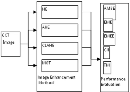
The architecture of the proposed method is given in Figure 1, where two modules are adopted to improve and test the quality of OCT image. The proposed enhancement method consists of two different modules: the former is image enhancement module and the latter is the performance indices evaluation module.
Improved Oct Image Enhancement using Clahe
Fig. 1.Process using the Proposed Preprocessing Frame work
A. Histogram Equalization (HE)
Histogram Equalization [13] is a technique inspatial domain, which is easy to implement. The HE modifies the image intensity that helps in improving the contrast level of an image. It is also defined as a transformation technique that improves the image contrast nature by proper redistribution of image gray level in a uniform way. The HE is expressed as in the following manner,
P(rk)=nk/n
where nk= 0,1,2,…,L-1 and rk are considered as the kth gray level and n is defined as the total number of pixels available in an image with gray level rk.
B. Adaptive Histogram Equalization (AHE)
AHE [13] technique transforms each pixel into a picture with a transformative function produced from the neighborhood area. It enhances an image's local contrast. The main purpose of this method is to improve both natural and medical images. But noise in homogeneous regions can be amplified. During image digitalization, this vagueness is shown by uncertain boundaries and colored values.
C. Contrast Limited Adaptive Histogram Equalization (CLAHE)
Originally, CLAHE [14] is used to improve the low-contrast medical images. The method divides the images into the associated areas and finds equalization for each area. This flatters the division of gray levels, thus increasing the visibility of the hidden features of the image. This paper equalizes the images individually in RGB color spaces. The standard CLAHE with transformed gray levels is expressed using following mathematical expression and this is expressed in terms of uniform distribution as below:
g
g
max
g
min
*
P f
g
min Where gmax is regarded as maximum pixel value, gmin is regarded as the minimum pixel value, P(f) is regarded as the cumulative probability distribution and g is regarded as the computed pixel value.
D. Maximization of Threshold (MOT)
Maximization of threshold method sets to increase the threshold range of lower pixels and maintains the threshold
value of higher threshold pixels. When the addition of two entropies attained its maximum by assuming the foreground and background as two different sources, the image is said to be optimally thresholded. The aspects define the entropic correlation and achieve the threshold that maximizes it. The maximization of the threshold was calculated using the two entropies which maximize the image and threshold optimally. The enhancement of OCT images used various parameters and correlation techniques to achieve the threshold which maximizes it. The algorithm is presented below,
Input: Filtered Result Output: Enhanced Image Procedure:
(3)
III. PERFORMANCEANALYSIS
In the proposed method, the image enhancement techniques are evaluated using the following performance indices, which are given below. The quantitative performance measures used are absolute mean brightness error, measure of enhancement by entropy, measure of enhancement, contrast improvement index and Tenengrad measurement. These measures are used to measure the performance of all the image enhancement techniques and the results obtained through these measures are used for the purpose of evaluation. These measures have its own properties that have the ability to measure the details of the OCT images and help to increase the image brightness and contrast nature.
A. ABSOLUTE MEAN BRIGHTNESS ERROR
(AMBE)
AMBE determines the brightness preservation [15], which is based on different brightness level between the original and enhanced image. Lower the value of AMBE implies improved preservation of image brightness. It is given by the following expression,
AMBE = |E(X) – E(Y)| (4) Where,
E(X) is referred as the input image average intensity and
E(Y) is referred as the enhanced image average intensity. B. Measure of Enhancement (EME)
EME [16] is defined as a measure of image enhancement method to test between the original and enhanced image. Consider an image x(n,m) that is said to split into two different blocks, namely k1 and k2. Here, the window wk,l(i, j) is maintained at the sizel1×l2, and {Φ}is defined as the
orthogonal transform class for enhancing the image with parameters: α, β, and λ. Using all these parameters, the EME is defined as follows:
2 1
max; ,
1 1
1 2 min; ,
1
max
20log
w
k k
k l w
l k k l
I
EME
k k
I
Where
I
max; ,w k lis the maximum value of image X(n,m) withinwk,l block and min; ,
w k l
I
is theminimum value of image
International Journal of Innovative Technology and Exploring Engineering (IJITEE) ISSN: 2278-3075, Volume-8 Issue-11, September 2019
regarded as sign function, where χ(x)=x, or −x, which depends on enhancement method.
C. Measure of Enhancement by Entropy (EMEE) EMEE [16] is defined as the image enhancement measure that uses the concept of entropy.Consider an image x(n,m) that is said to split into two different blocks, namely k1 and k2.
Here, the window wk,l(i, j) is maintained at the size l1×l2 and
hence the EMEE is defined as follows:
2 1max; , max; ,
1 1
1 2 min; , min; ,
1
log
w w
k k
k l k l
w w
l k k l k l
I
I
EME
k k
I
I
D. Contrast Improvement Index (CII)
CII [13] is considered as an important benchmark that helps in comparing the image enhancement technique performance. It is measured as a ratio of local contrast of enhanced image and input image, which is given by the following expression,
CII= Aenhanced/Aoriginal (7) where,
Aenhancedis expressed as the average local contrast level of an enhanced image with 3 × 3 window size and
Aoriginalis expressed as the average local contrast level of an input image with 3 × 3 window size
The increase in the value of CII shows improvement over image contrast nature.
E. Tenegrad Measurement (TM)
TM measure [13] is defined as the maximization of gradient measure. Consider an OCT image J, where the Tenengrad value is estimated using the gradient value ΔJ(x,y) and sobel filter derivative is used to estimate the partial derivatives with convolution kernels jx and jy. Therefore the gradient magnitude of TM is expressed as:
T x y
,
j
x
J x y
,
2
j
y
J x y
,
2 and hence the Tenengrad measure is estimated using the following expression:
,
2x y
TGD
T x y
Higher the value of TM, higher is the image quality and vice versa, which signifies that the image structural information is preserved.
IV. RESULTSANDDISCUSSION
The proposed method is implemented in MATLAB (R2015a) that runs on Intel (R) core (TM)-i5 2600, 3.4 GHz CPU. The implementation of OCT image enhancement techniques is carried out in MATLAB, where image processing toolbox is used to code the image enhancement techniques and quantitative performance measurement were calculated to test the given OCT input image contrast quality. To implement various image enhancement techniques, the proposed method uses OCT images for testing purpose. The image for input is shown in Figure 1 and the obtained results after the application of image enhancement techniquesare displayed from Figure 2 – Figure 5. The various image
enhancement techniques discussed above are used for testing the OCT image contrast quality.
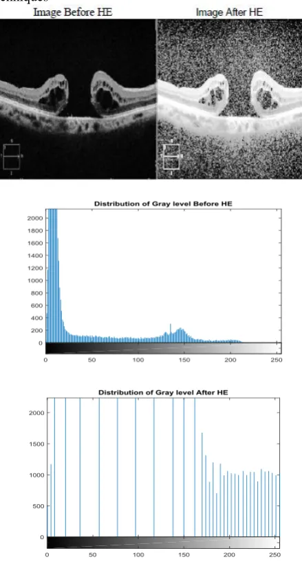
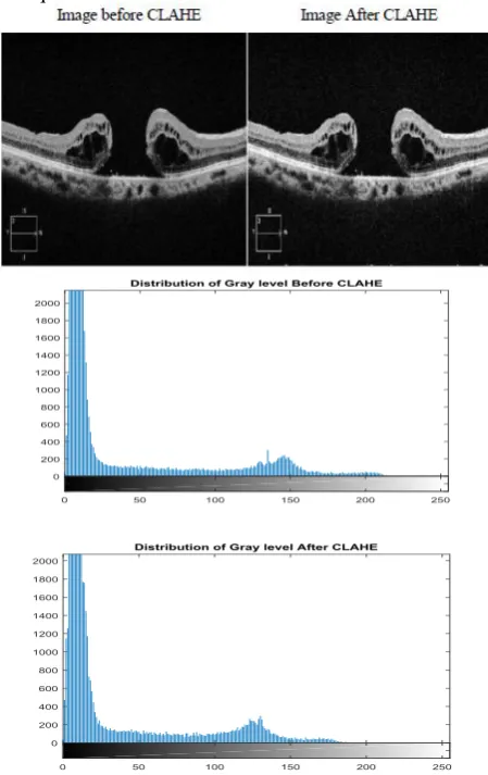
[image:3.595.312.532.212.624.2]After the application of image enhancement techniques, the proposed method obtains following results that are depicted in Figure 2 – Figure 5. Figure 2 shows the results of Histogram Equalization. The Figure 3 shows the results of Adaptive Histogram Equalization, Figure 4 shows the results of CLAHE and Figure 5 shows the results of Maximization of Threshold method. These figures show both input image and enhanced image, and input histogram and enhanced histogram. It is evident from these figures that the CLAHE provides highly enhanced results than the other techniques. Further, the histogram of CLAHE is improved than the other techniques
Fig. 2.Result of Histogram Equalization
[image:4.595.322.525.52.187.2]
Fig. 3.Result of Adaptive Histogram Equalization The histogram image of the AHE method shows a slight improvement in gray level distribution than the one present in Figure 2. This draws a conclusion that that the image obtained using AHE can provide the better enhancement than HE.
Fig. 4.Result of Maximization of Threshold
[image:4.595.313.538.279.636.2]The histogram image of the MOTmethod shows a very lesser improvement in gray level distributionthan the one present in Figure 2 and 3. This concludes that the images obtained by MOT provide the low converged output than the other techniques.
Fig. 5.Result of Contrast Limited Adaptive Histogram Equalization
[image:4.595.48.278.518.773.2]International Journal of Innovative Technology and Exploring Engineering (IJITEE) ISSN: 2278-3075, Volume-8 Issue-11, September 2019
Table- I: Performance Analysis
Enhancement
Techniques AMBE EME EMEE CII TM
HE 94.0009 0.5439 9.6565e-04 0.0035 7.2109e+04
AHE 23.0328 0.4329 9.6565e-04 0.0047 2.2243e+04
CLAHE 4.5351 0.1373 9.6565e-04 0.0063 1.0825e+04
MOT 9.7466 0.1922 8.1538e-04 8.8593e-04 3.2126e+04 The result showing comparison of various enhancement techniques in terms of different performance metrics is given in Table 1.It is observed that the CLAHE method attains better enhanced output of OCT image than the other techniques such as HE, AHE and MOT. The CLAHE attains reduced AMBE, EME, TM and increased CII than other techniques over OCT images.
V. CONCLUSION
In this paper, the objective is to determine a better image enhancement technique to improve the contrast nature of an OCT image. The various enhancement techniques implemented are HE, AHE, CLAHE and MoT. Further presents a comparative evaluation on enhancement techniques based on the performance indices like AMBE, EME, EMEE, CII and TM. The result shows that the CLAHE method achieves improved performance in terms of reduced AMBE (4.5351), reduced EME (0.1373), increased CII (0.0063) and reduced TM (1.0825e+04). The result shows that the CLAHE method performs better on OCT images than the other techniques, which proves the efficacy of CLAHE than other histogram equalization techniques. Further the claim that the application of Histogram based techniques helps in improving the contrast nature of OCT than other techniques like thresholding is proved by the present study.
ACKNOWLEDGMENT
We would like to thank Dr.M.Ravishankar, Director of the Nethralayam , Senior Consultant of the Rajan Eye Care Hospital, Honorary Doctor of the Lions Club International, Founder of the Uyiralayam and Advisor of the Self Help Women’s Association, Chennai for providing the Optical Coherence tomography Images.
REFERENCES
1. Negi, Shailendra Singh, and Yatendra Singh Bhandari. "A hybrid approach to image enhancement using contrast stretching on image sharpening and the analysis of various cases arising using histogram." International Conference on Recent Advances and Innovations in Engineering (ICRAIE-2014). IEEE, 2014.
2. Maini, Raman, and Himanshu Aggarwal. "A comprehensive review of image enhancement techniques." arXiv preprint arXiv:1003.4053 (2010).
3. Chen, ZhiYu, et al. "Gray-level grouping (GLG): an automatic method for optimized image contrast Enhancement-part I: the basic method." IEEE transactions on image processing 15.8 (2006): 2290-2302.
4. Bhattacharya, Saumik, Sumana Gupta, and Venkatesh K. Subramanian. "Localized image enhancement." 2014 Twentieth National Conference on Communications (NCC). IEEE, 2014. 5. Singh, Kuldeep, et al. "Contrast enhancement via texture region based
histogram equalization." Journal of modern optics 63.15 (2016): 1444-1450.
6. Makandar, Aziz, and Bhagirathi Halalli. "Breast cancer image enhancement using median filter and CLAHE." International Journal of Scientific & Engineering Research 6.4 (2015): 462-465.
7. Kharel, Nabin, et al. "Early diagnosis of breast cancer using contrast limited adaptive histogram equalization (CLAHE) and Morphology methods." 2017 8th International Conference on Information and Communication Systems (ICICS). IEEE, 2017.
8. Miri, Mohammad Saleh, et al. "Multimodal registration of SD-OCT volumes and fundus photographs using histograms of oriented gradients." Biomedical optics express 7.12 (2016): 5252-5267. 9. Du, Ya-ni, et al. "Self-adaptive histogram equalization image
enhancement based on canny operator." AOPC 2017: Optical Sensing and Imaging Technology and Applications. Vol. 10462. International Society for Optics and Photonics, 2017.
10. Miao, Yu, et al. "Application of the CLAHE Algorithm Based on Optimized Bilinear Interpolation in Near Infrared Vein Image Enhancement." Proceedings of the 2nd International Conference on Computer Science and Application Engineering. ACM, 2018. 11. Sahu, Sima, et al. "An approach for de-noising and contrast
enhancement of retinal fundus image using CLAHE." Optics & Laser Technology 110 (2019): 87-98.
12. Li, Ang, et al. "Automated segmentation and quantification of OCT angiography for tracking angiogenesis progression." Biomedical optics express 8.12 (2017): 5604-5616.
13. Puniani, Shruti, and Sankalap Arora. "Performance evaluation of image enhancement techniques." International Journal of Signal Processing, Image Processing and Pattern Recognition8.8 (2015): 251-262.
14. Singh, B., and Shailendra Patel. "Efficient medical image enhancement using CLAHE enhancement and wavelet fusion." International Journal of Computer Applications 167.5 (2017): 0975-8887. 15. Chen, Soong-Der, and Abd Rahman Ramli. "Minimum mean
brightness error bi-histogram equalization in contrast enhancement." IEEE transactions on Consumer Electronics49.4 (2003): 1310-1319.