Copyright © 2000, American Society for Microbiology. All Rights Reserved.
Functional Analysis of the
env
Open Reading Frame
in Human Endogenous Retrovirus IDDMK
1,2
22
Encoding Superantigen Activity
MATTHIAS LAPATSCHEK,1SUSANNE DU¨ RR,1ROSWITHA LO¨ WER,2CHRISTINE MAGIN,2
HERMANN WAGNER,1ANDTHOMAS MIETHKE1*
Institute of Medical Microbiology, Immunology and Hygiene, Technical University of Munich, 81675 Munich,1and
Paul Ehrlich Institute, 63225 Langen,2Germany
Received 5 January 2000/Accepted 26 April 2000
Mice harbor a family of endogenous retroviruses, the mouse mammary tumor viruses (MMTV), which
encode superantigens. These superantigens are responsible for the deletion of T cells expressing certain V
chains of the T-cell receptor in the thymus. Human T cells are able to recognize MMTV-encoded superantigens presented by human major histocompatibility complex class II-positive cells. Owing to this and to the similarity of the human and murine immune systems, it was speculated that human endogenous retroviruses
might also code for superantigens. Recently, it was reported that a proviral clone (IDDMK1,222) of the human
endogenous retrovirus family HTDV/HERV-K encodes a superantigen. The putative superantigen gene was
located within theenvregion of the virus. Stimulated by these findings, we amplified by PCR and cloned into
eucaryotic expression vectors open reading frames (ORFs) which were identical or very similar to IDDMK1,222.
When we transfected these vectors into A20 cells, a murine B-cell lymphoma, we were able to demonstrate mRNA expression and protein production. However, we did not find any evidence that the ORF stimulated
human or murine T cells in a V-specific fashion, the most prominent feature of superantigens.
Superantigens (Sags) are characterized as stimulating a large fraction of peripheral T cells expressing a certain Vchain of the T-cell receptor (TCR) (21). If they are expressed within the thymus, they induce a V-specific deletion of thymocytes (3). To activate T cells or delete thymocytes, Sags have to be presented by major histocompatibility complex class II (MHC-II)-positive cells (11, 12). They are produced by gram-positive bacteria, in particularStaphylococcus aureusandStreptococcus pyogenes(21), as well as by a family of murine endogenous retroviruses, the mouse mammary tumor viruses (MMTV) (1, 2). Until now no other retrovirus was shown convincingly to encode a Sag. Since human T cells are stimulated in a V -specific pattern by MMTV-encoded Sags presented by human MHC-II-positive cells (17), and since the human and murine immune systems are very similar, it was speculated that human endogenous Sags might also exist. By analogy to the murine situation, it was envisaged that these endogenous human Sags are encoded by endogenous retroviral elements which reside in the human genome. It was suggested repeatedly that the pos-tulated endogenous human Sags might be responsible for the initiation of human autoimmune diseases like systemic lupus erythematosus, multiple sclerosis, or type I diabetes mellitus. Indeed, in the murine model of experimental autoimmune encephalitis, relapsing paralysis can be triggered by bacterial Sags (7), and, in humans, expression of an endogenous retro-virus family (MSRV) was shown to be associated with the occurrence of multiple sclerosis (23). Furthermore, Conrad et al. showed that expression of a proviral sequence designated IDDMK1,222 of the human endogenous retrovirus family
HTDV/HERV-K was associated with the onset of type I dia-betes mellitus (10). This association, however, could not be
confirmed in other studies (18, 20, 22). Conrad et al. also demonstrated that IDDMK1,222 encodes a Sag function and
suggested that the Sag might be responsible for the initiation of type I diabetes mellitus, since the Sag activated V7⫹T cells in
a test system (10). V7 T-cell expansion was also detected in patients with type I diabetes mellitus (9).
Intrigued by the hypothesis that a human endogenous Sag might exist, we cloned the retroviral open reading frame (ORF) described by Conrad et al. and tested the activity of its product as a Sag. Here we report experiments to test whether a V-specific subset of human T cells was activated by the ORF product presented by murine A20 cells. To circumvent the problem of allogenic stimulation, which is unavoidable in a human-based test system, we had previously developed an as-say system consisting of syngeneic murine cells (30), including the B-cell lymphoma line A20 as an antigen-presenting cell (APC) and T cells. In this study we also took advantage of this test system to elucidate whether the endogenous retroviral ORF product represents a Sag, i.e., stimulates T cells in a V-specific fashion.
MATERIALS AND METHODS
Reagents and antibodies.Anti-Flag monoclonal antibody M2 (MAb) (catalog no. F3165) came from Sigma (Deisenhofen, Germany). The fluorescein isothio-cyanate (FITC)-labeled anti-human V7 MAb (clone ZOE) was purchased from Coulter (Hialeah, Fla.). The biotinylated anti-human CD3 MAb (clone UCHT1), the biotinylated anti-murine V8.1 and -8.2 MAb (clone MR5-2), the phycoerythrin (PE)-labeled anti-murine V10 MAb (clone B21.5), the biotinyl-ated anti-murine V14 MAb (clone 14-2), and the FITC-labeled anti-murine CD3εMAb (clone 145-2C11) were bought from Pharmingen (San Diego, Calif.). The hybridoma KT4, which produces an anti-murine V4 MAb, was kindly donated by H. Hengartner (Zu¨rich, Switzerland). The antibody was purified and biotinylated according to standard protocols. PE-conjugated streptavidin was purchased from Jackson ImmunoResearch (West Grove, Pa.), and staphylococ-cal enterotoxin B (SEB) (BT202) was from Toxin Technology (Sarasota, Fla.). Cell culture.The murine B-cell lymphoma A20 (16) cell line was cultured in Clicks RPMI medium (Seromed Biochrom, Berlin, Germany) with 10% fetal calf serum (FCS; BioWhittaker, Verviers, Belgium), 2 mM glutamine (Seromed Biochrom), 100 U of penicillin and 100g of streptomycin (Seromed Biochrom) * Corresponding author. Mailing address: Institute of Medical
Mi-crobiology, Immunology and Hygiene, Technical University of Mu-nich, Trogerstr. 9, 81675 MuMu-nich, Germany. Phone: 49/89/4140-4187. Fax: 49/89/4140-4868. E-mail: [email protected].
6386
on November 9, 2019 by guest
http://jvi.asm.org/
per ml, 50M 2-mercaptoethanol (GIBCO BRL, Paisley, Scotland), and 1g of indomethacine (Sigma) per ml.
Human T cells were isolated from a healthy blood donor’s buffy coat by Ficoll (density, 1.077; Seromed Biochrom) separation followed by negative selection via a magnetic cell sorting column with the Pan T-cell isolation kit (Miltenyi Biotec, Auburn, Calif.). The purity of the T-cell fraction was over 90%, as assessed by CD3-CD14 double staining and flow cytometry.
Murine lymphocytes were freshly prepared from the mesenteric lymph nodes of BALB/c mice (Harlan, Borchen, Germany) by teasing through a mesh metal sieve. Over 85% of the viable cells were CD3⫹T cells.
Plasmids and primers.pEGFP-N1 was kindly donated by H. Ha¨cker (Munich, Germany), pMMTV2Sag was a gift from W. Gu¨nzburg (Vienna, Austria), pCIneo-DMSag was generously provided by B. Conrad (Geneva, Switzerland), and pRK5 came from Pharmingen.
The putative Sag gene in the plasmid pHKputSag1 was amplified from the genome of a patient with type I diabetes mellitus using the primers HERV-1 (ACCCATCAGAGATGCAAAGAAAAGC) and R300 (CTTTACAAAGCAG TATTGCTGC), resulting in a PCR product of approximately 2.2 kb. To obtain the coding region of the putative Sag gene, a second PCR was performed using the primers 5⬘ Sag (ATCCGCGGATCCCACCATGGTAACACCAGTCACA TG) and 3⬘Sag (CTCGAGCTAATCTATAATAGTTCCGAA). The PCR prod-uct was cloned into the mammalian expression vector pRK5. The resulting plasmid, pHKputSag1, contained only the endogenous retrovirus Sag ORF with a Kozak sequence in the vector pRK5. The plasmid pHKputSag1-FLAG con-tains the Flag sequence but is otherwise identical to pHKputSag1.
Plasmid pHKputSag2 contained the same sequence of the endogenous retro-virus Sag ORF as pHKputSag1 but was cloned into theHindIII andXbaI sites of the vector pRC/CMV (Invitrogen, Carlsbad, Calif.) after amplification with prim-ers IDMM3 (TCCAAGCTTATGGTAACACCAGTCACATGG) and IDDM4 (ACGTCTAGACTAATCTATAATAGTTCCGAATTC). pHKputSag3 also con-tained the same endogenous Sag ORF but had a slightly different sequence (see Fig. 1). This plasmid was constructed as detailed in reference 10, relying on the same primers (IDDM1, GACTAAGCTTAAGAACCCATCAGAGATGC, and IDDM2, AGACTGGATCCGTTAAGTCGCTATCGACAGC) and sub-cloning into pCIneo (Promega, Madison, Wis.). Primers were purchased from TIB Molbiol (Berlin, Germany) and ARK Scientific Biosystems (Darmstadt, Germany).
Transfection.Twenty-four hours after passaging, 107A20 cells in 400l of
RPMI medium–10% FCS with 20g of plasmid DNA were electroporated in a 0.4-cm cuvette at 960F and 280 V using a gene pulser (Bio-Rad, Hercules, Calif.). Transfection efficiency was controlled with the pEGFP-N1 vector by flow cytometry 24 h posttransfection.
Detection of mRNA by reverse transcription and PCR.Transfected cells (5⫻
106A20 cells, 20g of plasmid DNA) were cultured overnight (37°C). After
centrifugation (300⫻gfor 5 min) the pellet was lysed with 600l of RLT (RNA lysis/thiocyanate) buffer (Qiagen, Hilden, Germany). RNA was extracted using the RNeasy kit (no. 74103) from Qiagen. The amount of RNA was quantified photometrically (260 nm). Afterward, any contaminating DNA was digested with DNase (20 U/2g of RNA, 10 min, 37°C; Roche, Mannheim Diagnostics GmbH, Mannheim, Germany), and cDNA was synthesized using 1l of oligo(dT) (10
M; Gibco Life Technology Inc., Rockville, Md.), 1l of hexamer random primer (10M; Gibco), 4l of 5⫻transcription buffer (Gibc), 2l of dithio-threitol (0.1 M; Gibco), 2l deoxynucleoside triphosphate (200M; Roche), 0.5
l of Rnasin (Promega), and 1l of Superscript II Plus (Gibco). The reaction mixture was incubated for 60 min at 37°C and subsequently heated to 95°C for 5 min.
Two microliters of the generated cDNA was used for PCR amplification to detect the transcribed retroviral ORF using the 5⬘primer 5⬘-Sag and the 3⬘
primer 3⬘-Sag. The PCR mixture consisted of 10l of deoxynucleoside triphos-phate (1 mM; Roche), 1.5l of each of the primers 5⬘-Sag and 3⬘-Sag (50M; TIB Molbiol), 29l of H2O, 1l ofPfuTurbo (Invitrogen), and 5l of 10⫻
buffer (Invitrogen). The cycle conditions were 94°C and 30 s for denaturation, 55°C and 60 s for annealing, and 72°C and 120 s for elongation. The PCR was run for 30 cycles. The PCR products were visualized with an ethidium bromide-stained 1% agarose gel.
Western blotting.Transfected cells (5⫻106A20 cells, 20g of plasmid DNA)
were cultured overnight (37°C) and subsequently lysed in NP-40 (1%, vol/vol). Cell lysates were separated by sodium dodecyl sulfate–12.5% polyacrylamide gel electrophoresis using Laemmli sample buffer (15.1 g of Tris per liter, 9.4 g of glycine per liter, 0.5% sodium dodecyl sulfate). After being transferred onto nitrocellulose by semidry electroblotting for 2 h (250 mA), the filters were blocked (5% milk powder in TBST [2.4 g of Tris per liter, 8 g of NaCl per liter, 0.1% Tween, pH 7.6], 1 h at 37°C and 1 h at room temperature) and incubated with the anti-Flag MAb M2 (diluted 1:1,000, overnight, 4°C). After three wash-ings with 1⫻TBST, goat anti-mouse serum coupled with peroxidase was added (diluted 1:5,000, 1 h, room temperature; Dianova, Hamburg, Germany). The blot was washed again three times with 1⫻TBST and visualized by using the en-hanced chemiluminescence reagent (NEN Life Science Products, Boston, Mass.) as described by the manufacturer.
Mixed-lymphocyte culture.Twenty-four hours after transfection, the A20 cells were arrested in growth by a 1-h incubation with 80-ng/ml mitomycin C. After three washings they were subsequently cocultured at a stimulator-responder ratio
of 2:1 or 1:1 with human T cells in RPMI medium containing 5% autologous human serum for a total culture period of 8 days with 10 U of interleukin-2 (IL-2) per ml added after 3 days of culture. The BALB/c lymphocytes were cocultured with the mitomycin-treated A20 cells at a stimulator-responder ratio of 2:1 in RPMI medium–10% FCS with 10 U of IL-2 per ml for 4 to 5 days.
FACS analysis.After the mixed-lymphocyte culture, viable T lymphocytes were retrieved from the coculture by gradient centrifugation with Ficoll at a density of 1.077 for human and 1.090 for murine lymphocytes. The cells were then stained with the anti-V-antibodies (diluted 1:100) mentioned above and counterstained with CD3 (diluted 1:100). In the case of biotinylated anti-bodies, a second staining step with streptavidin-PE (diluted 1:100) was added. The fluorescence-activated cell sorter (FACS) analysis was performed with a Facscalibur cytometer (Becton Dickinson, Franklin Lakes, N.J.).
Proliferation assay.The proliferative response of murine T cells was measured on day 4 or 5 by pulsing microcultures with 37 kBq of [3H]thymidine (NEN,
Zaventem, Belgium) for 8 h. Thereafter, cultures were harvested on fiberglass filters, and the amount of [3H]thymidine incorporation was determined by direct
beta counting (Matrix 96; Packard Instrument Co., Meriden, Conn.).
RESULTS
Cloning and sequencing of the ORF sequence.The putative
HTDV/HERV-K Sag was amplified from the genome of pe-ripheral blood lymphocytes from a patient with type I diabetes mellitus using the primers listed in Materials and Methods. PCR products of the appropriate size were obtained from patients as well as healthy blood donors (data not shown). The PCR products were cloned into the mammalian expression vector pRK5, resulting in the plasmid pHKputSag1; into the vector pRC/CMV, resulting in the plasmid pHKputSag2; and into the vector pCIneo, resulting in the plasmid pHKputSag3. As demonstrated in Fig. 1, the sequences of the ORFs encod-ing the putative Sag are identical or very similar to the one published by Conrad et al. (10), reflecting the multicopy nature of the endogenous retrovirus family HTDV/HERV-K. The plasmids and a plasmid containing the established Sag gene from MMTV2 were used to transiently transfect A20 cells for the functional characterization of the ORF. The different vec-tor backbones were used to exclude potential unspecific acti-vation of transfected cells due to nonphysiological high expres-sion of a foreign protein by potent eucaryotic promoters. Besides the MMTV2-encoded Sag, the bacterial Sag SEB, was included as a positive control.
Transcription and protein expression of the ORF in A20
cells.At first we verified that the plasmids were transcribed in
the transfected cells. A plasmid encoding the enhanced green fluorescent protein (EGFP) was used to measure the transfec-tion efficiency of A20 cells. As shown in Fig. 2, around 50% of all living cells were transfected. Using reverse transcription and PCR, we showed (Fig. 3) that A20 cells electroporated with the different plasmids expressed mRNA encoding the respective ORF products. Since there is no antibody available which would recognize the product of the ORFs, we inserted a Flag sequence at the 5⬘ end of the coding sequence, giving rise to the plasmid pHKputSag1-FLAG (Fig. 1). A protein with the predicted size of 18 kDa was detected by Western blotting using an anti-Flag MAb only in A20 cells transfected with pHKputSag1-FLAG (Fig. 4).
Test for Sag function using human T cells.Since the
postu-lated Sag function was described in humans, we initially stim-ulated human T cells with murine A20 cells transfected with the vectors described above, in a manner similar to a protocol described by B. Conrad (personal communication). Twenty-four hours after transfection, A20 cells were arrested in their growth and used to stimulate purified human T cells (see Materials and Methods). Because Conrad et al. had reported that human V7⫹T cells were induced to proliferate (10), this
human T-cell subpopulation was analyzed by FACS 8 days after stimulation of the culture. However, in comparison to the
VOL. 74, 2000 FUNCTIONAL ANALYSIS OF THE env ORF BORNE BY IDDMK1,222 6387
on November 9, 2019 by guest
http://jvi.asm.org/
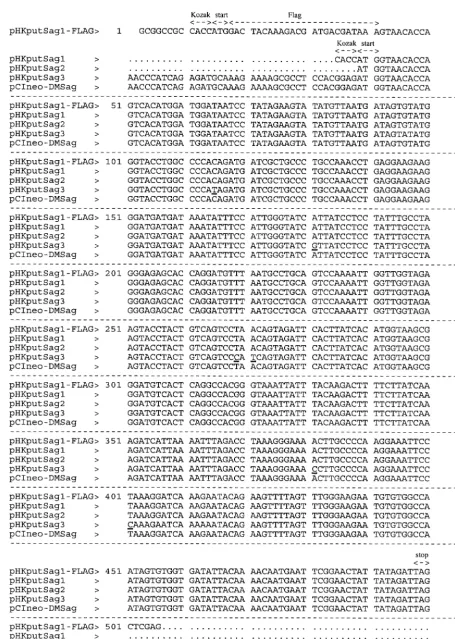
FIG. 1. Sequence comparison of the IDDMK1,222-borne ORF and ORFs cloned from different type I diabetes mellitus patients. Underlined residues indicate point
mutations in pHKputSag3.
on November 9, 2019 by guest
http://jvi.asm.org/
vector controls (pRK5, pRC/CMV, and pCIneo), we were un-able to observe any increase of the V7⫹T-cell population,
although, as depicted in Fig. 5, we tested a variety of different ORF-containing plasmids including the construct
pCIneo-DMSag, which was kindly provided by Conrad and which he had used in his studies.
Test of Sag function using the murine test system.All
es-tablished Sags of bacterial origin have been found to stimulate human as well as murine T cells. As mentioned earlier, the Sags encoded by MMTV stimulate human and murine T cells (17). The Vchains of the TCRs involved in the recognition of the different Sags are conserved between humans and mice (15, 17). At present there is no evidence that a species-specific Sag exists, i.e., a Sag which is active only in one species. We therefore used our well-defined murine test system, which cir-cumvents the problem of alloreactivity encountered in human-based test systems or of xenoreactivity observed in human- and murine-based test systems. In order to analyze whether the ORF product of IDDMK1,222 belongs to the Sag family,
syn-geneic murine peripheral T cells were stimulated with trans-fected A20 cells for 5 days and subsequently analyzed by FACS for a V-specific outgrowth of T cells. As already mentioned, the system was controlled by two established Sags: first, the exogenous bacterial Sag, SEB, which stimulates all murine V8⫹T cells (21), and second and more important, the Sag
ORF product of MMTV2, which stimulates all murine V14⫹
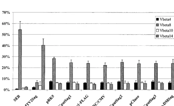
T cells (3, 14). The positive control plasmid bearing the ORF of MMTV2 was used in exactly the same way as all the plas-mids bearing ORFs of human endogenous retroviruses. A typ-ical result of the T-cell stimulation assay is demonstrated in Fig. 6. As was expected, SEB expanded all V8⫹T cells, which
resulted in a relative reduction of the other three tested T-cell subsets, consisting of V4-, -10-, and -14-positive T cells. The MMTV2-encoded Sag stimulated all V14⫹cells; again, the
[image:4.612.57.290.72.255.2]relative numbers of the other T-cell subsets were reduced. In contrast, none of the constructs encoding the putative endog-enous human Sag changed the Vrepertoire of peripheral T cells (Fig. 4), as determined by comparison with peripheral T cells which were stimulated by A20 cells transfected with the noncoding parental plasmids pRK5, pRC/CMV, and pCIneo.
[image:4.612.314.542.73.294.2]FIG. 2. Transfection efficiency of A20 cells. A20 cells were transfected with the EGFP-encoding plasmid pEGFP-N1. After 24 h the cells were analyzed by FACS for the expression of EGFP. Shadowed area, cells transfected with an empty vector; solid line, EGFP-transfected cells.
FIG. 3. Transcription of pHKputSag1, pHKputSag1-FLAG, and pCIneo-DMSag in A20 cells. A20 cells were transfected with pHKputSag1, pHKputSag1-FLAG, and pCIneo-DMSag (20g of DNA/20⫻106cells). After 24 h RNA was
prepared. After reverse transcription (⫹RT) and treatment with DNase, ORF cDNA was amplified by PCR. All three plasmids are transcribed (top panel). The omission of reverse transcription (⫺RT) prevents detection by PCR (middle panel), indicating that the PCR products were not derived from the transfected plasmid. The glyceraldehyde-3-phosphate-dehydrogenase gene (GAPDH) was also amplified to show that approximately equal amounts of RNA were used for reverse transcription and PCR amplification (bottom panel). The experiment was repeated twice with identical results.
FIG. 4. Translation of the Flag ORF in A20 cells. A20 cells were transfected with the indicated plasmids. After 24 h, cells were lysed with NP-40 and the cell lysate was analyzed by Western blotting using MAb M2, which is specific for the Flag epitope. A predicted 18-kDa band is visible in the case of the flagged ORF (pHKputSag1-FLAG). pCIneo-DMSag and pHKputSag1 served as negative controls. The experiment was repeated with identical results.
VOL. 74, 2000 FUNCTIONAL ANALYSIS OF THE env ORF BORNE BY IDDMK1,222 6389
on November 9, 2019 by guest
http://jvi.asm.org/
[image:4.612.58.291.382.639.2]Notably, the Sag-encoding construct kindly provided by B. Conrad also did not stimulate T cells in a V-specific manner. Since in this approach, we analyzed the expansion of a sub-stantial portion of but not the entire Vrepertoire, we might have missed the responding subset of T cells. Therefore, we
[image:5.612.137.471.73.305.2]also measured the induced proliferation of the peripheral T cells as a parameter for Sag response. As depicted in Fig. 7, only the control Sags, SEB and the ORF product encoded by MMTV2, stimulated T cells to proliferate above background levels (levels reached with the expression vectors lacking a
FIG. 5. Human peripheral blood V7⫹T cells do not expand upon stimulation with different ORF constructs. Purified human peripheral blood T cells (1.25⫻106
or 2.5⫻106cells/well) were stimulated with mitomycin C (80g/ml)-treated A20 cells (2.5⫻106cells/well) transfected with the indicated ORF-bearing constructs.
T cells were cultured for 8 days. IL-2 (10 U/ml) was added for the last 5 days. After double staining with an FITC-labeled anti-V7 MAb and PE-labeled anti-CD3 MAb, the percentage of V7⫹T cells was measured by FACS. pRK5 is the vector backbone for pHKputSag1 and pHKputSag1-FLAG; pRC/CMV is the backbone
for pHKputSag2; and pCIneo is the backbone for pHKputSag3 and pCIneo-DMSag. Stim:Resp, stimulator-responder ratio.
FIG. 6. Murine peripheral T cells are not stimulated V-specifically by different ORF constructs. A20 cells were transfected with the indicated plasmids (20g of DNA/107cells). After 24 h the cells were treated with mitomycin C (80g/ml) and used as APCs at a density of 5⫻106cells/well. Murine peripheral lymph node cells
were added (2.5⫻106cells/well) and after 5 days of culture in the presence of 10 U of IL-2 per ml, the cells were harvested. After double staining with an MAb specific
for the V4, V8, V10, or V14 chains of the TCR and with an MAb specific for CD3εprotein, the individual T-cell subpopulations were quantified by FACS (■, V4⫹;p, V8⫹;䊐, V10⫹;o, V14⫹T cells). Each bar represents the mean of at least five independent experiments; error bars indicate standard errors. pRK5 is
the vector backbone for pHKputSag1 and pHKputSag1-FLAG; pRC/CMV is the backbone for pHKputSag2; and pCIneo is the backbone for pHKputSag3 and pCIneo-DMSag.
on November 9, 2019 by guest
http://jvi.asm.org/
[image:5.612.128.480.449.665.2]coding sequence, pRK5, pRC/CMV, and pCIneo). None of the plasmids encoding the putative endogenous retrovirus Sag, including the plasmid supplied by Conrad, induced prolifera-tion of the T cells. In addiprolifera-tion, a more extensive analysis of the Vrepertoire, including the V4, -6, -7, -8, -9, -10, -13, and -14 T-cell subsets, also failed to show a V-specific expansion of T cells (data not shown).
MHC-II expression is not changed by the ORF.In order to
activate T cells, bacterial as well as retroviral Sags need to be presented by MHC-II-expressing APCs (1, 21). To exclude the possibility that, during transfection or due to the expression of the ORF itself, MHC-II molecules are modulated from the surface of A20 cells, the expression of MHC-II molecules 24 h after transfection was analyzed. Figure 8 and Table 1 show that the expression level of MHC-II molecules is not influenced by the ORF.
DISCUSSION
It is well known that many retroviral sequences are inherent to the human genome, including the B/D-type retrovirus-re-lated family HTDV/HERV-K, which consists of approximately 50 proviruses with full-length genomes (28). Although all pro-viruses sequenced so far are defective and noninfectious, ret-roviral proteins like reverse transcriptase and proteins of the envandgagregions are expressed in germ cell tumors (5, 13, 24, 26, 27, 29). Furthermore, the expression of HTDV/ HERV-K proviruses in germ cell tumor cell lines leads to the formation of retrovirus particles (6, 19). The existence of these retroviral products, in particular the possibility that such pro-teins might possess a Sag function, has stimulated the idea that they might be involved in triggering autoimmunity in humans.
A recent paper stated that a novel sequence of the human endogenous retrovirus family HTDV/HERV-K was associated with the onset of type I diabetes mellitus and that this sequence encoded a Sag which activated human V7⫹ T cells (10).
Stimulated by this hypothesis, we tried to repeat these inter-esting findings. Although we and others could not confirm that the development of type 1 diabetes depends on the specific expression of HTDV/HERV-K proviruses (18, 20, 22), in that
FIG. 7. Proliferation of murine T cells stimulated by different ORF-bearing plasmids. The experiment was performed as described for Fig. 6. After a culture period of 4 days, the cells were pulsed for 8 h with [3H]thymidine (37 kBq) and
the amount of incorporation of [3H]thymidine was determined. Each bar
repre-sents the mean of quadruplicates; error bars indicate standard errors. The
ex-periment was repeated twice with comparable results. the ORF. Transfected A20 cells were analyzed for the expression of MHC-IIFIG. 8. Expression of MHC-II molecules on A20 cells is not influenced by molecules 24 h after electroporation. Cells were stained with an FITC-labeled anti-MHC-II antibody and the expression level of MHC-II was recorded with the cytometer. Solid lines, isotype controls; shadowed areas, expression of MHC-II. Four representative histograms are shown; the other transfected cells expressed MHC-II at the same level (Table 1).
TABLE 1. MHC-II expression is not influenced by different ORF constructs
Constructa
MHC-II FITC fluorescence
(mean)
Coefficient of variation (SD/mean)
(%)
None 790.27 65.67
pMMTV2Sag 659.29 68.24
pRK5 640.52 69.67
pHKputSag1 624.55 68.55
pHKputSag1-FLAG 649.85 68.73
pRC/CMV 645.16 67.61
pHKputSag2 624.18 68.08
pCIneo 624.68 68.25
pHKputSag3 628.62 68.74
pCIneo-DMSag 659.11 70.61
aPlasmid used to transfect A20 cells.
VOL. 74, 2000 FUNCTIONAL ANALYSIS OF THE env ORF BORNE BY IDDMK1,222 6391
on November 9, 2019 by guest
http://jvi.asm.org/
study we could not exclude the possibility that the expressed sequences harbor a Sag function (20). In the present study we have cloned into eucaryotic expression vectors sequences iden-tical or very similar to the sequence published by Conrad et al. Together with an IDDMK1,222 expression clone kindly
pro-vided by Conrad, we tested these plasmids in several systems that would reveal a Sag function. None of the test systems indicated the existence of a Sag function in the HERV-K sequences, although the MMTV2-derived sequence exerted a Sag function similar to that of the bacterial Sag.
Several issues should be considered to ensure that the neg-ative results obtained were not caused by the experimental design. First, did we use the wrong species for the evaluation of the ORF as a Sag? Until now, all established Sags have stim-ulated T cells of different species, i.e., Sags are not species specific (17, 21). In order to avoid the stimulation of responder T cells by alloantigen (since the frequency of V segments of the TCR in peripheral blood lymphocytes seems to be influenced by HLA genes) (4), we chose to use a murine test system instead of the human test system used by Conrad et al. The system used here responded rapidly to the MMTV2-encoded Sag as well as to the bacterial Sag SEB (Fig. 6). Although one could expect to observe an expansion of murine T-cell subsets expressing the V4 or V10 chain of the TCR because these chains are closely related to the human V7 chain (8), we were unable to detect such an expansion with the IDDMK1,222
expression plasmid or with any related plasmid. To minimize the chance that the IDDMK1,222-borne ORF is the first
spe-cies-specific Sag, we used a xenogeneic system consisting of human T cells and murine A20 cells as APCs. As shown by Subramanyam et al., Sags can be presented by xenogeneic APCs (25).
In addition, when Conrad sent us the pCIneo-DMSag ex-pression plasmid for testing, he recommended A20 cells as the most convenient APCs and human T cells as the responder cell system, as described in this article. However, we were unable to detect any Sag function of the IDDMK1,222-borne ORF with
any of the systems tested.
Second, the APC used here was the transiently transfected B-cell line A20, whereas Conrad et al. utilized the transiently transfected monocytic cell line THP1 (10). They also used the stably transfected B-lymphoblastoid cell line Raji as the APC. Comparing both APCs revealed that the V7-specific expan-sion of human peripheral T cells induced by the Raji cell line was weaker than that induced by THP1 cells (10). Unfortu-nately, we did not receive the stable transfectants. In addition, we were unable to achieve a satisfactory transfection efficiency with the THP1 cell line, although we tested several reagents recommended by manufacturers (data not shown). The use of the B-cell line A20 as the APC might yield false-negative results if the expression of the transgene failed and if the cell line was not able to present a Sag. However, importantly, A20 cells can present the MMTV2-encoded Sag with the induction of a V14-specific expansion of T cells (Fig. 6). Furthermore, we were able to confirm that the putative Sags were indeed expressed in A20 cells, since mRNAs of the ORFs were tran-scribed (Fig. 3) and protein was produced (Fig. 4).
Third, the amount or the type of MHC-II molecules may be inadequate to present the putative Sag. THP1 cells had to be stimulated with gamma interferon to allow the upregulation of MHC-II molecules and presentation of the putative Sag (10). In contrast, the A20 cell line used in this study as APC did express high levels of MHC-II molecules and this did not change upon the transfection of different ORF constructs (Fig. 8; Table 1). The presentation of Sags is not MHC restricted. However, a hierarchy exists in that human HLA-DR class II
isotypes present Sags much better than HLA-DQ and HLA-DP isotypes (21). The same is true for their murine counterparts, since H2-IE is superior to H2-IA (2, 21). Since the A20 cell line is derived from BALB/c mice, the MHC-II isotypes H2-IE and H2-IA are expressed. Therefore, this cell line should be able to present Sags successfully to T cells, and this is shown here for SEB and the MMTV2-encoded Sag.
Fourth, could we have missed the murine T-cell subset, which was activated by the ORF, since we did not analyze the complete V repertoire? In that case, one should expect a relative reduction in the T-cell subsets tested. As shown in Fig. 6, we observed a relative reduction of the T-cell subsets V4, V10, and V14 in response to the expansion of V8 by SEB and a relative reduction of V4, V8, and V10 in response to the expansion of V14 by the MMTV2-encoded Sag. In addition, we were unable to induce a substantial prolifera-tion of murine T cells upon stimulaprolifera-tion by the putative endogenous retroviral Sag (Fig. 7), which also indicated that no undefined T-cell subset had expanded. In contrast, SEB-and pMMTV2Sag-transfected cells induced a significant proliferation of the murine responder T cells. To rule out that the endogenous retrovirus Sag might be the first spe-cies-specific Sag, we stimulated human T cells and again ob-tained no evidence that these ORF products activated a V -specific T-cell repertoire (Fig. 5). Surprisingly, the plasmid pCIneo-DMSag also showed no evidence of Sag activity.
Fifth, did the Flag in the pHKputSag1-FLAG construct im-pede Sag activity of the IDDMK1,222-borne ORF? We used a
flagged ORF to demonstrate translation of the ORF in A20 cells (Fig. 4). Since we were worried that the Flag influences the function of the protein, we added the Flag to either the 5⬘
or 3⬘end of the ORF. Both constructs as well as the unflagged ORF were negative in the T-cell stimulation assay (Fig. 5 and 6; also data not shown). However, in A20 cells transfected with the construct containing the Flag sequence at the 3⬘end of the ORF, we were unable to detect the formation of mRNA in a nonnested PCR approach, indicating that this construct was less efficiently transcribed than that with the 5⬘Flag (data not shown). One could argue that the 5⬘-flagged protein lost its Sag activity due to the Flag and that the normal ORF gave rise to an unstable message. However, we demonstrated that ORF mRNA levels were similar in A20 cells transfected with pHK-putSag1-FLAG, pHKputSag1, or pCIneo-DMSag (Fig. 3). Since we tested the flagged ORF together with four different unflagged ORF constructs in three different vectors and failed to detect Sag activity, we are convinced that the Flag is not responsible for our failure.
In summary we can only conclude from our data that the ORF located in theenvregion of IDDMK1,222 does not belong
to the family of Sags.
ACKNOWLEDGMENTS
This work was supported by the Bundesministerium fu¨r Bildung und Forschung grant 01 GB9403.
We thank Walter Gu¨nzburg and Hans Ha¨cker for donating MMTV2 Sag- and EGFP-expressing plasmids, respectively. We also thank Ber-nard Conrad for providing the plasmid pCIneo-DMSag.
REFERENCES
1.Acha-Orbea, H.1992. Retroviral superantigens. Chem. Immunol.55:65–86. 2.Acha-Orbea, H., and E. Palmer.1991. Mls—a retrovirus exploits the immune
system. Immunol. Today12:356–361.
3.Acha-Orbea, H., A. N. Shakov, L. Scarpellino, E. Kolb, V. Mu¨ller, A. Vessaz-Shaw, R. Fuchs, K. Blo¨chlinger, P. Rollini, J. Billotte, M. Sarafidou, H. R. MacDonald, and H. Diggelmann.1991. Clonal deletion of V14-bearing T cells in mice transgenic for mammary tumour virus. Nature350:207–211. 4.Akolkar, P. N., B. Gulwani-Akolkar, R. Pergolizzi, R. D. Bigler, and J. Silver.
on November 9, 2019 by guest
http://jvi.asm.org/
1993. Influence of HLA genes on T cell receptor V segment frequencies and expression levels in peripheral blood lymphocytes. J. Immunol.150:2761– 2773.
5.Boller, K., O. Janssen, H. Schuldes, R. R. To¨njes, and R. Kurth.1997. Characterization of the antibody response specific for the human endoge-nous retrovirus HTDV/HERV-K. J. Virol.71:4581–4588.
6.Boller, K., H. Konig, M. Sauter, N. Mueller Lantzsch, R. Lower, J. Lower, and R. Kurth.1993. Evidence that HERV-K is the endogenous retrovirus sequence that codes for the human teratocarcinoma-derived retrovirus HTDV. Virology196:349–353.
7.Brocke, S., A. Gaur, C. Piercy, A. Gautam, K. Gijbels, C. G. Fathman, and L. Steinman.1993. Induction of relapsing paralysis in experimental autoim-mune encephalomyelitis by bacterial superantigen. Nature365:642–644. 8.Clark, S. P., B. Arden, D. Kabelitz, and T. W. Mak.1995. Comparison of
human and mouse T-cell receptor variable gene segment subfamilies. Im-munogenetics42:531–540.
9.Conrad, B., E. Weidmann, G. Trucco, W. A. Rudert, R. Behboo, C. Ricordi, H. Rodriquez-Rilo, D. Finegold, and M. Trucco.1994. Evidence for supe-rantigen involvement in insulin-dependent diabetes mellitus aetiology. Na-ture371:351–355.
10. Conrad, B., R. N. Weissmahr, J. Boni, R. Arcari, J. Schupbach, and B. Mach. 1997. A human endogenous retroviral superantigen as candidate autoim-mune gene in type I diabetes. Cell90:303–313.
11. Dellabona, P., J. Peccoud, J. Kappler, P. Marrack, C. Benoist, and D. Mathis.1990. Superantigens interact with MHC class II molecules outside of the antigen groove. Cell62:1115–1121.
12. Fraser, J. D.1989. High-affinity binding of staphylococcal enterotoxins A and B to HLA-DR. Nature339:221–223.
13. Gotzinger, N., M. Sauter, K. Roemer, and N. Mueller-Lantzsch.1996. Reg-ulation of human endogenous retrovirus-K Gag expression in teratocarci-noma cell lines and human tumours. J. Gen. Virol.77:2983–2990. 14. Gunzburg, W. H., F. Heinemann, S. Wintersperger, T. Miethke, H. Wagner,
V. Erfle, and B. Salmons.1993. Endogenous superantigen expression con-trolled by a novel promoter in the MMTV long terminal repeat. Nature 364:154–158.
15. Herman, A., J. W. Kappler, P. Marrack, and A. M. Pullen.1991. Superan-tigens: mechanism of T-cell stimulation and role in immune responses. Annu. Rev. Immunol.9:745–772.
16. Kim, K. J., C. Kanellopoulos-Langevin, R. M. Merwin, D. H. Sachs, and R. Asofsky.1979. Establishment and characterization of BALB/c lymphoma lines with B cell properties. J. Immunol.122:549–554.
17. Labrecque, N., H. McGrath, M. Subramanyam, B. T. Huber, and R. P. Sekaly.1993. Human T cells respond to mouse mammary tumor virus-encoded superantigen: V beta restriction and conserved evolutionary fea-tures. J. Exp. Med.177:1735–1743.
18. Lan, M. S., A. Mason, R. Coutant, Q. Y. Chen, A. Vargas, J. Rao, R. Gomez,
S. Chalew, R. Garry, and N. K. Maclaren.1998. HERV-K10s and immune-mediated (type 1) diabetes. Cell95:14–16.
19. Lower, R., K. Boller, B. Hasenmaier, C. Korbmacher, N. Muller-Lantzsch, J. Lower, and R. Kurth.1993. Identification of human endogenous retroviruses with complex mRNA expression and particle formation. Proc. Natl. Acad. Sci. USA90:4480–4484.
20. Lower, R., R. R. Tonjes, K. Boller, J. Denner, B. Kaiser, R. C. Phelps, J. Lower, R. Kurth, K. Badenhoop, H. Donner, K. H. Usadel, T. Miethke, M. Lapatschek, and H. Wagner.1998. Development of insulin-dependent dia-betes mellitus does not depend on specific expression of the human endog-enous retrovirus HERV-K. Cell95:11–14.
21. Marrack, P., and J. Kappler.1990. The staphylococcal enterotoxins and their relatives. Science248:705–711.
22. Murphy, V. J., L. C. Harrison, W. A. Rudert, P. Luppi, M. Trucco, A. Fierabracci, P. A. Biro, and G. F. Bottazzo.1998. Retroviral superantigens and type 1 diabetes mellitus. Cell95:9–11.
23. Perron, H., J. A. Garson, F. Bedin, F. Beseme, G. Paranhos-Baccala, F. Komurian-Pradel, F. Mallet, P. W. Tuke, C. Voisset, J. L. Blond, B. Lalande, J. M. Seigneurin, B. Mandrand, and The Collaborative Research Group on Multiple Sclerosis.1997. Molecular identification of a novel retrovirus re-peatedly isolated from patients with multiple sclerosis. Proc. Natl. Acad. Sci. USA94:7583–7588.
24. Schommer, S., M. Sauter, H. G. Krausslich, B. Best, and N. Mueller-Lantzsch.1996. Characterization of the human endogenous retrovirus K proteinase. J. Gen. Virol.77:375–379.
25. Subramanyam, M., B. McLellan, N. Labrecque, R. P. Sekaly, and B. T. Huber.1993. Presentation of the Mls-1 superantigen by human HLA class II molecules to murine T cells. J. Immunol.151:2538–2545.
26. Tonjes, R. R., K. Boller, C. Limbach, R. Lugert, and R. Kurth.1997. Char-acterization of human endogenous retrovirus type K virus-like particles gen-erated from recombinant baculoviruses. Virology233:280–291.
27. To¨njes, R. R., C. Limbach, R. Lo¨wer, and R. Kurth.1997. Expression of human endogenous retrovirus type K envelope glycoprotein in insect and mammalian cells. J. Virol.71:2747–2756.
28. Tonjes, R. R., R. Lower, K. Boller, J. Denner, B. Hasenmaier, H. Kirsch, H. Konig, C. Korbmacher, C. Limbach, R. Lugert, R. C. Phelps, J. Scherer, K. Thelen, J. Lower, and R. Kurth.1996. HERV-K: the biologically most active human endogenous retrovirus family. J. Acquir. Immune Defic. Syndr. Hum. Retrovirol.13(Suppl. 1):S261–S267.
29. Vogetseder, W., A. Dumfahrt, P. Mayersbach, D. Schonitzer, and M. P. Dierich.1993. Antibodies in human sera recognizing a recombinant outer membrane protein encoded by the envelope gene of the human endogenous retrovirus K. AIDS Res. Hum. Retrov.9:687–694.
30. Wintersperger, S., S. Indraccolo, T. Miethke, W. H. Gunzburg, and B. Salmons.1994. A transient assay for gene expression studies in B lympho-cytes and its use for superantigen assays. BioTechniques16:882–886.
VOL. 74, 2000 FUNCTIONAL ANALYSIS OF THE env ORF BORNE BY IDDMK1,222 6393