Copyright © 2002, American Society for Microbiology. All Rights Reserved.
Mouse Model for the Equilibration Interaction between the Host
Immune System and Human T-Cell Leukemia Virus Type 1
Gene Expression
Rika A. Furuta,
1,2Kikuya Sugiura,
2,3Shigenari Kawakita,
4Takefumi Inada,
1,5Susumu Ikehara,
2,3Tadashi Matsuda,
4and Jun-ichi Fujisawa
1,2*
Department of Microbiology,1First Department of Pathology,3Department of Urology,4Department of Anesthesiology,5and
Transplantation Center,2Kansai Medical University, Moriguchi, Osaka 570-8506, Japan
Received 11 July 2001/Accepted 17 December 2001
To study the involvement of immune responses against Tax of human T-cell leukemia virus type 1 (HTLV-1) in the growth of and gene suppression in Tax-expressing tumor cells in vivo, we established a model system involving C57BL/6J mice and a syngeneic lymphoma cell line, EL4. When mice were immunized by DNA-based immunization with Tax expression plasmids, solid tumor formation upon subcutaneous inoculation of EL4 cells expressing green fluorescent protein-fused Tax (Gax) under the control of the HTLV-1 enhancer was strongly inhibited, and in vitro analysis showed that DNA immunization elicited cytotoxic T-lymphocyte (CTL) responses but not production of antibodies to Tax protein. Since EL4/Gax cells inoculated into DNA-immu-nized mice were not completely eradicated but were maintained as small solid tumors for a long period, there appeared to be a certain equilibrium between CTL activity and the growth of Gax-expressing cells. With such a balance, expression of the Gax gene in EL4/Gax cells was strongly suppressed. These results suggested that gene expression under the control of the HTLV-1 long terminal repeat and Tax is silenced in vivo, resulting in an equilibrium between viral expression and the host immune system. Such a balance would represent a status of persistent infection by HTLV-1 in virus-infected individuals during the latency period.
Human T-cell leukemia virus type 1 (HTLV-1) is the etio-logic agent for adult T-cell leukemia (ATL) (23, 34) and for a neuropathic disease, tropical spastic paraparesis/HTLV-1-as-sociated myelopathy (HAM/TSP) (3; M. Osame, M., K. Usuku, S. Izumo, N. Ijichi, H. Amitani, A. Igata, M. Matsu-moto, and M. Tara, Letter, Lancet i:1031–1032, 1986). HTLV-1 infects 10 to 20 million people worldwide; of whom 1 to 2% develop HAM/TSP and a further 2 to 3% develop ATL. The 40-kDa viral transactivator protein Tax triggers viral tran-scription (4, 6, 7, 25, 26) as well as induction of cellular genes including those for interleukin-2 (IL-2) and the IL-2 receptor (2, 11, 18), and the function of Tax is essential for HTLV-1 transformation of human T lymphocytes (5).
In addition, Tax protein plays a role as an immunodominant target antigen recognized by HTLV-1 specific cytotoxic T lym-phocytes (CTL) in most HTLV-1-infected individuals (14, 22). In HAM/TSP patients, a high virus load in peripheral blood lymphocytes is observed and a Tax-specific CTL response oc-curs at a high frequency (14), while low CTL activity has been reported for ATL patients (15). Therefore, a balance between Tax expression and Tax-specific CTL responses is thought to be an important determinant of the development of HTLV-1-related diseases (31). There has been controversy over whether Tax-specific CTL causes or prevents HTLV-1-related diseases, or whether a high viral load in the blood in HAM/TSP patients is a result or a cause of CTL. Recently, it was reported that there is a significant negative correlation between the
fre-quency of Tax-specific CTL and the percentage of HTLV-1-infected CD4⫹T cells in the peripheral blood of HAM/TSP
patients (10). These results seem to be in accord with the view that Tax-specific CTL protect against disease progression.
Furthermore, there remains another unanswered question as to the long latency before the onset of HTLV-1-related diseases. It has been suggested that the latent period involves mutations in the genomes of infected lymphocytes, some part of which have been attributed to the function of Tax protein (13, 32), and selection by the host immune system. In fact, it was demonstrated recently that the HTLV-1taxgene in most leukemic cells from ATL patients accumulated various types of mutations leading to viral escape from the host immune system (9). For analysis of events in such a latent period, it is necessary to establish an animal model in which the growth of HTLV-1-infected lymphocytes and the host immune system reach a certain balance in vivo. Since HTLV-1 cannot replicate well in mouse cells, a certain strain of rat (17) and SCID mice (27) only have been used as animal models of HTLV-1-related diseases. Mice, however, have a great advantages as model animals, and normal immune reactivity is required to investi-gate the onset of HTLV-1-related diseases.
To gain insights into the mechanisms underlying the long latency and onset of HTLV-1-related diseases, we established a simple animal model for investigating the interaction be-tween Tax-expressing cells and Tax-specific immune responses involving mice and syngeneic T lymphoma cells expressing Tax. Focusing on Tax and anti-Tax immune responses, we demon-strated that Tax expression under the control of the HTLV-1 long terminal repeat (LTR) was transiently suppressed in vivo, resulting in a kind of equilibrium between Tax-specific immune responses and the growth of tumor cells exhibiting very low
* Corresponding author. Mailing address: Department of Microbi-ology, Kansai Medical University, 10-15 Fumizono-cho, Moriguchi, Osaka 570-8506, Japan. Phone: 81-6-6993-9430. Fax: 81-6-6993-1668. E-mail: [email protected].
2703
on November 8, 2019 by guest
http://jvi.asm.org/
Tax expression. Expression of Tax in vivo resumed quickly when the cells were transferred to in vitro conditions. Since this situation greatly resembles virus expression in HTLV-1-infected T lymphocytes derived from ATL or HAM/TSP pa-tients, the mouse system that we established here will be a valuable model for analyzing the suppression mechanism for the proviral genome in vivo and the involvement of the host immune system in maintaining the long latency before the onset of HTLV-1-related diseases.
MATERIALS AND METHODS
Plasmid construction.To establish a mouse lymphoma cell line which ex-presses HTLV-1 Tax under the control of the HTLV-1 LTR, we used a murine leukemia virus (MLV)–HTLV-1 chimeric retrovirus vector. The parental MLV
vector pRx (30) comprises a cytomegalovirus enhancer/promoter unit at the 5⬘
end, a multicloning site (MCS) linked with a drug resistance gene,bsr, through
an internal ribosomal entry site (IRES), and an MLV LTR at the 3⬘end. The
cDNA of Tax was inserted at theBamHI site in the MCS, resulting in Tax
expression retrovirus vector pRTaxbsr. To replace the U3 region of the MLV
LTR with that of HTLV-1, a 273-bpNheI-XbaI fragment of the U3 region of the
MLV LTR was replaced with a 266-bpSmaI-NdeI fragment of U3 of the
HTLV-1 LTR. The resultant vector was designated pR3Taxbsr. To obtain
en-hanced green fluorescent protein (EGFP)-fused Tax DNA, anNcoI-PstI
(757-bp) EGFP fragment of pEGFP-C1 (Clontech, Palo Alto, Calif.) was inserted into
the 5⬘end of the Tax coding region in frame (we refer to EGFP-fused Tax as Gax
below). We also constructed an EGFP-expressing retrovirus vector (pREGFP) encoding EGFP cDNA in the MCS of pRx. Expression vector pCG-BL (8) was
used for vaccine construction. The wild-type and mutanttaxcDNAs were
in-serted into pCG-BL, resulting in pCG-Tax, pCG-d17/5, and pCG-Gax. In the
d17/5 Tax mutant, the sequence Pro113-Tyr114was changed to Gln-Asp-Cys, and
thus the function of Tax as a transcriptional activator was completely abolished (28). To confirm the transcriptional activity of mutant Tax(s), we constructed a reporter plasmid, pLTR-Luc, by inserting a 647-bp HTLV-1 LTR fragment (33) into the MCS site of the pGL3 vector (Promega, Madison, Wis.). As adjuvants for DNA immunization, the granulocyte-macrophage colony-stimulating factor (GM-CSF) expression vector (obtained from Hirofumi Hamada, Sapporo Med-ical College) and a CpG oligonucleotide were used in some experiments. The
sequence of the CpG oligonucleotide was as follows: 5⬘-TCCATGACGTTCCT
GACGTT-3⬘. All plasmid DNAs were purified by using an Endfree Plasmid Kit
(Qiagen, Hilden, Germany).
Cell lines.All cells used except EL4 and its derivatives were grown in Dul-becco’s modified Eagle medium supplemented with 10% fetal calf serum and
antibiotics. Carcinogen-induced lymphoma EL4 cells of C57BL/6 (H-2b) origin
were grown in RPMI 1640 supplemented with 10% fetal calf serum and antibi-otics. The mouse cell lines NIH 3T3 and EL4 and the human cell lines HeLa and 293T were purchased from the American Type Culture Collection. BOSC23 packaging cells (21) were also obtained from the American Type Culture Col-lection.
Retrovirus vectors. To produce a retrovirus vector, 3 g of pRTaxbsr,
pR3Taxbsr, or pR3Gaxbsr was transfected with 5⫻105BOSC23 packaging cells
by using Lipofectamine (GIBCO BRL, Gaithersburg, Md.). At 50 h posttrans-fection, the culture supernatants were collected in 15-ml tubes and centrifuged at
600⫻gat 4°C for 10 min to remove the cells. Aliquots of the clarified
super-natants were stored at⫺80°C until use. To determine the virus titers, 100l of
the culture supernatant of each vector was infected with 105NIH 3T3 cells in the
presence of 8g of Polybrene/ml. Two days postinfection, 5g of Blasticidin S
(Funakoshi, Tokyo, Japan)/ml was added with fresh growth medium. After 7 days of selection, colonies formed by surviving cells were counted and the titer of each virus vector was calculated. To compare the expression levels of Tax in the
infected cells, 105NIH 3T3 cells were infected with the same titer of RTaxbsr
and R3Taxbsr virus, and then the cells were collected for Western blotting.
Tax mutants.To confirm the expression of mutant Tax, d17/5, and Gax, 1.5g
of each pCG plasmid was transfected into 105293T cells with Lipofectamine.
Two days after transfection, cells were collected for Western blotting. For the
functional assay, 1.5g of each pCG plasmid and 0.5g of pLTR-Luc were
cotransfected into 105HeLa or 293T cells. Two days after transfection, the
luciferase activity of each cell lysate was measured by using the Luciferase Assay System (Promega).
Protein analysis.For Western blotting, transfected or infected cells were lysed in radioimmunoprecipitation assay buffer (150 mM NaCl, 50 mM
Tris-Cl, 0.1% sodium deoxycholate, 0.05% sodium dodecyl sulfate, 20M
phe-nylmethylsulfonyl fluoride, and 4g of aprotinin and leupeptin/ml). To
ex-tract proteins from a solid tumor, the tumor was cut into small pieces in liquid nitrogen and then homogenized in radioimmunoprecipitation assay buffer on
ice. Cell lysates were clarified by centrifugation at 13,000⫻gfor 5 min at 4°C,
and the concentrations of proteins were measured with bicinchoninic acid protein assay reagents (Pierce, Rockford, Ill.). The same amount of each protein was analyzed by SDS–12% polyacrylamide gel electrophoresis (PAGE), blotted onto a PVDF membrane (Bio-Rad, Hercules, Calif.), and then probed with a rabbit antiserum against the C terminus of Tax and horseradish peroxidase-conjugated protein A. Detection of a protein was performed by means of ECL (Amersham Pharmacia, Little Chalfont, Buck-inghamshire, United Kingdom). To distinguish live cells from dead ones, cells were first incubated with propidium iodide (PI; Sigma, St. Louis, Mo.) for 10 min at room temperature and then subjected to flow cytometry with a FAC-scan (Becton Dickinson, Mountain View, Calif.). Data were analyzed with CELLQuest software (Becton Dickinson).
Establishment of a Tax-expressing lymphoma cell line.A 0.1-ml volume of
either R3Gaxbsr, RTaxbsr, or REGFP virus was inoculated with 2⫻106EL4
cells into 0.9 ml of culture medium containing 0.8g of Polybrene (Sigma)/ml
followed by culturing for 4 h. After a wash with RPMI 1640, the cells were
suspended in 10 ml of growth medium. Two days postinfection, 20g of
Blas-ticidin S (Funakoshi)/ml was added to select cells. Two weeks after selection, the mixture of surviving cells was diluted and seeded into the wells of microtiter well plates to obtain single-cell clones. Expression of Gax or EGFP protein was assessed as the intensity of EGFP fluorescence under a fluorescent microscope, and a cell clone exhibiting high-level expression of Gax was further selected by higher intensity of GFP with a FACstar (Becton Dickinson). The cell lines transduced with either R3Gaxbsr, RTaxbsr, or REGFP virus were named EL4/ Gax, EL4/Tax, or EL4/EGFP, respectively.
Immunization protocol.Six-week-old female C57BL/6J (H-2b) mice were
pur-chased from CREA Japan Inc. and maintained under standard pathogen-free conditions in the animal facility of Kansai Medical University. Vaccination was
performed five times weekly by intramuscular (i.m.) injection of 50g of pCG
DNA in 100l of 0.9% NaCl into a hind thigh, with the animal under anesthesia.
In some experiments, we also used 10g of adjuvant DNA(s). For the protection
assay, immunized mice were then subcutaneously (s.c.) injected with 5⫻105
EL4/Gax or EL4 cells as a control. To analyze the protein expression of EL4/Gax
cells in vivo, 104EL4/Gax cells were intraperitoneally (i.p.) injected into
immu-nized mice. Control mice were not immuimmu-nized but were i.p. injected with 102EL4
cells. For cell-immunization, 105EL4, EL4/Tax, or EL4/EGFP cells in 100l of
saline were injected s.c. 2 weeks before i.p. challenge.
CTL assay. To prepare antigen-presenting cells (APCs), splenocytes of C57BL/6J mice were plated onto a dish and then cultured for 2 h at 37°C. After removal of floating cells by extensive washing with RPMI 1640, adherent cells were transfected with pCG-Tax with Lipofectamine. One day after transfection,
CD5⫹T cells were purified from the splenocytes of immunized mice on a MACS
column (Miltenyi Biotec, Gladbach, Germany), and cocultivation was started with the APCs. After 3 days, CTL activity against EL4/Gax cells was determined
by a 5-h51Cr release assay.
RT-PCR.A solid tumor in a sacrificed mouse was cut into small pieces and homogenized in Trisol (GIBCO BRL). The RNA extraction procedure fol-lowed the commercial instructions. Fifty micrograms of purified RNA was treated with 2 U of RNase-free DNase I (GIBCO BRL) at 37°C for 15 min, followed by phenol-chloroform extraction and ethanol precipitation. Two micrograms of RNA was subjected to reverse transcription (RT) using
Ri-vaTra Ace (Toyobo, Osaka, Japan) with oligo(dT)20–40(Amersham
Pharma-cia) as a primer. All PCRs were performed in 50-l reaction mixtures
com-prising 5l of the 50-times-diluted RT product, 1.25 U of ExTaqpolymerase
and its buffer (Takara, Kyoto, Japan), 200 mM deoxynucleoside
triphos-phates, and a primer set at 250M. The cycling conditions for all
amplifi-cations were 94°C for 5 min; 30 cycles of 94°C for 1 min, 55°C for 2 min, and 72°C for 1 min; and a final extension at 72°C for 5 min. The sequences of the primers for detection of Gax and glyceraldehyde-3-phosphate dehydrogenase
(G3PDH) were as follows: 5⬘-CGGATACCCAGTCTACGTGT-3⬘and 5⬘-G
AGCCGATAACGCGTCCATCG-3⬘for the Gax gene and 5⬘-ACCACAGT
CCATGCCATCAC-3⬘ and 5⬘-TCCACCACCCTGTTGCTGTA-3⬘ for the
G3PDH gene as an internal control. A 10-l volume of PCR products was
analyzed by 1.5% agarose electrophoresis. As a positive control, 50 fg of the pR3Gaxbsr plasmid was used as a template in one reaction.
on November 8, 2019 by guest
http://jvi.asm.org/
RESULTS
Construction of a Tax-expressing EL4 cell line.To establish an animal model for studying the interaction between Tax-expressing tumor cells and the host immune system, the lym-phoma cell line EL4 was infected with a Tax-encoding recom-binant retrovirus, and then the growth of the infected cells in syngeneic C57BL/6J mice and the gene expression in these cells were analyzed.
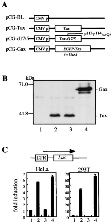
The Tax-encoding retrovirus vector, pR3Taxbsr, was con-structed from an MLV-based retrovirus vector, pRTaxbsr, by exchanging the U3 region of the 3⬘LTR with that of HTLV-1 (Fig. 1A). At the end of the recombinant retrovirus infection cycle, the HTLV-1 U3 sequence located in the 3⬘LTR of the pR3Taxbsr vector plasmid was copied to the 5⬘ LTR of the proviral genome in infected cells and functioned as a Tax-dependent enhancer of transcription. Both retroviral vector plasmids, pRTaxbsr and pR3Taxbsr, were transfected into an ecotropic-packaging cell line, BOSC23, and recombinant vi-ruses were recovered in the culture medium at 50 h posttrans-fection. Similar levels of Tax protein expression was observed in the packaging cells with the two vector plasmids (Fig. 1B, lanes 1 and 2). Tax-dependent transcriptional activation was confirmed in NIH 3T3 cells that had been infected with a similar titer of either recombinant retrovirus (Fig. 1B, lanes 3 and 4). About 100 drug-resistant cell clones were pooled, and Tax expression in these cells was analyzed by immunoblotting of the total-cell lysate. As shown in Fig. 1B, changing of the U3 region from that of MLV to that of HTLV-1 resulted in great enhancement of Tax expression in infected cells, indicating that the integrated proviral genome was activated by Tax pro-tein, as was observed in HTLV-1-infected cells.
Then, to conveniently monitor Tax expression in vivo, pR3Taxbsr was further modified to encode Gax by fusing the EGFP gene to the 5⬘end of the Tax-coding region, resulting in pR3Gaxbsr (Fig. 1A). The characteristics of Gax protein as a transactivator were shown to be retained in the luciferase assay by cotransfecting its expression vector with the HTLV-1 LTR Luc reporter plasmid into either HeLa or 293T cells (Fig. 2C). EL4 cells were infected with R3Gaxbsr recombinant virus, and Gax-transduced cell-clones were selected by the limiting-dilution method in the presence of blasticidin S. Cloned cells were screened for GFP intensity, and from one of the clones expressing high levels of Gax, a cell population of higher in-tensity was further enriched by means of a fluorescence-acti-vated cell sorter, resulting in EL4/Gax cells (Fig. 1C). We also established EL4-derived cell lines infected with either RTaxbsr or REGFP recombinant retrovirus, resulting in EL4/Tax or EL4/EGFP, respectively, as a control.
Effect of Tax DNA vaccine on EL4/Gax cells in vivo. To analyze the interaction of the host immune system with Tax-expressing tumor cells, EL4/Gax cells were injected s.c. into syngeneic C57BL/6J mice. Injection of 5⫻105EL4/Gax cells
caused the formation of a solid tumor at the injection site; the maximum size was reached within 2 to 3 weeks, and the mice died of multiorgan metastasis in 4 to 10 weeks (data not shown). Since it has been demonstrated that Tax protein re-tains strong CTL epitope activity, we examined whether or not the CTL activity against Tax protein affected the growth of Tax-expressing EL4 cells.
To induce CTL activity against Tax in mice, a DNA-medi-ated vaccine strategy was employed (Fig. 3A). Six-week-old female C57BL/6J mice were placed into four groups of five animals each and then immunized 5 times every week with 50
g of an expression vector plasmid, pCG-Tax, or its derivatives by i.m. injection. To enhance immune responses, 10g of an expression vector for GM-CSF (1, 29) and 10 g of a CpG oligonucleotide (16) were also injected. Mice in group A were
FIG. 1. (A) Design of retrovirus vectors. The parental vector, pRTaxbsr, has a cytomegalovirus enhancer/promoter unit (CMV p) at the 5⬘end, cDNA of Tax linked with a drug resistance gene,bsr, by an IRES, and an MLV LTR at the 3⬘end. The 273-bp U3 region of MLV LTR was replaced by a 266-bp fragment of HTLV-1 LTR, resulting in pR3Taxbsr. Then cDNA of EGFP was fused withtaxcDNA at the 5⬘ end, resulting in pR3Gaxbsr. (B) The protein expression of the pR and pR3 vectors was compared by Western blotting using an anti-Tax rabbit antiserum. BOSC23 cells were transfected with either pRTaxbsr or pR3Taxbsr by using Lipofectamine. NIH 3T3 cells were infected with the same titer of a supernatant of transfected BOSC23 cells. Proteins were harvested 50 h posttransfection and 48 h postinfection. Three micrograms of transfected BOSC23 cell lysates (lanes 1 and 2) and 17 g of infected NIH 3T3 cell lysates (lanes 3 and 4) were analyzed by SDS–12% PAGE, blotted onto PVDF membranes, and then probed with a rabbit antiserum against the C terminus of Tax. (C) Establishment a mouse lymphoma cell line expressing Gax. EL4 cells were infected with pR3Gaxbsr virus, selected with Blasticidin S, cloned by limiting dilution, and then sorted as to EGFP intensity. The Gax-expressing cell line was established and named EL4/Gax. We also established EL4/Tax and EL4/EGFP cell lines by infection with RTaxbsr and REGFP viruses, respectively (data not shown).
on November 8, 2019 by guest
http://jvi.asm.org/
[image:3.587.312.520.75.415.2]immunized with the control vector, pCG-BL, which expresses no protein. Mice in groups B and D were immunized with the wild-type Tax expresser, pCG-Tax, and group C mice were immunized with the Tax null mutant expresser, pCG-d17/5 (a linker insertion mutant of the Tax gene with amino acid changes of P113Y114to QAC). On day 35 after the first
im-munization, mice were challenged s.c. in a hind thigh with 5⫻
105EL4/Gax cells (groups A, B, and C) or EL4 cells (group D).
Fifteen days later, the mice were sacrificed, and the cellular and humoral immune responses were examined in vitro.
[image:4.587.61.254.73.456.2]On monitoring of the diameters of tumors with calipers every 3 days, we detected no visible solid tumors until day 6 postchallenge in mice immunized with either the wild type or the d17/5taxgene (groups B and C, respectively), while solid tumors became visible in 3 days after challenge in control mice (groups A and D). Although, at 1 week after challenge, tumors were visible in immunized mice as well, the sizes of the tumors seemed to reach a plateau thereafter (Fig. 3B). On day 15 postchallenge, the average diameters of tumors in groups B and C were 42.3 and 23.5%, respectively, of that in the control mice in group A. DNA immunization with the wild type Tax gene afforded no protection against parental EL4 cells (group
FIG. 2. DNA vaccine of HTLV-1 Tax and its mutant. (A) Sche-matic diagram of the expression vectors used for DNA immunization. The eukaryotic expression vector pCG-BL expresses no structural pro-teins. Wild-type and mutanttaxcDNAs were inserted into pCG-BL, resulting in pCG-Tax, pCG-d17/5, and pCG-Gax. In pCG-d17/5, the
113Pro-114Tyr sequence was changed to Gln-Asp-Cys. (B) Protein
ex-pression from each vector in human kidney 293T cells. Cells were transfected with pCG-BL (lane 1), pCG-Tax (lane 2), pCG-d17/5 (lane 3), or pCG-Gax (lane 4), and then Western blot analysis was per-formed as described in the legend to Fig. 1. (C) Transactivation of the HTLV-1 LTR by Tax or Tax mutants. Either HeLa (left) or 293T (right) cells were cotransfected with pLTR-Luc, which is an HTLV-1 LTR reporter plasmid, and one of the pCG vectors by using Lipo-fectamine. Lanes correspond to those in panel B. Two days posttrans-fection, cells were lysed and luciferase activity was measured. Data shown are averages for three independent transfections.
FIG. 3. (A) Mice were placed in four groups, A to D, and immu-nized with 50g of pCG vector 5 times by i.m. injection. Mice in group A were immunized with pCG-BL. In groups B and D, mice were vaccinated with pCG-Tax, while mice in group D were challenged with control tumor cells, EL4, after immunization. Mice in group C were immunized with a vector expressing the Tax null mutant d17/5. All mice in groups B to D were also injected with 10g of a GM-CSF-expressing vector and a CpG oligonucleotide. Two weeks after five DNA injections, mice were challenged s.c. with 5⫻105lymphoma
cells. (B) The diameters of tumors at the injection sites were deter-mined by using vernier calipers on the indicated days after challenge with EL4/Gax or EL4 cells. Data represent averages for five mice. (C) Wet weights of tumors taken from sacrificed mice 50 to 52 days postimmunization (about 2 weeks after tumor challenge) were deter-mined. Mouse groups correspond to those in panel A.
on November 8, 2019 by guest
http://jvi.asm.org/
D), thus indicating that tumor growth was inhibited by Tax-specific immunity.
We also excised solid tumors from mice and determined the wet weight on the same day (Fig. 3C). The tumor weights in either Tax -or d17/5-immunized mice were significantly lower than those in control mice. Immunization with wild-type Tax DNA appeared not to inhibit the growth of parental EL4 cells. In vivo protection assays showed no significant difference between vaccination with the wild-type Tax DNA and vaccina-tion with the d17/5 mutant Tax DNA with regard to tumor protection. These data excluded the possibility that Tax expres-sion in vivo might have an indirect effect on the host immune system by inducing cytokine production in DNA-transfected tissue cells, instead supporting the idea that some peptide epitope(s) on Tax protein is involved in the immune response to suppress the growth of EL4/Gax cells. Furthermore, the fact that direct injection of the expression plasmid for a null mutant of Tax protein successfully induced the protective immune response against Tax-expressing tumor cells suggests a possible practical application of the mutant gene.
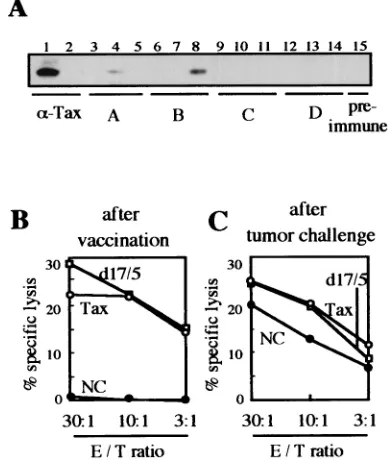
Cellular and humoral immunity with DNA vaccination.To investigate immune responses induced by the DNA vaccine, we examined the antibody responses and CTL activities of the animals used in the experiment for which results are shown in Fig. 3. Sera of three mice in each group and one preimmune mouse were analyzed by immunoblotting (Fig. 4A). Two serum specimens from groups A and B showed positive signals (Fig. 4A, lanes 4 and 8). These responses were possibly due to the EL4/Gax challenge cells, because no antibody responses were observed just after DNA immunization (data not shown), and the positive signal detected in lane 4 was obtained for a group A mouse that had not been immunized with the Tax gene. Thus, we concluded that DNA vaccination with Tax DNA does not induce strong antibody production.
To investigate the role of cell-mediated immune responses in tumor suppression, we examined the potential of T cells derived from the same mice to lyse EL4/Gax cells in a 51Cr
release assay. Assays were performed 1 week after the last booster DNA immunization and 15 days after challenge with tumor cells. As shown in Fig. 4B, both wild-type Tax and mutant d17/5 DNA vaccination induced good CTL activity, but control DNA (pCG-BL) vaccination had no effect on CTLs. On the other hand, mice immunized with the pCG-BL control vector showed a relatively high response of CTLs at 2 weeks after s.c. inoculation of tumor cells. This could be explained by the possibility that EL4/Gax challenge cells induced this CTL response as well as the occasional humoral response against Tax protein (Fig. 4A, lane 4), but the induction of immune responses appeared to be too late to protect mice from the vigorous growth of tumor cells. Based on the results of these assays of both humoral and cellular immune responses, we concluded that CTL activity against Tax is responsible for the elimination of EL4/Gax cells in mice immunized with the DNA vaccine.
Reduced expression of Gax in a solid tumor.In mice with Tax DNA vaccination, a small solid tumor was found to remain at the injected site without tumor growth. To determine what kind of cells was maintained in vivo with or without DNA vaccination, expression of Gax protein in solid tumors was analyzed. Three weeks after challenge, mice were sacrificed
under anesthesia, and tumors were excised and placed in liquid nitrogen. Proteins and total RNA were isolated from these samples and analyzed by Western blotting (Fig. 5A) and RT-PCR (Fig. 5B), respectively.
[image:5.587.322.516.77.307.2]Expression of Gax protein was scarcely observed in the tu-mor samples analyzed (two specimens for each group; Fig. 5A, lanes 1 to 4), while the amount of Gax protein in EL4/Gax cells that had been cultured in vitro without a drug remained un-changed for the same period as in the case of in vivo inocula-tion (lane 6). However, upon longer exposure, bands became visible for tumor samples, indicating a very low level of Gax expression in vivo. RT-PCR analysis also confirmed Gax-spe-cific mRNA expression in tumors of EL4/Gax cells (Fig. 5B). Interestingly, little difference was observed in the intensity of amplified bands between RNA samples from EL4/Gax cells grown in vitro and those grown in vivo. This could be due to a certain bias of PCR, however, so further analysis is required. Nevertheless, the low level of Tax expression in tumor cells suggested that such low expression enabled the tumor cells to evade Tax-specific CTLs, but it was enough to maintain CTL
FIG. 4. Humoral and cellular immune responses determined in vitro for DNA-immunized and EL4/Gax-challenged mice. (A) Sera were collected from three mice in each group and one preimmune mouse. 293T cells were transfected with pCG-Gax with Lipofectamine, and then cell lysates were used for blotting with Gax antigen. A lysate from nontransfected cells was used in lane 2. Sera used for blotting were as follows: lanes 1 and 2, anti-Tax rabbit antiserum; lanes 3 to 5, mice in group A (pCG-BL vaccinated and EL4/Gax challenged); lanes 6 to 8, mice in group B (pCG-Tax vaccinated and EL4/Gax chal-lenged); lanes 9 to 11, mice in group C (pCG-d17/5 vaccinated and EL4/Gax challenged); lanes 12 to 14, mice in group D (pCG-Tax vaccinated and EL4 challenged); lane 15, preimmune mouse. (B and C) CTL responses to EL4/Gax cells were determined with different effector/target (E/T) ratios at 7 days after DNA immunization (B) and 15 days after EL4/Gax cell challenge (C). T cells purified from spleno-cytes of mice from groups A (NC), B (Tax), and C (d17/5) were incubated for 3 days with syngeneic dendritic cells transfected with pCG-Tax by using Lipofectamine. Subsequently, CTL activity against EL4/Gax cells was determined by means of a 5-h51Cr release assay.
on November 8, 2019 by guest
http://jvi.asm.org/
activity against Tax in vivo. In accordance with this notion, an extensively growing tumor in a control mouse and a very small tumor with restricted growth in a vaccinated mouse expressed similar levels of Gax protein at 3 weeks after tumor inocula-tion, when CTL activity against Tax protein was already ob-served in both control and vaccinated mice. Thus, the growth of cells per se did not reflect the expression level of Tax; rather, the host immune response against Tax caused the suppression of Gax expression in target tumor cells.
Suppression of Gax gene expression in the peritoneal cavity.
The reduced expression of Gax in EL4/Gax cells in vivo could be a consequence of negative selection by the immune system that eliminates cells with high Gax expression, leaving geneti-cally silenced clones alive. To facilitate examination of the mechanism, EL4/Gax cells were inoculated into the peritoneal cavities of vaccinated C57BL/6J mice to monitor the
expres-sion of Gax protein in tumor cells. Mice were immunized four times weekly with the CpG oligonucleotide and with expres-sion vector plasmids for Tax and GM-CSF and were then challenged by i.p. injection of 104EL4/Gax cells. Three weeks
after inoculation, ascitic fluid was collected from these mice and analyzed for Gax expression in cells by flow cytometry. Compared with that in an in vitro culture, expression of Gax decreased dramatically in ascitic fluids, as observed in a solid tumor (Fig. 6A and B).
To determine whether the lack of Gax expression is due to transient silencing of transcription or is a result of negative selection for permanently dormant clones by the immune system, the EL4/Gax cells from ascitic fluids were trans-ferred to in vitro conditions. Gax expression was monitored every 3 h, and it was found that the expression in these cells had already started to increase by 3 h after the beginning of incubation; the maximum level was reached at 48 h (Fig. 6C). This result strongly supports the hypothesis that the sup-pression of Gax exsup-pression is caused by transient silencing of transcription and not by selection of genetically low-expression clones.
Effects of the immune reaction on Gax gene suppression in the peritoneal cavity. To analyze the effect of Tax-specific immunity on transient Gax gene suppression, EL4/Gax cells were i.p. inoculated into nonimmunized or Tax-immunized mice, and then Gax expression was monitored by flow cytom-etry. To induce immune responses against Tax, s.c. injection of EL4/Tax cells, instead of DNA vaccination, was employed as an immunization protocol. Six-week-old C57BL/6J mice were s.c. injected with 105EL4 or EL4/Tax cells 2 weeks before i.p.
challenge with 2⫻ 107 EL4/Gax cells. Two days after
chal-lenge, ascitic fluids were collected from each mouse and cells were stained with PI to exclude dead cells. In a naive (nonim-munized) mouse, 2 days of i.p. incubation resulted in little decrease in Gax expression compared with that in in vitro-cultured cells (Fig. 7A and B). When EL4/Gax cells were inoculated into mice immunized with the parental EL4 cells, a further slight decrease in the Gax expression level was ob-served (Fig. 7C). In contrast, in the peritoneal cavity of a mouse immunized with EL4/Tax cells, more cells became PI positive, indicating specific killing by Tax-specific immunity, and the level of Gax expression in living cells decreased greatly (Fig. 7D). Thus, Tax immunization resulted in the rapid re-duction of Gax expression at the cellular level, although the partial decrease in Gax expression observed in nonimmunized mice could be due to natural immunity or to any changes in the growing condition. The intensity of Gax expression appeared as two peaks 2 days after i.p. inoculation, but i.p. incubation for an additional 2 days left a single peak of lower intensity (Fig. 7E). Since in vitro culture of these cells for 3 h resulted in the reexpression of Gax (Fig. 7F), most of the population express-ing lower levels of Gax in the peritoneal cavity were not dead but living cells. These results strongly suggested that EL4/Gax cells with higher Gax expression were rapidly killed in Tax-immunized mice, while lower expressers survived. It was also confirmed that Gax gene modulation in vivo was not due to negative selection of genetically silenced EL4/Gax cell clones, but to cells whose Gax genes were transiently suppressed, while unsuppressed EL4/Gax cell clones were quickly killed by the host immune system.
FIG. 5. Expression of Gax in vivo. In this experiment, mice were immunized with either pCG-BL (⫺) or pCG-Tax (Tax), with the schedules described in the legend to Fig. 3A. Solid tumors formed by EL4/Gax cells in mice were removed, and then proteins and RNAs were extracted as described in Materials and Methods. (A) Western blotting with anti-Tax serum. Two samples from each group (lanes 1 to 4) were analyzed. As negative and positive controls, respectively, ly-sates from cultured EL4 (lane 5) and EL4/Gax (lane 6) cells were blotted. The upper panel shows results with a 3-min film exposure, while the lower panel shows results with a 30-min exposure. (B) RT-PCR of RNA samples from tumors. Lane 1, size markers. RNAs used for RT-PCR were derived from tumors in mice vaccinated with pCG-BL (lane 2), pCG-Tax (lane 3), or pCG-d17/5 (lane 4), or from in vitro EL4/Gax cell cultures (lanes 5 and 7). In lane 6, pR3Gaxbsr was used as a positive control. In lane 7, RT-PCR was performed without reverse transcriptase as a negative control. In the upper panel, a PCR primer set was used to detect Tax, while in the lower panel, PCR was performed with another primer set to detect mouse G3PDH as an internal control.
on November 8, 2019 by guest
http://jvi.asm.org/
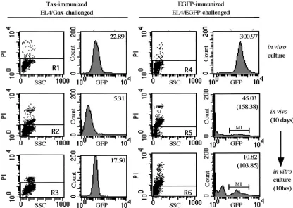
Transient Gax gene suppression. To determine whether transient exogenous gene suppression occurs generally or not, in vivo gene suppression and in vitro recovery were examined with another exogenous gene product. Six-week-old C57BL/6J mice were immunized by s.c. injection with either 105 EL4/Tax cells or 105 EL4/EGFP cells 2 weeks
before i.p. challenge with 2 ⫻107 EL4/Gax or EL4/EGFP
cells, respectively. Ten days after challenge, cells in ascitic fluids were stained with PI and then subjected to flow cy-tometry. A part of the collected cells from the peritoneal cavity was cultured in vitro for 10 h and also analyzed by flow cytometry. In EL4/Gax cells inoculated into the peri-toneal cavities of Tax-immunized mice, transient Gax gene suppression was observed repeatedly (Fig. 8, left panels). When a mouse was immunized with EGFP and then chal-lenged with EL4/EGFP cells, the major population of cells was PI positive and the GFP intensity of the PI-negative population was dramatically decreased (Fig. 8, right panels). The mean fluorescence of EGFP, even in the EGFP-positive fraction
(M1), had not recovered after10 h of in vitro culture (Fig. 8, bottom right panel), whereas in vitro culture of EL4/Gax cells isolated from ascitic fluids resulted in reexpression of Gax (from 5.31 to 17.50 in mean fluorescence value [Fig. 8, left histograms]). These data demonstrated that EL4 cells express-ing exogenous antigens were killed in the peritoneal cavities of immunized mice, at least in both the cases of Tax and EGFP. However, recovery of expression upon in vitro culture was not a general event for exogenous gene products but rather specific to the Tax gene.
It was further suggested that the transient silencing of the proviral genome in HTLV-1-infected lymphocytes helped virus-positive cells to evade eradication by the host immune system, in which cytotoxic T cells specific to the Tax protein are mainly involved. The suppression and reexpression of Gax transcription in this mouse system are in good agree-ment with the phenomenon observed in HTLV-1-infected CD4⫹lymphocytes isolated from ATL and HAM/TSP
pa-tients, in which the virus expression suppressed in
periph-FIG. 6. Protein expression of EL4/Gax cells in ascitic fluid. Mice were vaccinated with 50g of pCG-d17/5, 10g of a GM-CSF expression vector, and 10g of a CpG oligonucleotide four times weekly. Two weeks after the last immunization, 104EL4/Gax cells were i.p. injected.
EL4/Gax cells were collected from ascitic fluids and transferred to in vitro conditions at 3 weeks after challenge. Expression of Gax proteins was analyzed before challenge (A) and just after collection from ascitic fluids (B). (C) Reversion of Gax expression after transfer to in vitro conditions. From the time point just after collection of cells from the peritoneal cavity (t⫽0 h), Gax expression was analyzed by flow cytometry every 3 h. Gax expression started to show reactivation in 3 h and reached a level almost equivalent with that before in vivo incubation within 12 h.
on November 8, 2019 by guest
http://jvi.asm.org/
[image:7.587.91.490.77.427.2]eral blood resumes soon after the transfer of cells to in vitro culture conditions. Although a high provirus load was ob-served in the peripheral blood of HAM/TSP patients, it was shown that virus replication was strongly suppressed in vivo
(10). Therefore, the mouse system established here will pro-vide a suitable model for investigating the apparent equilib-rium in vivo between virus expression and the host immune system in individuals persistently infected with HTLV-1.
FIG. 7. Effects of the immune responses on Gax gene suppression in vivo. C57BL/6J mice were immunized by s.c. injection of either saline (B), 105EL4 cells (C), or 105EL4/Tax cells (D and E) cells 2 weeks before i.p. challenge with 2⫻107EL4/Gax cells. Cells were harvested from an
in vitro culture (A), from mice 2 days (B to D) or 4 days (E) after challenge, and after 3 h of in vitro culture following 4 days of incubation in the peritoneal cavity (F). The collected cells were stained with PI and then subjected to flow cytometry analysis. (Left panels) PI staining; (right panels) histograms of GFP fluorescence of the PI-negative population (R1 to R6). Numbers in the right panel indicate mean fluorescence.
on November 8, 2019 by guest
http://jvi.asm.org/
DISCUSSION
In this study, we established a simple mouse model for in-vestigating the role of Tax-specific immune responses with regard to protection from Tax-expressing tumor cell growth. In C57BL/6J mice immunized with either the wild-type or the null mutant Tax-expressing plasmid, growth of a syngeneic T-cell lymphoma expressing EGFP-fused Tax (EL4/Gax) was strongly inhibited, while the parental EL4 cells grew rapidly to form the largest solid tumor (Fig. 3). This inhibition was Tax specific, because immunization with the empty plasmid vector did not protect mice from EL4/Gax tumor growth. The im-mune response involved in protection from Gax-expressing tumor cell growth consisted mainly of cellular immunity, since the CTL response, but not antibody production against Tax protein, was observed in mice which had been immunized with Tax DNA. Therefore, we concluded that DNA immunization by direct injection of Tax expression plasmids elicited protec-tive immunity against Tax-expressing tumor cell growth through induction of anti-Tax CTL.
The role of Tax protein in cellular transformation has been well studied (32). The mechanisms of onset of HTLV-1-related diseases, however, have been in contention. In particular, the role of the immune response against Tax is the subject of a key argument; that is, are Tax-specific CTL a protective factor or one of the causes of the diseases? In the case of HAM/TSP and
other autoimmune syndromes involving HTLV-1 infection, there have been several reports insisting that Tax is the cause of the diseases. Recently, Hanon et al. showed a significant negative correlation between the frequency of Tax-specific CD8⫹ T cells and the percentage of CD4⫹ T cells among
peripheral blood mononuclear cells of patients infected with HTLV-1 (10). These data suggest the possibility of a protective function of Tax-specific CTL. In this context, it is of note that a mutant Tax protein lacking transforming ability could suc-cessfully induce CTL. The results obtained in this study indi-cate the practical possibility of a DNA vaccination procedure for prophylactic purposes to reduce the onset rates of HTLV-1-related diseases.
[image:9.587.84.500.75.371.2]Recently, Ohashi et al. reported that Tax DNA vaccine was effective in their rat model (20). In this model, Tax DNA vaccination shortened the period for rejecting syngeneic HTLV-1-immortalized cells, which are able to grow only in syngeneic nu/nu rats. Their results are also consistent with the availability of a DNA vaccine for prophylactic use. However, to assess this possibility, the indicator cells should remain in vivo for a long period. In this respect, EL4/Gax cells stayed at the injected site even in extensively immunized mice in our model system. Thus, it is easy to monitor the fate of EL4/Gax cells and to analyze immune responses in a mouse that has a normal immune system.
FIG. 8. Gene suppression of EGFP. C57BL6 mice were immunized by s.c. injection of either 105EL4/Tax cells (left) or 105EL4/EGFP cells
(right) 2 weeks before i.p. challenge with 2⫻107EL4/Gax (left) or EL4/EGFP (right) cells. Cells were collected from an in vitro culture (top),
just after 10 days of incubation in the peritoneal cavity (middle), or after 10 h of in vitro culture following 10 days of incubation in the peritoneal cavity (bottom). GFP fluorescence was analyzed in the PI-negative population (R1 to R6). Top numbers in histograms indicate mean fluorescence, and numbers in parentheses indicate the mean fluorescence of the M1 region.
on November 8, 2019 by guest
http://jvi.asm.org/
The average size of EL4/Gax tumors in control mice was substantially lower than that of EL4 tumors in mice immunized with Tax DNA. These data suggest that some immune re-sponse was also elicited in naive mice after challenge with EL4/Gax cells (Fig. 4), and in vitro analysis of CTL activity supported this explanation. In agreement with this, previous studies demonstrated the presence of strong CTL epitopes on Tax protein (12, 14). Thus, mice bearing EL4/Gax tumors should express abundant CTL activity against Tax, irrespective of previous immunization with Tax DNA, and the difference in tumor growth between immunized and nonimmunized mice can be attributed to the initial number of input tumor cells, which should be smaller in immunized mice because of the cytotoxicity of preexisting CTL. Consistent with this explana-tion, inoculation of an increased number of EL4/Gax cells resulted in failure in protecting the immunized mice from tumor growth (data not shown).
In addition to the number of tumor cells, the expression level of the target antigen in each tumor cell should also de-termine the fate of an inoculated cell. In this regard, it is of note that the expression level of Gax remained extremely low in vivo (Fig. 5). Since tumor cells with almost undetectable Gax expression did not grow in immunized mice, such a low level of Gax might be sufficient for targeting by Tax-specific CTLs. This seems to be in accord with the observation that HTLV-1 pro-virus is transcriptionally silent within a high proportion of T lymphocytes in the peripheral blood of HTLV-1-infected pa-tients (19, 24). These facts imply that there is some equilibrium between antigen expression and the host immune system in the peripheral blood of individuals persistently infected with HTLV-1. Furthermore, it has been postulated that the evasion of such equilibrium by escape mutations of thetaxgene and/or suppression of the host immune system leads to the develop-ment of ATL (9).
The low level of Gax expression in vivo was also confirmed by an experiment in which EL4/Gax cells were transferred to the peritoneal cavity of a Tax DNA immunized mouse (Fig. 6A and B). Although expression of Gax was almost undetectable after 15 days of incubation in the peritoneal cavity, it was restored quickly upon culturing in vitro (Fig. 6C). These data strongly indicated that the expression of Gax was silenced rather transiently, probably through an enhancer/promoter re-gion of HTLV-1 in vivo. This situation greatly resembles the suppression and reexpression of the HTLV-1 provirus genome in virus-positive CD4⫹ lymphocytes from HTLV-1-infected
patients. Hanon et al. attributed the suppression of virus ex-pression in infected cells in vivo to Tax-specific CTL, since the addition of a drug, concanamycin A, that inhibits the function of CTL in an in vitro culture of HTLV-1-positive T lympho-cytes resulted in enhancement of the recovery from suppres-sion of virus expressuppres-sion (10). As the mechanism of gene silenc-ing remains unclear, the i.p. EL4/Gax inoculation system will provide a strong tool for investigating the mechanism of tran-scriptional suppression in vivo.In this context, it is of interest whether the silencing of gene expression involves any intracel-lular signal transduction or chemical modifications such as CpG methylation of genomic DNA, which has been implicated in most silencing events.
We demonstrated that the higher Gax expressers were rap-idly killed in a Tax-immunized mouse but not in a
nonimmu-nized mouse (Fig. 7), suggesting that Tax-specific immune re-sponses play some role, if not all, in the transient suppression in vivo. Although we have not determined what kind of cells directly kill EL4/Gax cells in the peritoneal cavity, preliminary experiments showed that more T cells had infiltrated into the ascitic fluid of a Tax-immunized mouse, while more NK cells were observed in the ascitic fluid of a naive mouse after chal-lenge with EL4/Gax cells (data not shown). We also demon-strated that transient suppression of exogenous gene products was not a general phenomenon, because, although EGFP ex-pression in EL4/EGFP cells was suppressed in an immuniza-tion-specific manner, the expression was not recovered upon in vitro culturing. Therefore, transient gene suppression of Gax seemed to be a specific event with regard to the balance be-tween viral gene expression and antivirus immunity.
We developed a simple but very useful mouse model system for investigating the events during the asymptomatic stage of HTLV-1 infection, in which there is a certain equilibrium be-tween the host immune surveillance mechanism and the growth of HTLV-1-infected lymphocytes with limited expres-sion of virus antigen. Studies on the involvement of the im-mune system in the control of disease progression related to HTLV-1 infection with such a simple mouse model will pro-vide valuable information for protecting infected people from the onset of the diseases.
ACKNOWLEDGMENTS
This study was supported by a grant from the Ichiro Kanehara Foundation, a grant from the Japanese Private School Foundation, and a grant from the “Haiteku Research Center” of the Ministry of Education.
We thank M. Inaba for technical advice and N. J. Halewood for language assistance.
REFERENCES
1.Abe, J., H. Wakimoto, Y. Yoshida, M. Aoyagi, K. Hirakawa, and H. Hamada.
1995. Antitumor effect induced by granulocyte/macrophage colony-stimulat-ing factor gene-modified tumor vaccination: comparison of adenovirus- and
retrovirus-mediated genetic transduction. J. Cancer Res. Clin. Oncol.121:
587–592.
2.Ballard, D. W., E. Bohnlein, J. W. Lowenthal, Y. Wano, B. R. Franza, and W. C. Greene.1988. HTLV-I tax induces cellular proteins that activate the
B element in the IL-2 receptor␣gene. Science241:1652–1655.
3.Bangham, C. R., S. Daenke, R. E. Phillips, J. K. Cruickshank, and J. I. Bell.
1988. Enzymatic amplification of exogenous and endogenous retroviral se-quences from DNA of patients with tropical spastic paraparesis. EMBO J.
7:4179–4184.
4.Cann, A. J., J. D. Rosenblatt, W. Wachsman, N. P. Shah, and I. S. Chen.
1985. Identification of the gene responsible for human T-cell leukaemia virus
transcriptional regulation. Nature318:571–574.
5.Chen, I. S., A. J. Cann, N. P. Shah, and R. B. Gaynor.1985. Functional relation between HTLV-II x and adenovirus E1A proteins in transcriptional
activation. Science230:570–573.
6.Felber, B. K., H. Paskalis, C. Kleinman-Ewing, F. Wong-Staal, and G. N. Pavlakis.1985. The pX protein of HTLV-I is a transcriptional activator of its
long terminal repeats. Science229:675–679.
7.Fujisawa, J., M. Seiki, T. Kiyokawa, and M. Yoshida.1985. Functional activation of the long terminal repeat of human T-cell leukemia virus type I by atrans-acting factor. Proc. Natl. Acad. Sci. USA82:2277–2281. 8.Fujisawa, J., M. Toita, T. Yoshimura, and M. Yoshida.1991. The indirect
association of human T-cell leukemia virus Tax protein with DNA results in
transcriptional activation. J. Virol.65:4525–4528.
9.Furukawa, Y., R. Kubota, M. Tara, S. Izumo, and M. Osame.2001. Existence of escape mutant in HTLV-I tax during the development of adult T-cell
leukemia. Blood97:987–993.
10.Hanon, E., S. Hall, G. P. Taylor, M. Saito, R. Davis, Y. Tanaka, K. Usuku, M. Osame, J. N. Weber, and C. R. Bangham.2000. Abundant tax protein
expression in CD4⫹T cells infected with human T-cell lymphotropic virus
type I (HTLV-I) is prevented by cytotoxic T lymphocytes. Blood95:1386–
1392.
on November 8, 2019 by guest
http://jvi.asm.org/
11.Inoue, J., M. Seiki, T. Taniguchi, S. Tsuru, and M. Yoshida.1986. Induction of interleukin 2 receptor gene expression by p40x encoded by human T-cell
leukemia virus type 1. EMBO J.5:2883–2888.
12.Jacobson, S., H. Shida, D. E. McFarlin, A. S. Fauci, and S. Koenig.1990.
Circulating CD8⫹cytotoxic T lymphocytes specific for HTLV-I pX in
pa-tients with HTLV-I associated neurological disease. Nature348:245–248.
13.Jin, D. Y., F. Spencer, and K. T. Jeang.1998. Human T cell leukemia virus type 1 oncoprotein Tax targets the human mitotic checkpoint protein
MAD1. Cell93:81–91.
14.Kannagi, M., S. Harada, I. Maruyama, H. Inoko, H. Igarashi, G. Ku-washima, S. Sato, M. Morita, M. Kidokoro, M. Sugimoto, S. Funahashi, M. Osame, and H. Shida. 1991. Predominant recognition of human T cell
leukemia virus type I (HTLV-I) pX gene products by human CD8⫹cytotoxic
T cells directed against HTLV-I-infected cells. Int. Immunol.3:761–767.
15.Kannagi, M., S. Matsushita, H. Shida, and S. Harada.1994. Cytotoxic T cell response and expression of the target antigen in HTLV-I infection.
Leuke-mia8(Suppl. 1):S54-S9.
16.Krieg, A. M., A. K. Yi, S. Matson, T. J. Waldschmidt, G. A. Bishop, R. Teasdale, G. A. Koretzky, and D. M. Klinman.1995. CpG motifs in bacterial
DNA trigger direct B-cell activation. Nature374:546–549.
17.Kushida, S., M. Matsumura, H. Tanaka, Y. Ami, M. Hori, M. Kobayashi, K. Uchida, K. Yagami, T. Kameyama, T. Yoshizawa, H. Mizusawa, Y. Iwashita, and M. Miwa.1993. HTLV-1-associated myelopathy/tropical spastic para-paresis-like rats by intravenous injection of HTLV-1-producing rabbit or
human T-cell line into adult WKA rats. Jpn. J. Cancer Res.84:831–833.
18.Leung, K., and G. J. Nabel.1988. HTLV-1 transactivator induces
interleu-kin-2 receptor expression through an NF-B-like factor. Nature333:776–
778.
19.Moritoyo, T., S. Izumo, H. Moritoyo, Y. Tanaka, Y. Kiyomatsu, M. Nagai, K. Usuku, M. Sorimachi, and M. Osame.1999. Detection of human T-lympho-tropic virus type I p40 Tax protein in cerebrospinal fluid cells from patients with human T-lymphotropic virus type I-associated myelopathy/tropical
spastic paraparesis. J. Neurovirol.5:241–248.
20.Ohashi, T., S. Hanabuchi, H. Kato, H. Tateno, F. Takemura, T. Tsukahara, Y. Koya, A. Hasegawa, T. Masuda, and M. Kannagi.2000. Prevention of adult T-cell leukemia-like lymphoproliferative disease in rats by adoptively transferred T cells from a donor immunized with human T-cell leukemia
virus type 1 Tax-coding DNA vaccine. J. Virol.74:9610–9616.
21.Pear, W. S., G. P. Nolan, M. L. Scott, and D. Baltimore.1993. Production of high-titer helper-free retroviruses by transient transfection. Proc. Natl. Acad.
Sci. USA90:8392–8396.
22.Pique, C., F. Connan, J. P. Levilain, J. Choppin, and M. C. Dokhelar.1996. Among all human T-cell leukemia virus type 1 proteins, Tax, polymerase,
and envelope proteins are predicted as preferential targets for the
HLA-A2-restricted cytotoxic T-cell response. J. Virol.70:4919–4926.
23.Poiesz, B. J., F. W. Ruscetti, A. F. Gazdar, P. A. Bunn, J. D. Minna, and R. C. Gallo.1980. Detection and isolation of type C retrovirus particles from fresh and cultured lymphocytes of a patient with cutaneous T-cell lymphoma.
Proc. Natl. Acad. Sci. USA77:7415–7419.
24.Richardson, J. H., P. Hollsberg, A. Windhagen, L. A. Child, D. A. Hafler, and A. M. Lever. 1997. Variable immortalizing potential and frequent virus latency in blood-derived T-cell clones infected with human T-cell leukemia
virus type I. Blood89:3303–3314.
25.Sodroski, J., C. Rosen, W. C. Goh, and W. Haseltine.1985. A transcriptional activator protein encoded by the x-lor region of the human T-cell leukemia
virus. Science228:1430–1434.
26.Sodroski, J. G., C. A. Rosen, and W. A. Haseltine.1984. Trans-acting tran-scriptional activation of the long terminal repeat of human T lymphotropic
viruses in infected cells. Science225:381–385.
27.Stewart, S. A., G. Feuer, A. Jewett, F. V. Lee, B. Bonavida, and I. S. Chen.
1996. HTLV-1 gene expression in adult T-cell leukemia cells elicits an NK cell response in vitro and correlates with cell rejection in SCID mice.
Virol-ogy226:167–175.
28.Suzuki, T., H. Hirai, J. Fujisawa, T. Fujita, and M. Yoshida.1993. Atrans
-activator Tax of human T-cell leukemia virus type 1 binds to NF-B p50 and
serum response factor (SRF) and associates with enhancer DNAs of the
NF-B site and CArG box. Oncogene8:2391–2397.
29.Turner, J. G., J. Tan, B. E. Crucian, D. M. Sullivan, O. F. Ballester, W. S. Dalton, N. S. Yang, J. K. Burkholder, and H. Yu.1998. Broadened clinical utility of gene gun-mediated, granulocyte-macrophage colony-stimulating factor cDNA-based tumor cell vaccines as demonstrated with a mouse
my-eloma model. Hum. Gene Ther.9:1121–1130.
30.Wakimoto, H., Y. Yoshida, M. Aoyagi, K. Hirakawa, and H. Hamada.1997. Efficient retrovirus-mediated cytokine-gene transduction of primary-cul-tured human glioma cells for tumor vaccination therapy. Jpn. J. Cancer Res.
88:296–305.
31.Wodarz, D., M. A. Nowak, and C. R. Bangham. 1999. The dynamics of
HTLV-I and the CTL response. Immunol. Today20:220–227.
32.Yoshida, M.2001. Multiple viral strategies of HTLV-1 for dysregulation of
cell growth control. Annu. Rev. Immunol.19:475–496.
33.Yoshida, M., J. Inoue, J. Fujisawa, and M. Seiki.1989. Molecular mecha-nisms of regulation of HTLV-1 gene expression and its association with
leukemogenesis. Genome31:662–667.
34.Yoshida, M., M. Seiki, K. Yamaguchi, and K. Takatsuki.1984. Monoclonal integration of human T-cell leukemia provirus in all primary tumors of adult T-cell leukemia suggests causative role of human T-cell leukemia virus in the
disease. Proc. Natl. Acad. Sci. USA81:2534–2537.