of liposomes delivered siRNA and
effects on siRNA engagement
with target mRNA and silencing in
cancer cells
Abdullah Alshehri
1, Anna Grabowska
2& Snow Stolnik
1Design of an efficient delivery system is a generally recognised bottleneck in translation of siRNA technology into clinic. Despite research efforts, cellular processes that determine efficiency of siRNA silencing achieved by different delivery formulations remain unclear. Here, we investigated the mechanism(s) of cellular internalisation of a model siRNA-loaded liposome system in a correlation to the engagement of delivered siRNA with its target and consequent silencing by adopting siRNA molecular beacon technology. Probing of cellular internalisation pathways by a panel of pharmacological inhibitors indicated that clathrin-mediated (dynamin-dependent) endocytosis, macropinocytosis (dynamine independent), and cell membrane cholesterol dependent process(es) (clathrin and caveolea-independent) all play a role in the siRNA-liposomes internalization. The inhibition of either of these entry routes was, in general, mirrored by a reduction in the level of siRNA engagement with its target mRNA, as well as in a reduction of the target gene silencing. A dramatic increase in siRNA engagement with its target RNA was observed on disruption of endosomal membrane (by chloroquine), accompanied with an increased silencing. The work thus illustrates that employing molecular beacon siRNA technology one can start to assess the target RNA engagement – a stage between initial cellular internalization and final gene silencing of siRNA delivery systems.
Delivery of siRNA to the cytoplasm of target cells is a promising therapeutic approach for the treatment of a wide range of diseases1. However, the therapeutic potential of siRNA has not yet been realized due to the need for an appropriate delivery system2. The optimal siRNA delivery system should be non-toxic, protect siRNA from RNase degradation, facilitate intracellular uptake followed by escape from endosome vesicles into the cytosol, and finally encourage effective gene silencing3. Cationic liposomes have been reported as one of the extensively used non-viral delivery systems4,5 exploited in a delivery of different nucleic acids, including siRNA. In order to engineer a well-designed siRNA liposome formulation, it is important to understand the cellular internalisation and processing mechanisms of the delivery system in order to achieve efficient cellular uptake and evade the destructive or recycling cellular pathways6. However, despite the extensive efforts to translate siRNA-liposome technology to clinic, processes that govern interaction with cells, cellular internalisation mechanisms, and intra-cellular trafficking remain poorly understood7,8 to be exploited in rational design and engineering of efficient siRNA delivery systems.
In this study we selected to use pharmacological inhibitors to study cellular transport mechanisms, as their application is well established in the field9. We do acknowledge the fact that these can exert multiple cellular effects and that siRNA knockdown of endocytosis pathway-selective proteins could be advocated as an alterna-tive10. However, the latter approach is less well established, has its own drawbacks11 and is technically impractical in the present study.
1Division of Molecular Therapeutics and Formulation, School of Pharmacy, University of Nottingham, Nottingham,
NG7 2RD, UK. 2Cancer Biology, Division of Cancer and Stem Cells, School of Medicine, Queen’s Medical Centre,
University of Nottingham, Nottingham, NG7 2RD, UK. Correspondence and requests for materials should be addressed to S.S. (email: [email protected])
The present work exploits a relatively new siRNA molecular beacon technology, initially introduced as nucleic acid analytical probes that recognize and report the presence of specific nucleic acids in homogeneous solutions12, and underused in a field of siRNA delivery. siRNA molecular beacons are hairpin shaped single stranded nucleic acid probes with conjugated fluorophore and quencher that fluoresce only upon hybridization with its target mRNA molecule when the loop region hybridizes with the target, opens up and hence separates the conjugated fluorophore and the quencher. Recently application of the technology to study cellular pharmacokinetics and pharmacodynamics of siRNA intracellularly was pioneered by a seminar work in RJ Lee’s group13. The present study applies the technology to assess an intermittent point - the siRNA engagement with the target mRNA - in the study aiming to establish a corroboration between cellular internalisation mechanism(s), engagement in the RISC machinery, and silencing efficiency of liposomally delivered siRNA.
Results
Liposome formulation optimization and Cy3-Annexin V/Propidium Iodide Cytotoxicity
Study.
The optimization of physicochemical properties of siRNA loaded liposomes used in this study is summarized in Supplementary Information (Figs S1–S3). Empty liposomes with average particle size of ~80– 100 nm were formulated by a classical ‘film hydration’ method with a range of DC-Chol to DOPE ratios (Fig. S1). Incorporation of siRNA during the liposomes fabrication, rather than subsequent addition, resulted in a good incorporation efficiency at optimized N/P ratios; for instance, the N/P ratio 3.125:1 resulted in the absence of free siRNA in the gel retardation assay (Fig. S2). An increase in the average particle size of the liposomes, with resultant sizes in the approximately 200–300 nm size range, was observed on siRNA incorporation (Fig. S3). Zeta potential values of the siRNA-liposomes were dependent on the N/P ratios used, and increased as the N/P ratio was increased; for example values in order of approximately +20 to +30 mV for N/P 3.125:1 to 12.5:1 were meas-ured for the liposomes with DC-Chol:DOPE ratio of 1:1.Toxicity of empty liposomes, as determined by MTS and PI/Annexin tests, is summarized in the Supplementary Information (Figs S4 and S5). Total lipid content applied to cells was found a crucial toxicity factor with, in general, applied concentrations above 1 mM causing a significant toxicity (EC50 in 24.0 to 8.7 mM
[image:2.595.111.552.47.277.2]analysis, siRNA-liposomes (at 1 µg/well siRNA, Fig. S7) were applied to A549-luc cells for 4 hours, removed and the assessment of luciferase activity conducted at 24, 48, 72, and 96 hours post-transfection. (No significant effects of non-targeting siRNA or effects of siRNA applied as ‘free’, unformulated solution on the luciferase activity were observed in a preliminary experiment, Fig. S7). The data (Fig. S8) indicate the highest significant decrease (p < 0.001) in luciferase activity at 48 hours post-transfection, with the recovery of luciferase activity at the later time points. All subsequent experiments were hence performed at 48 hour time point. Biophysical attributes of siRNA liposomal formulation used in all subsequent experiments are summarized in Table 1.
Mechanisms of Cellular Internalisation of
Cy3siRNA-Liposomes.
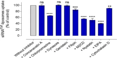
We used pharmacological inhibi-tors of endocytosis to assess cellular processing of siRNA-liposomes. The effect of endocytosis inhibition on the cellular uptake of cy3siRNA-liposomes in A549 cells was examined by flow cytometry following the application ofconcanavalin A (100 μg/ml), chlorpromazine (20 μg/ml), dynasore (20 μg/ml), genistein (15 μg/ml), filipin (20 μg/ ml), MßCD (300 μg/ml), nystatin (20 μg/ml), EIPA (0.5 μg/ml), and cytochalasin D (2 μg/ml). Concertation opti-mization of used inhibitors is shown in Supplementary Information (Fig. S9) and, when available, ligands known to selectively use a specific entry pathway (transferrin for clathrin and choleratoxin B for caveolae mediated endo-cytosis) were used as positive controls to confirm the effect of applied inhibitors (Fig. S10). The data show that the inhibitory effect is increased with a corresponding increase in the concentration of inhibitors tested. Regarding inhibition of macropinocytosis by EIPA and cytochalasin D, as dextran may not to be a solely specific ligand for this internalisation pathway14, we tested these inhibitors at three different sub-EC
50 concentrations to eliminate
possible artefacts of a singular concentration, as shown in Figure S11. The levels cell viability, determined as cell metabolic activity by MTS assay, at the above listed concentrations of endocytosis inhibitors applied are summa-rized in Supplementary Information (Tbl. S1). Tested at corresponding experimental conditions, these data allow comparisons of treated with control sets of data in Figs 2–4. Cellular internalization of liposomes was, in addition to flow cytometry, visualized by confocal microscopy (e.g. Fig. S12). Experimental data are summarized in Fig. 2.
Considering first an inhibition of clathrin-mediated pathway, concanavalin A - an inhibitor that interferes with the signalling of transmembrane G protein-coupled receptors located on the surface of cells, and in that way prevents the assembly of clathrin coated pits and impairs their movement through the cell membrane - had no significant effect (p > 0.05) on the internalisation of Cy3siRNA-liposomes by A549 cells (Fig. 2). In contrast,
chlorpromazine inhibition of clathrin pathway, which acts by a different mechanism of preventing formation of clathrin-coated pits at the plasma membrane, demonstrated a significant effect (p < 0.0001) on the internaliza-tion, reducing it to approximately 66% of that in untreated cells.
The inhibition of dynamin by dynasore, an inhibitor with the ability to block the GTPase activity of dynamin and hence detachment from the membrane in dynamin-dependent endocytosis in cells, exhibited no significant effects (p > 0.05) on the internalisation of cy3siRNA-liposomes. Genistein, as tyrosine kinase and caveolae pathway
inhibitor, did not produce a statistically significant effect on the internalisation of cy3siRNA-liposomes. However,
inhibition of caveolae-mediated endocytosis by filipin showed a moderate, statistically significant reduction Figure 2. The effect of a panel of pharmacological inhibitors on the internalisation of cy3siRNA-liposomes
in A549 cells. Cellular uptake of cy3siRNA-liposomes in absence (‘without inhibitor’ control) and presence of
inhibitors, assessed by flow cytometry (MFI). Data represent the mean ± SD (N = 2, n = 4). **, *** and ****
[image:3.595.156.401.46.118.2] [image:3.595.156.359.166.267.2]in the internalisation of cy3siRNA-liposomes (p = 0.0001). MßCD inhibitor, which depletes cell membrane
associated cholesterol, reduced the internalisation of cy3siRNA-liposomes significantly (p < 0.0001). Another
inhibitor that acts on membrane cholesterol, nystatin, also demonstrated a significantly reduced liposomes internalisation (p < 0.0001), 51% of untreated cells. EIPA inhibits Na+/H+ exchange, consequently lowering the sub-membranous pH of the cell cytoplasm, and is normally used to investigate the role of macropinocyto-sis pathway. Results show that EIPA was able to significantly reduce the internalisation of cy3siRNA-liposomes
(p < 0.0001). Moreover, the inhibition of endocytosis by cytochalasin D, another macropinocytosis inhibitor, showed a moderate, but statistically significant reduction (p < 0.01).
Effect of endocytosis inhibitors on engagement of liposomes delivered
luc-siRNA-molecular
beacon with target mRNA.
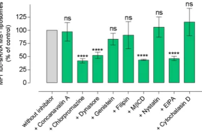
Flow cytometry data summarized in Fig. 3 demonstrate effects of different pharmacological inhibitors on the luc-siRNA-molecular beacon engagement (in terms of the inhibitor’s effect on measured engagement, i.e. cell associated fluorescence, relative to untreated control) with target luciferase mRNA in A594-luc cells following application of tested liposomal formulation. On the whole, the levels of siRNA engagement with its target mRNA follow the trend obtained for inhibition of liposomes internalization in Fig. 2, with the exceptions of dynasore and nystatin. For example, a pronounced inhibition of the internalization was seen for chlorpromazine, MβCD and EIPA treatments (Fig. 2), and the same inhibitors produced a statistically highly significant reduction in the molecular beacon siRNA engagement (statistical significance as indicated in Fig. 3), while concavalin A, genistein and cytochalasin did not produce a statistically significant reductions in either cellular internalization or siRNA engagement with target mRNA (statistical significance as indicated). Confocal micrographs in Fig. 3 visualize the presence of engaged, hence green luc-siRNA based molecular bea-con in A549-luc cells for some tested systems. The fluorescence broadly appears to be localised in the perinuclear region of the cells.Figure 4. The effect of pharmacological inhibitors on the luciferase knockdown by luc-siRNA-liposomes in A549-luc cells. Luc-siRNA-liposomes prepared at an N/P ratio 3.125:1, DC-Chol:DOPE ratio of 1:1, applied at 1 μg of luc-siRNA per well and 1 mM total lipid content. The luciferase activity assessed after 48 hours. Luciferase knockdown relative to the control (cells without inhibitors as 100%), data represents the mean ± SD (N = 2, n = 4), **** indicate a significant difference between the results (p < 0.0001) and ns indicates the difference is a non-statistically significant (p > 0.05) compared to the control.
[image:4.595.158.357.45.175.2] [image:4.595.154.363.308.411.2]Effect of endocytosis inhibitors on siRNA-liposomes silencing.
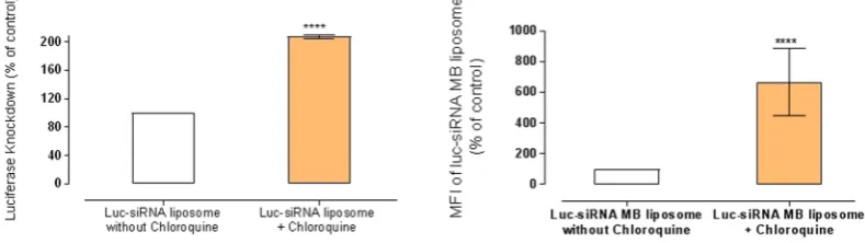
Figure 4 presents the effect of endocytosis inhibition on silencing of luciferase protein expression in A549-luc cells. The general data profile appears to follow trends in Figs 2 and 3; the main feature being the effect of dynasore; it statistically significantly (p < 0.0001) reduced molecular beacon siRNA engagement with its target mRNA (Fig. 3) and produced a signif-icant luciferase silencing effect (p < 0.0001), despite no significant effect (p > 0.05) on the cellular uptake (Fig. 2). A highly significant reduction of the silencing effect was measured for chlorpromazine, MßCD and EIPA treat-ments (statistical significance as indicated in Fig. 4), whilst concavalin A, genistein, nystatin and cytochalasin D did not produce a significant reduction in the silencing effect (all p > 0.5).Figure 5 shows the effect of chloroquine (100 μg/ml) on the luciferase silencing by luc-siRNA-liposomes in A549-luc cells. The data clearly demonstrate that chloroquine treatment statistically significantly (p < 0.0001) increases both the target RNA engagement and luciferase knockdown, relative to the cells not treated with chlo-roquine; the results show approximately 7-fold increases in fluorescence signal of the ‘luc-siRNA based MB’ in comparison to control and approximately 2-fold increase in the silencing.
Discussion
Despite extensive research in the design of formulations to deliver siRNA, the cellular internalisation pathways of different siRNA formulations, including liposomes, and engagement of delivered siRNA with its target mRNA, have not been extensively studied to provide a mechanistic insight on cellular processing and, consequently, define requirements for, and functionalities needed of, an optimized siRNA delivery formulation15. A substantial body of literature concludes that siRNA-liposomes enter cells via endocytosis pathways7,16,17. More specifically, it has been proposed that cationic liposomes (containing helper lipid as in the present study) are internalised by the clathrin-mediated pathway, leading to a relatively successful gene knockdown. The success is generally attrib-uted to the interactions of charged lipids with the anionic components of endosomal membrane, and the effects of helper lipid on the cellular membranes structure, leading to the material escape from endosomal vesicles18. It has also been suggested that the caveolae-mediated pathway plays an essential role in a successful gene delivery, due to avoiding the delivery systems’ ‘entrapment’ in endosomes and lysosomes19. In either cases, further cellular processes leading to siRNA silencing remain to be deciphered. In that context, involvement of the intracellularly present (delivered) siRNA with its target mRNA is scarcely studied to bridge delivery (cellular internalization) stage with the outcome - siRNA silencing.
Here we investigated the internalisation pathways of siRNA-liposomes in a conjunction with assessments of ‘down-stream’ processes: siRNA engagement with its target mRNA in the cell cytosol and eventual gene silencing effect. We applied highly sensitive and sequence specific siRNA molecular beacon technology, used in molecular biology field for, for instance, gene expression analysis, gene copy determination, sequence detection in qPCR, and recently pioneered to assessment of intracellular siRNA engagement with its target mRNA13. We sought to investigate if the application of this technology would provide further insight into understanding of intercellular processing of delivered siRNA.
Initially we made a considerable effort to fabricate a ‘model’ siRNA loaded liposomes with ‘optimized’ bio-physical properties (Table 1); particle size appropriate for endocytosis, i.e. diameter in ~200 nm range, and surface charge of approximately +20 to +30 mV - typical features of siRNA delivery liposomal systems - and that their application to cells in culture, under optimized conditions, did not induce evident toxic effects or membrane damage. Moreover, a panel of inhibitors was selected and a certain endocytosis pathways probed via different inhibition mechanisms, as described in the results section.
In essence, when data presented in Figs 2, 3 and 4 are taken together a clear corroboration appears between trends for cellular uptake, engagement with target mRNA, and siRNA induced luciferase silencing as cells are treated with different pathways inhibitors; nystatin and dynasore treatments are exceptions, as will be discussed later.
[image:5.595.154.549.50.161.2]seen in the absence of the inhibitor, whilst concavalin A did not produce any significant effect. In our previous work, concanavalin A (at 100 μg/ml) reduced internalisation of siRNA-polyplexes with modified chitosan poly-mers by A549 cells to approximately 50% of untreated control20, while chlorpromazine was reported to reduce the internalisation DOTAP lipoplexes, also in A549 cells21. However, to the best of our knowledge, there is no a report on the use of both of these inhibitors of clathrin-mediated endocytosis in one study to make comparisons and possibly explain the apparently contradictory effects. One could hypothesise that the different outcomes observed could possibly be due to different mechanisms these inhibitors use to affect the clathrin-mediated pathway22,23, but a further investigation would be needed to decipher the reason(s).
Treatment with dynasore, to inhibit dynamin-dependent entry routes, showed that, despite no significant effect on the liposomes cellular uptake, the internalized siRNA engagement with its target mRNA was reduced to ~52%, and silencing to ~51% of the respective untreated controls. Dynasore is a pharmacological inhibitor of dynamin, a GTP-dependent enzyme that forms helical ‘collar’ around the neck of newly formed cell membrane invagination inducing a scission of endocytic vesicles from the cell membrane; it plays an essential role in both caveolin and clathrin-coated pathways and some lipid raft-mediated processes24. The data presented here could indicate that, although internalization of the liposomes occurred in the presence of dynasore, a significant reduction in the molec-ular beacon siRNA engagement with target mRNA occured due to inhibition of endocytic vesicles scission from the cell plasma membrane which, in turn, prevented their further intracellular trafficking and hence downstream effects of the internalized siRNA. It should be noted however that other cellular entry pathways not dependent on dynamin would not be inhibited by dynasore, and their possible involvement (as discussed later) would result in the liposomal uptake and ‘downstream’ effects (siRNA engagement with mRNA and silencing) observed. Taken together with the data on chlorpromazine inhibition, as well as chloroquine treatment (Fig. 5), this would point to a conclusion that dynamine dependent i.e. a vesicular processes (clathrin and/or caveole-mediated) could play an important role in internalization of the siRNA-liposomes and consequent cellular effects. One should however appreciate that chloro-quine treatment has been reported to inhibit clathrin-dependent endocytosis and that the effects observed in Fig. 5
can only be taken as an overall balance of its influences25.
Probing of caveolae-mediated endocytosis however, by applying ‘classical’ inhibitors, genistein (tyrosine kinase inhibitor) and filipin (binds to membrane cholesterol), revealed that this pathway does not play a sig-nificant role in the siRNA-liposomes internalization, and does not affect siRNA engagement with target mRNA or luciferase silencing. This would hence point to the major role of clathrin-mediated pathway. Involvement of clathrin pathway in the cellular uptake of positively charged nanoparticles, as is the case with liposomes used in the present study (Table 1), would be in line with a body of literature concluding that these systems are inter-nalised through clathrin-mediated endocytosis26,27, although some studies advocate the entry via caveolae- and clathrin-independent endocytosis28, macropinocytosis29 or multiple pathways including caveolae-mediated endocytosis30.
Methyl-β-cyclodextrin, MβCD, is a cholesterol-solubilising agent that ‘extracts’ cholesterol from the plasma membrane by forming soluble cholesterol inclusion complexes31, thus changing the structure of cholesterol rich domains within the membrane32; it reportedly disrupts a number of processes involving cholesterol including lipid rafts, macropinocytosis33 as well as caveolae-mediated uptake34. With this broad spectrum of action in mind, its significant reduction of the liposomes internalization (~55% of untreated control), translated into a significant reduction of both siRNA engagement (~43%) and silencing effect (~11% of untreated control), indi-cates an important role of cholesterol and the cell membrane structure on the internalization of the tested lipos-omes. Similarly, cellular uptake of liposomes was significantly reduced in the presence of MβCD in two cell lines, COS-7 and HUVEC in another study35. As indicated above, cholesterol involvement is reported in a clas-sical caveolae-dependent endocytosis, but also in macropinocytosis and in much less understood clathrin and caveolae-independent processes, making an unequivocal conclusion difficult. However, treatments with other two compounds, filipin and nystatin, that act based on their binding to the membrane cholesterol, rather than cholesterol depletion from the membrane, resulted in a moderate or significant reduction of cellular uptake (to ~85% for filipin and to ~51% for nystatin), but with no measureable effects on either siRNA engagement or luciferase silencing. Similarly another study on DNA/DOTAP lipoplexes demonstrated that filipin had no sig-nificant effect on their internalisation36. These two inhibitors are normally considered to principally influence caveolae-mediated internalization, and the results on this work would be in line with this proposition37,38. In addition, effects of the latter two inhibitors also eliminate influences of a possible disruptive effect of MβCD on the liposomes used in the study, and significant effects on liposomes internalization. It would require further mechanistic study to ascertain possible complexation of CD-cholesterol with MβCD, and if interactions of siRNA with CD-cholesterol diminish such complexation.
Considering other endocytic pathways, EIPA (5-(N-ethyl-N-isopropyl)-amiloride) is generally accepted to inhibit macropinocytosis by inhibiting the Na+/H− exchange located in membranes in a dynamin-independent way. The significant reduction of liposomes uptake to 40%, siRNA engagement to 46%, and luciferase silencing to 2% of relevant untreated controls all illustrate a major role of this internalization pathway on the siRNA delivery and knockdown of tested liposomal formulation. The prominent role of macropinocytosis seen in this study would be in line with a recent report where a down-regulation of macropinocytosis components (Cdc42 and Rac1) in HeLa cells decreased the internalization of siRNA-loaded cationic lipid nanoparticles by ~80%, whereas inhibition caveolae-dependent endocytosis and clathrin pathway (contrary to the present study) had little impact on cell entry39. (The possible effects of cell types used in the studies should be noticed to account for observed differences in the clathrin inhibition20,39,40). Similarly, it has been reported that the internalisation of liposomes was reduced significantly by EIPA in murine bladder tumour cells (MBT-2)41, whilst it did not have an effect on the uptake of liposomes in Zebrafish ZFL cells42.
Our data point to an inefficient escape from endocytic vesicles, also previously demonstrated for lipid nanopar-ticles mediated siRNA delivery whereby, by directly detecting colloidal-gold parnanopar-ticles conjugated to siRNAs, authors estimated that escape of siRNAs from endosomes into the cytosol occurs at low efficiency, at a level of 1–2%47.
The pronounced effect of chloroquine in our work may not be unexpected, based on the above discussion, assuming that clathrin-mediated internalization and macropinocytosis eventually associate with the lyso-somes45,48 however some reports conclude that contents in the macropinosomes may not merge with late lyso-somes49,50. Another point to consider is from work by Wittrup et al.51 who demonstrated that siRNA release into cell cytosol occurs during narrow ‘window of opportunity’ from maturing endosomes, and that no release occurs from late endosomes or lysosomes. They postulated that an initiated autophagy process, due to endosomal dam-age, may be a potential reason for the latter observation but, by inhibiting autophagy, did not manage to enhance siRNA release and silencing, hence concluding that there are other ‘actors’ in the play.
Nevertheless, the chloroquine treatment in the present work points to the importance of an efficient endoso-mal escape in the design of siRNA delivery systems internalized via clathrin-dependent pathway and, possibly, macropinocytosis. The data also demonstrate that measuring the molecular beacon florescence was able to assess a level of intracellularly ‘engaged’ siRNA with the target mRNA following the endosomolytic disruption and hence can be used as a non-invasive assay to assess the efficacy of endosomolytic functionality of siRNA delivery systems.
Conclusions
In summary, here we present our work into following and connecting stages of therapeutic siRNA delivery from the initial pathways of cell entry, to engagement with its target RNA, and to the consequent siRNA silenc-ing effect. A clear example of monitorsilenc-ing an increased siRNA engagement with target RNA on enhanced lyso-somal escape (on chloroquine treatment) illustrates an application of the siRNA-molecular beacon technology in drug delivery field as a non-destructive method to study siRNA intercellularly. The work is underway to, from essentially qualitative information at this stage, obtain quantitative information on siRNA ‘cellular phar-macokinetics’ following application of different siRNA delivery systems and hence provide comparisons and inform design specifications.
Methods
Preparation of siRNA-Liposomes.
The formulation of siRNA-liposomes was prepared using the thin-film hydration method with different molar ratio of DC-Chol (cationic lipid) to DOPE (neutral lipid) and with N/P ratio (cationic lipid/siRNA) of 3.125:1. The formulation was passed through an extruder equipped with 0.2-μm polycarbonate membrane filters for 21 times to produce unilamellar liposomes and to reduce the particle size of the formulation.Annexin V-Propidium Iodide Assay.
Annexin V/PI assay was performed on the A549 cells to differentiate between the apoptotic, necrotic and live cells. A549 cells were seeded onto a 24-well plate at a density of 5 × 10⁴cells per well and incubated overnight. The cells were then incubated with liposomes samples for 4 hrs, while those incubated with normal media (DMEM) were used as negative controls. Annexin V/PI assay was conducted according to the manufacturer’s instructions. The cells were harvested and assessed by flow cytometry a Beckman Coulter MoFlo (minimum 10,000 cells/sample), and data was analysed using Weasel Software Version 3.0.2 (The Walter and Eliza Hall Institute of Medical Research, Melbourne Australia).
Assessment of siRNA-Liposomes Cellular Uptake and Luciferase Protein Silencing.
A549 and A549-luc cells were seeded onto 24-well plates at a density of 5 × 10⁴ cells per well and cultured overnight in0.5 mL of DMEM. Cells were then washed with PBS and the samples of siRNA-liposome formulations containing siRNA were added to the cells in OptiMEM
®
serum-reduced media and incubated for 4 hrs. Cellular uptake was assessed after 4 hrs using flow cytometry (minimum 10,000 events, Beckman Coulter MoFlo). The assessment of luciferase activity was performed after 24, 48, 72, and 96 hrs using the luciferase assay system (Promega) accord-ing to the manufacturer’s protocol.Confocal Microscopy.
A549 and A549-luc cells were cultured overnight in an 8-well chamber slide at a density of 3 × 10⁴ cells per chamber. Cells were then treated with cy3-siRNA liposomes (in A549 cells) or 6-FAM‘luc-siRNA based Molecular Beacon’ liposomes (in A549-luc cells) for 4 hrs. Cells were then washed gently with PBS and then fixed with 4% paraformaldehyde for 10 min at room temperature. Washing the cells with PBS was repeated before the cells were incubated for 10 min at room temperature with DRAQ5 (1:5000) to stain the nuclei. Finally, cells were washed once again with PBS and one drop of mounting media was added. Slides were stored at 4 °C until viewed using a Zeiss LSM510 confocal microscope.
Statistical Analysis.
All data are displayed as mean ± standard deviation with three repeats. Student’s t-tests were performed for comparisons of two group means, whilst one way analysis of variance (ANOVA) followed by Bonferroni post-hoc test was applied for comparison of three or more group means. P value of < 0.05 was consid-ered statistically significant. ****, ***, ** and * display p < 0.0001, p < 0.001, p < 0.01 and p < 0.05, respectively, whereas ‘ns’ indicates non-significant.References
1. Kumar, L. D. & Clarke, A. R. Gene manipulation through the use of small interfering RNA (siRNA): from in vitro to in vivo
applications. Advanced Drug Delivery Reviews 59, 87–100 (2007).
2. Hughes, J., Yadava, P. & Mesaros, R. Liposomal siRNA delivery. Methods in Molecular Biology 605, 445–459 (2010). 3. Oh, Y.-K. & Park, T. G. siRNA delivery systems for cancer treatment. Advanced Drug Delivery Reviews 61, 850–862 (2009). 4. Torchilin, V. P. Recent advances with liposomes as pharmaceutical carriers. Nature Reviews. Drug Discovery 4, 145–60 (2005). 5. Miller, A. D. Nonviral liposomes. Methods in Molecular Medicine 90, 107–137 (2004).
6. Vranic, S. et al. Deciphering the mechanisms of cellular uptake of engineered nanoparticles by accurate evaluation of internalization using imaging flow cytometry. Particle and Fibre Toxicology 10, 1–16 (2013).
7. Lu, J. J., Langer, R. & Chen, J. A novel mechanism is involved in cationic lipid-mediated functional siRNA delivery. Molecular Pharmaceutics 6, 763–771 (2009).
8. Kanasty, R. L., Whitehead, K. A., Vegas, A. J. & Anderson, D. G. Action and reaction: the biological response to siRNA and its delivery vehicles. Molecular Therapy 20, 513–524 (2012).
9. Iversen, T. G., Skotland, T. & Sandvig, K. Endocytosis and intracellular transport of nanoparticles: Present knowledge and need for future studies. Nano Today 6, 176–185 (2011).
10. Vercauteren, D. et al. The use of inhibitors to study endocytic pathways of gene carriers: optimization and pitfalls. Molecular Therapy 18, 561–569 (2010).
11. Dutta, D. & Donaldson, J. G. Search for inhibitors of endocytosis: Intended specificity and unintended consequences. Cellular Logistics 2, 203–208 (2012).
12. Tyagi, S. & Kramer, F. R. Molecular beacons: probes that fluoresce upon hybridization. Nature Biotechnology 14, 303–308 (1996). 13. Zhou, C. et al. Comparative cellular pharmacokinetics and pharmacodynamics of siRNA delivery by SPANosomes and by cationic
liposomes. Nanomedicine: Nanotechnology, Biol. Med. 9, 504–513 (2013).
14. Li, L. et al. The effect of the size of fluorescent dextran on its endocytic pathway. Cell Biol. Int. 39, 531–539 (2015).
15. Kanasty, R., Dorkin, J. R., Vegas, A. & Anderson, D. Delivery materials for siRNA therapeutics. Nature Materials 12, 967–77 (2013). 16. Schroeder, A., Levins, C. G., Cortez, C., Langer, R. & Anderson, D. G. Lipid-based nanotherapeutics for siRNA delivery. Journal of
Internal Medicine 267, 9–21 (2010).
17. Ziello, J. & Huang, Y. Cellular Endocytosis and Gene Delivery. Molecular Medicine 16, 222–229 (2010).
18. Hui, S. W. et al. The role of helper lipids in cationic liposome-mediated gene transfer. Biophysical Journal 71, 590–599 (1996). 19. Kiss, A. L. & Botos, E. Endocytosis via caveolae: Alternative pathway with distinct cellular compartments to avoid lysosomal
degradation? Journal of Cellular and Molecular Medicine 13, 1228–1237 (2009).
20. Capel, V., Vllasaliu, D., Watts, P. & Stolnik, S. Insight into the relationship between the cell culture model, cell trafficking and siRNA silencing efficiency. Biochemical and Biophysical Research Communications 477, 260–265 (2016).
21. Rejman, J., Bragonzi, A. & Conese, M. Role of clathrin- and caveolae-mediated endocytosis in gene transfer mediated by lipo- and polyplexes. Molecular Therapy 12, 468–474 (2005).
22. Qian, Z. M., Li, H., Sun, H. & Ho, K. Targeted drug delivery via the transferrin receptor-mediated endocytosis pathway.
Pharmacological Reviews 54, 561–587 (2002).
23. Tang, H., Nishishita, T., Fitzgerald, T., Landon, E. J. & Inagami, T. Inhibition of AT1 receptor internalization by concanavalin A blocks angiotensin II-induced ERK activation in vascular smooth muscle cells. Involvement of epidermal growth factor receptor proteolysis but not AT1 receptor internalization. The Journal of Biological chemistry 275, 13420–13426 (2000).
24. Maniak, M. Endocytosis (ed. Marsh, M.) 78–93 (Oxford University Press, 2001).
25. Chen, C.-L. et al. Inhibitors of clathrin-dependent endocytosis enhance TGF signaling and responses. Journal of Cell Science 122, 1863–1871 (2009).
26. Fröhlich, E. The role of surface charge in cellular uptake and cytotoxicity of medical nanoparticles. International Journal of Nanomedicine 7, 5577–5591 (2012).
27. Kou, L., Sun, J., Zhai, Y. & He, Z. The endocytosis and intracellular fate of nanomedicines: Implication for rational design. Asian Journal of Pharmaceutical Sciences 8, 1–8 (2013).
28. Perumal, O. P., Inapagolla, R., Kannan, S. & Kannan, R. M. The effect of surface functionality on cellular trafficking of dendrimers.
Biomaterials 29, 3469–3476 (2008).
29. Harush-Frenkel, O., Rozentur, E., Benita, S. & Altschuler, Y. Surface charge of nanoparticles determines their endocytic and transcytotic pathway in polarized MDCK cells. Biomacromolecules 9, 435–443 (2008).
30. Sahay, G., Alakhova, D. Y. & Kabanov, A. V. Endocytosis of nanomedicines. Journal of Controlled Release 145, 182–195 (2010). 31. Rodal, S. K. et al. Extraction of cholesterol with methyl-beta -cyclodextrin perturbs formation of clathrin-coated endocytic vesicles.
Molecular Biology of the Cell 10, 961–974 (1999).
32. Zidovetzki, R. & Levitan, I. Use of cyclodextrins to manipulate plasma membrane cholesterol content: Evidence, misconceptions and control strategies. Biochimica et Biophysica Acta - Biomembranes 1768, 1311–1324 (2007).
33. Grimmer, S., van Deurs, B. & Sandvig, K. Membrane ruffling and macropinocytosis in A431 cells require cholesterol. Journal of Cell Science 115, 2953–62 (2002).
34. Kitchens, K. M., Kolhatkar, R. B., Swaan, P. W. & Ghandehari, H. Endocytosis inhibitors prevent poly(amidoamine) dendrimer internalization and permeability across caco-2 cells. Molecular Pharmaceutics 5, 364–369 (2008).
35. Huth, U. S., Schubert, R. & Peschka-Süss, R. Investigating the uptake and intracellular fate of pH-sensitive liposomes by flow cytometry and spectral bio-imaging. Journal of Controlled Release 110, 490–504 (2006).
Nanotechnology 5, 1625–1636 (2014).
45. Dominska, M. & Dykxhoorn, D. M. Breaking down the barriers: siRNA delivery and endosome escape. Journal of Cell Science 123, 1183–1189 (2010).
46. Caracciolo, G. & Caminiti, R. Do DC-Chol/DOPE-DNA complexes really form an inverted hexagonal phase? Chemical Physics Letters 411, 327–332 (2005).
47. Gilleron, J. et al. Image-based analysis of lipid nanoparticle–mediated siRNA delivery, intracellular trafficking and endosomal escape. Nat. Biotechnol. 31, 638–649 (2013).
48. Lönn, P. et al. Enhancing Endosomal Escape for Intracellular Delivery of Macromolecular Biologic Therapeutics. Sci. Rep. 6, 32301 (2016).
49. West, M. A. et al. Distinct endocytotic pathways in epidermal growth factor stimulated human carcinoma A431 cells. J Cell Biol 109, 2731–2739 (1989).
50. Hewlett, L. J. et al. The coated pit and macropinocytic pathways serve distinct endosome populations. J Cell Biol 124, 689–703 (1994).
51. Wittrup, A. et al. Visualizing lipid-formulated siRNA release from endosomes and target gene knockdown. Nat. Biotechnol. 33, 870–876 (2015).
Acknowledgements
The authors acknowledge studentship received by A Alshehri from King Abdulaziz City for Science and Technology (KACST), Saudi Arabia.
Author Contributions
A.A. and S.S. participated in designing the experimental work and review the manuscript. A.A. performed all experimental work. A.G. provided A549-luc cells and participated in designing luciferase study. All authors read and approved the final manuscript.
Additional Information
Supplementary information accompanies this paper at https://doi.org/10.1038/s41598-018-22166-3. Competing Interests: The authors declare no competing interests.
Publisher's note: Springer Nature remains neutral with regard to jurisdictional claims in published maps and institutional affiliations.
Open Access This article is licensed under a Creative Commons Attribution 4.0 International License, which permits use, sharing, adaptation, distribution and reproduction in any medium or format, as long as you give appropriate credit to the original author(s) and the source, provide a link to the Cre-ative Commons license, and indicate if changes were made. The images or other third party material in this article are included in the article’s Creative Commons license, unless indicated otherwise in a credit line to the material. If material is not included in the article’s Creative Commons license and your intended use is not per-mitted by statutory regulation or exceeds the perper-mitted use, you will need to obtain permission directly from the copyright holder. To view a copy of this license, visit http://creativecommons.org/licenses/by/4.0/.