Dissertation on
A MORPHOMETRIC AND RADIOLOGICAL STUDY
OF GREATER SCIATIC NOTCH IN HIP BONE FOR
SEX DETERMINATION
Submitted in partial fulfillment for
M.D. DEGREE EXAMINATION BRANCH- XXIII, ANATOMY
Upgraded Institute of Anatomy
Madras Medical College and Rajiv Gandhi Government General Hospital,
Chennai - 600 003
THE TAMILNADU Dr.M.G.R. MEDICAL UNIVERSITY CHENNAI – 600 032
TAMILNADU
CERTIFICATE
This is to certify that this dissertation entitled“A MORPHOMERTIC
AND RADIOLOGICAL STUDY OF GREATER SCIATIC NOTCH IN HIP
BONE FOR SEX DETERMINATION”is a bonafide record of the research work done by Dr. P. MYTHILI,Post graduate student in the Institute of
Anatomy, Madras Medical College and Rajiv Gandhi Government General
Hospital,Chennai-03, in partial fulfillment of the regulations laid down by
The Tamil Nadu Dr.M.G.R. Medical University for the award of M.D.
Degree Branch XXIII-Anatomy, under my guidance and supervision during
the academic year from 2015-2018.
The Dean,
Madras Medical College &
Rajiv Gandhi Govt. General Hospital, Chennai
Chennai – 600003. Dr. SudhaSeshayyan,M.B.B.S., M.S.,
Director & Professor, Institute of Anatomy, Madras Medical College, Chennai– 600 003.
Dr. B. Chezhian, M.B.B.S., M.S.,
Professor,
ACKNOWLEDGEMENT
I wish to express exquisite thankfulness and gratitude to my most respected teachers Dr.SudhaSeshayyan, Director and Professor and my guide
Dr.B. Chezhian, Professor Institute of Anatomy, Madras Medical College,
Chennai – 3, for their invaluable guidance, persistent support and quest for perfection which has made this dissertation take its present shape.
I am thankful to Dr.R. Jayanthi, M.D., FRCP (Glasg), Dean, Madras Medical College, Chennai – 3 for permitting me to avail the facilities in this college for performing this study.
I am thankful to Dr. R.Ravi, Director and Professor, Banard Institute of Radiology for permitting me to avail radiographs for the study.
My heartfelt thanks to, Dr.V.Lokanayaki and Dr.B.Santhi,
Associate Professors, Dr.V.Lakshmi, Dr.T.Anitha, Dr.P.Kanagavalli, Dr.J.Sreevidya, Dr.Elamathi Bose, Dr.S.Arrchana, Dr.B.J.Bhuvaneshwari,
Dr.B.Mohanapriya, Dr.S.Keerthi, Dr.P.R.Prefulla, Dr.M.K. Punitharani,
Dr. M. Bama, Dr. K. Lavanya Devi, Assistant Professors, Dr.N.Sridharan, & Mrs. Nirmala Devi, Tutor, Institute of Anatomy, Madras Medical College, Chennai – 3 for their valuable suggestions and encouragement throughout the study.
I extend my heartfelt thanks to my colleagues Dr. H. GeethaSangeetha, Dr.S.Elavarkuzhali and Dr. P. Soundarya, for their constant encouragement and unstinted co-operation.
I am especially thankful to Mr.R.A.C.Mathews and Mr. E.Senthilkumar, technicians, who extended great support for this study and all other staff members including Mr.Jagadeesan, Mr.Maneesh,Mrs.Gangammal and Mr. Devaraj for helping me to carry out the study.
I thank my parents in law who have showered their choicest blessings on me and supported me in my every step.
I am grateful beyond words to my husband Dr.Senthil Kumar and my
son S. MidhulSai who in all possible ways supported me in making this study a reality.
PLAGIARISM CERIFICATE
This is to certify that this dissertation work titled “A
MORPHOMERTIC AND RADIOLOGICAL STUDY OF GREATER SCIATIC
NOTCH IN HIP BONE FOR SEX DETERMINATION”of the candidate
DR. P. MYTHILI with registration Number 201633003for the award of
M.D in the branch of ANATOMY.I personally verified the urkund.com
website for the purpose of plagiarism Check. I found that the uploaded
thesis file contains from introduction to conclusion pages and result shows
0%percentageof plagiarism in the dissertation.
LEGEND
GSN
-
Greater Sciatic Notch
RT
-
Right
LT
-
Left
ANT
-
Anterior
CONTENTS
SL.NO TITLE PAGE NO
1. INTRODUCTION 1
2. AIM OF THE STUDY 5
3. REVIEW OF LITERATURE 7
4. EMBRYOLOGY 20
5. MATERIALS AND METHODS 23
6. OBSERVATION 30
7. DISCUSSION 79
8. CONCLUSION 99
1
INTRODUCTION
Humans are the only mammals who are unique among the primates, to assume an upright posture and bipedal mode of locomotion. The human pelvis became shorter and bowl shaped over the years. The body weight is transmitted through the acetabulum to lower limb and at the same time mediates the propulsive thrust from the lower limb to the body in standing position. In sitting position the body weight is transmitted through the ischial tuberosity.
The word pelvis is derived from the Latin word which means “basin”. The pelvic girdle is a ring of bone consisting of two hip bones in front and at the sides, and the sacro coccygeal part of vertebral column behind.
The hip bone (innominate bone) is a large, irregular bone.The sciatic notch may be referred to by any one of several names: the greater sciatic notch, the ilio-sciatic notch, the great sacro-ilio-sciatic notch 15, or, less commonly, incisura ischiadia major27 .It consists of three parts namely-ilium, ischium, and pubis.
GSN
2
the posterior superior iliac spine to the upper end of the posterior border of ischium. The medial border extends from the iliac crest to the iliopubic eminence on the sacro pelvic surface. The gluteal surface is the outer surface of ilium. The inner surface is further divided into iliac fossa and sacropelvic surface.
The ischium is very thick and lies below and posterior to the acetabulum. It comprises of body and a ramus. The body of the ischium presents with two ends (upper and lower), three borders (anterior, lateral, posterior) and three surfaces – femoral, dorsal, and pelvic. The upper end forms the postero inferior 2/5th of acetabulum. The lower end is occupied by the caudal part of the ischial tuberosity. The anterior border is formed by the posterior margin of the obturator foramen. The lateral border is notched in the upper part close to the lower margin of acetabulum. The posterior border is continuous above with the posterior border of ilium and presents with triangular projection, the ischial spine, which divides the border into greater sciatic notch above and lesser sciatic notch below.
The pubis is anteroinferior part of the hip bone. It forms the anterior 1/5th of the acetabulum. It comprises of body and superior and inferior rami. The body is quadrilateral and presents with three surfaces (anterior, posterior, and medial). The superior ramus extends from the body of the pubis to the acetabulum; the inferior ramus extends from the body to the ramus of ischium and form conjoint ischiopubic ramus.
MALE PELVIS
FEMALE PELVIS
3
long bones 6. However, it is generally accepted that the hip bone displays the greatest degree of sexual dimorphism in humans, making it the ideal bone for sex determination9.
Generally the difference between male and female pelvis are
• Male pelvis is heavier with prominent muscular markings than female. • Iliac fossa is more concave in female than male.
• The ischio-pubic ramus is thick and everted due to stronger attachment of muscle of penis in male.
• Acetabular cavity is large in male when compared to females.
• Obturator foramen is large and oval in male, in female it is small and triangular.
• Pre auricular sulcus is prominent in females.
• The pelvic inlet is heart shaped in males and round in females.
• The pelvic cavity is longer and more conical than females. The pelvic outlet is wider in females.
• The sub pubic angle is narrower and measures about 50 to 60 degree in males with the inturned ischial spine.
• The greater sciatic notch is narrower in males compared to females.
4
notch of the hip bone is trabecular which is encased by two layers of compact bone and can survive for long time because of its high density. The greater sciatic notch has shown higher survivability rate and it is used to determine sex of individual.
. The greater sciatic notch lies on the posterior border of the innominate bone, and is formed mostly by the ilium but also by the ischium. The ilium and ischium, along with the pubis, achieve complete fusion as late as 17 years of age. The notch function as a gateway for the pyriformis muscle, the gluteal vessels, and superior and inferior gluteal nerves, the sciatic nerve, the internal pudendal nerves and vessels and the nerve to obturator internus and quadrates femoris7.
5
AIM OF THE STUDY
To analyze the morphology and morphometry of greater sciatic notch for sex determination.
Determination of sex is an important step in development of the biological profile in human osteology for analyzing a forensic case or in an archaeological population.
The methods of sex determination of human skeleton vary from visual assessment to metric analyses of sexually dimorphic traits.
Skull, Hip bone and long bones show features of Sex dimorphism. Krogman (1973)23 in his study for determining the sex of the individual found an
accuracy of 80% in long bones,90% in skull and 95% in pelvis.
The pelvic girdle is the most accurate region of the skeleton for sex determination. Female pelvis is wider with wide pelvic inlet, wide sub pubic angle and wide greater sciatic notch as it has to accommodate the large foetal head during pregnancy and parturition2.
6
PARAMETERS STUDIED ARE:
1. Width of the greater sciatic notch 2. Depth of the greater sciatic notch 3. Width of the anterior segment of notch
4. Width of the posterior segment of sciatic notch 5. Index I - Depth x 100 / Width
6. Index II - Anterior segment of Width x 100 / Width 7. Index III - Posterior segment of Width x 100 / Width 8. Total angle of greater sciatic notch
7
REVIEW OF LITERATURE
WIDTH OF THE GREATER SCIATIC NOTCH (GSN)
1. Verneau45 (1875), in his studies on GSN in adult hip bones stated that “The Greater sciatic notch was narrower and shallower in males than in females”. 2. Letterman27 (1941) in his study on GSN in 426 adult hip bones observed that
“the width is wider in females than in males”.
3. Washburns47 (1949) in his study on GSN in 152 adult hip bones found that “the width of GSN in female was 1 cm wider than in males”.
4. Davivongs V. et al8 (1963) in their study on GSN in 100 adult hip bones of Australian population reported that “the males had narrower GSN compared to females”.
5. Jovanovic et al20 (1965) in their study on GSN in 102 adult hip bones found that “the males had narrower GSN and females had wider GSN”.
6. Day et al47 (1975) observed in their study on GSN in 59 adult hip bones found that “the males have narrower GSN notch than females”.
7. Shamer Singh et al40 (1978) in their study on GSN in 200 hip bones (120 M and 80 F) stated that “the mean width of GSN in right and left side were 44.30mm and 45.22mm in males and 48.27mm and 47.40mm in females”. 8. Kelley22 (1979) studied the morphometry of GSN in 400 adult hip bones and
stated that “the females had wider GSN than in males”.
8
10. M.Steyna et al42 (2003), studied GSN in 115 adult hip bones of South African population and found that “the females have wider GSN compared to males”.
11. Liknur Ari28 (2005) in his study on GSN in 26 adult male hip bones of Nicea population found that “the mean width of GSN was 46mm and 46.92mm in right and left side respectively”.
12. Brenna Kay Blanchard5 (2010) in his study on GSN in adult hip bones in California university stated that “A wide greater sciatic notch more likely belongs to female than a male, while a narrow notch will be found only in males”.
13. Shiaval shah et al38 ( 2011) in their study on GSN in 268 hip bones(174 M and 94 F) in Gujarat population reported that, “the average width of greater sciatic notch were 27 to 53 mm in males and 32 to 53 mm in females”. 14. Kathleen Ann Satterlee Blake11 (2011) reported in his study on GSN in 187
(90 males and 97 females) adult hip bones found that “the shape and angle of GSN was useful in sex determination”.
15. Partha Pratim Mukhopadhyay33 (2012) in his study on GSN in 30 adult hip bones in Kolkata population observed that “the mean width of greater sciatic notch in males and females were 43.9mm and 49.1 mm respectively”.
16. Salim49 (2012) in his study on GSN in 101 hip bones from University of Mexico reported observed that the “the mean width of GSN in male and female were 40.78mm and 38.63mm respectively”.
9
18. Rajashree Sheelawant Raut et al35 (2013) in their study on GSN in 183 adult hip bones of Aurangabad population stated that “the mean width in males and females were 35.72mm and 43.5mm respectively”.
19. Sanjeev Kumar Jain et al37 (2013) in their study on GSN in 46(24 M and 22 F) hip bones of Moradabad population reported that “the mean width of GSN in males and females were 33.8mm and 45mm respectively.”
20. Abhishek Prasad Sinha41 (2014) studied GSN in 149(75 M and 74 F) adult hip bones of Dehradun population and found that “the mean width of notch in males and females were 49.64mm and 54.35mm respectively.”
21. Apoorva Tripathi et al3 (2014) in their study on GSN in 100 dry adult hip bones (M 49 and F 51) of Haryana population stated that “the mean width of GSN in right and left were 44.30mm and 45.20mm in males and 48.27mm and 48.27mm in females”.
22. Gautam Kumar Singh 13(2014) in his study on 258(142M and 116F) adult hip bones stated “the mean width of GSN in right and left side were 39mm and 39.11mm in males and 43.5mm and 43.7mm in females”.
23. Mohammed Muzammil Ahmed et al1 (2015) in their study on GSN in adult hip bones 50 (31M and 19F) and stated that “the males have narrow sciatic notch and females have wider greater sciatic notch, the average value of chilotic line is more in males than in females”
24. Jaber Gharehdaghi et al19 (2017) in his study on GSN in 225 male adult hip bones observed that “the mean width of notch in right and left side was 45.1mm and 44.4 mm respectively”.
10
26. Lakshmi TA et al25 (2014) in their study on GSN in 104 (50 M and 54 F) adult hip bones found that “the mean width of GSN in male and female were 53.27mm and 58.24mm respectively”.
DEPTH OF GREATER SCIATIC NOTCH
1. Shamer Singh et al40 (1978) in their study on GSN in 200(120 M and 80 F) hip bones has stated that “the mean depth in right and left side were 25.93mm and 25.62 mm in males, and 25.86mm and 24.97mm in females” 2. Patriquin et al34 (2002) conducted study on GSN in 200 adult hip bones of
South African Population and found that “the mean depth for males and females were 26.55 mm and 26.68mm respectively”.
3. Liknur Ari28 (2005) in his study on GSN in 26 adult hip bones of Nicea population observed that “the mean depth of male in right and left side were 25.23mm and 28.07mm respectively”.
4. Shiaval shah et al38 (2011) in their study on GSN in 268 (174 M and 94F) adult hip bones of Gujarat population stated that, “the mean depth measured in males and females were 24.14mm and 22.27mm respectively”.
5. Suma Dnyanesh et al43 (2013) in their study on GSN in 100 (57M and 43F) adult hip bones of Karnataka population found that “ the mean depth of hip bones of right and left side were 32.34mm and 31.73mm in males and 31.28mm and 33.61mm in females”.
6. Rajashree Sheelawant Raut et al35 (2013) in their study on GSN in 183 adult hip bones of Aurangabad population stated that “the mean depth in males and females were 27.06mm and 25.41 mm respectively”.
11
right and left side were 25.6 mm and 26.5mm in males and 24.7mm and 24.2 mm in females”.
8. Apoorva Tripathi et al 3(2014) in their study on GSN in 100 dry adult hip bones ( 49M and 51F) stated that “the mean depth in right and left were 25.93mm and 25.62mm in males and 25.86mm and 24.97mm in females”. 9. Gautam Kumar Singh13 (2014) conducted study on GSN in 258(142M
and116F) hip bones and found that “ the mean depth of right and left side were 39.1mm and 39.11mm in males and 36.0mm and 36.4mm in females”. 10. Devadas32 (2017) in his study on GSN in 50(25 M and 25 F) hip bones from
dead fetuses stated that “the depth of greater sciatic notch averages from 65mm to 90mm in males and 48mm to 74mm in females”.
POSTERIOR SEGMENT OF GREATER SCIATIC NOTCH
1. Shamer Singh et al 40 (1978) examined GSN in 200 (120 M and 80 F) hip bones and stated that “the mean width of posterior segment in right and left side were 6.06mm and 6.38 mm in males, and 16.39mm and 15.57mm in females”
2. Patriquin et al34 (2002) in their study on GSN in 200 hip bones of South African White and Black Population reported that “the mean width of posterior segment is 15.56 mm in females and 9.31mm in males”.
3. Liknur Ari28 (2005) in his study on GSN in 26 adult hip bones of Nicea population observed that “the mean width of posterior segment in male in right and left side 20.30mm and 20.61mm respectively”.
12
posterior segment measured in males and females were 6.83mm and 12.48mm respectively”.
5. Rajashree Sheelawant Raut et al35 (2013) in their study on GSN in 183(125 M and 58F) adult hip bones found that “the mean width of posterior segment is 8.92 mm in male and 17.93 mm in female respectively”.
6. Suma Dnyanesh et al43 (2013) in their study on GSN in 100 (57M and 43F) hip bones of Karnataka population has observed that “the mean width of posterior segment of hip bones of right and left side were 11.40mm and 11.00mm in males and 21.92mm and 21.67mm in females”.
7. Sanjeev Kumar Jain et al37(2013) in their study on GSN in 46 hip bones stated that “the mean width of posterior segment in right and left side were 28.9 mm and 28.0mm in males and 24.7mm and 22.0mm in females”.
8. Apoorva Tripathi et al3 (2014) in their study on GSN in 100 dry adult hip bones found that “the mean width of posterior segment of hip bone of right and left side were 6.06mm and 6.38mm in males and 15.57mm and 16.37mm in females”.
INDEX I OF GREATER SCIATIC NOTCH
1. Hideo Takahashi et al44 (1959) in their study on GSN in 165(104 M and 61 F)adult hip bone of Japanese population found that “ the Index I for males and females were 48.15 and 58.22 respectively”.
13
3. Liknur Ari28 (2005) in his study on GSN in 26 adult male hip bones stated that “the Index I in male in right and left side 55.51 and 61.02 right and left sides respectively”.
4. Shival Shah et al38 ( 2011) in their study on GSN in 268(174 male and 94 F ) dry adult hip bones of Gujarat population stated that, ”the Index I measured in males and females were 63.48 and 52.54 respectively”.
5. Gohil Dilip et al14 (2013) in their study on GSN in 108 adult hip bones observed that “the mean Index I of male on right and left side measured 66.58 and 69.32, in female right and left side were 58.17 and 59.15 respectively”.
6. Sanjeev Kumar Jain et al37 (2013) in their study on GSN in 46 hip bones (24 M and 22 F ) stated that “the Index I for male right and left side were 74 and 78, in female right and left was 55 and 57 respectively.
7. Apoorva Tripathi et al3 (2014) in their study on GSN in 100 dry adult hip bones ( 49M and 51F) reported that “the Index I for males in right and left were 65.12 and 64.44 ,in females right and left side were 53.69 and 53.63 respectively”.
8. Devadas32 (2017) in his study on GSN in 50(25 M and 25 F) hip bones and stated that “the Index I averages from 65mm to 90mm in males and 48mm to 74mm in females ”.
INDEX III OF GREATER SCIATIC NOTCH
14
2. Shamer Singh et al40 (1978) in their study on GSN in 200 adult hip bones (120 M and 80 F) mentioned that “the Index III in males right and left side were 14.61 and 15.00, in females right and left side were 33.7 and 32.81 respectively.”
3. Liknur Ari28 (2005) in his study on GSN in stated that “the mean Index III in male in right and left side were 55.51 and 61.02 respectively”.
4. Shiaval Shah et al38 (2011) in their study on GSN in 268(174 M and 94 F) adult hip bones of Gujarat population observed that,” the Index III measured in males and females were 17.52 and 29.14 respectively”.
5. Gohil Dilip et al14 (2013) in their study on GSN in 108 adult hip bones found that “the mean Index III in male right and left side were 28.98 and 31.46, in female right and left side were 44.04 and 44.24 respectively”.
6. Sanjeev Kumar Jain et al37 (2013) in their study on GSN in 46 hip bones reported that “the Index III in male right and left were 85 and 82, in female right and left side were 55 and 50 respectively”.
7. Apoorva Tripathi et al3 (2014) in their study on GSN in 100 adult hip bones ( 49M and 51F) observed that “the Index III for males in right and left side were 14.61 and 15, in female were 33.70mm and 32.81mm respectively”.
TOTAL ANGLE OF GREATER SCIATIC NOTCH
1. Hanna and Washburn46 (1953) in his study on GSN in 224 adult hipbones stated that “the total angle is 50.4 degree in males and 74.4 degree in females”.
15
3. Shamer Singh et al40 (1978) ) in their study on GSN in 200 adult hip bones (120 M and 80 F) mentioned that “the total angle in males right and left side were 65 and 60 degrees, in females were 82 degree and 83 degrees respectively.”
4. Shiaval Shah ET al38 (2011) in their study on GSN in 268(174 male and 94 F) adult hip bones of Gujarat population stated that, the total angle of greater sciatic notch for males and females were 69.45 and 82.85 respectively”. 5. Abhishek Prasad Sinha et al41(2013) in their study on GSN in 154 adult hip
bones (79M and 74 F) stated that the “the mean angle in males and females were 51.47and 68.43 degrees respectively”.
6. Rajashree Sheelawant Raut et al35 (2013) in their study on GSN in 183 adult hip bones (125M and 58 F) reported that “the total angle in males and females were 62.91 and 79.93 degrees respectively”.
7. Krupa Daniel et al24 (2014) in their study on GSN in 100 adult hip bones stated that “the total angle of greater sciatic notch in males and females were 71 and 74 degrees respectively”.
8. Apoorva Tripathi et al3 (2014) in their study on GSN in 100 adult hip bones ( 49M and 51F) and stated that “the total angle for males in right and left were 65.31 and 66.15 degrees, in females right and left side were 82.76 and 83.35 degrees respectively”.
POSTERIOR ANGLE OF GRATER SCIATIC NOTCH
16
2. Shamer Singh et al40 (1978) in their study on GSN in 200 adult hip bones (120 M and 80 F) reported that “the posterior angle in males right and left sided were 12.78 and 13.34 degrees, in females right and left side were 32.24 and 31.88 degrees respectively”.
3. Shiaval Shah et al38 ( 2011) in their study on GSN in 268(174 M and 94 F ) of Gujarat population mentioned that the posterior angle of greater sciatic notch of hip bone were found significantly higher in females and more than 2.5 times than males . For males with an average of 13.06, while in females with an average of 32.15 degrees.
4. Rajashree Sheelawant Raut et al35 (2013) in their study on GSN in 183 adult hip bones (125M and 58 F) stated that “the posterior angle of greater sciatic notch of hip bone were found to be 18.39 and 43.77 degrees in males and females respectively”.
5. Suma Dhyanesh et al43 (2013) conducted their study on GSN in 100 (57M and 43F) adult hip bones and stated that “ In male the mean posterior angle were found to be 19.17 and 19.41 degrees on right and left side respectively whereas in females it was 34.32 and 31.89 degrees respectively”.
17
RADIOLOGICAL STUDY
1. WIDTH OF GREATER SCITIC NOTCH
2. S. C. Okeseimiema et al31 (2012) had conducted their study on GSN in 518 (259M and 259 F) X ray pelvis of South Nigerian population and stated that “mean width for males and females were 42.24mm and 50.73mm respectively”.
3. Berjina Farooq Naqshi et al4 (2016) in their study on GSN in 50 (25M and 25F) radiographs of pelvis of North Indian Population mentioned that “mean width for males and females were 55.4mm and 63.3mm respectively”.
4. Z Alizadeh et al48 (2013) in their study on GSN in 64(32M and 32F) radiographs of North Indian population found that “ mean width on right side for males and females were 56.1mm and 52.0mm and on the left side were 55.7mm and 62.9mm respectively”.
2. DEPTH OF GREATER SCIATIC NOTCH
1. S. C. Okeseimiema et al31 (2012) had conducted study on GSN in 518 (259M and 259 F) X ray pelvis of South Nigerian population and reported that “the mean depth of Greater Sciatic Notch in males and females were 15.60mm and 14.91mm respectively”.
2. Berjina Farooq Naqshi et al4 (2016) in their study on GSN in 50 (25M and 25F) radiographs of pelvis of North Indian Population observed that“ the mean depth of Greater Sciatic Notch in males and females were 21.4mm and 24.1 mm respectively” .
18
of Greater Sciatic Notch on right side for males and females were 20.9mm and 23.3 mm and left side were 23.0mm and 25.1 mm respectively” .
3. POSTERIOR SEGMENT OF GREATER SCIATIC NOTCH
1. S. C. Okeseimiema et al31 (2012) had conducted study on GSN in 518 (259 M and 259 F) X ray of pelvis of South Nigerian population and stated that “the mean posterior segment of Greater Sciatic Notch of males and females were 14.65mm and 21.39mm respectively”.
2. Berjina Farooq Naqshi et al4 (2016) in their study on GSN in 50 (25M and 25F) radiographs of pelvis of North Indian Population reported that “the mean posterior segment of Greater Sciatic Notch of males and females were 22.9mm and 29.2mm respectively”.
3. Z Alizadeh et al48 (2013) in their study on GSN in 64(32M and 32F) radiographs of North Indian population observed that “the mean posterior segment of Greater Sciatic Notch on right side for males and females were 22.7mm and 28.5mm and left side were 2.24mm and 2.95 mm.
4. INDEX I OF GSN
1. S. C. Okeseimiema et al31 (2012) had conducted their studies on GSN in 518 (259M and 259 F) X ray of pelvis in South Nigerian population and stated that “the values of Index I for males and females were 38.81 and 30.10 and males had significantly higher index I compared to the females.
19
3. Z Alizadeh et al48 (2013) in their study on GSN in 64(32M and 32F) radiographs of North Indian population observed that “the mean values of Index I on right side for males and females were 32.71 and 37.12 and on the left side were 41.96 and 39.98”.
5. INDEX III OF GSN
1. 1. S. C. Okeseimiema et al31 (2012) had conducted study on GSN in 518 (259M and 259 F) X ray of pelvis of South Nigerian population and stated that “the mean Values of Index II for males and females were 34.55 and 42. 18 respectively”.
2. Berjina Farooq Naqshi et al4 (2016) in their study on GSN in 50 (25M and 25F) radiographs of pelvis of North Indian Population observed that” the mean values of Index II for males and females were 41.44 and 46.16 respectively”.
20
EMBRYOLOGY
The bony pelvis is made up of the sacrum and coccyx and a pair of hip bone. The entire pelvis develops from somatopleuric mesoderm. Around sixth week of development at the site of future bone formation the mesenchymal cells begin to condense and differentiate into chondrocytes. Chondrocytes form a cartilage model for prospective bone. The pelvic bones are endochondrally developed, where cartilaginous template of the bone is later infiltrated by bone tissue, creating centers of ossification. The three elements of the innominate, the ischium, ilium, and pubis, begin from cartilaginous templates and their own individual primary centers of ossification. Additional or secondary ossification centers forms and represent areas of continued bone growth.
At 5th week-the pelvic element and lower limb becomes apparent in radiographs.
At 8th week-ossification center for ilium, pubis and ischium begins separately around the acetabulum.
Ilium grows towards the sacrum associated with the development of iliac crest, anterior superior iliac spine, ischial spine and notches.
Figure No 4 Development of Hip Bone
1 2 3
4 5 6
21
Subsequently, formation of obturator foramen was evident by 9th week
At 12 th week- The three bones are becomes visible by ultrasound.
The ilium is the first bone to appear and begin to ossify at eight to nine weeks. The ischium begins to ossify twelve to fourteen weeks. The pubis ossifies last around sixteen to twenty weeks, often after the ischium has completely ossified.
Differential growth of the male and female pelvis depends mainly on the hormones, particularly testosterone. This hormonal influence on the developing embryo begins around 8th to 9th week as it progress towards sex differentiation. The action of hormones continues up to 20 weeks of development.
At 24 weeks- Male and female pelvic dimorphic features are clearly evident with specific differences found in the angle and width of the sciatic notch, sub pubic angle.
Childhood Growth and Development
All three elements of the innominate are well formed and recognizable at birth with the distinguishing characters. For example, the anterior and posterior iliac spines of the ilium can be easily identified. Most of the changes arising in childhood occur to the ilium and ischium.
22
development During childhood, size differences between the male and female pelvis decrease, while the sexual dimorphic features becomes more evident.
At puberty, when hormones surge occurs, further differentiation of male and female pelvis takes place. During Pubertal period there is a rapid growth of musculoskeletal system to prepare the body for its adult form and it is the time of pelvic remodeling. Growth of pelvis continues throughout puberty. Several secondary ossification centers appear for pubic symphysis, iliac crest, anterior inferior iliac spine, and ischial ramus and tuberosity, fusing at varying times.
23
MATERIALS AND METHODS
STUDY MATERIALS:
1. 24 Cadavers (13 male and 11 Female) 2. 50 Digital X-rays (25 Male and 25 Female) 3. Sodium Hydroxide Crystals
4. Hydrogen Peroxide Solution 5. Metal brush
6. Digital Vernier Calipers 7. Inextensible thread 8. Protractor
9. Plain paper 10. Illuminator 11. Marker pen
STUDY METHODS
A. Dissection method B. Radiological study
INCLUSION CRITERIA:
The bones taken for study were of known sex with intact greater sciatic notch in cadavers.
24
EXCLUSION CRITERIA:
Deformed bones and malformed bones are not taken in bone study. Pathological X rays are not taken for radiological study.
SPECIMEN COLLECTION
Hip bones from the 10 male and 10 female embalmed adult cadavers from the Institute of Anatomy, Madras Medical College, Chennai-600003.
A. DISSECTION METHOD
25
and 2 female hip bones were damaged. Further processing was done in 40 hip bones (20 males and 20 females).
The hip bone were processed by the following steps 1) Preparation of the solution
For the preparation of the caustic soda solution, 400 grams of sodium hydroxide crystals were dissolved in 4 liters of water.
2) Preparation of the bone
The soft tissue and the muscles were dissected as much as possible exposing the bare surface of the bone. Care was taken to clear out the muscles near the greater sciatic notch.
3) Procedure
The pelvic bone was then immersed in caustic soda solution for 24 hours. The caustic soda solution soften the muscles and the ligaments that were
closely applied to the bone that are resistant to removal by conventional dissection.
After the removal from the caustic soda solution, they were scrubbed thoroughly with a metal brush to remove softened muscles and ligaments. The hip bone was immersed in hydrogen peroxide solution for 2 to 3 days. The hip bone dried in sunlight.
26 PARAMETERS STUDIED ARE
A - Ischial spine
B - Piriform tubercle or the posterior inferior iliac spine C - Deepest point in the greater sciatic notch
O - Point where the C meets the AB perpendicularly
The width of greater sciatic notch was measured using digital vernier calipers, other parameters were measured after reconstruction of greater sciatic notch on the paper and measured with help of scale and protractor.
1. Width of the greater sciatic notch (AB)
Figure No.6 Reconstruction of GSN on paper
A
B
27 2. Depth of the greater sciatic notch( OC)
This is measured after reconstruction of greater sciatic notch on paper, depth was measured by taking a deepest point on notch (C) and a perpendicular line drawn to AB.
3. Width of the anterior segment(OA)
The distance between the ischial spine to the point O. 4. Width of the posterior segment(OB)
The distance between the piriform tubercle or the posterior inferior iliac spine to the point O.
5. Index I= depth / Width x 100.
6. Index II= Anterior segment / Width x100. 7. Index III= Posterior segment / Width x100. 8. Total angle of greater sciatic notch
Construction of the triangle on paper. Angle is measured with help of protractor. <ACB denotes total angle
28
RADIOLOGICAL STUDY
50 Standard pelvic radiographs Antero posterior view (25 male and 25 female) aged between 18 to 75 years were used for the study. The study was conducted in Barnard Institute of Radiology, Madras Medical College, Chennai 600003.
Radiographic measurements were taken by placing X-ray on the illuminator. Measurements were taken using vernier calipers. A marker was used to mark these points for clear visualization.
The definition of these measurements were taken from the literature and were selected on the basis of their being good discriminators in previous studies.
Point A - ischial spine Point B - piriform tubercle
Point C -deepest point in greater sciatic notch
Point O - point where the line drawn from the point C meets AB
The following parameters were measured 1. Width of the greater sciatic notch(AB)
The distance between the point A and point B Measured using vernier calipers
Figure No.8 Reconstruction of GSN
B
B
A
A
C
C
O
29 3. Width of the anterior segment (AO)
The distance between point A and point 4. Width of the posterior segment (OB)
The distance between point O and point B 5. Index I
Depth (OC) x 100 / width (AB) 6. Index II
Anterior segment width (AO) x 100 / width (AB) 7. Index III
Figure No 9. Male Hip Bones (RT) Reterived from Cadaver
Figure No.11 Female Hip Bones (RT) Retreived from Cadaver
30
OBSERVATIONS
The measurements for the study were taken from the cadaveric specimen of dry hip bones and also from the radiographs of pelvis
1. WIDTH OF THE GREATER SCIATIC NOTCH
A. MALE
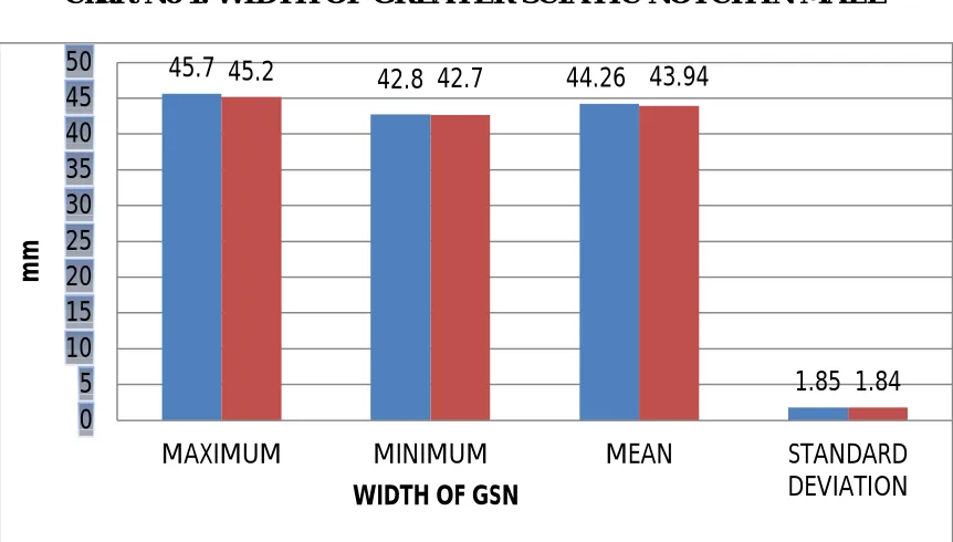
[image:55.595.94.525.497.742.2]The width of GSN in male right and left side were 44.26mm and 43.94mm with standard deviation of 1.85 and 1.85 were observed respectively.
Table No 1. WIDTH OF GREATER SCIATIC NOTCH IN MALE
Chart No 1. WIDTH OF GREATER SCIATIC NOTCH IN MALE
45.7 42.8 44.26
1.85
45.2 42.7 43.94
1.84 0
5 10 15 20 25 30 35 40 45 50
MAXIMUM MINIMUM MEAN STANDARD
DEVIATION
m
m
WIDTH OF GSN
MALE MAXIMUM WIDTH(mm)
MINIMUM WIDTH (mm)
MEAN WIDTH
(mm)
STANDARD DEVIATION
RIGHT SIDE 45.7 42.8 44.26 1.85
31
B. FEMALE
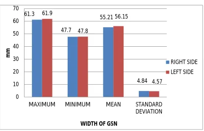
[image:56.595.94.510.401.670.2]The width of GSN of females in right side and left side were 55.21mm and 56.15mm with standard deviation of 4.84 and 4.57 were observed respectively.
Table No 2. WIDTH OF GREATER SCIATIC NOTCH IN FEMALE
FEMALE MAXIMUM WIDTH (mm)
MINIMUM WIDTH (mm)
MEAN WIDTH
(mm)
STANDARD DEVIATION
RIGHT SIDE 61.3 47.7 55.21 4.84
LEFT SIDE 61.9 47.8 56.15 4.57
Chart No 2. WIDTH OF GREATER SCIATIC NOTCH IN FEMALE
61.3
47.7
55.21
4.84 61.9
47.8
56.15
4.57
0 10 20 30 40 50 60 70
MAXIMUM MINIMUM MEAN STANDARD
DEVIATION
m
m
WIDTH OF GSN
RIGHT SIDE
32
C.COMPARISON OF THE WIDTH OF GSN
Table No 3.WIDTH OF RIGHT SIDE GSN
GENDER WIDTH (N=10) MEAN DIFFERENCE 95% CONFIDENCE
INTERVAL
P-VALUE¶ MEAN(mm) STD.
DEVIATION LOWER UPPER
MALE 44.26 1.85
-10.95 -14.39 -7.51 <0.001 FEMALE 55.21 4.84
Table No 4. WIDTH OF LEFT SIDE GSN
GENDER WIDTH (N=10) MEAN DIFFERENCE 95% CONFIDENCE
INTERVAL
P-VALUE¶ MEAN(mm) STD.
DEVIATION LOWER UPPER
MALE 43.94 1.68
-12.21 -15.44 -8.98 <0.001 FEMALE 56.15 4.57
Independent Sample T-test
Chart No 3. COMPARISON OF THE WIDTH OF GSN
44.26 43.94 55.21 56.15 0 10 20 30 40 50 60
RIGHT SIDE LEFT SIDE
m
m
WIDTH OF GSN
MALE
33 It is observed that there is
There is no significant difference between right and left side for both the sexes.
There is significant difference between males and females with mean difference of 12 mm.
‘p’ value was less than 0.001 which is statistically significant, helpful for sex determination.
Wider width of GSN was observed in females than in males.
2. DEPTH OF GREATER SCIATIC NOTCH A. MALE
The depth of GSN in male of right side and left side were 28.66mm and 26.66mm with standard deviation of 3.79and 5.08 were observed respectively.
Table No 5. DEPTH OF GREATER SCIATIC NOTCH IN MALE
MALE MAXIMUM DEPTH(mm) MINIMUM DEPTH (mm) MEAN DEPTH (mm) STANDARD DEVIATION
RIGHT SIDE 33.1 23 28.86 3.79
LEFT SIDE 35.1 20 26.66 5.08
Chart No 4. DEPTH OF GREATER SCIATIC NOTCH IN MALE
33.1 23 28.86 3.79 35.1 20 26.66 5.08 0 5 10 15 20 25 30 35 40
MAXIMUM MINIMUM MEAN STANDARD DEVIATION
m
m
DEPTH OF GSN
MALE RIGHT SIDE
34
B. FEMALE
The depth of GSN in female right and left side were 30mm and 28.6mm with standard deviation of 4.21 and 4.57 were observed respectively
Table No 6. DEPTH OF GREATER SCIATIC NOTCH IN FEMALE
FEMALE MAXIMUM DEPTH (mm)
MINIMUM DEPTH (mm)
MEAN DEPTH (mm)
STANDARD DEVIATION
RIGHT SIDE 40 25.3 30 4.21
LEFT SIDE 39 26.2 28.6 4.57
Chart No 4: DEPTH OF GREATER SCIATIC NOTCH IN MALE
40
25.3
30
4.21 39
26.2
28.6
4.57
0 5 10 15 20 25 30 35 40 45
MAXIMUM MINIMUM MEAN STANDARD DEVIATION
m
m
DEPTH OF GSN
FEMALE RIGHTSIDE
35
C.COMPARISON OF DEPTH OF GSN
Table No 7. DEPTH OF GSN RIGHT SIDE
GENDER DEPTH (N=10) MEAN DIFFERENCE 95% CONFIDENCE
INTERVAL P-VALUE¶ MEAN(mm) STD.
DEVIATION LOWER UPPER
MALE 28.86 3.79
-1.16 -4.92 2.60 0.525
FEMALE 30.02 4.21
Independent Sample T-test
Table No 7. DEPTH OF GSN LEFT SIDE
GENDER DEPTH (N=10) MEAN DIFFERENCE 95% CONFIDENCE
INTERVAL P-VALUE¶ MEAN(mm) STD.
DEVIATION LOWER UPPER
MALE 26.66 3.79
-2.00 -4.92 2.60 0.525
FEMALE 28.6 4.21
Independent Sample T-test
Chart No 6. COMPARSION OF DEPTH OF GSN
28.66 26.66 30.02 28.6 24 25 26 27 28 29 30 31
RIGHT SIDE LEFT SIDE
m
m
DEPTH OF GSN
36 It is observed that there is
No significant difference between right and left side for both the sexes. Mean difference of 1.16mm was noted between males and females in depth
of GSN with no significant difference between them.
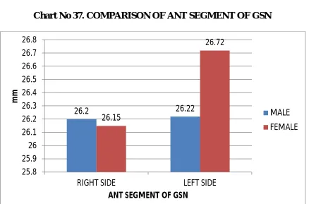
3. ANTERIOR SEGMENT OF GREATER SCIATIC NOTCH A. MALE
The anterior segment of GSN in right side and left side were 30.99mm and 31.09mm with standard deviation in right and left side were 3.75 and 3.57 respectively
Table No 9. ANT SEGMENT OF GREATER SCIATIC NOTCH IN MALE
MALE MAXIMUM ANT SEG(mm) MINIMUM ANT SEG(mm) MEAN ANT SEG(mm) STANDARD DEVIATION
RIGHT SIDE 36.1 24 30.99 3.75
LEFT SIDE 36.8 26 31.09 3.57
Chart No 7. ANT SEGMENT OF GREATER SCIATIC NOTCH IN MALE
36.1 24 30.99 3.75 36.8 26 31.09 3.57 0 5 10 15 20 25 30 35 40
MAXIMUM MINIMUM MEAN STANDARD
DEVIATION
m
m
ANT SEGMENT OF GSN
MALE RIGHT SIDE
37
B. FEMALE
The anterior segment of GSN in female right side and left side were 39.14mm and 39.97mm with standard deviation in right and left side were 4.87 and 4.40 respectively
Table No 9. ANT SEGMENT OF GREATER SCIATIC NOTCH IN FEMALE
FEMALE
MAXIMUM ANT SEG
(mm)
MINIMUM ANT SEG(mm)
MEAN ANT SEG(mm)
STANDARD DEVIATION
RIGHT SIDE 46.2 30.6 39.14 4.87
LEFT SIDE 46.3 30.6 39.97 4.57
Chart No 8. ANT SEGMENT OF GSN IN FEMALES
46.2
30.6
39.14
4.87 46.3
30.6
39.97
4.57
0 5 10 15 20 25 30 35 40 45 50
MAXIMUM MINIMUM MEAN STANDARD
DEVIATION
m
m
38
C. COMPARISON OF ANTERIOR SEGMENT OF GSN
Table No.11 ANT SEGMENT OF GSN RIGHT SIDE
GENDER
ANTERIOR SEGMENT
(N=10) MEAN
DIFFERENCE
95% CONFIDENCE
INTERVAL P-VALUE¶ MEAN(mm) STD.
DEVIATION LOWER UPPER
MALE 30.99 3.75
-8.15 -12.23 -4.07 0.001
FEMALE 39.14 4.87
Table No.12 ANT SEGMENT OF GSN LEFT SIDE
GENDER
ANTERIOR SEGMENT
(N=10) MEAN
DIFFERENCE
95% CONFIDENCE
INTERVAL P-VALUE¶ MEAN(mm) STD.
DEVIATION LOWER UPPER
MALE 31.09 3.57
-8.88 -12.64 -5.12 <0.001
FEMALE 39.97 4.40
Independent Sample T-test
Chart No 9. COMPARISON OF ANT SEGMENT OF GSN
30.99 31.09 39.14 39.97 0 5 10 15 20 25 30 35 40 45
RIGHT SIDE LEFT SIDE
m
m
ANT SEGMENT OF GSN
MALE
39 It was observed that
There is no significant difference between right and left side of males and females.
Mean difference of 8mm is noted between the males and females in the anterior segment of GSN.
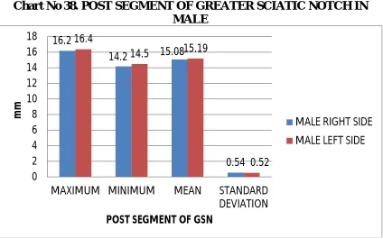
4. POSTERIOR SEGMENT OF GREATER SCIATIC NOTCH A. MALE
The posterior segment of greater sciatic notch in right and left side were 11.86mm and 11.77mm with standard deviation in right and left side were 1.33 and 1.49 respectively
Table No 13. POST SEGMENT OF GREATER SCIATIC NOTCH IN MALE MALE MAXIMUM POST SEG(mm) MINIMUM POST SEG (mm) MEAN POST SEG (mm) STANDARD DEVIATION
RIGHT SIDE 14.3 10 11.86 1.33
LEFT SIDE 14.1 10.1 11.77 1.49
Chart No 10.POST SEGMENT OF GSN IN MALE 14.3 10 11.86 1.33 14.1 10.1 11.77 1.49 0 2 4 6 8 10 12 14 16
MAXIMUM MINIMUM MEAN STANDARD
DEVIATION
m
m
POST SEGMENT OF GSN
40
C. FEMALE
The posterior segment of GSN in right side and left side were 16.20mm and 16.18mm with standard deviation in right and left side were 2.59 and 2.64 respectively
Table No 14.POST SEGMENT OF GREATER SCIATIC NOTCH IN FEMALE
FEMALE
MAXIMUM POST SEG
(mm)
MINIMUM POST SEG
(mm)
MEAN POST SEG (mm)
STANDARD DEVIATION
RIGHT SIDE 18.20 10.5 16.20 2.59
LEFT SIDE 19.3 9.9 16.18 2.64
Chart No 11. .POST SEGMENT OF GSN IN FEMALE
18.2
10.5
16.2
2.59 19.3
9.9
16.18
2.64
0 5 10 15 20 25
MAXIMUM MINIMUM MEAN STANDARD
DEVIATION
m
m
41
C.COMPARISON OF POSTERIOR SEGMENT OF GSN
Table No 15. POST SEGMENT OF GSN RIGHT SIDE
GENDER
POSTERIOR SEGMENT
(N=10) MEAN
DIFFERENCE
95% CONFIDENCE
INTERVAL P-VALUE¶ MEAN(mm) STD.
DEVIATION LOWER UPPER
MALE 11.86 1.33
-4.34 -6.27 -2.41 <0.001
FEMALE 16.20 2.59
Table No 15. POST SEGMENT OF GSN SIDE LEFT SIDE
GENDER
POSTERIOR SEGMENT
(N=10) MEAN
DIFFERENCE
95% CONFIDENCE
INTERVAL P-VALUE¶ MEAN(mm) STD.
DEVIATION LOWER UPPER
MALE 11.77 1.49
-4.41 -6.43 -2.39 <0.001
FEMALE 16.18 2.64
Chart No 12. COMPARISON OF POSTERIOR SEGMENT OF GSN
It was observed that
There is significant difference between male and females of 4.41mm in posterior segment of GSN.
11.86 11.77 16.2 16.18 0 5 10 15 20
RIGHT SIDE LEFT SIDE
m
m
POST SEGMENT OF GSN
MALE
42
No significant difference between right and left side of post segment of GSN in both males and females.
4. INDEX I OF GREATER SCIATIC NOTCH
On calculating the index I using formula Index I= Depth/Widthx100, it was found out in male right and left side is 62.54 and 60.54 with standard deviation of 10.65 1nd 10.95 respectively
Table No 17. INDEX I OF GREATER SCIATIC NOTCH IN MALE
MALE MAXIMUM INDEX I
MINIMUM INDEX I
MEAN INDEX I
STANDARD DEVIATION
RIGHT SIDE 73.7 50.1 62.54 10.65
LEFT SIDE 74.1 49.1 60.54 10.95
Chart No 13. INDEX I OF GREATER SCIATIC NOTCH IN MALE
73.7
50.1
62.54
10.65 74.1
49.1
60.54
10.95
0 10 20 30 40 50 60 70 80
MAXIMUM MINIMUM MEAN STANDARD
DEVIATION
INDEX I OF GSN
MALE RIGHT SIDE
43
B.FEMALES
On calculating the index I using formula Index I= Depth/Widthx100, it was found out in right and left side is 54.09 and 51.51 with standard deviation of 8.95 and 7.11 respectively
Table No 18. INDEX I OF GREATER SCIATIC NOTCH IN FEMALE
FEMALE MAXIMUM INDEX I
MINIMUM INDEX I
MEAN INDEX I
STANDARD DEVIATION
RIGHT SIDE 67 41.5 54.09 8.95
LEFT SIDE 61.4 40.9 51.51 7.11
Chart No.14 INDEX I OF GREATER SCIATIC NOTCH IN FEMALE
67
41.5
54.09
8.95 61.4
40.9
51.51
7.11
0 10 20 30 40 50 60 70 80
MAXIMUM MINIMUM MEAN STANDARD
DEVIATION
44
C.COMPARISON OF INDEX I OF GREATER SCIATIC NOTCH
Table No 15. INDEX I OF GSN RIGHT SIDE
GENDER INDEX I(N=10) MEAN DIFFERENCE 95% CONFIDENCE
INTERVAL P-VALUE¶ MEAN STD.
DEVIATION LOWER UPPER
MALE 62.54 10.65
8.45 -0.79 17.69 0.071 FEMALE 54.09 8.95
Independent Sample T-test
Table No 16. INDEX I OF GSN LEFT SIDE
GENDER INDEX I(N=10) MEAN DIFFERENCE 95% CONFIDENCE
INTERVAL P-VALUE¶ MEAN STD.
DEVIATION LOWER UPPER
MALE 60.54 10.95
9.03 0.35 17.71 0.042 FEMALE 51.51 7.11
Independent Sample T-test
Chart No 15. COMPARISON OF INDEX I OF GREATER SCIATIC NOTCH 62.54 60.54 54.09 51.51 0 10 20 30 40 50 60 70
RIGHT SIDE LEFT SIDE
INDEX I OF GSN
MALE
45 It is observed that
There is no significant difference between right and left sides of Index I of GSN in both sexes.
Significant difference is noted between males and females.
6. INDEX II OF GREATER SCIATIC NOTCH A.MALE
On calculating the index II using formula Index II= Anterior segment/Widthx100, it was found out in male right and left side is 70.23 and 70.58 with standard deviation of 6.60 and 6.22 respectively
Table No 21. INDEX II OF GREATER SCIATIC NOTCH IN MALE
MALE MAXIMUM INDEX II
MINIMUM INDEX II
MEAN INDEX II
STANDARD DEVIATION
RIGHT SIDE 76.8 66.1 70.23 6.60
LEFT SIDE 77.4 59.3 70.58 6.22
Chart No 16. INDEX II OF GREATER SCIATIC NOTCH IN MALE
76.8
66.1 70.23
6.6 77.4
59.3
70.58
6.22
0 10 20 30 40 50 60 70 80 90
MAXIMUM MINIMUM MEAN STANDRAD
DEVIATION
INDEX II OF GSN
MALE RIGHT SIDE
46
B.FEMALE
On calculating the index II using formula Index II= Anterior segment/Widthx100, it was found out in right and left side is 70.6 and 71.90 respectively with standard deviation of 5.08 and 5.42 respectively.
Table No 22. INDEX II OF GREATER SCIATIC NOTCH IN FEMALE
FEMALE MAXIMUM INDEX II
MINIMUM INDEX II
MEAN INDEX II
STANDARD DEVIATION
RIGHT SIDE 81.9 62.3 70.60 5.08
LEFT SIDE 82.3 64 71.90 5.42
Chart No 17. INDEX II OF GREATER SCIATIC NOTCH IN FEMALE
81.9
62.3
70.6
5.08 82.3
64
71.9
5.42
0 10 20 30 40 50 60 70 80 90
MAXIMUM MINIMUM MEAN STANDRAD
DEVIATION
47
C.COMPARISON OF INDEX II OF GREATER SCIATIC NOTCH
Table No 23. INDEX II OF GSN RIGHT SIDE
GENDER
INDEX II (N=10)
MEAN DIFFERENCE
95% CONFIDENCE
INTERVAL P-VALUE¶ MEAN STD.
DEVIATION LOWER UPPER
MALE 70.23 6.60
-0.37 -5.91 5.17 0.890 FEMALE 70.60 5.08
Table No 24. INDEX II OF GSN LEFT SIDE
GENDER
INDEX II (N=10)
MEAN DIFFERENCE
95% CONFIDENCE
INTERVAL P-VALUE¶ MEAN STD.
DEVIATION LOWER UPPER
MALE 70.58 6.22
-1.32 -6.80 4.16 0.619 FEMALE 71.90 5.42
Independent Sample T-test
Chart No.18. COMPARISON OF INDEX II OF GREATER SCIATIC NOTCH 70.23 70.58 70.6 71.9 69 69.5 70 70.5 71 71.5 72 72.5
RIGHT SIDE LEFT SIDE
INDEX II OF GSN
MALE
48 It is observed that
There is no significant difference between right and left side of both sexes. There is no much statistically significant difference between male and
female.
7. INDEX III OF GREATER SCIATIC NOTCH A.MALE
On calculating the index III using formula Index III= Posterior
segment/Widthx100, it was found out in right and left side were 26.83 and 26.82 with standard deviation of 3.54 and 3.75 respectively
Table No 25. INDEX III OF GREATER SCIATIC NOTCH IN MALE
MALE MAXIMUM INDEX III
MINIMUM INDEX III
MEAN INDEX III
STANDARD DEVIATION
RIGHT SIDE 33.7 23.1 26.83 3.54
LEFT SIDE 33.6 22.5 26.82 3.75
Chart no 19. INDEX III OF GSN IN MALE
33.7
23.1 26.83
3.54 33.6
22.5
26.82
3.75
0 5 10 15 20 25 30 35 40
MAXIMUM MINIMUM MEAN STANDARD DEVIATION
INDEX III OF GSN
MALE RIGHT SIDE
49
B.FEMALE
On calculating the index III using formula Index III= Posterior segment /Widthx100,it was found out in right and left side is 29.44 and 29.22 with standard deviation of 5.08 and 4.62 respectively
Table No 26. INDEX III OF GREATER SCIATIC NOTCH IN FEMALE
FEMALE MAXIMUM
INDEX III
MINIMUM INDEX III
MEAN INDEX III
STANDARD DEVIATION
RIGHT SIDE 37.9 18.6 29.44 5.08
LEFT SIDE 35.9 17.6 29.22 4.62
Chart No 20. INDEX III OF GSN IN FEMALE
37.9
18.6
29.44
5.08 35.9
17.6
29.22
4.62
0 5 10 15 20 25 30 35 40
MAXIMUM MINIMUM MEAN STANDARD
DEVIATION
50
C. COMPARISON OF INDEX III OF GREATER SCIATIC NOTCH
Table No 27. INDEX III OF GSN RIGHT SIDE
GENDER
INDEX III (N=10)
MEAN DIFFERENCE
95% CONFIDENCE
INTERVAL P-VALUE¶ MEAN STD.
DEVIATION LOWER UPPER
MALE 26.83 3.54
-2.61 -6.72 1.50 0.199 FEMALE 29.44 5.08
Independent Sample T-test
Table No 28. INDEX III OF GSN LEFT SIDE
GENDER
INDEX III (N=10)
MEAN DIFFERENCE
95% CONFIDENCE
INTERVAL P-VALUE¶ MEAN STD.
DEVIATION LOWER UPPER
MALE 26.82 3.95
-2.40 -6.46 1.66 0.231 FEMALE 29.22 4.67
Chart no 21.COMPARISON OF INDEX III OF GREATER SCIATIC NOTCH
26.83 26.82 29.44 29.22 25.5 26 26.5 27 27.5 28 28.5 29 29.5 30
right side left side
INDEX III OF GSN
male
51 It is observed that
There is no significant difference between right and left side of both sex Significant difference of 2.5 is noted between males and females
8. TOTAL ANGLE OF GREATER SCIATIC NOTCH
A.MALE
On construction of triangle, total angle measured in males right and left side were 56.00 and 56.20 degrees with standard deviation of 7.06 and 7.39.
Table No 29. TOTAL ANGLE OF GREATER SCIATIC NOTCH IN MALE
MALE MAXIMUM TOTAL ANGLE MINIMUM TOTAL ANGLE MEAN TOTAL ANGLE STANDARD DEVIATION
RIGHT SIDE 71 48 56.00 7.06
LEFT SIDE 68 49 56.20 7.39
Chart No 22. TOTAL ANGLE OF GREATER SCIATIC NOTCH IN MALE
71 48 56 7.06 68 49 56.2 7.39 0 10 20 30 40 50 60 70 80
MAXIMUM MINIMUM MEAN STANDARD DEVIATION
D
EG
R
EE
TOTAL ANGLE OF GSN
MALE RIGHT SIDE
52
B.FEMALE
On construction of triangle, total angle measured in females right and left side were 65.10 and 65.40 degrees with standard deviation of 10.13 and 6.59 respectively.
Table No 30. TOTAL ANGLE OF GREATER SCIATIC NOTCH IN FEMALE
FEMALE MAXIMUM TOTAL ANGLE MINIMUM TOTAL ANGLE MEAN TOTAL ANGLE STANDARD DEVIATION
RIGHT SIDE 88 52 65.10 10.13
LEFT SIDE 79 54 65.40 6.59
Chart No 23. TOTAL ANGLE OF GREATER SCIATIC NOTCH IN FEMALE
88 52 65.1 10.13 79 54 65.4 6.59 0 10 20 30 40 50 60 70 80 90 100
MAXIMUM MINIMUM MEAN STANDARD
DEVIATION
D
EG
R
EE
53
C. COMPARSION OF TOTAL ANGLE OF GSN
Table No 31. TOTAL ANGLE OF GSN RIGHT SIDE
GENDER
TOTAL ANGLE (N=10)
MEAN DIFFERENCE
95% CONFIDENCE INTERVAL
P-VALUE
MEAN STD.
DEVIATION LOWER UPPER
MALE 56.00 7.06
-.9.10 -15.34 0.34 0.008 FEMALE 65.10 8.13
Independent Sample T-test
Table No 32. TOTAL ANGLE OF GSN LEFT SIDE
GENDER
TOTAL ANGLE (N=10)
MEAN DIFFERENCE
95% CONFIDENCE INTERVAL
P-VALUE
MEAN STD.
DEVIATION LOWER UPPER
MALE 56.20 7.39
-9.20 -15.78 -2.62 0.009 FEMALE 65.40 6.59
Chart No 24. COMPARSION OF TOTAL ANGLE OF GSN
On observation it was found that
Males had narrower angle compared to females
56 56.2 65.1 65.4 50 52 54 56 58 60 62 64 66 68
RIGHT SIDE LEFT SIDE
D
EG
R
EE
TOTAL ANGLE ANGLE OF GSN
MALE
54
9. POSTERIOR ANGLE OF GREATER SCIATIC NOTCH
A.MALE
On construction of triangle, posterior angle were measured in males right and left side 22 and 24.4 degree with standard deviation of 3.94 and 4.09 respectively.
Table No 33. POSTERIOR ANGLE OF GREATER SCIATIC NOTCH IN MALE
MALE MAXIMUM POST ANGLE
MINIMUM POST ANGLE
MEAN POST ANGLE
STANDARD DEVIATION
RIGHT SIDE 28 17 22 3.94
LEFT SIDE 30 19 24.40 4.09
Chart No 25. POSTERIOR ANGLE OF GREATER SCIATIC NOTCH IN MALE
28
17
22
3.94 30
19
24.4
4.09
0 5 10 15 20 25 30 35
MAXIMUM MINIMUM MEAN STANDRAD DEVIATION
D
EG
R
EE
POST ANGLE OF GSN
MALE RIGHT SIDE
55
B.FEMALE
On construction of triangle, posterior angle were measured in females right and left side 65.10 and 65.40 degree with standard deviation of 4.79 and 2.69 respectively
Table No 34. POSTERIOR ANGLE OF GREATER SCIATIC NOTCH IN
FEMALE
FEMALE MAXIMUM POST ANGLE
MINIMUM POST ANGLE
MEAN POST ANGLE
STANDARD DEVIATION
RIGHT SIDE 39 25 30.9 4.79
LEFT SIDE 36 29 32.10 2.69
Chart No 26. POSTERIOR ANGLE OF GREATER SCIATIC NOTCH IN FEMALE
39
25
30.9
4.79 36
29 32.1
2.69
0 5 10 15 20 25 30 35 40 45
MAXIMUM MINIMUM MEAN STANDRAD
DEVIATION
D
EG
R
EE
56
C. COMPARISON
Table No 35. POST ANGLE OF GSN RIGHT SIDE
GENDER
POSTERIOR ANGLE
(N=10) MEAN
DIFFERENCE
95% CONFIDENCE INTERVAL
P-VALUE
MEAN STD.
DEVIATION LOWER UPPER
MALE 22.00 3.94
-8.90 -13.02 -4.78 <0.001 FEMALE 30.90 4.79
Independent Sample T-test
Table No 36. POST ANGLE OF GSN LEFT SIDE
GENDER
POSTERIOR ANGLE
(N=10) MEAN
DIFFERENCE
95% CONFIDENCE INTERVAL
P-VALUE
MEAN STD.
DEVIATION LOWER UPPER
MALE 24.40 4.09 -7.70 -10.95 -4.45 <0.001 FEMALE 32.10 2.69
Independent Sample T-test
Chart No 28. COMPARISON OF POSTERIOR ANGLE OF GSN
It is observed that
There is significant difference between right and left side of both sex Significant difference of 8 degree is noted between males and females
22 24.4 30.9 32.1 0 5 10 15 20 25 30 35
RIGHT SIDE LEFT SIDE
D
EG
R
EE
POST ANGLE OF GSN
MALE
57
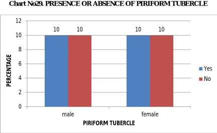
[image:82.595.93.525.486.751.2]10. PRESENCE OR ABSENCE OF PIRIFORM TUBERCLE
Table No 37. PRESENCE OR ABSENCE OF PIRIFORM TUBERCLE RIGHT
SIDE
PRESENCE OR ABSENCE OF PIRIFORM TUBERCLE
GENDER
TOTAL
P-VALUE
MALE FEMALE
YES 5(50%) 4(40%) 9(45%)
0.999
NO 5(50%) 6(60%) 11(55%)
TOTAL 10(100%) 10(100%) 20(100%)
Chi-Square test
Table No 38. PRESENCE OR ABSENCE OF PIRIFORM TUBERCLE RIGHT SIDE
PRESENCE OR ABSENCE OF PIRIFORM TUBERCLE
GENDER
TOTAL
P-VALUE
MALE FEMALE
YES 5(50%) 4(40%) 9(45%)
0.999
NO 5(50%) 6(60%) 11(55%)
TOTAL 10(100%) 10(100%) 20(100%)
Chi-Square test
Chart No29. PRESENCE OR ABSENCE OF PIRIFORM TUBERCLE
10 10 10 10
58 RADIOLOGICAL MEASUREMENTS
1. WIDTH OF GREATER SCIATIC NOTCH
A.MALE
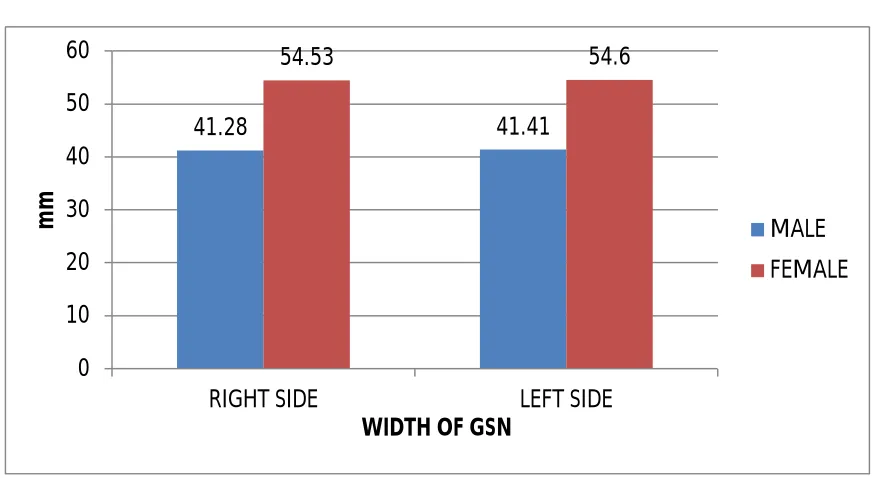
On observing the measurements of greater sciatic notch in X-ray of pelvis, it was found that the width of the notch in male right and left side is 44.4mm and 44.4mm respectively.
Table No 39. WIDTH OF GREATER SCIATIC NOTCH IN MALE
MALE MAXIMUM WIDTH(mm)
MINIMUM WIDTH (mm)
MEAN WIDTH (mm)
STANDARD DEVIATION
RIGHT SIDE 44.4 39.1 41.28 1.64
LEFT SIDE 44.4 39.2 41.41 1.50
Chart No 30. WIDTH OF GREATER SCIATIC NOTCH IN MALE
44.4
39.1 41.28
1.64 44.4
39.2 41.41
1.5 0
5 10 15 20 25 30 35 40 45 50
MAXIMUM MINIMUM MEAN STANDARD DEVIATION
m
m
WIDTH OF GSN
MALE RIGHT SIDE
59
B.FEMALE
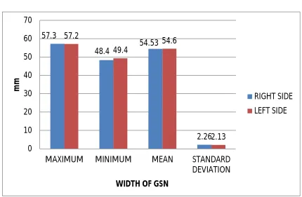
[image:84.595.93.523.384.665.2]On observing the measurements of greater sciatic notch in X-ray of pelvis, it was found that the width of the notch in right and left side is 54.53mm and 54.60mm with standard deviation of 2.26 and 2.13 respectively
Table No 40. WIDTH OF GREATER SCIATIC NOTCH IN FEMALE
FEMALE MAXIMUM WIDTH (mm)
MINIMUM WIDTH (mm)
MEAN WIDTH
(mm)
STANDARD DEVIATION
RIGHT SIDE 57.3 48.4 54.53 2.26
LEFT SIDE 57.2 49.4 54.60 2.13
Chart No-31. WIDTH OF GREATER SCIATIC NOTCH IN FEMALE
57.3
48.4 54.53
2.26 57.2
49.4
54.6
2.13 0
10 20 30 40 50 60 70
MAXIMUM MINIMUM MEAN STANDARD DEVIATION
m
m
WIDTH OF GSN
RIGHT SIDE
60
[image:85.595.86.535.135.694.2]C.COMPARISON OF WIDTH OF GSN
Table No 41. WIDTH OF G SN RIGHT SIDE
GENDER WIDTH (N=25) MEAN DIFFERENCE 95% CONFIDENCE
INTERVAL
P-VALUE¶ MEAN(mm) STD.
DEVIATION LOWER UPPER
MALE 41.28 1.64
-13.25 -14.38 -12.13 <0.001 FEMALE 54.53 2.26
[image:85.595.91.528.449.688.2]Independent Sample T-test
Table No 42. WIDTH OF GSN LEFT SIDE
GENDER WIDTH (N=25) MEAN DIFFERENCE 95% CONFIDENCE
INTERVAL
P-VALUE¶ MEAN(mm) STD.
DEVIATION LOWER UPPER
MALE 41.41 1.50 -13.19 -14.24 -12.14 <0.001 FEMALE 54.60 2.13
Independent Sample T-test
Chart No-32. COMPARISON OF WIDTH OF G SN
It is observed that
There is no difference between the right and left side of both sex
41.28 41.41 54.53 54.6 0 10 20 30 40 50 60
RIGHT SIDE LEFT SIDE
m
m
WIDTH OF GSN
MALE
61
There is significant difference between the width of both males and females
When comparing the bone and radiological study, there is significant difference in width between males and female in both studies.
2. DEPTH OF GREATER SCIATIC NOTCH A.MALE
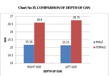
On observing the measurements of greater sciatic notch in X-ray of pelvis, it was found that the depth of the notch in male right and left side is 25.13mm and 25.75mm with standard deviation of 0.60 and 1.16respectively
Table No 43. DEPTH OF GREATER SCIATIC NOTCH IN MALE
MALE MAXIMUM DEPTH (mm)
MINIMUM DEPTH (mm)
MEAN DEPTH (mm)
STANDARD DEVIATION
RIGHT SIDE 26.3 24.2 25.18 0.60
LEFT SIDE 26.2 24.2 24.75 1.16
Chart No33. DEPTH OF GREATER SCIATIC NOTCH IN MALE
26.3
24.2 25.18