BMC Infectious Diseases 2002,
2 x
Research article
Development of real-time NASBA assays with molecular beacon
detection to quantify mRNA coding for HHV-8 lytic and latent
genes
Abeltje M Polstra*, J Goudsmit and M Cornelissen
Address: Department of Human Retrovirology, Academic Medical Center, University of Amsterdam, Amsterdam, The Netherlands E-mail: Abeltje M Polstra* - [email protected]; J Goudsmit - [email protected]; M Cornelissen - [email protected] *Corresponding author
Abstract
Background: Human herpesvirus-8 (HHV-8) is linked to the pathogenesis of Kaposi's sarcoma (KS), and the HHV-8 DNA load in peripheral blood mononuclear cells (PBMC) is associated with the clinical stage of KS. To examine the expression of HHV-8 in PBMC, four HHV-8 mRNA specific NASBA assays were developed
Methods: We have developed four quantitative nucleic acid sequence-based amplification assays (NASBA-QT) specifically to detect mRNA coding for ORF 73 (latency-associated nuclear antigen, LANA), vGCR (a membrane receptor), vBcl-2 (a viral inhibitor of apoptosis) and vIL-6 (a viral growth factor). The NASBA technique amplifies nucleic acids without thermocycling and mRNA can be amplified in a dsDNA background. A molecular beacon is used during amplification to enable real-time detection of the product. The assays were tested on PBMC samples of two AIDS-KS patients from the Amsterdam Cohort.
Results: For all four assays, the limit of detection (LOD) of 50 molecules and the limit of quantification (LOQ) of 100 molecules were determined using in vitro transcribed RNA. The linear dynamic range was 50 to 107 molecules of HHV-8 mRNA. We found HHV-8 mRNA expression in
9 out of the 10 tested samples.
Conclusion: These real-time NASBA assays with beacon detection provide tools for further study of HHV-8 expression in patient material.
Background
Homosexual men infected with HIV-1 are at increased risk for developing AIDS-related Kaposi's sarcoma (AIDS-KS) due to co-infection with HHV-8, a gamma herpes virus. Also known as Kaposi's sarcoma associated herpes virus, HHV-8 was first discovered in 1994 in KS-affected tissue [1]. Since then it also has been associated with the devel-opment of multicentric Castleman's disease and primary
effusion lymphoma (PEL) [2–5]. HHV-8 DNA has been detected in both tissues and bodily fluids [6,7]. Studies in-itially focused on HHV-8 and its association with KS, transmission of HHV-8, and the natural history of HHV-8 infection. As shown by Renwick et al. and Jacobsen et al. [8,9] seroconversion for HHV-8 during HIV infection in-creases the risk of developing KS, implying that HIV-1 has an impact on HHV-8. Laboratory assays developed for the
Published: 4 September 2002
BMC Infectious Diseases 2002, 2:18
Received: 24 May 2002 Accepted: 4 September 2002
This article is available from: http://www.biomedcentral.com/1471-2334/2/18
detection of HHV-8 include serologic assays such as im-munofluorescence antibody assays [10–13], immunoblot assays [10], and various enzyme immunoassays [14–16]. PCR-based assays have also been used to detect HHV-8 DNA [1]. With the serologic assays, sensitivity and specif-icity are always problematic. No single assay is completely sensitive and specific [17].
We would prefer a method using viral RNA or DNA as a marker for the presence of HHV-8. Though DNA is an ob-vious choice because HHV-8 is a DNA virus, we have cho-sen RNA, because it indicates activity of the virus and may thus be a better prognostic marker. Active infected cells produce more RNA than DNA. In situ hybridisation has shown that most HHV-8 infected cells in KS lesions are la-tently infected [18,19]. but lytic HHV-8 infection is clearly present in a small proportion of cells [20–22].
A method for the detection of RNA is RT-PCR. To increase sensitivity an additional amplification step can follow the RT-PCR. This method is time consuming and also increas-es the likelihood of false positive reactions due to cross-contamination. Nucleic acid sequence-based amplifica-tion (NASBA) is a single-step isothermal RNA-specific am-plification process [23] that amplifies mRNA in a dsDNA background [24]. NASBA has proved successful in the de-tection of various mRNAs [24–26] and in the dede-tection of both viral [23] and bacterial [27] RNA in clinical samples.
By combining the standard NASBA technology [28] with a molecular beacon that anneals during amplification to the target sequence, a real-time detection system is gener-ated [29]. This paper describes our development of real-time NASBA assays to quantify four functionally different genes of HHV-8: ORF 73, a latent gene, vGCR, a constitu-tively active gene, and the lytic genes vBcl-2 and vIL-6. To normalise the input mRNA we developed a fifth NASBA for a constitutively expressed mRNA, U1A mRNA, and standardised the specific HHV8 mRNAs to the expression of U1A mRNA.
Methods
HHV-8 genes
Our first assay was developed for ORF 73 and is based on the amplification of a 192 bp fragment situated within the gene. ORF 73 encodes the major immunogenic latency-as-sociated nuclear antigen (LANA) of HHV-8. LANA is es-sential for maintenance of this virus in latently infected cells [30,31]. It is expressed only in such cells and thus gives a good indication of latent HHV-8 infection. In ad-dition, LANA interacts with the tumour suppressor pro-tein p53 and represses its transcriptional activity [32].
Our second assay was developed for vGCR, G-protein cou-pled receptor, and amplifies a 168 bp fragment. A receptor
that binds several CXC and CC chemokines, vGCR ap-pears to be constitutively active [33], although some stud-ies indicate that it is expressed only during lytic replication [34]. It may contribute to the pathogenesis of KS by increasing the release of cellular growth factors such as VEGF [33]. It transforms rodent fibroblasts and induces expression of angiogenic factors [35].
The final two assays were developed for the genes vBcl-2 and vIL-6. The assay for vBcl-2 amplifies a 216-bp frag-ment, and for 6 the fragment is 186 bp. vBcl-2 and vIL-6 are both early lytic genes, as their expression increases within 10 hours after induction [9].
vBcl-2 is a member of the Bcl-2 family, and functional studies indicate that vBcl-2 prevents Bax-mediated apop-tosis and thus is an anti-apoptotic protein [36,37]. It is primarily active during lytic replication and its expression pattern suggests that it may function to prolong the sur-vival of cells in which lytic infection is present.
vIL-6 is a secreted cytokine that maintains proliferation of IL-6-dependent mouse and human mycelia cell lines [21,38,39] and prevents apoptosis [21,38,40].
NASBA
A NASBA reaction is based on the simultaneous activity of avian myeloblastosis virus (AMV) reverse transcriptase (RT), RNase H and T7 RNA polymerase with two oligonu-cleotide primers to produce amplification of the desired fragment more than 1012 fold in 90 to 120 minutes
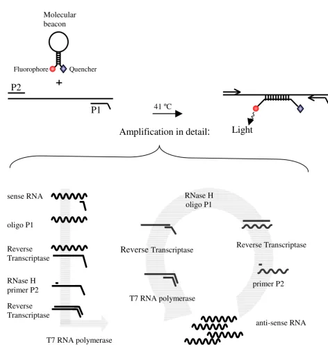
Figure 1
NASBA amplification reaction NASBA amplification reaction with the P1 (anti-sense) – P2 (sense) oligonucleotide primer set. The overhang on the P1 encodes the promoter sequence for the T7 RNA polymerase. A molecular beacon with a fluoro-phore and a quencer with the NASBA amplification reaction generates a real-time detection system.
Quencher
P1
P2
+
41 ºC
Light
oligo P1
Reverse
Transcriptase
RNase H
primer P2
Reverse
Transcriptase
T7 RNA polymerase
sense RNA
Reverse Transcriptase
Reverse
Transcriptase
RNase H
oligo P1
anti-sense RNA
T7 RNA polymerase
primer P2
Molecular
beacon
Amplification in detail
:
Standard RNA
The NASBA quantification method is based on a standard curve with a known input of RNA. For this input we used
in vitro RNA that was transcribed from four different plas-mids. These plasmids were generated by cloning a specific PCR product for each of the four target genes, using prim-er sets located on the bordprim-ers of the diffprim-erent genes. Prim-er sequences are shown in table 1. Input for the PCR was the nucleic acids of the BCP-1 cell line (a cell line contain-ing HHV-8). The sizes of the four different PCR fragments were 595 bp, 926 bp, 473 bp, and 589 bp for ORF 73, vGCR, vBcl-2 and vIL-6, respectively (table 1). Each PCR fragment was cloned into the TOPO-TA plasmid (Invitro-gen/ Lifetechnologies, Carlsbad, USA), and after all inserts were verified by sequencing, the fragments were cloned in a plasmide, pBluescript SK (Stratagene, La Jolla, USA). In vitro RNA was generated from these plasmids using T7 or T3 RNA polymerase, depending on the orientation of the fragment, and was treated with DNase to remove the plas-mid. The Q-RNA was purified by RNeasy (Qiagen, Hilden, Germany), quantified spectrophotometrically, and checked by eye on agarose gel. The resulting in vitro RNA was used for the four different standard curves.
Real-time amplification system
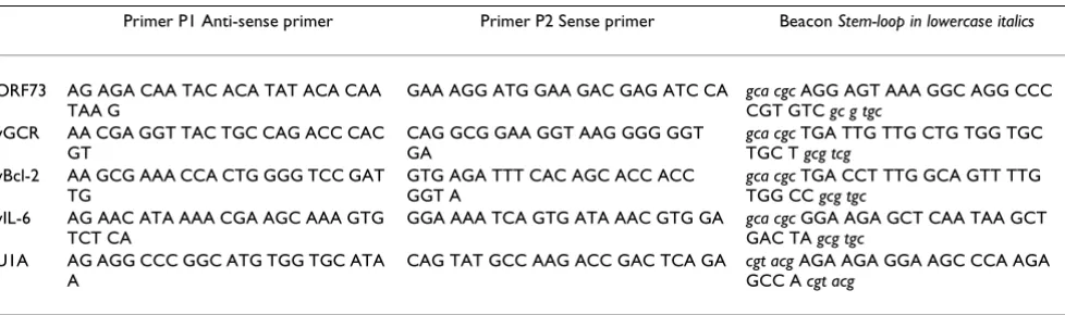
NASBA amplification requires a sense and an anti-sense primer, and for real-time detection a unique beacon is added to the reaction. We developed molecular beacons that could hybridise with the known sequences of the four HHV-8 genes, as shown in table 2. All the beacons have 6-fluorescine (6-FAM) as the fluorescent label at the 5' end and a (4-(dimethylamino) phenyl) azo) benzoic acid (Dabcyl) as universal quencher at the 3' end. A BLAST analysis revealed that in each case, neither the primers nor the beacon shared significant homology with any known nucleotide sequences other than the target gene. The qual-ity of the different beacons was tested with a melting curve (from 80°C to 20°C) and acryl amide gel electrophoresis.
Each reaction consisted of 5 ml of the sample RNA and 10
[image:4.612.56.550.99.188.2]ml of NASBA reaction mix. This mix consisted of 80 mM Tris-HCl [pH 8.5], 24 mM MgCl2, 140 mM KCl, 1.0 mM DTT, 2.0 mM of each dNTP, 4.0 mM each of ATP, UTP and CTP, 3.0 mM GTP, and 1.0 mM ITP in 30% DMSO. This solution also contained the anti-sense and sense primers for amplification and the molecular beacons used for de-tection. The final concentration for the primes was 0.1 mM and for the beacons 40 nM.
Table 1: Primers for PCR fragment in plasmids
5' primer 3' primer PCR fragment size
ORF 73 agcccaccaggagataataca tcatttcctgtggagagtccc 595 bp vGCR gcggatatgactactctggaaact gaggctttggaagagaccgt 926 bp vBcl-2 atggacgaggacgttttgcct cccaatagcgctgtcattct 473 bp vIL-6 ggttcaagttgtggtctctctt ggagtcacgtctgggatagagt 589 bp
Table 2: Sequences of the primers and beacons for each of the assays
Primer P1 Anti-sense primer Primer P2 Sense primer Beacon Stem-loop in lowercase italics
ORF73 AG AGA CAA TAC ACA TAT ACA CAA TAA G
GAA AGG ATG GAA GAC GAG ATC CA gca cgc AGG AGT AAA GGC AGG CCC CGT GTC gc g tgc
vGCR AA CGA GGT TAC TGC CAG ACC CAC GT
CAG GCG GAA GGT AAG GGG GGT GA
gca cgc TGA TTG TTG CTG TGG TGC TGC T gcg tcg
vBcl-2 AA GCG AAA CCA CTG GGG TCC GAT TG
GTG AGA TTT CAC AGC ACC ACC GGT A
gca cgc TGA CCT TTG GCA GTT TTG TGG CC gcg tgc
vIL-6 AG AAC ATA AAA CGA AGC AAA GTG TCT CA
GGA AAA TCA GTG ATA AAC GTG GA gca cgc GGA AGA GCT CAA TAA GCT GAC TA gcg tgc
U1A AG AGG CCC GGC ATG TGG TGC ATA A
CAG TAT GCC AAG ACC GAC TCA GA cgt acg AGA AGA GGA AGC CCA AGA GCC A cgt acg
[image:4.612.61.550.241.386.2]The reaction mixtures were incubated at 65°C for 5 min, and after cooling to 41°C for 5 min to allow for primer an-nealing, 5 ml of enzyme mix was added by pipetting into the lids of the microtubes and subsequently spinning down. This mix contained, per reaction, 375 mM sorbitol, 2.1 mg BSA, 0.08 U RNase H, 32 U T7 RNA polymerase and 6.4 U AMV reverse transcriptase. Reactions were incu-bated at 41°C for 120 min in a fluorometer (Cytofluor 4000; Perkin-Elmer, Wellesley, Mass.). The RNA ampli-cons generated in the NASBA process are detected by mo-lecular beacons [29]. These beacons generate a fluorescent signal during amplification when hybridising with their target. The fluorescence is measured continuously during amplification for real-time monitoring (i.e., as the reac-tion proceeds). A threshold is determined by setting the fluorescence emitted in the first 5 measure points as a background. The time-point at which a reaction rises above this threshold of detection and becomes positive is determined. This time-to-positivity (TTP) principle is sim-ilar to real-time PCR [43]. A calibration curve with 50, 102, 103, 104, 105, 106 and 107 molecules HHV-8 mRNA
was included in each experiment. The number of mRNA copies per sample input can be extrapolated from the standard curve. De Baar et al showed that it is possible to use ttp values for quantification without the use of an in-ternal calibrator [44].
RNA quantification
For standardisation of the amount of RNA input the U1A assay was used in each experiment. U1A mRNA encodes for one of the proteins of the U1 snRNP [45,46]. Because U1A is constitutively expressed, the amount of U1A mRNA measured in the samples is an indication of total RNA input. The mean of 6 assays (variation 0.6 log) was taken as the input RNA amount of the sample, and sample input RNA was normalised accordingly. The NASBA con-ditions for the U1A assay were the same as those for the HHV-8 assay, with the exception of the final concentra-tion of the primer (0.2 mM) and the beacon (50 nM).
HHV-8 DNA assay
The DNA fractions of the PBMC samples were tested for the presence of HHV-8 DNA using a nested PCR for ORF 73 described by Goudsmit et al. [47]. As a quantification of the amount of DNA input, a limited dilution PCR for the cellular CCR5 gene was used. With limited dilution, an estimation of copies of HHV-8 DNA was made in rela-tion to the amount of cells. For both the ORF 73 and the CCR5 nested PCR, the detection level was 5 molecules in-put per reaction.
Patient samples
To test our assays in vivo, we selected PBMC samples from two participants of the Amsterdam Cohort Studies. De-scribed elsewhere in detail [48,49], they were both
HIV-1-infected men with Kaposi's sarcoma but had very different disease development. Patient 1 was a 30-year-old man who was demonstrated to be HIV-1-seropositive in 1995. In July 1996 he was diagnosed with KS. He did not re-spond to anti-retroviral or chemotherapy and died in Jan-uary of 1997 as a result of severe infiltration of KS in both lungs. Patient 2 is a 36-year-old man who visited our out-patient clinic in 1992 with an increasing number of KS-re-lated skin lesions. After he started anti-retroviral therapy several lesions disappeared and complete remission was gradually reached over the course of two years. These two patients were chosen in large part for the diversity in their course of Kaposi's sarcoma. We tested three samples for patient 1 and seven samples for patient 2 collected over time, all taken after diagnosis of KS.
After the frozen PBMC samples were thawed the cells were collected and resuspended in Trizol™ buffer to isolate RNA and DNA simultaneously, according to the manufac-turer's recommendations. Precipitated RNA was redis-solved in 50 ml H2O. For each time-point, a known amount of cells (7.6–11.1 * 106 cells) were isolated. With
high input of total RNA we found that background RNA can disturb the amplification if present in large amounts and it was necessary to dilute the samples. To have a broad range in the dilution for the RNA quantification in the NASBA experiments, the samples were diluted 10,100, and 1000 times. Of these diluted samples 5 ml was used per reaction, and 10 ml of reaction mix was added. U1A mRNA was used as an external standard to compare quan-tification signals among the different samples.
Results
Sensitivity and specificity
These newly developed assays can be used quantitatively by testing a standard curve of samples with a known amount of mRNA molecules within the same experiment as the unknown samples, then extrapolating the results to the standard curve. Typical amplification curves could be plotted in which an increase of fluorescence was observed until most of the molecular beacon had hybridised with the synthesised amplicons and the fluorescence reached a maximum level.
Figure 2
Standard curves in vitro RNA Figure 2 a-d. Relationship of time-to-positivity (TTP) to HHV-8 mRNA copy number. The number of mRNA molecules present in the reaction is indicated on the x axis and the ttp value in indicated on the y axis. For the data obtained with 50, 102 to 107 molecules, the values of TTP are the mean of five replicates of independent experiments.
For the U1A the range of RNA input was 103 to 108. Error bars indicate the standard deviation for the values of ttp. The solid
line was obtained by linear regression analysis of the data from 50 to 107 molecules, and the dotted lines indicate the 95%
con-fidence intervals for the regression. Inserts a-d: Amplification plot of a 10-fold dilution serial dilution of in vitro RNA for ORF 73, vGCR, vBcl-2 and vIL-6. The amount of input RNA 1 * 107, 1 * 106, 1 * 105, 1 * 104, 1 * 103, 1 * 102, 50 and 0 molecules.
Insert e: Amplification plot of a 10-fold dilution serial dilution of U1A in vitro RNA, the amount of input RNA 1 * 108, 1 * 107, 1
* 106, 1 * 105, 1 * 104, 1 * 103 and 0 molecules.
The dilution series of in vitro RNA showed a detection lev-el of 50 molecules input. For accurate quantification the required level is 102 molecules input per assay. The line is
calculated with linear regression analysis. Besides the lim-it of detection (LOD), the limlim-it of quantification (LOQ) can be determined. For ORF 73, the LOD is 50 molecules input (84% pos.) while the LOQ is 102 molecules per
re-action. The linear quantification of the ORF 73 assay was determined to be between 102 to 107 copies RNA per
re-action (R 2 = 0.96, p < 0.001, fig. 2a). The sensitivity of all
four assays was studied using five serial dilutions. The lin-ear regression analyses for the other assays are shown in figure 2b,2c,2d,2e. The assay for vGCR has a very small standard deviation, and the amplification is linear from 107 to 50 molecules input per reaction (R 2 > 0.99, p <
0.001, fig. 2b). The LOD is 50 molecules (55% pos.) and the LOQ is 102 molecules per reaction. The assays for both
vBcl-2 and vIL-6 are weaker but still within a workable range. As above, the linear quantification was between 102 to 107 copies RNA per reaction (R 2 = 0.94 and R 2 =
0.95 respectively, p < 0.001, fig. 2c,2d). For both assays, the LOD is 50 molecules (72% and 50% pos. respectively) and the LOQ is 102 molecules per reaction. U1A has a
lin-ear quantification range of 103 to 108 molecules (R2 =
0.99, p < 0.001, fig 2e). The assay for U1A had a sensitivity of only 103 copies input/reaction, which is feasible due to
the high expression level of U1A.
The specificity of the five assays was tested by spiking each reaction with 106 molecules of in vitro RNA that the
prim-ers and beacon were not designed for. For example, ORF 73 in vitro RNA did not give a signal with the vGCR assay (data not shown).
Robustness
In replicate independent experiments, the time-to-positiv-ity (TTP) value had a linear relationship with the loga-rithm of the amount of in vitro RNA added in the reaction. Subsequent experiments used serial 10-fold dilutions of in vitro RNA (from 107 to 102 and 50 molecules per reaction)
as a standard curve to determine the amount of HHV-8 RNA in samples. The experiments were done on different days and by different people with no significant variation in results, which confirmed the robustness of the assays and also their ease of performance.
Patient samples
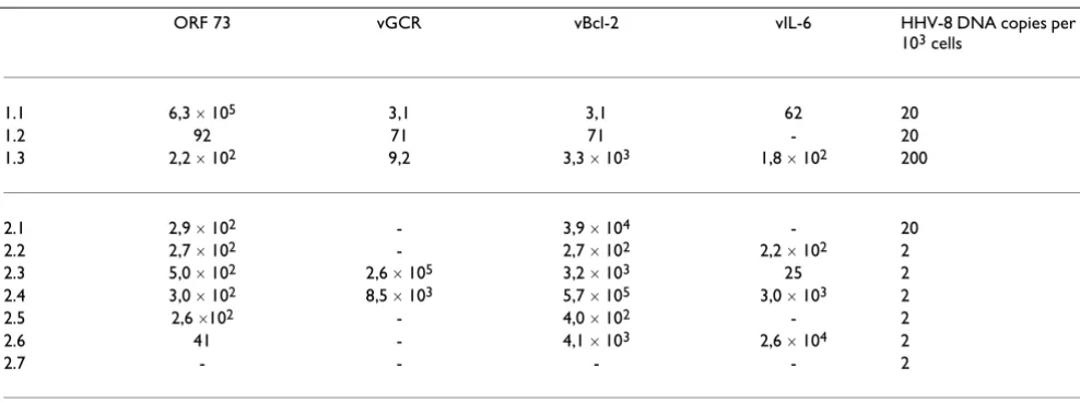
To address the functionality and quality of the HHV-8 as-says in vivo, they were tested on PBMC samples collected after KS diagnosis from two participants in the Amster-dam Cohort Studies. In total, 10 frozen samples were an-alysed. As a positive control, all samples were tested for the presence of HHV-8 DNA with a nested PCR and were found to be positive (see Materials and Methods). The copies of HHV-8 DNA varied between 2 and 2 * 102 per
103 cells (table 3). A relation has been shown between the
HHV-8 DNA levels in PBMC and the extent of Kaposi's sarcoma [50]. A possible correlation between DNA levels and mRNA expression should be investigated.
[image:7.612.57.551.98.282.2]The results of the HHV-8 assays are shown in table 3. Of the 10 samples, 9 were positive for ORF 73, whereas only half were positive for vGCR. While vBcl-2 was measured in 9 out of 10 samples, vIL-6 was detected in approximate-ly half of the samples. All the samples that were positive for ORF 73 were also positive for vBcl-2. Surprisingly the samples were positive for a latent and a lytic gene at the same time. The variation in expression levels is large both
Table 3: The ratio mRNA of (HHV-8 / U1A) ´ 107
ORF 73 vGCR vBcl-2 vIL-6 HHV-8 DNA copies per 103 cells
1.1 6,3 ´ 105 3,1 3,1 62 20
1.2 92 71 71 - 20
1.3 2,2 ´ 102 9,2 3,3 ´ 103 1,8 ´ 102 200
2.1 2,9 ´ 102 - 3,9 ´ 104 - 20
2.2 2,7 ´ 102 - 2,7 ´ 102 2,2 ´ 102 2
2.3 5,0 ´ 102 2,6 ´ 105 3,2 ´ 103 25 2
2.4 3,0 ´ 102 8,5 ´ 103 5,7 ´ 105 3,0 ´ 103 2
2.5 2,6 ´102 - 4,0 ´ 102 - 2
2.6 41 - 4,1 ´ 103 2,6 ´ 104 2
2.7 - - - - 2
among the different assays and among the samples. No clear trend in the expression of HHV-8-specific mRNA can be distinguished, based on HHV-8 DNA levels, patient, or disease status.
Discussion
In general the NASBA assay has several advantages over the PCR technique [51]. The amplification procedure of the NASBA assay is entirely isothermal and is conducted at 41°C; it does not require thermal cycling instrumenta-tion. The NASBA product is single-stranded and can there-fore be readily detected by hybridisation analysis.
Our four real-time NASBA assays offer these benefits and also the five features that define validation of any assay: accuracy, sensitivity, precision, robustness, and specificity. With the use of standard curves in each experiment, accu-racy is achieved. The sensitivity is determined with a linear standard curve. The limit of detection (LOD) and the limit of quantification (LOQ) are determined. For all the HHV-8 assays, the LOD is 50 molecules per reaction and LOQ was 100 molecules per reaction. To achieve linear ampli-fication requires at least 5 concentrations on the linear portion of the curve. For the HHV-8 assays, the linear range spans 7 concentrations and for U1A it spans 6 con-centrations. Quantification was achieved within this line-ar range of at least six orders of magnitude (102 to 107
molecules per reaction). Within this linear range quantifi-cation of at least six orders of magnitude (102 to 107
mol-ecules per reaction) was achieved.
The precision of the assays was measured as the standard deviation and the confidence interval (fig. 2). We used 5 replicates of 7 different concentrations of the standard curve. Robustness was determined by having different people perform the experiments on different days. Specif-icity was shown with spiking experiments. The lower de-tection level of the assays was approximately 50 copies per reaction. The assays work equally well in vivo as in vitro
e.g., for quantification of HHV-8 RNA in PBMCs.
The onset of KS is associated with a high HHV-8 load in PBMC [19] and this indicates that HHV-8 infected circu-lating cells play an important role in KS development [20,52,53]. HHV-8 can infect circulating B cells, mono-cytes/macrophages, T cells and KS-like spindle cell pro-genitors [54–56]. In KS lesions B cells are rare or absent, but monocytes macrophages are abundantly present [52], this suggest that circulating monocytes may recruit the vi-rus into tissues or transmit the vivi-rus to other cells. Since lymphocytes and monocytes infiltrating KS lesions can be productively infected by HHV-8 [20,22], it is important to look at active infection in the PBMC. PBMCs of two AIDS-KS patients were selected and analysed for the presence of HHV-8 mRNA.
In 90% of our samples, ORF 73 mRNA and vBcl-2 mRNA were detected, whereas for vIL-6 mRNA and vGCR mRNA were present in 60% and 50%, respectively. The expres-sion levels of all four genes varied widely. Comparing the different expression levels of the four HHV-8 genes within one sample or comparing expression levels of one gene in one patient did not show an apparent tendency. No clear trend can be distinguished based on the HHV-8 DNA lev-els, patient or disease status. Nevertheless the finding of expression of different HHV8 specific genes in PBMCs is notable in itself, as it has not been described before. HHV-8 RNA has been found in tissues, endothelial cells and spindle cells [18,57,58]. In 90% of fresh KS-affected tis-sues, HHV-8 RNA has been detected with RT-PCR [59]. In situ hybridisation and LANA immunohistochemistry have confirmed HHV-8 gene expression in KS spindle cells within KS lesions [18,58,60–62]. Expression of lytic HHV-8 genes has also been documented in monocytes and macrophages in KS lesions [20].
Conclusion
The assays that were developed work well for the detection and quantification of HHV-8 specific mRNA. Although no clear trend in the expression pattern could be distin-guished the finding of expression of different HHV8 spe-cific genes in PBMCs is important in itself. As this suggests that HHV-8 replication takes place in the PBMC. The PB-MCs we used were obtained from two patients. To draw conclusions about the clinical and prognostic utility of these assays more patients and patient samples need to be analysed. However, as the assays are able to quantify HHV-8 mRNA in PBMC samples we feel that these real-time NASBA assays with beacon detection provide tools for further study of HHV-8 expression in patient material.
Competing interests
None declared.
Authors' contributions
AP carried out the assay development and validation, pre-formed the NASBA experiments and drafted the manu-script. JG participated in the study design. MC participated in the study design and carried out its supervision.
Acknowledgements
We thank Remco van den Burg, Margreet Bakker and Eveline Timmermans for their technical assistance, Bob van Gemen for his advice on assay design and Lucy Phillips for editorial review.
References
1. Chang Y, Cesarman E, Pessin MS, Lee F, Culpepper J, Knowles DM, Moore PS: Identification of herpesvirus-like DNA sequences in AIDS-associated Kaposi's sarcoma.Science 1994, 266: 1865-1869
3. Cesarman E, Moore PS, Rao PH, Inghirami G, Knowles DM, Chang Y: In vitro establishment and characterization of two acquired immunodeficiency syndrome-related lymphoma cell lines (BC-1 and BC-2) containing Kaposi's sarcoma-associated herpesvirus-like (KSHV) DNA sequences.Blood 1995, 86: 2708-2714
4. Soulier J, Grollet L, Oksenhendler E, Cacoub P, Cazals-Hatem D, Bab-inet P, D'Agay M-F, Clauvel J-P, Raphael M, Degos L, et al: Kaposi's sarcoma-associated herpesvirus-like DNA sequences in mul-ticentric Castleman's disease.Blood 1995, 86:1276-1280 5. Rettig MB, Ma HJ, Vescio RA, Pold M, Schiller G, Belson D, Savage A,
Nishikubo C, Wu C, et al: Kaposi's sarcoma-associated herpes-virus infection of bone marrow dendritic cells from multiple myeloma patients.Science 1997, 276:1851-1854
6. LaDuca JR, Love JL, Abbott LZ, Dube S, Freidman-Kien AE, Poiesz BJ: Detection of human herpesvirus 8 DNA sequences in tissues and bodily fluids.Journal of Infectious Diseases 1998, 178:1610-1615 7. Stamey FR, Patel MM, Holloway BP, Pellett PE: Quantitative, fluor-ogenic probe pcr assay for detection of human herpesvirus 8 dna in clinical specimens.J Clin Microbiol 2001, 39:3537-3540 8. Renwick N, Halaby T, Weverling GJ, Dukers NHTM, Simpson GR,
Coutinho RA, Lange LMA, Schulz TF, Goudsmit J: Seroconversion for human herpesvirus 8 during HIV infection is highly pre-dictive of Kaposi's sarcoma.AIDS 1998, 12:2481-2488
9. Jenner RG, Alba MM, Boshoff C, Kellam P: Kaposi's sarcoma-asso-ciated herpesvirus latent and lytic gene expression as re-vealed by DNA arrays.Journal of Virology 2001, 75:891-902 10. Gao SJ, Kingsley L, Li M, Zheng W, Parravicini C, Ziegler J, Newton
R, Rinaldo CR, Saah A, Phair J, et al: KSHV antibodies among Americans, Italians and Ugandans with and without Kaposi's sarcoma.Nat Med 1996, 2:925-928
11. Kedes DH, Operskalski E, Busch M, Kohn R, Flood J, Ganem D: The seroepidemiology of human herpesvirus 8 (Kaposi's sarco-ma-associated herpesvirus): distribution of infection in KS risk groups and evidence for sexual transmission.Nat Med
1996, 2:918-924
12. Lennette ET, Blackbourn DJ, Levy JA: Antibodies to human her-pesvirus type 8 in the general population and in Kaposi's sar-coma patients.Lancet 1996, 348:858-861
13. Koelle DM, Huang ML, Chandran B, Vieira J, Piepkorn M, Corey L: Frequent detection of Kaposi's sarcoma-associated herpes-virus (human herpesherpes-virus 8) DNA in saliva of human immu-nodeficiency virus-infected men: clinical and immunologic correlates.J Infect Dis 1997, 176:94-102
14. Chatlynne LG, Lapps W, Handy M, Huang YQ, Masood R, Hamilton AS, Said JW, Koeffler HP, Kaplan MH, Friedman-Kien A, et al: Detec-tion and titraDetec-tion of human herpesvirus-8-specific antibodies in sera from blood donors, acquired immunodeficiency syn-drome patients, and Kaposi's sarcoma patients using a whole virus enzyme-linked immunosorbent assay.Blood 1998, 92: 53-58
15. Davis DA, Humphrey RW, Newcomb FM, O'Brien TR, Goedert JJ, Straus SE, Yarchoan R: Detection of serum antibodies to a Ka-posi's sarcoma-associated herpesvirus-specific peptide.J In-fect Dis 1997, 175:1071-1079
16. Smith MS, Bloomer C, Horvat R, Goldstein E, Casparian JM, Chandran B: Detection of human herpesvirus 8 DNA in Kaposi's sarco-ma lesions and peripheral blood of husarco-man immunodeficien-cy virus-positive patients and correlation with serologic measurements.J Infect Dis 1997, 176:84-93
17. Spira TJ, Lam L, Dollard SC, Meng YX, Pau CP, Black JB, Burns D, Cooper B, Hamid M, Huong J, et al: Comparison of serologic as-says and PCR for diagnosis of human herpesvirus 8 infection. J Clin Microbiol 2000, 38:2174-2180
18. Staskus KA, Zhong W, Gebhard K, Herndier B, Wang H, Renne R, Beneke J, Pudney J, Anderson DJ, Ganem D, et al: Kaposi's sarco-ma-associated herpesvirus gene expression in endothelial (spindle) tumor cells.Journal of Virology 1997, 71:715-719 19. Decker LL, Shankar P, Khan G, Freeman RB, Dezube BJ, Lieberman J,
Thorley DA-Lawson: The Kaposi sarcoma-associated herpesvi-rus (KSHV) is present as an intact latent genome in KS tissue but replicates in the peripheral blood mononuclear cells of KS patients.Journal of Experimental Medicine 1996, 184:283-288 20. Blasig C, Zietz C, Haar B, Neipel F, Esser S, Brockmeyer NH,
Tsch-achler E, Colombini S, Ensoli B, Sturzl M: Monocytes in Kaposi's
sarcoma lesions are productively infected by human herpes-virus 8.Journal of Virology 1997, 71:7963-7968
21. Moore PS, Boshoff C, Weiss RA, Chang Y: Molecular mimicry of human cytokine and cytokine response pathway genes by KSHV.Science 1996, 274:1739-1744
22. Orenstein JM, Alkan S, Blauvelt A, Jeang K-T, Weinstein MD, Ganem D, Herndier B: Visualization of human herpesvirus type 8 in Kaposi's sarcoma by light and transmission electron micros-copy.AIDS 1997, 11:F35-F45
23. Kievits T, Van Gemen B, Van Strijp D, Schukkink R, Dircks M, Adri-aanse H, Malek L, Sooknanan R, Lens P: NASBA(TM) isothermal enzymatic in vitro nucleic acid amplification optimized for the diagnosis of HIV-1 infection.Journal of Virological Methods
1991, 35:273-286
24. Heim A, Grumbach IM, Zeuke S, Top B: Highly sensitive detection of gene expression of an intronless gene: Amplification of mRNA, but not genomic DNA by nucleic acid sequence based amplification (NASBA). Nucleic Acids Research 1998, 26:2250-2251
25. Blok MJ, Goossens VJ, Vanherle SJV, Top B, Tacken N, Middeldorp JM, Christiaans MHL, Van Hooff JP, Bruggeman CA: Diagnostic value of monitoring human cytomegalovirus late pp67 mRNA ex-pression in renal-allograft recipients by nucleic acid se-quence-based amplification.Journal of Clinical Microbiology 1998, 36:1341-1346
26. Darke BM, Jackson SK, Hanna SM, Fox JD: Detection of human TNF-alpha mRNA by NASBA(TM). Journal of Immunological Methods 1998, 212:19-28
27. Morre SA, Sillekens P, Jacobs MV, van Aarle P, de Blok S, Van Gemen B, Walboomers JM, Meijer CJ, van den Brule AJ: RNA amplification by nucleic acid sequence-based amplification with an inter-nal standard enables reliable detection of Chlamydia tracho-matis in cervical scrapings and urine samples.J Clin Microbiol
1996, 34:3108-3114
28. Van Gemen B, Wiel VP, Van Beuningen R, Sillekens P, Jurriaans S, Dries C, Schoones R, Kievits T: The one-tube quantitative HIV-1 RNA NASBA: Precision, accuracy, and application. PCR Methods & Applications 1995, 4:S177-S184
29. Leone G, Van Schijndel H, Van Gemen B, Kramer FR, Schoen CD: Molecular beacon probes combined with amplification by NASBA enable homogeneous, real-time detection of RNA. Nucleic Acids Research 1998, 26:2150-2155
30. Ballestas ME, Chatis PA, Kaye KM: Efficient persistence of extra-chromosomal KSHV DNA mediated by latency-associated nuclear antigen.Science 1999, 284:641-644
31. Cotter MA, Robertson ES: The latency-associated nuclear anti-gen tethers the Kaposi's sarcoma-associated herpesvirus ge-nome to host chromosomes in body cavity-based lymphoma cells.Virology 1999, 264:254-264
32. Friborg J Jr, Kong W, Hottiger MO, Nabel GJ: p53 inhibition by the LANA protein of KSHV protects against cell death.Nature
1999, 402:889-894
33. Arvanitakis L, Geras-Raaka E, Varma A, Gershengorn MC, Cesarman E: Human herpesvirus KSHV encodes a constitutively active G-protein-coupled receptor linked to cell proliferation. Na-ture 1997, 385:347-349
34. Kirshner JR, Staskus K, Haase A, Lagunoff M, Ganem D: Expression of the open reading frame 74 (G-protein-coupled receptor) gene of Kaposi's sarcoma (KS)-associated herpesvirus: Impli-cations for KS pathogenesis.Journal of Virology 1999, 73: 6006-6014
35. Bais C, Santomasso B, Coso O, Arvanitakis L, Raaka EG, Gutkind JS, Asch AS, Cesarman E, Gerhengorn MC, Mesri EA: G-protein-cou-pled receptor of Kaposi's sarcoma-associated herpesvirus is a viral oncogene and angiogenesis activator. Nature 1998, 391:86-89
36. Sarid R, Sato T, Bohenzky RA, Russo JJ, Chang Y: Kaposi's sarcoma-associated herpesvirus encodes a functional Bcl-2 homo-logue.Nature Medicine 1997, 3:293-298
38. Nicholas J, Ruvolo VR, Burns WH, Sandford G, Wan X, Ciufo D, Hen-drickson SB, Guo H-G, Hayward GS, Reitz MS: Kaposi's sarcoma-associated human herpesvirus-8 encodes homologues of macrophage inflammatory protein-1 and interleukin-6. Na-ture Medicine 1997, 3:287-292
39. Burger R, Neipel F, Fleckenstein B, Savino R, Ciliberto G, Kalden JR, Gramatzki M: Human herpesvirus type 8 interleukin-6 homo-logue is functionally active on human myeloma cells.Blood
1998, 91:1858-1863
40. Neipel F, Albrecht J-C, Ensser A, Huang Y-Q, Jian J, Li , Kien AE, Fleck-enstein B: Human herpesvirus 8 encodes a homolog of inter-leukin-6.Journal of Virology 1997, 71:839-842
41. Compton J: Nucleic acid sequence-based amplification.Nature
1991, 350:91-92
42. Tyagi S, Kramer FR: Molecular beacons: Probes that fluoresce upon hybridization.Nature Biotechnology 1996, 14:303-308 43. Higuchi R, Fockler C, Dollinger G, Watson R: Kinetic PCR
analy-sis: Real-time monitoring of DNA amplification reactions. Bio-Technology 1993, 11:1026-1030
44. De Baar MP, van Dooren MW, de Rooij E, Bakker M, Van Gemen B, Goudsmit J, De Ronde A: Single rapid real-time monitored iso-thermal RNA amplification assay for quantification of hu-man immunodeficiency virus type 1 isolates from groups M, N, and O.J Clin Microbiol 2001, 39:1378-1384
45. Maniatis T, Reed R: The role of small nuclear ribonucleoprotein particles in pre-mRNA splicing.Nature 1987, 325:673-678 46. Maxwell ES, Fournier MJ: The small nucleolar RNAs.Annu Rev
Bi-ochem 1995, 64:897-934
47. Goudsmit J, Renwick N, Dukers NHTM, Coutinho RA, Heisterkamp S, Bakker M, Schulz TF, Cornelissen M, Weverling GJ: Human her-pesvirus 8 infections in the Amsterdam Cohort Studies (1984–1997): Analysis seroconversions to ORF65 and ORF73.Proceedings of the National Academy of Sciences of the United States of America 2000, 97:4838-4843
48. Prins JM, Sol CJA, Renwick N, Goudsmit J, Veenstra J, Reiss P: Fa-vourable effect of chemotherapy on clinical symptoms and human herpesvirus-8 DNA load in a patient with Kaposi's sarcoma presenting with fever and anemia.European Journal of Clinical Microbiology & Infectious Diseases 1999, 18:499-502
49. Wit FWN, Sol CJA, Renwick N, Roos MTL, Pals Van ST, Leeuwen R, Goudsmit J, Reiss P: Regression of AIDS-related Kaposi's sarco-ma associated with clearance of husarco-man herpesvirus-8 from peripheral blood mononuclear cells following initiation of antiretroviral therapy [2].AIDS 1998, 12:218-219
50. Campbell TB, Borok M, Gwanzura L, MaWhinney S, White IE, Ndem-era B, Gudza I, Fitzpatrick L, Schooley RT: Relationship of human herpesvirus 8 peripheral blood virus load and Kaposi's sarco-ma clinical stage.AIDS 2000, 14:2109-2116
51. Romano JW, Shurtliff RN, Dobratz E, Gibson A, Hickman K, Markham PD, Pal R: Quantitative evaluation of simian immunodeficien-cy virus infection using NASBA technology. J Virol Methods
2000, 86:61-70
52. Fiorelli V, Gendelman R, Sirianni MC, Chang HK, Colombini S, Markham PD, Monini P, Sonnabend J, Pintus A, Gallo RC, et al: gam-ma-Interferon produced by CD8+ T cells infiltrating Kaposi's sarcoma induces spindle cells with angiogenic phenotype and synergy with human immunodeficiency virus-1 Tat pro-tein: an immune response to human herpesvirus-8 infection? Blood 1998, 91:956-967
53. Sirianni MC, Vincenzi L, Fiorelli V, Topino S, Scala E, Uccini S, Angeloni A, Faggioni A, Cerimele D, Cottoni F, et al: gamma-Interferon production in peripheral blood mononuclear cells and tumor infiltrating lymphocytes from Kaposi's sarcoma patients: correlation with the presence of human herpesvirus-8 in pe-ripheral blood mononuclear cells and lesional macrophages. Blood 1998, 91:968-976
54. Mesri EA, Cesarman E, Arvanitakis L, Rafii S, Moore MA, Posnett DN, Knowles DM, Asch AS: Human herpesvirus-8/Kaposi's sarco-ma-associated herpesvirus is a new transmissible virus that infects B cells.J Exp Med 1996, 183:2385-2390
55. Sirianni MC, Vincenzi L, Topino S, Scala E, Angeloni A, Gonnella R, Uccini S, Faggioni A: Human herpesvirus 8 DNA sequences in CD8+ T cells.J Infect Dis 1997, 176:541
56. Sirianni MC, Uccini S, Angeloni A, Faggioni A, Cottoni F, Ensoli B: Cir-culating spindle cells: correlation with human herpesvirus-8 (HHV-8) infection and Kaposi's sarcoma.Lancet 1997, 349:255
57. Ascherl G, Hohenadl C, Monini P, Zietz C, Browning PJ, Ensoli B, Sturzl M: Expression of human herpesvirus-8 (HHV-8) encod-ed pathogenic genes in Kaposi's sarcoma (KS) primary le-sions.Adv Enzyme Regul 1999, 39:331-339
58. Sturzl M, Blasig C, Schreier A, Neipel F, Hohenadl C, Cornali E, Ascherl G, Esser S, Brockmeyer NH, Ekman M, et al: Expression of HHV-8 latency-associated T0.7 RNA in spindle cells and en-dothelial cells of AIDS-associated, classical and African Ka-posi's sarcoma.Int J Cancer 1997, 72:68-71
59. Huang YQ, Li JJ, Zhang WG, Feiner D, Friedman-Kien AE: Tran-scription of human herpesvirus-like agent (HHV-8) in Kapo-si's sarcoma.J Clin Invest 1996, 97:2803-2806
60. Sturzl M, Wunderlich A, Ascherl G, Hohenadl C, Monini P, Zietz C, Browning PJ, Neipel F, Biberfeld P, Ensoli B: Human herpesvirus-8 (HHV-8) gene expression in Kaposi's sarcoma (KS) primary lesions: an in situ hybridization study.Leukemia 1999, 13(Suppl 1):S110-S112
61. Davis MA, Sturzl MA, Blasig C, Schreier A, Guo HG, Reitz M, Opal-enik SR, Browning PJ: Expression of human herpesvirus 8-en-coded cyclin D in Kaposi's sarcoma spindle cells.J Natl Cancer Inst 1997, 89:1868-1874
62. Luppi M, Barozzi P, Maiorana A, Trovato R, Marasca R, Morselli M, Cagossi K, Torelli G: Expression of cell-homologous genes of human herpesvirus-8 in human immunodeficiency virus-neg-ative lymphoprolifervirus-neg-ative diseases.Blood 1999, 94:2931-2933
Pre-publication history
The pre-publication history for this paper can be accessed here:
http://www.biomedcentral.com/1471-2334/2/18/prepub
Publish with BioMed Central and every scientist can read your work free of charge
"BioMedcentral will be the most significant development for disseminating the results of biomedical research in our lifetime."
Paul Nurse, Director-General, Imperial Cancer Research Fund
Publish with BMCand your research papers will be:
available free of charge to the entire biomedical community peer reviewed and published immediately upon acceptance cited in PubMed and archived on PubMed Central yours - you keep the copyright
[email protected] Submit your manuscript here:
http://www.biomedcentral.com/manuscript/