2
Commentary
Mouse models for breast cancer
Lothar Hennighausen
National Institute of Diabetes, Digestive and Kidney Diseases, Bethesda, Maryland, USA
Received: 15 November 1999
Revisions requested: 22 November 1999 Revisions received: 30 November 1999 Accepted: 1 December 1999
Published: 17 December 1999
Breast Cancer Res2000, 2:2–7
© Current Science Ltd
APC = adenomatous polyposis coli; BLG = beta lactoglobulin; ER = estrogen receptor; LTR = long terminal repeat; min = multiple intestinal neo-plasia; MMTV = mouse mammary tumor virus; MT = metallothionein; WAP = whey acidic protein.
Introduction
Recent advances in gene targeting technologies in the mouse have taken us one leap closer to understanding the genetic pathways that operate during normal mammary gland development and tumorigenesis. The possibility to delete or mutate genes specifically in mammary epithelial cells and at predetermined time points permits investiga-tors to analyze the fates of defined cell types in the absence of confounding systemic effects. Gene deletion (knockout) and transgenic mice, both alone and in combi-nation, can be used to address specific questions in developmental and cancer biology. The genetic ablation of steroid (estrogen and progesterone) and peptide (pro-lactin, epidermal growth factor) hormone receptors and their ligands has provided a deep insight into their function during ductal and alveolar development and has shed light on their redundancy and parallel pathways. Finally, the deletion of transcription factors, including those that mediate peptide hormone signaling, has revealed distinct roles in epithelial cell proliferation, differentiation, and death (for a detailed assessment of genetic approaches to study mammary development, see [1]). Rather than describing individual models (an array of mouse models will be presented in depth in the January 2000 issue of the journal Oncogene), herein I discuss some of the lessons we have learned during the past 15 years from the mice models that are at hand, and the technological hurdles we now encounter. Like in many explorations, the initial con-cepts, approaches, and tools are rather crude and need to be further developed and refined as new information streams in and new hypotheses are articulated. On the basis of this need I present contemporary approaches that should aid our quest to identify and understand molecular pathways of pathogenesis.
Experiments conducted by Philip Leder and coworkers 15 years ago represent a milestone in breast cancer research [2]. They fused the long terminal repeat (LTR) of the mouse mammary tumor virus (MMTV) to the human c-myc proto-oncogene and incorporated this hybrid gene into mice. These transgenic mice expressed the human myc protein in their mammary glands, which resulted in the development of breast tumors [2]. This landmark paper helped to establish an entirely new research arena poised to identify genetic pathways that control breast cancer. After decades of research on tissue culture cells, both federal and private funding agencies saw the opportunity to extend investigations into settings that more closely resembled the human condition. Fifteen years after the study by Leder and coworkers, research by Deng (a former student of Leder) and coworkers set another mile-stone towards this goal. These investigators succeeded in inactivating the breast cancer gene Brca1 specifically in mammary epithelial cells of mice, and they demonstrated that mammary tumors coincided with genome instability [3]. The distinct lesson learned from these studies was that the wrongful expression of an oncogene and the inac-tivation of a tumor suppressor gene in mice can cause cancer, just like in humans. However, the mycand Brca1 mice differ in two fundamental aspects from the human situation. In the myc mice oncogene activity occurs as early as puberty, whereas in humans genetic changes leading to cancer may occur later in life. The appearance of tumors in Brca1conditional mice depends on the loss of both alleles, whereas in humans only one BRCA1allele is altered (for discussion, see [4]).
3
molecular and pharmacological agents to treat and prevent cancer. Over the past 15 years almost 100 mouse models have been generated that permit the investigation of defined aspects of tumorigenesis. The impact of trans-genic mouse models on breast cancer research was the topic of recent conferences in Annapolis, Maryland (March 3–5, 1999) and Bar Harbor, Maine, USA (The Jackson Laboratory Conference on ‘Cancer of the Mammary Gland’, October 5–8, 1999) [5]. It is fair to say that not a single model by itself covers the full spectrum of this disease, but that individual models address distinct aspects. Each transgene targets different signaling path-ways outside and inside the mammary cell, and disrupts these pathways at different time points during develop-ment. In addition, the concomitant disruption of some physiologic parameters provided insight into the cellular requirements for cellular transformation to occur.
At the Annapolis conference, pathologists and basic researchers convened and assessed different mouse models. Specifically, they asked the following key ques-tion: How similar are mouse models to the human condi-tion? A panel of nine medical and veterinary pathologists with expertise in mammary gland biology reviewed material representing more than 90% of the mouse models. A nomenclature was developed and recommendations for future analyses were drafted. The consensus report from the Annapolis meeting, including the ‘Annapolis guide-lines’ will be published in an upcoming issue of the journal Oncogene[6]. It is suggested that the Annapolis nomen-clature is adopted by the research community and in fed-erally funded research. In addition, the recent development of a web-based interactive histology atlas [7] now permits the comparison of high-resolution images from mouse models and human breast cancer, and researchers in different locations can view, discuss, anno-tate, and compare histologic images in real time. The his-tology atlas in conjunction with the database for genetically engineered mice [8] will provide in depth infor-mation on genetic pathways in human breast cancer and corresponding mouse models.
In comparing the biology from human breast tumors with that of mammary tumors in genetically engineered mice, the Annapolis pathologists identified similarities and differences (Table 6 in [6]). Among the similarities identified are as follows: molecular lesions that cause breast cancer in humans can also cause cancer in genetically engineered mice; lesions in both species display similar morphologic patterns; multi-hit kinetics of cancer development; mammary cancers in both species are metastatic; and mammary cancer is frequently hormone independent. Among the dif-ferences are as follows: some molecular lesions that cause mammary cancer in mice have not been found in human mammary cancer; the morphology of most mouse tumors does not resemble that of common human cancers; some
transgenes in mouse appear to be associated with single-hit kinetics; most mouse tumors metastasize to the lung, whereas most human tumors metastasize to the lymph nodes; and half of the human cancers are hormone independent, but most mouse tumors are hormone depen-dent. Although many transgenic mice display dissimilarities to the human condition, it is likely that their usefulness extends into understanding molecular pathways that lead to cancer initiation and progression. For example, the viral oncogene that encodes the SV40 T antigen cannot be linked to human breast cancer, but the respective trans-genic mice provide insight into cell-cycle control during hyperplasia and tumor progression (see below).
Models at hand
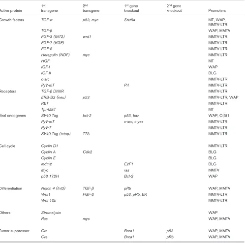
Many of the models that have been used to date are pre-sented in Table 1, and will be discussed in detail in a special edition of the journal Oncogene, which will be published in January, 2000. It is necessary to consider several variables in assessing a genetically engineered model. Most notably, the nature of the transgene deter-mines the developmental and/or tumorigenic phenotype. The regulatory region controlling transgene expression defines the cell type affected and the temporal onset of the phenotype. Because there is experimental evidence to suggest that tumor progression is a multistep process probably involving different signaling pathways, researchers have generated mice that carry more than one transgene. Again, the pioneering study came from the lab-oratory Leder and coworkers in 1987 [9], and demon-strated the synergism between the oncoproteins myc and ras. Gain of function studies in transgenic mice that carry growth regulators or oncogenes address only one aspect of tumorigenesis, and the role of tumor suppressor genes and the presence of endogenous hormonal signaling cannot be ignored. Experimentally these issues are addressed through the deletion of tumor suppressor genes and the genetic ablation of endogenous hormone signaling pathways.
appear-4
ance of mammary hyperplasia and tumors is among the pre-dominant phenotypes in these mice.
The second lesson focuses on the positional and temporal effect of an oncogenic stimulus. Depending on the pro-moter, the transgene is activated in ductal and alveolar cells
[image:3.612.58.556.117.611.2][MMTV-LTR, C(3)1], preferentially in alveolar cells [whey acidic protein (WAP), beta lactoglobulin (BLG)], or in a large variety of cells (MT). In addition, high activity of MMTV-driven transgenes can be detected earlier than WAP-controlled genes. The consequences of such differences are exemplified by the int3/notch4gene, in that MMTV-int3 Table 1
Description of genetically engineered mice that develop mammary hyperplasias and tumors
1st 2nd 1stgene 2ndgene
Active protein transgene transgene knockout knockout Promoters
Growth factors TGF-α p53, myc Stat5a MT, WAP,
MMTV-LTR
TGF-β WAP, MMTV
FGF-3 (INT2) wnt1 MMTV-LTR
FGF-7 (KGF) MMTV-LTR
FGF-8 MMTV-LTR
Heregulin (NDF) myc MMTV-LTR
HGF MT
IGF-I WAP
IGF-II BLG
c-src MMTV-LTR
PyV-mT Prl MMTV-LTR
Receptors TGF-βDNIIR MMTV-LTR
ERB-B2 (neu) p53 MMTV-LTR, WAP
RET MMTV-LTR
Tpr-MET MT
Viral oncogenes SV40 Tag bcl-2 p53, bax WAP, C(3)1
PyV-mT c-src, c-yes MMTV-LTR
PyV-T MMTV-LTR
SV40 Tag (tetop) TTA MMTV-LTR
Cell cycle Cyclin D1 MMTV-LTR
Cyclin A Cdk2 BLG
Cyclin E BLG
mdm2 E2F1 BLG
Myc ras MMTV
p53 172H Bcl-2 WAP
Differentiation Notch 4 (Int3) TGF-β pRb WAP, MMTV
Wnt1 FGF-3 p53, pRb, ER MMTV-LTR
Wnt 10b MMTV-LTR
Others Stromelysin WAP
Ras myc WAP, MMTV
Tumor suppressor Cre Brca1 p53 WAP, MMTV
Cre Brca1 pRb WAP, MMTV
mice have an early onset of tumorigenesis and do not form alveolar structures, whereas WAP-int3mice develop tumors later and exhibit lobuloalveolar compartments.
The third lesson centers on the identification of parallel and interconnected pathways through the generation of bitransgenic and gene deletion mice. Synergism of two different oncoproteins revealed two-hit kinetics and paral-lel pathways (eg ras and myc, and transforming growth factor-αand myc), and the deletion of Stat5a in the back-ground of transforming growth factor-α transgenic mice linked the epidermal growth factor receptor and the Jak2/Stat5 pathway in tumor progression.
Lessons from mouse mammary tumor virus
and its ‘tagged’ genes
The first mouse strains that had a high incidence of mammary tumors were not transgenic mice, but rather were certain inbred strains developed more than 60 years ago at the Jackson Laboratory in Bar Harbor, Maine. Bit-tner’s original demonstration of the ‘milk factor’ in mouse strains that had a high incidence of mammary tumors led to the discovery of the MMTV [10]. Proviruses of MMTV are integrated in the mouse genome and, as somatic ‘genetic mutagens’, they have the capacity to activate jux-taposed cellular genes, which can function in some cases as oncogenes. Originally MMTV was used as a ‘molecular tag’ to identify those genes that had been disrupted as a consequence of proviral insertion [11]. Several classes of molecules were identified and they include Wnt proteins, members of the Fgf family, and cell fate proteins of the Notch type (reviewed in [12]). The first protein to be iden-tified in MMTV-induced tumors was Wnt1, a protein that signals through a receptor called Frizzled (Fz) and the β -catenin pathway. The Wnt1 signaling molecule has played an exceptional role in our understanding of the synergy between signaling pathways. MMTV-wnt1 transgenic mice, which develop hyperplasia and tumors early in life, have been bred with many other transgenic and gene knockout mice, and a wealth of information on signaling pathways has emerged. For example, as with other trans-genes, wnt1 synergizes with Fgf signals, but it does not depend on the presence of the estrogen receptor (ER)-α, suggesting that tumor progression is independent of estrogen. More recently, however, a second form of the ER was discovered and its role remains obscure. In addi-tion, p53-mediated cell death has been demonstrated in wnt1-induced tumors (see The cell cycle, below).
The lesson learned from studies with the MMTV (viral infections and transgenic experiments) centers on cooper-ating pathways that are operative during tumor progres-sion [12]. In particular, the infection of wnt1 transgenic mice with MMTV has led to the identification that members of the Fgf family are the preferred cooperative partners in the dysregulation of normal growth control. The
infection of new and improved mouse models, such as the conditional Brca1mice, with MMTV may result in the iden-tification of additional growth regulators that are relevant to human breast cancer. Although this approach has not recently yielded new genes in the context of the wnt1 transgenic mice, the outcome with other transgenic and knockout mice cannot be predicted. Because the fre-quency with which the common integration sites for MMTV are rearranged by the virus in mammary tumors is dependent on the host strain [12], it could also be possi-ble to use this system to identify genetic modifiers.
The cell cycle
Disrupting the cell cycle is an obvious strategy for a tumor cell to escape growth control. A variety of oncogenes do precisely this, and have therefore been choice genes for expression in transgenic mice. Some of them have obvious links to human cancers, such as those that encode myc and cyclins; the use of others, though, such as the viral oncogenes, is less direct. It was vital to use viral onco-genes in the early days of transonco-genesis, however, because they disrupt key nuclear and cytoplasmic signaling path-ways that are operative in human cancer. These studies provide critical insight into global growth control networks. Mice that express the SV40 T antigen led to an under-standing of cell-cycle regulation during tumor progression and provided compelling evidence that both p53 -depen-dent and p53-independent pathways are operative in mammary tissue.
The tumor suppressor gene p53 is mutated in approxi-mately 50% of primary human breast cancers. Its role in mouse models has been addressed through the expres-sion of viral oncogenes that bind to and thus inactivate p53. However, SV40 T antigen dismantles the cell cycle through binding to, and thus the inactivation of several key regulators, including p53and pRb, which makes it difficult to dissect the contribution of individual components. In order to address the role of p53 specifically, researchers deleted one or two copies of the gene in the presence of different transgenes, including myc, ras, and wnt. In general, the absence of one or two p53 alleles did not accelerate the formation of mammary tumors, but it did accelerate tumorigenesis in other organs, such as the sali-vary gland. The only acceleration of mammary tumors in transgenic mice in the absence of functional p53 was observed in context of the wnt1 transgene, suggesting that p53-dependent cell death is critical in this genetic framework. Although the presence of p53is not critical for cell death in mammary tissue that proceeds during involu-tion, it may well contribute to cell death after the introduc-tion of genomic lesions. Deleintroduc-tion of both alleles of the Brca1 gene from mammary tissue leads to tumors after approximately 1 year [3]. The concomitant deletion of one copy of p53 accelerates tumor formation, which is often
6
Hormonal signaling and cancer
It is well established that the presence of estrogen is a risk factor for mammary tumorigenesis. However, its exact mol-ecular action and the entirety of signaling pathways that are affected is not understood. The availability of the ER-α and ER-β knockout mice, in conjunction with transgenic oncomice should provide some of the answers. Studies with ER-α-null mice and the wnt1oncogene have demon-strated that the presence of a strong oncogenic stimulus does not require the synergism of the ER-α. Experiments with less potent oncoproteins and natural lesions, such as the deletion of the Brca1gene, will provide further insight into the modulatory role of estrogens. Prolactin signals through the Jak2/Stat5 pathway is required for functional development of mammary tissue. A role of prolactin in tumor initiation and/or progression had been proposed, and recent experiments using transgenic mice and both prolactin-null and Stat5a-null mice have confirmed this.
The course ahead
The tidal wave of transgenic studies has provided a wealth of information about molecular pathways and cancer physi-ology. These studies have also revealed problems inherent in transgenic mice and technical challenges that have to be met. The challenges come in different categories, which include the variable biology of mouse strains, the different expression pattern of transgenes, and the development of new technologies to control multiple genes simultaneously. There is no longer any doubt that the nature of the mouse strain can greatly influence the latency and even the type of the tumor caused by the transgenic oncoprotein. This was not an apparent problem in the early days of transgenesis (mice were generated in only a few inbred backgrounds and in C57BL/6 × SJL hybrids), when investigators studied mice that carried individual transgenes. More recently, however, investigators have studied mice that carry several transgenes and gene deletion mutations that are normally generated through complex breeding strate-gies. This resulted in the introduction of the 129 strain background, which clearly behaves in a different manner from that of the classic FVB/N transgenic strain. Concerted effort is being made by investigators and centers, such as the Jackson Laboratory, to breed all transgenic and gene knockout strains into the 129 and C57BL/6 background, accelerated through the use of speed congenics. The dis-covery of distinct strain differences also provides an oppor-tunity to identify modifier genes in a defined setting that is not possible in humans. The power of such systems has been demonstrated with the adenomatous polyposis coli (APC)/multiple intestinal neoplasia (min) locus. By crossing the Apcmin/+ locus from the C57BL/6J strain into other
inbred strains, strong variations in adenoma multiplicity were observed.
Biologic challenges include the dissection of the role of individual cell types in mammary tissue in the process of
tumor progression. Gene knockout and transplant studies have revealed a cross-talk between the stroma and the epithelium (both compartments themselves consist of several cell types). At this point, however, the choice of promoters to target transgenes is restricted to those that are specific to epithelial cells. In addition to the LTR of the MMTV, promoters from milk protein genes (WAP, β -lac-toglobulin, β-casein) and the C3(1) promoter have been used to control transgenes. Expression of these control elements is targeted to the mammary epithelium and enhanced by lactogenic hormones. As a result, in many cases the tumor latency is slightly shorter in multiparous mice. In addition, the temporal – and perhaps spatial – activity of these promoters is distinct, which determines the target of the oncogenic stimulus. It is time to initiate a search for promoters that target transcription preferentially to stroma cells (adipocytes and fibroblasts) in the mammary gland. Since mammary stroma probably has unique features that distinguish it from adipocytes within other organs, it will be necessary to identify genes with expression that is specific to this compartment. It is experi-mentally possible to clear the epithelium from the mammary fat pad, and thus identify genes that are expressed within the stroma at different developmental stages. One ongoing effort of the Mammary Genome Program is the identifica-tion of expressed sequence tags that are expressed in stromal structures [13]. The large-scale expressed sequence tag programs currently underway in the mouse may be the best way to identify genes with expression that is confined (or preferential) to the mammary stroma.
for the first time to delete (and reactivate) genes in the cell-specific and time-specific manner. Traditional knock-out experiments based on embryonic stem cell-based gene targeting permitted the identification of ‘early’ gene functions, but not those that were suspected subsequent to the observed defect. For example, in the absence of functional Stat5a, mammary development is abrogated and mice do not lactate [17]. These studies did not provide any information regarding whether Jak2/Stat5a signaling is required for the maintenance of lactation, however. Using temporal tools it should now be possible to maintain Stat5 function throughout pregnancy and delete it after established lactation.
One major hurdle centers on the simultaneous inactivation of several members of a given gene family, such as cell survival factors from the bcl-2 family. Because these genes are found at different locations, a knockout approach is inherently difficult. To modulate expression from several genes simultaneously it may be necessary to revisit the antisense strategy, and to develop appropriate transgenic vectors.
After 15 years of innovative, intensive and productive research the mammary community has identified genetic pathways of breast cancer, and therapeutic and preventive compounds are now being tested in mouse models [18]. Our understanding of the pathways that control normal mammary physiology in the mouse and human is still rudi-mentary, and we are only at the beginning of the road to replicating human cancer in mice. Whereas researchers in the 20th century focused on the identification of signals
and genetic pathways that control mammary development, researchers in the 21stcentury will need to focus on the
interphase of normal physiology and cancer.
Acknowledgements
The author thanks the members of his laboratory, and Bob Cardiff and Priscilla Furth for continuously establishing new challenges and thoughtful discussions.
References
1. Hennighausen L, Robinson GW: Think globally, act locally: the making of a mouse mammary gland. Genes Dev1998, 12:449–455. 2. Stewart TA, Pattengale PK, Leder P: Spontaneous mammary adenocarcinomas in transgenic mice that carry and express MTV/myc fusion genes. Cell1984, 38:627–637.
3. Xu X, Wagner KU, Larson D, et al: Conditional mutations of Brca1 in mammary epithelial cells results in blunted ductal morphogenesis and tumor formation. Nature Genet1999, 22:37–43.
4. Gusterson B, Howard B, Crook T, Tennent B: http://breast-cancer-research.com/vol1no1/02jul99/dispatch/1, 1999.
5. Annapolis Guidelines (http://mammary.nih.gov/Annapolis-guidelines) 6. Cardiff RD, Anver MR, Gusterson BA, et al: The mammary pathology
of genetically engineered mice: the consensus report and recom-mendation from the Annapolis meeting.Oncogene2000 (in press). 7. Interactive Histology Atlas (http://histology.nih.gov).
8. Genetically engineered mice (http://cancermodels.nih.gov).
9. Sinn E, Muller W, Pattengale P, et al: Coexpression of MMTV/v-Ha-ras and MMTV/c-myc genes in transgenic mice: synergistic action of oncogenes in vivo. Cell1987, 49:465–475.
10. Bittner JJ: Science 1936, 34:162.
11. Varmus HE: Cancer Surv1982, 1:309–320.
12. Callahan R, Smith G: MMTV-induced mammary tumorigenesis: gene discovery, progression to malignancy and cellular pathways.
Oncogene2000 (in press).
13. Biology of the Mammary Gland (http://mammary.nih.gov).
14. Ewald D, Li M, Efrat S, et al: Time sensitive reversal of hyperplasia in transgenic mice expressing SV40 T antigen. Science 1996, 273:1384–1386.
15. Utomo ARH, Nikitin AY, Lee W-H: Temporal, spatial, and cell type-specific control of Cre-mediated DNA recombination in transgenic mice.Nature Biotech1999, 17:1091–1096.
16. Wagner KU, Wall RJ, St. Onge L, et al: Cre mediated deletion in the mammary gland.Nucleic Acids Res1997, 25:4323–4330. 17. Liu X, Robinson GW, Wagner KU, et al: Stat5a is mandatory for
adult mammary gland development and lactogenesis.Genes Dev 1997, 11:179–186.
18. Bearss DJ, Subler MA, Hundley JE, Troyer DA, Salinas RA, Windle JJ: Genetic determinants of response to chemotherapy in transgenic mouse mammary and salivary tumors.Oncogene2000 (in press).
Author’s affiliation:Laboratory of Genetics and Physiology, National Institute of Diabetes, Digestive and Kidney Diseases, National Institutes of Health, Bethesda, Maryland, USA
Correspondence:Lothar Hennighausen, Laboratory of Genetics and Physiology, Building 8, Room 101, National Institute of Diabetes, Digestive and Kidney Diseases, National Institutes of Health, Bethesda, MD 20892, USA. Tel: +1 301 496 2716; fax: +1 301 480 7312; e-mail: [email protected]