“A COMPARATIVE STUDY ON ELECTIVE LAPAROSCOPIC CHOLECYSTECTOMY WITH AND
WITHOUT ANTIMICROBIAL THERAPY”
THE TAMILNADU Dr. MGR MEDICAL UNIVERSITY
In partial fulfilment of the regulations for the Award of the degree A COMPARATIVE STUDY ON ELECTIVE
LAPAROSCOPIC CHOLECYSTECTOMY WITH AND WITHOUT ANTIMICROBIAL THERAPY”
Dissertation Submitted to
THE TAMILNADU Dr. MGR MEDICAL UNIVERSITY
Chennai-600 032
In partial fulfilment of the regulations for the Award of the degree
M.S. (General Surgery) Branch – I
MADRAS MEDICAL COLLEGE CHENNAI
MAY - 2019
A COMPARATIVE STUDY ON ELECTIVE LAPAROSCOPIC CHOLECYSTECTOMY WITH AND
WITHOUT ANTIMICROBIAL THERAPY”
THE TAMILNADU Dr. MGR MEDICAL UNIVERSITY
CERTIFICATE
This is to certify that, the dissertation entitled “A COMPARATIVE STUDY ON ELECTIVE LAPAROSCOPIC CHOLECYSTECTOMY WITH AND WITHOUT ANTIMICROBIAL THERAPY” is the bonafide work done by DR.A.SURESHKUMAR, during his M.S. (General Surgery) course 2016-2019, done under my supervision and is submitted in partial fulfilment of the requirement for the M.S.(BRANCH-I) General Surgery of The Tamil Nadu Dr.MGR Medical University, May 2019 examination.
GUIDE HOD
Prof. M.ALLI M.S., DGO Prof. M.ALLI M.S., DGO
DEAN Professor & Director i/c
Institute of General Surgery Madras Medical College
Chennai – 03.
Professor & director i/c Institute of General Surgery
Madras Medical College Chennai – 03.
DR. R. JAYANTHI M.D., FRCP, THE DEAN
Madras Medical College & Rajiv Gandhi Government
General Hospital
DECLARATION
I solemnly declare that this dissertation “A COMPARATIVE STUDY ON ELECTIVE LAPAROSCOPIC CHOLECYSTECTOMY WITH AND WITHOUT ANTIMICROBIAL THERAPY” was prepared by me at Institute of General surgery, madras medical college and RAJIV GHANDHI GOVERNMENT GENERAL HOSPITAL, CHENNAI under the guidance and supervision of PROF.M.ALLI.M.S.,D.G.O, professor of general surgery, institute of general surgery, madras medical college, Chennai. This dissertation is submitted to the Tamil Nadu DR.MGR Medical University, Chennai in fulfillment of the university regulation for the award of the degree M.S.General Surgery (branch 1).
DR.A.SURESHKUMAR, M.B.B.S, Post Graduate in General Surgery
CERTIFICATE – II
This is to certify that this dissertation work titled “A COMPARATIVE STUDY ON ELECTIVE LAPAROSCOPIC CHOLECYSTECTOMY WITH AND WITHOUT ANTIMICROBIAL THERAPY” of the candidate Dr.A.SURESHKUMAR with registration Number 221611015 for the award of M.S degree in the branch of General Surgery. I personally verified the urkund.com website for the purpose of plagiarism Check. I found that the uploaded thesis file contains from introduction to conclusion pages and result shows 11% percentage of plagiarism in the dissertation.
ACKNOWLEDGEMENT
First, I would like to extend my sincere thanks and appreciation towards all our patients for their willingness to co-operate with the study.
My inexpressible gratitude to my mentor, PROF.M.ALLI, M.S, D.G.O, Professor and Unit Chief, institute of General Surgery, Madras Medical College, chennai, for her constant encouragement and skillful guidance at each step of the preparation of this work. Her enthusiasm, zeal for perfection and eagerness for exploring the depth of learning helped me a lot to understand various aspects of the subject. It was only due to her constant inspiration, efforts and suggestions that this study waspossible.
With great respect, I express my gratitude to, Professor and Head. Department of Surgery, who with his vast experience and enthusiasm helped me through my dissertation work.
I sincerely thank my assistant professors
INDEX
S.NO CONTENTS PAGE NO
1 INTRODUCTION 1
2 AIMS AND OBJECTIVES 2
3 REVIEW OF LITERATURE 3
4 METHODOLOGY 64
5 RESULTS 66
6 DISCUSSION 75
7 CONCLUSION 79
8 BIBLIOGRAPHY 80
9
ANNEXURE
i. Information Sheet ii. Patient Consent Form iii. Questionnaire
iv. Tamil Consent Form
LIST OF ABBREVIATIONS USED
CHD Common Hepatic Duct
CBD Common BileDuct
LC Laparoscopic Cholecystectomy
CCK Cholecystokinin
HDL High Density Lipoproteins
TPN Total Parenteral Nutrition
USG Ultrasonography
YAG Yttrium Aluminium Garnet
COPD
Chronic Obstructive Pulmonary Disease
UDCA Ursodeoxycholic acid
CDCA Chenodeoxycholicacid
RHA Right Hepatic Artery
INTRODUCTION
AIM
To compare the impact of single dose of prophylactic intravenous antibiotic at induction of anaesthesia alone with intravenous antibiotic continued in the post operative period in terms of post-operative infection related complication.
OBJECTIVE
1. To avoid unnecessary long post operative antibiotic regimen
2. To reduce the hospital cost hence we can improve the cost effectiveness
REVIEW OF LITERATURE HISTORICAL REVIEW
Archaeological excavations demonstrating the presence of gallstones in young Egyptian women have confirmed that cholelithiasis has plagued mankind for over 2000 years.[1]
Alexander of Tralles (525-605), a physician of the Byzantine Empire, was one of the first to mention gall stones, describing calculi in human livers.[2]
Gordon Taylor (1937) suggested that the first clinical description of gallstone disease was recorded in the 4th century BC. Despite description of liver and gallbladder, recognition of the gallstones was not recorded until 5th century. Credit is given to Greek Physician Alexandra. His description of concretions within bile ducts is almost certainly that of gallstones.
Vesalius gave an accurate description of human gallstones, concluding that they represented a disease and describing some of their consequences.
Joenisius was credited for the first successful cholecystolithotomy in 1676, but the apparently extracted gallstones from a biliary fistula of the abdominal wall following spontaneous drainage of the abscess.
Cholecystotomy was reported and recommended by Jean- Louis Petit in 1743 after he had mistakenly opened the gall bladder when attempting to drain what he thought was an abdominal wall abscess.
began to evolve, John Bobbs, an Indian surgeon and others attempted to perform cholecystolithotomy, removing the stone from the gallbladder and leaving the organ in situ. [3] This proved to be effective in ameliorating acute symptoms, physicians were disappointed by the recurrence of symptoms in many of these patients.
In 1882, Karl Langenbunch, a noted German surgeon performed the first successful cholecystectomy.[4]
During the last 100 years, open cholecystectomy has remained the gold standard for the definitive management of patients with symptomatic cholelithiasis.[5]
LAPAROSCOPIC CHOLECYSTECTOMY
SURGICAL SITE INFECTIONS:
Surgical site infections (SSIs) are infections present in any location along the surgical tract after a surgical procedure. In 1992 the Surgical Wound Infection Task Force published a new set of definitions for wound infections that included changing the term to SSI. Unlike surgical wound infections, SSIs involve postoperative infections occurring at any level (incisional or deep) of a specific procedure. SSIs are divided into incisional superficial (skin, subcutaneous tissue), incisional deep (fascial plane and muscles), and organ/space related (anatomic location of the procedure itself).Examples of organ/space SSIs include intra-abdominal abscesses, empyema, and mediastinitis. SSIs are the most common nosocomial infection in our population and constitute 38% of all infections in surgical patients. By definition, they can occur anytime from 0 to 30 days after the operation or up to 1 year after a procedure that has involved the implantation of a foreign material (mesh, vascular graft, prosthetic joint, and so on). Incisional infections are the most common; they account for 60% to 80% of all SSIs and have a better prognosis than organ/space-related SSIs do, with the latter accounting for 93% of SSI-related mortalities.
Surgical site infections (SSIs) are a real risk associated with any surgical procedure and represent a significant burden in terms of patient morbidity and mortality, and cost to health services around the world. Surgical wound infection is a common postoperative complication and causes significant postoperative morbidity and mortality, prolongs hospital stay, and adds between 10% and 20% to hospital costs.
Surgical site infections are the 3rd most common post op infection in surgical patients after urinary tract and respiratory tract infections. Surgical site infections are usually secondary to inoculation of bacteria from patients own endoflora (eg. Anterior nares, mouth, rectum) and less often from the environment.
Definition of SSI
skin and soft tissues, such as peritoneum and bone. Surgical site infection is classified into superficial site infection and organ or space infection.
Types of SSI
• Superficial Incisional SSI
• Deep Incisional SSI
CRITERIA FOR DEFINING A SURGICAL SITE INFECTION (SSI) Superficial Incisional SSI
Infection occurs within 30 days after the operation and Infection involves only skin or subcutaneous tissue of the incision and at least one of the following:
1. Purulent drainage, with or without laboratory confirmation, from the superficial incision.
2. Organisms isolated from an aseptically obtained culture of fluid or tissue from the
superficial incision.
3. At least one of the following signs or symptoms of infection: pain or tenderness, localized swelling, redness, or heat and superficial incision is deliberately opened by surgeon, unless incision is culture-negative.
4. Diagnosis of superficial incisional SSI by the surgeon or attending physician.
Do not report the following conditions as SSI:
1. Stitch abscess (minimal inflammation and discharge confined to the points of suture penetration).
2. Infection of an episiotomy or newborn circumcision site. 3. Infected burn wound.
Deep Incisional SSI
Infection occurs within 30 days after the operation if no implant† is left in place or within 1 year if implant is in place and the infection appears to be related to the operation and infection involves deep soft tissues (e.g., fascial and muscle layers) of the incision and at least one of the following:
1. Purulent drainage from the deep incision but not from the organ/space component of the surgical site.
2. A deep incision spontaneously dehisces or is deliberately opened by a surgeon when the patient has at least one of the following signs or symptoms: fever (>38ºC), localized pain, or tenderness, unless site is culture-negative.
3. An abscess or other evidence of infection involving the deep incision is found on direct examination, during reoperation, or by histopathologyic or radiologic examination.
4. Diagnosis of a deep incisional SSI by a surgeon or attending physician.
Notes:
1. Report infection that involves both superficial and deep incision sites as deep incisional SSI.
2. Report an organ/space SSI that drains through the incision as a deep incisional SSI.
Organ/Space SSI
related to the operation and infection involves any part of the anatomy (e.g., organs or spaces), other than the incision, which was opened or manipulated during an operation and atleast one of the following:
1. Purulent drainage from a drain that is placed through a stab wound‡ into the organ/space.
2. Organisms isolated from an aseptically obtained culture of fluid or tissue in the organ/space.
3. An abscess or other evidence of infection involving the organ/space that is found on direct examination, during reoperation, or by histopathologic or radiologic examination.
CDC Classification of Surgical Wounds
Classification Criteria
Clean Elective, not emergency, non-traumatic,
primarily closed; no acute inflammation; no break in technique; respiratory, gastrointestinal, biliary and genitourinary tracts not entered
Clean-contaminated Urgent or emergency case that is otherwise clean; elective opening of respiratory, gastrointestinal, biliary or genitourinary tract with minimal spillage (e.g. appendectomy) not encountering infected urine or bile; minor technique break.
Contaminated Non-purulent inflammation; gross spillage from gastrointestinal tract; entry into biliary or genitourinary tract in the presence of infected bile or urine; major break in technique; penetrating trauma <4 hours old; chronic open wounds to be grafted or covered
Dirty Purulent inflammation (e.g. abscess); preoperative perforation of respiratory,
gastrointestinal, biliary or genitourinary tract; penetrating trauma >4 hours old
Antibiotic prophylaxis
including Staphylococcus aureus, coagulase-negative staphylococci (usually
Staphylococcus epidermidis), and Enterococcus spp. With surgery of the head
and neck, (when pharyngoesophageal structures are entered) or intestinal surgery, enteric aerobic (e.g. Escherichia coli) and anaerobic (e.g. Bacteroides
fragilis) bacteria may cause SSIs. However, it is only the surgical incision that
colitis, nosocomial infections other than SSI, and the emergence of multi-drug-resistant pathogens. Both pneumonia and catheter-related infections have been associated with prolonged antibiotic prophylaxis, as has the emergence of SSI caused by methicillin-resistant Staphylococcus aureus
Timing of antibiotic prophylaxis:
A prospective observational study using logistic regression to analyse data collected from patients undergoing elective clean or clean-contaminated surgery at a teaching hospital examined the timing of antibiotic prophylaxis administration as a risk factor for SSI.(60)
antibiotic administration was delayed after the surgical incision (z score = 2.00,
P <0.05 Wilcoxon test).
Evidence statement – timing of antibiotic prophylaxis
There is evidence that administration of antibiotic prophylaxis up to 2 hours preoperatively is associated with the lowest rates of infection in clean and clean contaminated surgery.
Recommendations on antibiotic prophylaxis Give antibiotic prophylaxis to patients before:
• clean surgery involving the placement of a prosthesis or implant • clean-contaminated surgery
• contaminated surgery.
they will need antibiotic prophylaxis, and afterwards if they have been given antibiotics during their operation.
EMBRYOLOGY
ANATOMY
The gallbladder is a pear-shaped sac lying on the visceral surface of the right lobe of the liver in a fossa between the right and quadrate lobes.
It has:
• a rounded end (fundus of gallbladder), which may project from the inferior border of theliver,
• a major part in the fossa (body of gallbladder), which may be against the transverse colon and the superior part of theduodenum;
• anarrowpart(neckofgallbladder)withmucosalfoldsformingthespiralfo ld.
The gallbladder varies from 7 to 10 cm in length and from 2.5 to 3.5 cm in width. A moderately distended gallbladder has a capacity of 50 to 60 ml of bile.
Hartmann's pouch is an asymmetrical bulge of the infundibulum that lies close to the gallbladder's neck. It is a common site for a gallstone to lodge. The neck points in a cephalad and dorsal direction to join the cysticduct.
The gallbladder wall consists of five layers. The innermost layer is the epithelium, and the other layers are the lamina propria, smooth muscle, perimuscular subserosal connective tissue, and serosa. The gallbladder has no muscularis mucosa or submucosa. The lamina propria contains nerve fibers, vessels, lymphatics, elastic fibers, loose connective tissue, and occasional mast cells and macrophages. The muscle layer is a loose arrangement of circular, longitudinal, and oblique fibers without well-developed layers.
Rokitansky-Aschoff sinuses are invaginations of epithelium into the lamina propria, muscle, and subserosal connective tissue. The ducts of Luschka are tiny bile ducts found around the muscle layer on the hepatic side of the gallbladder. [12]
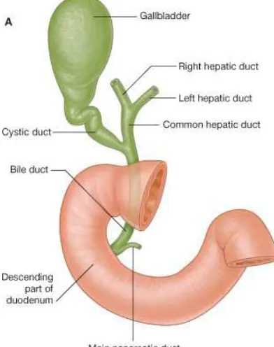
Anatomy of biliary tract
cystic duct contains a variable number of mucosal folds, similar to those found in the neck of the gallbladder. Although referred to as valves of Heister, these spiral folds do not have a valvular function.
[image:31.595.214.410.424.672.2]The common bile duct runs between the layers of the lesser omentum, lying anterior to the portal vein and to the right of the hepatic artery. Passing behind the first part of the duodenum in a groove on the back of the head of the pancreas, it enters the second part of the duodenum. The duct runs obliquely through the posterior-medial wall, usually joining the main pancreatic duct to form the Ampulla of Vater (1720). The ampulla makes the mucous membrane bulge inwards to form an eminence: the duodenal papilla. In about 10-15% of subjects the bile and pancreatic ducts open separately into theduodenum.
The dimensions of the common bile duct depend on the technique used. At operation it is about 0.5-1.5 cm in diameter. Using endoscopic cholangiography, it is usually less than 11 mm and values greater than 18 mm are pathological. By ultrasound the values are less, the common bile duct being 2-5 mm and values greater than this are abnormal.[13]
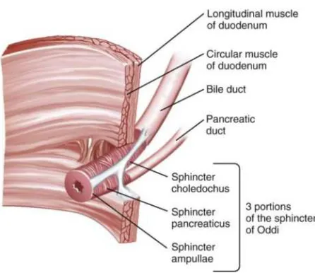
[image:32.595.207.430.330.523.2]The duodenal portion of the common bile duct is surrounded by a thickening of both longitudinal and circular muscle fibres derived from the intestine. This is called the sphincter of Oddi. [14]
Figure 4: Anatomy of the sphincter of Oddi Calot’s Triangle
with accurate identification of al
Blood supply
The gallbladder receives blood from the
hepatic artery is large, tortuous and variable in its anatomical relationships. Smaller blood vessels
[image:33.595.230.439.102.278.2]venous drainage is into the
[image:33.595.225.412.485.700.2]Figure 6: Arterial blood supply of the gall bladder with accurate identification of all structures within this triangle.
Figure 5: Calot’s triangle
The gallbladder receives blood from the cystic artery. This branch of the hepatic artery is large, tortuous and variable in its anatomical relationships. Smaller blood vessels enter from the liver through the gallbladder fossa. The venous drainage is into the cystic vein and hence into the portal venous system.
Figure 6: Arterial blood supply of the gall bladder l structures within this triangle.[12]
This branch of the hepatic artery is large, tortuous and variable in its anatomical relationships. enter from the liver through the gallbladder fossa. The and hence into the portal venous system.
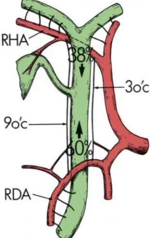
The arterial blood supply to the supra-duodenal bile duct is generally by two main (axial) vessels, which run beside the bile duct in the 3’o’ clock and 9’o’ clock position. [15] These are supplied predominantly by the retro-duodenal artery from below, and the right hepatic artery from above, although many other vessels contribute. This pattern of arterial supply would explain why vascular damage results in bile duct stricturing.
Figure 7 : Arterial blood supply of the extrahepatic biliary tree
Lymphatics
Nerve supply
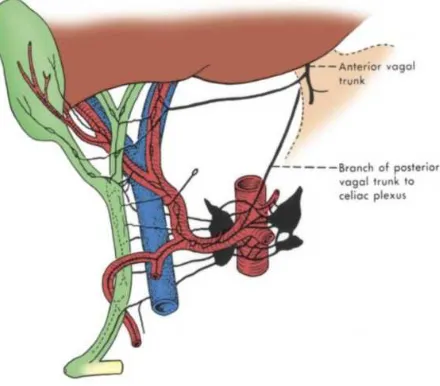
[image:35.595.207.427.168.361.2]The gallbladder and bile ducts are liberally supplied with nerves, from both the parasympathetic and sympathetic system. [16]
Figure 8: Nerve supply to the extrahepatic bile tree.
Laparoscopic anatomy [17]
The advent and popularity of LC has led to a new look and insights into the biliary anatomy especially of the Calot’s triangle area and the term “laparoscopic anatomy” has actually found a place even in anatomy texts.
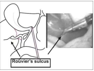
Finally the ‘posterior’ or ‘reverse’ dissection of the Calot’s triangle, which is popular during LC, again gives a different view of the area and since the gallbladder is view of the area and since gallbladder is flipped over during the method may lead to further anatomical distortion. The Rouviere’s sulcus is fissure on the liver between the right lobe and caudate process and is clearly seen during a LC during a posterior dissection in majority of patients.
It corresponds to the level of the porta hepatic where the right pedicle enters the liver. It has been recommended that all dissection be kept to a level above (or anterior) to this sulcus to avoid injury to the bileduct.
[image:36.595.222.412.519.664.2]Also, this being an “extrabiliary” reference point it does not get affected by distortion due to pathology. Similarly, a clear delineation of the junction of cystic duct with the gallbladder along with the demonstration of a space between the gallbladder and the liver clear of any structure other than the cystic artery (safety window or critical window) is also recommended as an essential step to prevent bile duct injury.
GALLSTONES INCIDENCE:
Gallstones are the most common biliary pathology. The incidence of biliary calculous disease varies widely throughout the world. By the age of 75, about 35% of women and 20% of men would have developed gallstones. The incidence of gallstone disease in Asia is considerable and constitutes a problem of enormous magnitude. The incidence of cholesterol gallstones is increasing in Asia for the reasons that may be related to environmental and dietary considerations.
Most patients with gallstones are asymptomatic and only about 10% will have developed symptoms five years after discovery. In a functioning gall bladder, most of the gall stones are cholesterol stones. Gall stone disease is a relatively common problem in our country particularly in North India. It is estimated that more than sixty percent of these patients have cholesterol stones. Recent studies from south India have highlighted pigment and mixed variety of gall stones to be more common (> 90 %) in contrast to cholesterol stones.
RISK FACTOR ASSOCIATED WITH GALLSTONE FORMATION:[20] 1.Cholesterolstones
Age > 40years
a.Female sex (2-3 times the risk inmen)
b.Pregnancy (risk increases with number ofpregnancies) c.Estrogen containingOCPs.
ii. Genetic or ethnicvariation iii.High fat, low fiberdiet
iv.Obesity
v. Hyperlipidaemia
vi. Bile salt loss ( Ileal disease or resection; Crohn’s disease) vii. Cysticfibrosis
viii. Anti-hyperlipidaemic drugs ( Clofibrate ) ix.Impaired gall bladderemptying
a.Truncalvagotomy b.Type-1diabetes c.Octreotide
d.Total parenteralnutrition
e.Starvation or rapid voluntary weightloss 2.Pigmentstones
i.Haemolyticdisease ii.Biliarystasis
iii.Biliaryinfection
India, Cholesterol stones form the majority of gallstones. In contrast to this, pigment stones are more frequent in South India.
CLINICAL FEATURES:
Clinical Presentation of Gallstones:
1.Asymptomatic. 2.Biliarycolic.
i. Right subcostal or epigastric pain radiating to back or lower pole ofscapula lasting for 20 minutes to 6hours.
ii.Associated with vomiting, brought on by (any)food. iii.May disturb sleep.
3.Flatulent dyspepsia.
4.Acute Cholecystitis - Calculous (as opposed to Acalculous)/ Empyema gallbladder/ Gangrenous Gallbladder.
i.Severe pain and tenderness in right subcostal region - Murphy sign - pain on palpation of the right upper quadrant when the patient inhales.
ii.Fever and leucocytosis.
5.Chronic calculous Cholecystitis - repeated episodes of right hypochondrial pain with/without fever and vomiting.
7.Mucocele - Heaviness in upper abdomen; palpable lump. 8.Choledocholithiasis with extra-hepatic cholestasis. 9.Biliary pancreatitis.
10.Gallstone ileus.
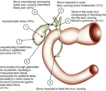
[image:40.595.133.502.280.598.2]11.Gallbladder perforation. 12.Gallbladder carcinoma.
The percentages indicate the approximate frequencies of complications that occur in untreated patients, based on natural history data. The most frequent outcome is for the patient with a stone to remain asymptomatic throughout life (1). Biliary pain (2), acute cholecystitis (3), cholangitis (5), and pancreatitis (5) are the most common complications. Mirizzi's syndrome (4), cholecystoenteric fistula (6), Bouveret's syndrome (6), and gallbladder cancer (7) are relatively rare.[22]
INVESTIGATIONS:
A variety of diagnostic modalities are available for the patient with suspected disease of the gallbladder and the bile ducts. In 1924, the diagnosis of gallstones was improved significantly by the introduction of oral cholecystography by Graham and Cole. For decades it was the mainstay of investigation for gallstones. In the 1950’s biliary scintigraphy was developed, and later trans hepatic and endoscopic retrograde cholangiography, allowing imaging of the biliary tract. Later ultrasonography, computed tomography (CT), and magnetic resonance imaging (MRI), vastly improved the ability to image the biliary tract.[24]
6.Magnetic Resonance Cholangio Pancreatography(MRCP). 7.Endoscopic Retrograde Cholangio Pancreatography(ERCP). 8.Hepato biliary Scintigraphy.
Blood investigations:
Nearly 50% of patients with symptomatic gallstone disease will have abnormal transaminases. An elevated white blood cell count alerts the clinician to the possibility of acute cholecystitis, a condition requiring more urgent treatment. Serum lipase and amylase levels are helpful in cases of diagnostic uncertainty or suspected concurrent pancreatitis. Coagulation parameter results measured by prothrombin (PT) and activated partial thromboplastin time (aPTT) might be abnormal in the severely jaundiced patient due to dysfunction in vitamin K absorption.
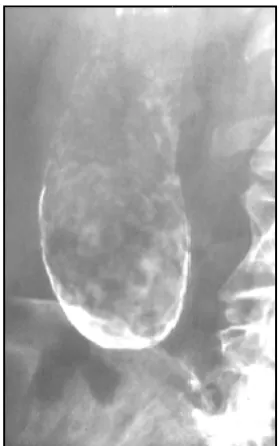
Plain X-ray abdomen:
Figure 17A: Plain radiograph showing Figure 17B: Porcelain gall bladder radio-opaque stones in the gallbladder
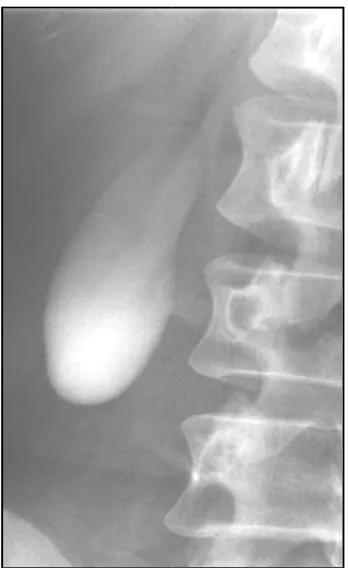
Oral Cholecystography:
Normal Oral cholecystogram Cholecystogram showing multiple gallstones
Figure 18: Oral cholecystography Cholangiography:
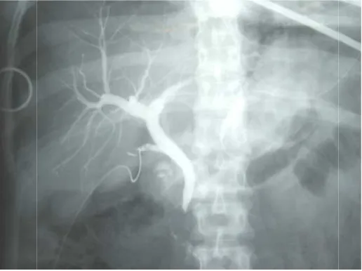
Routine cholangiography during laparoscopic cholecystectomy has been advocated to confirm anatomy and thus prevent ductal injury.
Figure 19: Normal intraoperative cholangiogram. Contrast can be seen clearly entering the cystic duct, flowing into the common bile duct and up into the
Ultrasonography:
An ultrasound is the initial investigation of any patient suspected o disease of the biliary
patient to radiation, and can be performed.
Ultrasonography can detect stones larger than 1 to 2 mm in diameter; can rule out alternative causes of right
abscess; and can suggest the presence of common bile duct dilatation.
Figure 19: Normal intraoperative cholangiogram. Contrast can be seen clearly entering the cystic duct, flowing into the common bile duct and up into the
hepatic ducts
An ultrasound is the initial investigation of any patient suspected o disease of the biliary tree. It is non-invasive, painless, does
and can be performed.
Ultrasonography can detect stones larger than 1 to 2 mm in diameter; can rule out alternative causes of right upper quadrant pain,
abscess; and can suggest the presence of common bile duct stones by showing Figure 19: Normal intraoperative cholangiogram. Contrast can be seen clearly
entering the cystic duct, flowing into the common bile duct and up into the
An ultrasound is the initial investigation of any patient suspected of does not submit the
Findings include gallstones or sludge and one or more of the following conditions:
1. Gallbladder wall thickening (>2-4 mm) - False positive wall thickening found in acute hepatitis, portal hypertension, hypo albuminemia, ascites, congestive cardiac failure and carcinoma.[26]
2. Gallbladder distention (diameter >4 cm, length >10 cm). 3. Pericholecystic fluid from perforation or exudates.
4. Air in the gallbladder wall (indicating gangrenous cholecystitis or emphysematous cholecystitis).
Its disadvantage is that it is operator be underestimated.
Figure 20(A): Typical ultrasonographic appearance of cholelithiasis. A gallstone is present within the lumen of the gallbladder (GB), casting an acoustic shadow.
Figure 20(B): Cholelithiasis in the setting of acute cholecystitis. Multiple gallstones can be seen within the gallbladder lumen with associated acoustic shadowing. In addition, the gallbladder wall is thickened (arrowheads).
Computed Tomography: Computed tomography cholelithiasis. Obvious although they may be Although this test is
does not provide important location of biliary dilatation and pancreas. In general,
Its disadvantage is that it is operator-dependent, number of stones may
ure 20(A): Typical ultrasonographic appearance of cholelithiasis. A gallstone is present within the lumen of the gallbladder (GB), casting an
Figure 20(B): Cholelithiasis in the setting of acute cholecystitis. Multiple gallstones can be seen within the gallbladder lumen with associated acoustic shadowing. In addition, the gallbladder wall is thickened
Computed Tomography:
mography scan are not a first-line test for Obvious gallstones frequently are missed
be seen as incidental finding, if they are densely
not particularly sensitive for identifying gallstones, it important information regarding the nature, extent, and the location of biliary dilatation and masses in and around the biliary
general, this test provides more useful information than dependent, number of stones may
ure 20(A): Typical ultrasonographic appearance of cholelithiasis. A gallstone is present within the lumen of the gallbladder (GB), casting an
Figure 20(B): Cholelithiasis in the setting of acute cholecystitis. Multiple gallstones can be seen within the gallbladder lumen with associated acoustic shadowing. In addition, the gallbladder wall is thickened
for the diagnosis of by routine CT, densely calcified. particularly sensitive for identifying gallstones, it
[image:47.595.134.500.133.260.2]cause other than choldocholithiasis. Limiting factors for CT scanning include patient exposure to ionizing radiation and cost.
Normal CT Scan of gall bladder
[image:48.595.231.411.399.534.2]CT image demonstrates a large gallstone (arrow) in the gallbladder.
Figure 21: CT image of gall bladder Magnetic Resonance Cholangio Pancreatography (MRCP):
but native high signal intensity of fluid on imaging of the biliary
MRI cholangiography for detecting choledocholithiasis is over 90 %.
Endoscopic Retrograde Cholangio Pancreatography (ERCP): Using a side
cannulated and a cholangiogram performed using fluoroscopy. The procedure requires intravenous sedation
endoscopic retrograde the ampullary region
possibility of therapeutic uncomplicated gallstone
particular when associated with obstructive jaundice, cholangitis, or pancreatitis, ERCP is
Once the endoscopic cholangiogram has sh
igh signal intensity of fluid on T2-weighted
[image:49.595.213.418.176.385.2]biliary tree. A preliminary study has found that the sensitivity of MRI cholangiography for detecting choledocholithiasis is over 90 %.
Figure 22: Normal MRCP image
ic Retrograde Cholangio Pancreatography (ERCP):
Using a side-viewing endoscope, the common bile duct can be cannulated and a cholangiogram performed using fluoroscopy. The procedure requires intravenous sedation for the patient. The
retrograde cholangiography (ERC) include direct region and direct access to the distal common bile
therapeutic intervention. The test is rarely gallstone disease, but for stones in the common particular when associated with obstructive jaundice, cholangitis, or
is the diagnostic and often therapeutic procedure
Once the endoscopic cholangiogram has shown ductal stones, weighted images permits has found that the sensitivity of MRI cholangiography for detecting choledocholithiasis is over 90 %. [27]
ic Retrograde Cholangio Pancreatography (ERCP): [28]
viewing endoscope, the common bile duct can be cannulated and a cholangiogram performed using fluoroscopy. The
sphincterotomy and duct cleared of stones.
Figure 23: Endoscopic Retrograde cholangiopancreatography demonstrating stone obstructing the common bile duct (arrow).
stone extraction can be performed, and the common bile duct cleared of stones.
Figure 23: Endoscopic Retrograde cholangiopancreatography demonstrating stone obstructing the common bile duct (arrow).
stone extraction can be performed, and the common bile
Hepatobiliary Scintigraphy:
Nuclear cholescintigraphy permits the rapid assessment of gallbladder function in a patient with suspected acute cholecystitis.[29] The short-lived isotope technetium- 99m is bound to one of several iminodiacetic acids (such as hydoxyiminodiacetic acid- HIDA or diisopropyl iminodiacetic acid - DISIDA) that are excreted into the bile ducts. Gamma rays emitted by the tracer are used to make an image of the bile ducts and gallbladder. Failure of the tracer to enter the gallbladder suggests obstruction of the neck of the gallbladder, as occurs in acute cholecystitis.
Figure 24: Cholescintigraphy demonstrating an obstructed cystic duct characteristic of acute cholecystitis.
The failure of the gallbladder to be visualized as a hot spot within 30 to 60 minutes constitutes a positive result and implies obstruction of the cystic duct.
MANAGEMENT OF GALLSTONE DISEASES
NON-OPERATIVE MANAGEMENT [30]
Medical treatment of gallstone disease was first proposed by Schiff in Italy in 1873. Dabney of Virginia first reported the effective treatment of gallstones with bile acids in 1876.Despite these initial reports, the use of medical dissolution treatment did not gain acceptance until large clinical series were reported in the 1970s.
Contact dissolution of gallstones with solvents and percutaneous cholecystolithotomy techniques also have been reported, but these modalities have not proved superior to oral dissolution, shock-wave lithotripsy, or laparoscopic cholecystectomy and have been abandoned. The mainstay of current non-surgical treatment of gallstone disease is oral dissolution with ursodeoxycholic acid, with or without extracorporeal shock-wave lithotripsy.
1.DissolutionTherapy
The rationale for oral dissolution therapy is the reversal of the condition that led to formation of cholesterol gallstones, namely, the supersaturation of bile with cholesterol. Cholesterol stones dissolve if the surrounding medium is capable of solubilizing the cholesterol in the stones. Both chenodeoxycholic
acid and ursodeoxycholic acid dissolve gallstones by decreasing biliary
Chenodeoxycholic acid was the first bile acid used for gallstone dissolution but has been abandoned because of side effects, including diarrhea and increased serum aminotransferase and cholesterol levels. Ursodeoxycholic acid is well tolerated and is currently used in oral dissolution regimens. Oral dissolution therapy should be considered for patients with uncomplicated gallstone disease, including those with mild, infrequent biliary pain. In addition, the gallbladder must function and the cystic duct must be patent to allow unsaturated bile and stones to clear from the gallbladder. Oral dissolution therapy works only on cholesterolstones.
Table 3: Selection Criteria for Oral Bile Acid DissolutionTherapy
Stage disease Of gallstone
• Symptomatic (biliary pain) without complications
Gallbladder function
• Opacification of gallbladder on oral cholecystography (patent cystic duct)
• Normal result of stimulated cholescintigraphy (normal GBemptying)
• Normal result of functional ultrasonography(normal
Stone characteristics
• Radiolucent on radiography
• Isodense or hypodense to bile and absence of calcification on CT scan
• Diameter <6 mm (optimal) or 6-10 mm(acceptable)
Ursodeoxycholic acid (ursodiol) is the preferred drug for oral dissolution treatment. It is taken in a dose of 10 to 15 mg/kg of body weight per day. Nighttime dosing is more effective and is associated with better patient compliance than mealtime dosing. Treatment should continue until stone dissolution is documented by two consecutive negative ultrasonograms at least one month apart.
2.Extracorporeal shock wave lithotripsy(ESWL)
the size, microcrystalline structure, and architecture of the stone.
Because shock-wave lithotripsy is usually combined with oral dissolution therapy, patient selection criteria for shock-wave lithotripsy are similar to those for oral dissolution treatment. Gallbladder function and cystic duct patency are required and are demonstrated by oral cholecystography, functional ultrasonography, or stimulated cholescintigraphy. Lithotripsy should be considered only for patients with mild, uncomplicated biliary pain. Pregnant patients and patients on anticoagulants should not undergo lithotripsy. Because only cholesterol stones are reliably cleared by the addition of oral dissolution therapy, stones should have radiographic features, such as radiolucency, suggestive of cholesterol stones.
Side effects of lithotripsy include petechiae of the skin at the site of shock-wave delivery (8%), hematuria (4%), and liver hematomas (<1%). No long-term liver biochemical abnormalities have been noted. Biliary pain develops in approximately one third of patients; cystic duct obstruction develops in 5%; and complications of stone passage, such as biliary pancreatitis, develop in less than 2%.
OPERATIVE MANAGEMENT
Cholecystectomy is one of the most common major abdominal operations. Cholecystectomy can be performed by open and laparoscopic methods.
The indications for cholecystectomy are the same for both techniques.[31] These include:
1.Symptomatic gallstones causing i.Repeated episodes of biliarypain ii.Mucocele of thegallbladder
iii.Choledocholithiasis with extra-hepaticcholestasis iv.Biliarypancreatitis
v.Gallstoneileus
2. Cholecystitis and its complications - acute calculous / acalculous cholecystitis, chronic cholecystitis, empyema gallbladder, gangrenous cholecystitis, gallbladder perforation.
3.Asymptomatic cholelithiasis: Laparoscopic cholecystectomy is indicated in asymptomatic cholelithiasis in certain selectiveindications:
i.Patients undergoing bariatricsurgery. ii.Diabetics.
iii.Renaltransplantation. iv.Children.
4. ‘Gallstone dyspepsia’: Patients with ‘classic’ biliary pain without evidence of gallstone may benefit from cholecystectomy. Biliary dyskinesia can be detected by objective measurements of changes in gallbladder volumes(ejection fraction <35%) or reproduction of the pain on consumption of fatty meal or cholecystokinin infusion. These patients may benefit from surgery. However, one should warn the patient that 20-30% of patients operated for dyspepsia have a persistence of symptoms.
5.Gallbladderpolyps.
Traditionally, open cholecystectomy has been the gold standard for all patients with symptomatic gallstonedisease.[32]
LAPAROSCOPIC CHOLECYCTECTOMY [39,40] Indications:
The indications for laparoscopic cholecystectomy remain the same as for open cholecystectomy.
Contra-indications:
1.Patients unfit for general anaesthesia. 2.Significant portal hypertension. 3.Uncorrectable coagulopathy.
Pre-operative Work-up:
1.Routine blood tests, including liver function tests. 2.Ultrasonography.
3.Upper GI endoscopy - to identify patients with acid peptic disorders or hiatus hernia
4.DVT prophylaxis in high risk patients.
Drawbacks of laparoscopic cholecystectomy:
1. The incidence of bile duct injuries is more as compared to open cholecystectomy.
2. The operating time required for laparoscopy cholecystectomy is more as compared to open method.
Advantages:
1. Laparoscopic cholecystectomy is associated with a lower risk of surgical site infection than open method, even after adjustment for other risk factors.
2. Post operative pulmonary function was impaired less after laparoscopic than after open cholecystectomies.
LAPARASCOPY CHOLECYSTECTOMY
Anaesthesia:
General anaesthesia is the preferred anaesthetic method for patients undergoing most therapeutic laparoscopic surgical procedures. Two advantages of general anaesthesia as compared to other types of anaesthesia are two folds: 1.It allows for complete control of the patient’s ventilation, which might
otherwise be compromised by systemic absorption of CO2 and increased diaphragmatic pressure from the pneumoperitoneum.
2.It enables complete relaxation of the abdominal wall muscles necessary for adequately maintaining pneumoperitoneum.
Patient Position and Room Set-up:
North American Approach: The patient is kept supine in anti-Trendelenburg position (150 head up tilt) with left lateral tilt (15-200). This ensures that the bowel and omentum falls down and medially, away from the operative site. The operating surgeon and camera surgeon stand on the left of the patient while the assistant surgeon stands on the right of the patient. The monitor is kept beyond the right shoulder of the patient facing the operating surgeon. An additional monitor may be kept beyond the left shoulder of the patient for the assistant surgeon.
subcostally.
French/European Approach: The patient is in semi-lithotomy anti-Trendelenburg position with the legs in Allen stirrups such that the thighs are almost parallel to the ground to avoid interference with the manipulations of the operating instruments. The operating surgeon stands between the legs of the patient with the camera surgeon on the right of the patient and the assistant on the left of the patient.
[image:61.595.112.500.465.660.2]The camera port placement remains the same as in the North American approach. Epigastric port (5 mm) is placed to allow retraction by the assistant. The right hand working port (10 mm) is placed in the left hypochondrium or in the midline between the camera port and the epigastric port. The left hand working port (5 mm) is placed in the right hypochondrium.
Figure 43: Differences between typical North American practice (a) and typical European practice (b) with respect to the placement of the trocars and the
instruments inserted through each port.
Technique:
1. Pneumoperitoneum and portplacement:
finger and ulnar border of the right palm is propped against the abdomen.
[image:63.595.242.408.257.434.2]The abdominal wall is lifted midway between the pubic symphysis and umbilicus by the left hand .The Veress needle is inserted either at a 45 degree caudal angle to the abdominal wall (in the asthenic or minimally obese patient) or perpendicular (in the markedly obese patients).
Figure 44: Veress needle insertion
The needle is connected to the insufflator and CO2 is instilled at a rate of 1 L/min. The opening pressure recorded on the insufflator should be < 10 mmHg. Initial pressures of 10 mmHg or higher may indicate the placement of the needle in the pre- peritoneal or other closed space. Upon insufflating approximately 1L of CO2, increased tympany in all four quadrants of the abdomen is confirmed, and the flow rate may be increased. Although high flow insufflators are designed to deliver flow rates of up to 8 to 10 L/min, the maximum flow rate through the small caliber Veress needle is approximately 2.5 L/min. Once the intra-abdominal pressure has reached 15 mmHg, generally requiring 3 to 6 L of CO2, the Veress needle is removed, and the trocar is inserted through the same site.
The trocar is grasped firmly in the palm of one hand and inserted using gently firm pressure while elevating the abdominal wall with the other hand or with towel clips. Once the port is in, the inner trocar is removed, leaving the outer cannula and sheath in place. Return of CO2 gas is confirmed by opening either the stopcock or flapper valve on the port and then connecting the insufflation line to the sheath. The video telescope is inserted and a general inspection of the peritoneal cavity, including underlying viscera and retroperitoneum, is carried out to assess for visceral injury.
The epigastric port (10 mm) is inserted in the midline just below the liver edge or the costal margin, whichever is lower. The trocar is thrust in a rotatory movement so that it pierces the fascia and reaches the pre-peritoneal space. Then, it is turned right so that it enters the peritoneum at the base of the falciform ligament. This maneuver serves two purposes: (a) The trocar avoids injuring a vessel which sometimes runs in the free edge of the falciform ligament. (b) The instruments through this port do not suffer interference from a falciform ligament hanging in front ofthem.
The mid-clavicular port (5 mm) is introduced at the same level, i.e., just below the liver edge or the costal margin, whichever is lower, right over the fundus of the gallbladder.
The lateral most port (5 mm) is introduced at the same level and just anterior to the lateral peritoneal attachment of the ascending colon.
Additional ports are sometimes required and may be placed as follows:
A. Left lumbar 5 or 10 mm for three prong or flat blade retractor for downward traction of the colon, omentum and duodenum. This maneuver gives wide exposure of thehilum.
Figure 45: Port position for laparoscopic cholecystectomy
2. Initialdissection:
The fundus of the gallbladder is held with a ratcheted grasper and retracted by the assistant in a cranial direction, which lifts the right lobe of the liver and exposes the Calot’s triangle and hilum of the liver.
Fig 46: Dissection of cystic duct Figure 47: Dissection of cystic artery
3. Dissection of cholecysto-hepatic triangle:
An atraumatic (dolphin-nosed) non-locking grasper is introduced through the left hand working port to hold the infundibulum and retract it downwards and to the right. Thus, the hepatocystic triangle is widened and opened up and the structures are placed under tension. By retracting the infundibular grasper laterally, the anterior aspect of the Calot’s triangle is exposed. By retracting the infundibular grasper antero-medially, the posterior aspect of the Calot’s triangle is exposed.
careful to remain on the gallbladder side. The infundibular grasper is moved inferolaterally and superomedially (flagtechnique) to aid this dissection on the anterior and posterior surface of cholecysto-hepatic triangle respectively. The cholecysto-hepatic triangle is thus exposed.
Fig 48: Dissection of cystic pedicle Fig 49: Dissection of cystic duct by blunt dissection
4. Identification of the cystic duct and artery:
Now comes the most critical step of the operation - the identification of the cystic duct and artery. There are two well-described methods for ductal identification in laparoscopic cholecystectomy.
as the cystic duct widens to become the gallbladder infundibulum. Often this is referred to as seeing a funnel shape i.e. the gallbladder should be seen to funnel down to terminate in the cystic duct. The infundibular method is the one usually found in texts describing the technique of laparoscopic cholecystectomy.
The second method is the “critical view of safety” technique, which was described in 1995.[42] This method requires complete dissection of the cholecystohepatic triangle and separation of the base of the gallbladder infundibulum from the liver bed. The anatomic rationale for identification of the cystic structures results from the fact that there are two, and only two, structures entering the gallbladder, which is otherwise still attached only by the upper part of the liver bed. The triangle of Calot is dissected free of all tissue except for cystic duct and artery, and the base of the liver bed is exposed. When this view is achieved, the two structures entering the gallbladder can only be the cystic duct and artery. It is not necessary to see the common bileduct.[43]
The cystic duct is identified at the junction with the gallbladder (safety zone) and followed down for an adequate length for cholangiography if desired. It is not always necessary to identify and dissect out the cystic-common duct junction (danger zone).
potential avulsion of the cystic artery off the right hepatic artery. The cystic node of Lund sometimes overlies the cystic artery. Attention is given to identify any unusual vascular or biliary tree anomalies. The main trunk of the cystic artery should be ligated and divided. Widely placed anterior and posterior branches areclipped individually and divided. Blind application of clips within the Calot’s triangle should be avoided.
[image:70.595.110.526.445.635.2]Both the cystic duct and the cystic artery are clipped, two clips on the cystic duct side and one clip on the gallbladder side. Though it is desirable to divide the artery before the duct, in selected situations, duct needs to be divided to expose cystic artery, hepatic artery, etc, and care is taken not to give excessive traction till the cystic artery is clipped and divided.
5. Detachment of the gallbladder from the liver:
The gallbladder can be detached from the liver bed using a variety of instruments - spatula with monopolar cautery, hook with monopolar cautery, scissors with monopolar cautery or Harmonic Scalpel. Surgeon’s experience and familiarity with a particular device is the most important aspect of choosing the best instrument for this purpose.
Care should be taken to stay away from the porta hepatis and the liver bed and to avoid perforating the gallbladder. The infundibular grasper is used to elevate the gallbladder and alternately twist it to the left (medial rotation) and to the right (lateral rotation). A hook cautery is very useful for this phase of the operation.
Figure 52: Dissection of gallbladder from its bed
6. Extraction of thegallbladder:
The extraction of the gallbladder can be carried out through the umbilicus or the epigastric port. A claw-shaped gallbladder extraction forceps is introduced and used to grasp the neck of the gallbladder. The forceps, cannula and the neck of the gallbladder are pulled out of the skin opening.If the gallbladder is too distended, the neck is opened and suction cannula is inserted to suck out the bile and if necessary, the stones are debulked through fragmentation by using a sponge holder. If the gallbladder is thick, preventing its extraction fascial incision is extended to facilitate its removal.
7. Final inspection andirrigation:
8. Drainage andClosure:
If a drain is needed, it can be placed through the lateral-most port. A size 14F Romovac tube which goes through a 5 mm trocar is usually sufficient. If larger drainage tube is needed, it should be placed inside the peritoneal cavity through the epigastric port and brought out by a grasper through the lateral-most port in a reverse fashion.
[image:73.595.135.307.470.650.2] [image:73.595.217.487.472.652.2]The trocars are removed under direct vision to check that there is no bleeding from the trocar sites. Pneumoperitoneum is evacuated. The fascia of the 10 mm ports is closed with vicryl suture using port closure needle. Fascial closure is not required for the 5 mm ports. Skin closure is done using 3-0 vicryl subcuticular stitch/skinclip.
COMPLICATIONS: 1) Trocar injuries:
2) Bleeding:
METHODOLOGY
The present study is a comparative study of 394 cases of
cholelithiasis who undergone laparoscopic cholecystectomy in the
institute of general surgery, MMC & RGGGH, Chennai, during study
period of may 2017 to October 2018. These cases were selected based on
inclusion criteria and were randomized using software after taking valid
informed consent (Annexure)
INCLUSION CRITERIA
Adults > 18 years of age undergoing elective laparoscopic cholecystectomy for cholelithiasis
EXCLUSION CRITERIA 1. Cholangitis
2. Acute cholecystitis
3. Lap converted open cholecystectomy 4 Recent onset acute cholecystitis
examination was done.
Pre-operative work up included a complete blood count, blood sugar, blood urea, serum creatinine, liver function tests, hepatitis profile, X-ray chest and ultrasound of abdomen. Ultrasonogram was routinely performed on all patients to confirm the clinical diagnosis of cholelithiasis with number of calculus and size of calculus, gall- bladder wall thickness (>4mm was considered abnormal), pericholecystic collection.
A routine pre-anaesthetic checkup was done. A fully explained well informed consent was taken. A nasogastric tube was placed in all cases for gastric decompression to prevent trocar injury. All patients received prophylactic pre-op antibiotics (Inj. Cefotaxim 1gm IV).
RESULTS
A total of 394 patients eligible for the study were selected. All the patients who undergone elective laparoscopic cholecystectomy categorised into study group and control group. Study group receiving prophylactic intravenous antibiotic (1gm cefotaxim) at the time of induction of anaesthesia alone. Control group receiving prophylactic intravenous antibiotic at the time of induction of anaesthesia which will be continued in the post operative period till discharge. Patients were followed in the post operative period with regard to surgical site infections.
AGE INCIDENCE
Study group (N=197)
Control group (N=197)
Characteristics n % N % p value
AGE (in years)
30 to 39 98 49.7 117 59.4
p<0.05
40 to 49 68 34.5 60 30.5
Mean age in the study group is 41 years, in the control group is 38 years, the age group of patients ranges from 30 to 58 years. In study group 49.7% of patients between 30 to 39 years of age. In control group 59.4% of patients from 30 to 39 years of age. patients are allocated in the study and control without statistically significant.
0 20 40 60 80 100 120 140
30 to 39 40 to 49 50 and above
#
c
a
se
s
Age group
STUDY
SEX INCIDENCE:
STUDY (N=197)
CONTROL(N=19 7)
Characteristics n % n % p value
SEX
Male 114 57.9 124 62.9
p>0.05
Female 83 42.1 73 37.1
COMORBIDITIES INCIDENCE:
STUDY (N=197)
CONTROL(N=197)
Characteristics n % n % p value
DIABETES MELLITUS
Diabetic 25 12.7 29 14.7
p<0.05 non
diabetic
172 87.3 168 85.3
In the study group 25 patients are diabetic in the control group 29 patients are diabetic. When analysed statistically no significant association between the presence of diabetes and wound infection could be obtained.
0 20 40 60 80 100 120 140 160 180 200 STUDY CONTROL A xi s T it le Axis Title
Single dose (N=197)
Multi dose (N=197)
Characteristics N % n % p value
Hypertension Hypertensive 19 9.6 11 5.6 p>0.05 non
hypertensive
178 90.4 186 94.4
In the study group 19 patients are hypertensive which is 9.6%. in the control group 11 patients are hypertensive which is 5.6%. when analysed statistically no significant association between the presence of hypertension and wound infection could be obtained
0 20 40 60 80 100 120 140 160 180 200 STUDY CONTROL A xi s T it le Axis Title
PRESENTING COMPLAINTS INCIDENCE:
STUDY (N=197)
CONTROL (N=197)
Characteristics N % N % p value
presenting complaint
abdominal pain
157 79.7 128 65.0 p>0.05
Asymptomati c
10 5.1 42 21.3
Dyspepsia 30 15.2 26 13.2
Indigestion 0 0.0 1 0.5
0 50 100 150 200
POST OPERATIVE COMPLICATIONS
STUDY (N=197)
CONTROL (N=197)
N % N %
Complications Developed 8 4.2 6 3.0 P<0.05 not
developed
190 95.8 191 97.0
STUDY(N=197) CONTROL(N=197)
complication n % N %
Fever 1 1.5 1 0.5
Superficial infections (pus discharge from port
site)
7 3.6 5 2.5
deep infection 0 0 0 0
Seroma formation 0 0 0 0
Post operative complications are monitored. In study group 1 patient was developed fever, in the control group 1 patient developed fever.in this study surgical site infections were taken into account. In the study group 7 patients (3.6%) developed pus discharge from port site which is considered as superficial infections ,in the control group 5 patients (2.5%) developed pus discharge. In all cases deep infections are ruled out by doing ultrasonography. There is no seroma formation in both study and control group. I concluded that surgical site infection in the single IV antiobiotic group is 3.6% where as in the control group, in which IV antibiotics were continued in the post operative period till discharge is 2.5%.
0 1 2 3 4 5 6 7 8
fever superficial infections
DISCUSSION
It is well documented that prophylactic antibiotic coverage of most ‘clean contaminated’ surgical procedures can significantly prevent infectious complications, including wound infections, thereby affecting the overall mortality and morbidity. However the benefit of antibiotic prophylaxis in other ‘clean surgical procedures, such as laparoscopic cholecystectomy, has been questionable. The low rate of wound infections and the straight forward treatment, if they occur at all, are the main arguments against routine antibiotic coverage during laparoscopic cholecystectomy. Laparoscopic cholecystectomy is an elective clean operations, and the post operative wound infections would be very low. Prophylaxis in clean operations has been shown to be of value in other areas of surgery such as trauma and vascular surgery but in laparoscopic cholecystectomy, its benefits remains uncertain. Due to the unknown impact on bacterial resistance, waldvogel and associates suggested that the routine use of antibiotic prophylaxis should be discouraged.
and 65% in the control group. In my study 12.7% of patients in study group and 14.7% of patients in the control group were diabetic and 9.6% of patients in the study group and 5.6% of patients in the control group were hypertensive. There are several risk factors that are significantly associated with an increased incidence of infective complications in patients who undergo elective laparoscopic cholecystectomy, one of them is the presence of diabetic mellitus. Out of 197 patients in the study group 7 of them developed pus discharge from port site with incidence of about 3.6% and in the control group 5 patients out of 197 patients are developed pus discharge from port site with incidence of about 2.5%. all others had completely healed wound. These differences yielded a P>0.05 which is statistically insignificant, thereby illustrating that the rates of wound infection in patients given only a single shot of iv antiobiotic, and in patients given continuous post operative iv antibiotics is statistically insignificant.
In a randomized controlled trial on 417 patients undergoing laparoscopic cholecysectomy, conducted by Gaur and Pujahari11, they reported an overall infection rate of 2.2 %, which is consistent with the results obtained in our study. All the infections healed before the availability of culture and sensitivity report without any specific therapy.
prophylactic antibiotics are two major factors for decreasing the incidence of septic complications after biliary tract surgery.
In another study conducted by Mahmoud and associates to assess the role of antibiotic prophylaxis in elective laparoscopic cholecystectomy, they stated that antibiotic prophylaxis does not prevent wound infection in elective laparoscopic cholecystectomy. This is probably due to the fact that Mahmoud and associates excluded all patients with associated co-morbidities, like diabetes mellitus, hypertension etc. from their study.
They also concluded that the use of antibiotic prophylaxis is preferred to be restricted to high-risk patients such as patients with associated co-morbidities like diabetes mellitus. The rate of post-operative wound infection in our study was low (0.41%) and there was no significant difference between wound infection in patients receiving prophylactic antibiotics and post-operative antibiotics. This can be attributed to the following reasons.
• Good surgical technique
• Better handling of tissues
• Strict adherence to aseptic precautions • Experienced laparoscopic surgeons
sometimes difficult to clean. Also, it may be due to the routine protocol of our unit to extract the gall bladder through the umbilical port. Colizza and associates14 also stated that the umbilicus is the commonest site for sepsis in elective laparoscopic choelcystectomy.