Patterns of muscle coordination during
dynamic glenohumeral joint elevation: An
EMG study
David H. Hawkes1☯, Omid A. KhaiyatID2☯*, Anthony J. Howard3☯, Graham J. KempID4‡,
Simon P. Frostick1‡
1 Musculoskeletal Science Research Group, Institute of Translational Medicine, University of Liverpool,
Liverpool, United Kingdom, 2 School of Health Sciences, Liverpool Hope University, Liverpool, United Kingdom, 3 Academic Department of Trauma and Orthopaedic Surgery, Leeds General Infirmary, Leeds, United Kingdom, 4 Department of Musculoskeletal Biology, Institute of Ageing and Chronic Disease, University of Liverpool, Liverpool, United Kingdom
☯These authors contributed equally to this work. ‡ These authors also contributed equally to this work.
Abstract
The shoulder relies heavily on coordinated muscle activity for normal function owing to its limited osseous constraint. However, previous studies have failed to examine the sophisti-cated interrelationship between all muscles. It is essential for these normal relationships to be defined as a basis for understanding pathology. Therefore, the primary aim of the study was to investigate shoulder inter-muscular coordination during different planes of shoulder elevation. Twenty healthy subjects were included. Electromyography was recorded from 14 shoulder girdle muscles as subjects performed shoulder flexion, scapula plane elevation, abduction and extension. Cross-correlation was used to examine the coordination between different muscles and muscle groups. Significantly higher coordination existed between the rotator cuff and deltoid muscle groups during the initial (Pearson Correlation Coefficient (PCC) = 0.79) and final (PCC = 0.74) stages of shoulder elevation compared to the mid-range (PCC = 0.34) (p = 0.020–0.035). Coordination between the deltoid and a functional adducting group comprising the latissimus dorsi and teres major was particularly high (PCC = 0.89) during early shoulder elevation. The destabilising force of the deltoid, during the initial stage of shoulder elevation, is balanced by the coordinated activity of the rotator cuff, latissimus dorsi and teres major. Stability requirements are lower during the mid-range of elevation. At the end-range of movement the demand for muscular stability again increa-ses and higher coordination is seen between the deltoid and rotator cuff muscle groups. It is proposed that by appreciating the sophistication of normal shoulder function targeted evi-dence-based rehabilitation strategies for conditions such as subacromial impingement syn-drome or shoulder instability can be developed.
a1111111111 a1111111111 a1111111111 a1111111111 a1111111111 OPEN ACCESS
Citation: Hawkes DH, Khaiyat OA, Howard AJ,
Kemp GJ, Frostick SP (2019) Patterns of muscle coordination during dynamic glenohumeral joint elevation: An EMG study. PLoS ONE 14(2): e0211800.https://doi.org/10.1371/journal. pone.0211800
Editor: Chunfeng Zhao, Mayo Clinic Minnesota,
UNITED STATES
Received: August 7, 2018
Accepted: January 21, 2019
Published: February 8, 2019
Copyright:©2019 Hawkes et al. This is an open access article distributed under the terms of the
Creative Commons Attribution License, which permits unrestricted use, distribution, and reproduction in any medium, provided the original author and source are credited.
Data Availability Statement: An anonymised data
set is available as a Supporting Information file and accompanies the article.
Funding: The authors received no specific funding
for this work.
Competing interests: The authors have declared
Introduction
Shoulder pathology, such as subacromial impingement syndrome or shoulder instability, is prevalent and has a substantial impact on patient’s quality of life [1,2]. Normal shoulder func-tion, essential for many activities of daily living, requires the integration of strength, range of motion and muscular endurance. Understanding this is the basis for understanding pathology.
The glenohumeral (GH) joint is a multi-axial ball-and-socket synovial joint which, in combi-nation with the acromioclavicular, sternoclavicular and scapulothoracic articulations, facilitates movement of the upper limb. Its minimal bony constraint permits a remarkable range of motion but results in translation of the humeral head on the glenoid fossa following contraction of the powerful shoulder girdle muscles [3,4]. It has long been recognised that the rotator cuff muscles are crucial in limiting this translation and maintaining GH joint stability [5–7]: their contraction ‘stiffens’ the joint, establishing a stable fulcrum for arm movement [8]. However, more recently coordinated activity of all the major shoulder muscle groups during a functional shoulder elevation task has been shown, implying a wider muscular contribution to stability [9].
Muscular force generation must ensure a stable GH fulcrum whilst permitting the necessary net torque required to achieve movement. Inadequate stabilisation results in excessive transla-tion of the humeral head on the glenoid fossa, which can lead to symptomatic subluxatransla-tion or dislocation. Conversely, an over-stabilised system wastes energy [10]. The shoulder is particu-larly reliant on coordinated or balanced muscular activity to ensure that the humeral head remains centred on the glenoid fossa. The destabilising effects of the prime moving muscles are balanced by the synchronous activity of muscles whose anatomical arrangement means they can exert a stabilising effect for that particular movement. Indeed, aberrant muscle coor-dination is proposed to have a central role in the development of atraumatic shoulder instabil-ity [11] and shoulder impingement syndrome [12]. A full understanding of normal shoulder muscular coordination is essential as a basis to understand these pathologies and to develop effective evidence-based treatment strategies.
Muscle coordination is dynamic, as stability requirements vary according to task-specific demands [13]. The coordination between muscles can also change during a movement as the resultant force vector about the joint evolves [14]. Inherent muscle redundancy allows re-dis-tribution of activity among the shoulder complex, which will further alter inter-muscular coor-dination [15]. These dynamic relationships illustrate the sophistication of normal shoulder function and current research strategies must reflect this.
Several studies have described muscle activation during single planar shoulder movements [6,16–19]. However these principally focus on the level of muscle activation. Less attention has been given to the important concept of muscle coordination and we are not aware of any previous reports describing muscle coordination of the entire shoulder girdle and how this might vary dynamically during movement progression. This is a significant deficiency within the literature given the reliance of the shoulder on coordinated muscle activity, and the role in which aberrant coordination may play in the development of shoulder pathology.
The primary aim of this study, therefore, is to use EMG to define muscle activation patterns and dynamic inter-muscular coordination in healthy subjects during multi-planar shoulder elevation.
Methods
Participants
females. Mean age was 28.4±8.5 years, mean height 1.70±0.06m and mean mass 68.6±11.1kg. The study had Local Research Ethics Committee ((NRES Committee North West–Liverpool Central) approval and informed written consent was obtained from all participants.
EMG measurement
EMG signals were recorded using a TeleMyo 2400 G2 Telemetry System (Noraxon Inc., zona, USA) during the testing protocol described below. MyoResearch XP (Noraxon Inc., Ari-zon, USA) was used for signal acquisition and off-line analysis. Bipolar, disposable, self-adhesive pre-gelled Ag/AgCl dual surface electrodes (Noraxon Inc., Arizona, USA) were used to record the activity of the anterior deltoid (AD), middle deltoid (MD), posterior deltoid (PD), upper trapezius (UT), middle trapezius (MT), lower trapezius (LT), serratus anterior (SA), teres major (TM), latissimus dorsi (LD) and pectoralis major (PM). The skin was pre-pared with an abrasive paste (Nuprep: Weaver and Company, Aurora, CO, USA). Electrodes were placed in parallel to the muscles fibres according to accepted anatomical locations [20–
22]. The size of the surface electrodes (conducting area 10 mm diameter; inter-electrode dis-tance 20 mm) conformed to international guidance. The use of appropriately sized electrodes placed using anatomical criteria accepted in the literature limited the impact of cross-talk. A reference electrode was positioned on the acromion. Bipolar disposable hook wire electrodes (Nicolet Biomedical, Division of VIASYS, Madison, USA) were inserted aseptically for the intra-muscular recording of the supraspinatus (SSP), infraspinatus (ISP), subscapularis (SUBS) and rhomboid major (RM) [23,24].
Pre-amplified leads differentially amplified the detected signal (common mode rejection ratio (CMRR)>100dB; input impedance>100Mohm; gain 500dB). The detected signals were digitalised (sampling rate 3000Hz) and band pass filtered ([10. . .500]Hz for surface electrodes and [10. . .1500]Hz for fine wire electrodes). EMG data acquisition conformed to international standards [25]. Manual muscle testing confirmed correct electrode placement and signals were excluded if they were of poor quality.
EMG testing protocol
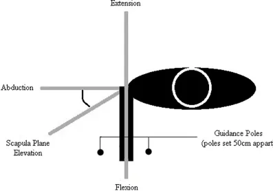
The testing protocol was performed with participants standing on a wooden board with their elbow extended, forearm in neutral rotation and their feet shoulder-width apart. Pre-marked axes were drawn on the board to guide shoulder movement between two guidance poles (50cm apart) in 4 different planes: flexion (sagittal plane movement); scapular plane elevation (movement 30oanterior to the coronal plane); abduction (coronal plane movement); exten-sion (sagittal plane movement) (Fig 1). Ten continuous cycles of maximal shoulder elevation and depression were performed in each movement plane. Participants were instructed to move in a smooth and continuous manner. The pace was governed by a metronome set at 1 beat every 2 seconds (s) such that each cycle took 4s to complete (2s for elevation and 2s for depression). The movement was performed between guidance poles to ensure that subjects maintained the correct plane. An opportunity for practice prior to EMG recording ensured familiarity with the movement task.
Data management and statistical analysis
This normalisation method has previously been accepted with the literature as a technique to compare relative (rather than absolute) differences in activity levels between movements [26,27]. Amplitude normalisation to the mean was performed for two reasons. First, the pri-mary aim of this work was to examine inter-muscular coordination, which requires the study of muscle activation patterns rather than absolute amplitudes. Second, whilst normalisation to a maximal voluntary contraction (MVC) is popular, using an isometric contraction to normal-ise EMG data obtained during a dynamic task may be inappropriate, as an MVC might not represent the maximum activation of the muscle at lengths other than those at which the MVC was performed [28–30].
[image:4.612.176.573.78.356.2]Time normalisation was performed to allow ensemble (synchronous) averaging of the EMG data from the 10 movement cycles, thus allowing point-to-point comparison of activity. Shoulder elevation (100 periods) and depression (100 periods) were normalised individually to eliminate the problem of inter-subject variations in kinematic patterns and ensure consistent comparisons. Data for the muscle groups were calculated by ensemble-averaging the activity of the individual component muscles: deltoid (AD, MD, PD); rotator cuff (SSP, ISP, SUBS); SA/UT (SA, UT); MT/LT (MT and LT) and LD/TM (LD and TM). The deltoid muscles were combined as all accepted prime movers during shoulder elevation. The rotator cuff muscles were combined as they are predominantly described in the literature as humeral head stabilis-ers. The LD/TM were combined as both exert an adducting moment at the GH joint, the MT/ LT as they adduct and depress the scapula, and the SA/UT as they elevate and upwardly rotate the scapula. It is acknowledged that that combining muscles into groups is artificial and an oversimplification as the rationale given above does not consider direction specific activation [31,32]. However, it was not felt possible to simultaneously conceptualise all permutations of individual muscle combinations. Strategies to simplify the data set therefore had to be sought.
Fig 1. Overhead schematic of the testing protocol indicating the different planes of movement studied.
The mean signal amplitude was calculated in addition to the timing of peak muscle ampli-tude. Time of peak amplitude was chosen as a parameter for muscle sequencing over an onset-offset analysis due to the continuous nature of the movement protocol. Results are expressed as a mean±SD or standard error of the mean (SEM) as appropriate. A paired t-test was used to compare amplitude during arm elevation and depression for each movement plane. A repeated measures analysis of variance (ANOVA) was used to compare muscle activity and timing across the different movement planes. A p value of�0.05 was accepted as significant.
Cross correlation is an established method for comparing EMG signals and was used to quantify the coordination between different muscles and muscle groups [33,34]. The cross-correlation coefficient is a measure of the similarity in shape between two curves, being sensi-tive in general to similarities and differences in temporal characteristics and when overall tim-ing is similar to similarities and differences in shape. The Pearson Correlation Coefficient (PCC) normalises the covariance of two signals with respect to the product of the square root of their variances (R = 1 indicates an exact agreement between signal). The PCC was used to assess the coordination between pairs of muscles. The PCC comparing all muscles was calcu-lated for epochs of 20 data points progressively throughout the movements in order to assess dynamic muscular coordination between pairs of muscles of muscle groups.
Results
Activation
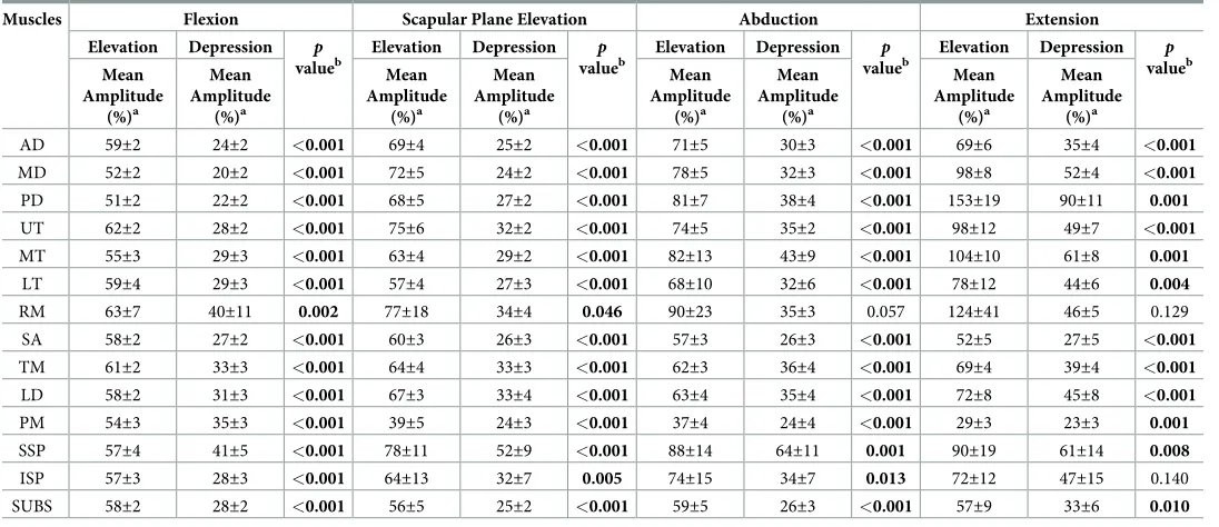
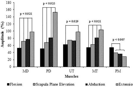
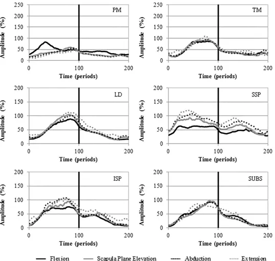
Table 1gives the mean EMG amplitude for each muscle during the different planes of move-ment. Significantly higher activity was seen during arm elevation as compared to depression for all muscles studied except for RM during abduction and extension and SUBS during exten-sion. Significant differences in signal amplitude were seen across all movements for MD, PD and MT. In addition, UT was significantly more active during extension than flexion and PM significantly more active during flexion as compared to the other movements (Fig 2). There were no significant differences in muscle activation between the movements for AD, LT, SA, TM, LD, SSP, ISP, SUBS, RM. Figs3and4depict the activation patterns for the individual muscles. The supplementary files reports the mean amplitude data disaggregated by gender (S1,S2,S3andS4Tables).
Timing
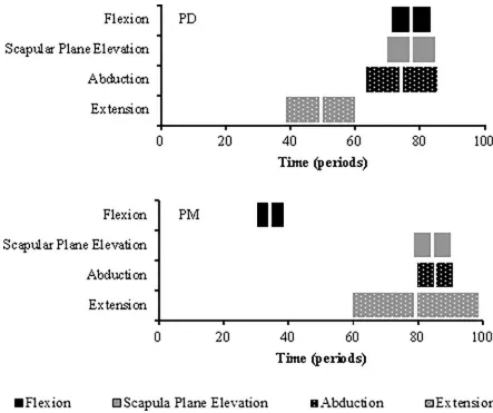
Peak activity of PD is seen significantly earlier in extension than in flexion (p<0.001), scapular plane elevation (p<0.001) and abduction (p = 0.001). Peak activity of PM is seen significantly earlier in flexion compared to scapular plane elevation (p = 0.004), abduction (p = 0.003) and extension (p = 0.011) (Fig 5). No muscles exhibited a double peak in their activation profile.
Muscular coordination
Fig 6illustrates the dynamic muscular coordination between the deltoid and rotator cuff mus-cle groups during shoulder elevation in the different planes of movement. Considering all movement planes together, coordination between the muscle groups was significantly higher during the initial (PCC = 0.79) and final (PCC = 0.74) stage of shoulder elevation compared to the mid-range (PCC = 0.34) (p = 0.020–0.035).
different movement planes is presented within the supplementary data files (S5,S6,S7andS8
Tables).
Discussion
The shoulder is a complex system which relies on coordinated muscle activity to maintain sta-bility [5–7]. Aberrant shoulder muscle coordination is considered to be an important aetiolo-gical component of shoulder impingement syndrome and shoulder instability [11,12]. A recent randomised control trial has questioned the role of surgery in subacromial impinge-ment syndrome and highlighted the important of rehabilitation [35]. However, an intimate knowledge of normal shoulder inter-muscular relationships is required if the pathological movement patterns of patients are going to be treated effectively with physiotherapy.
The results of the study are discussed below, firstly with respect to those muscles that act on the glenohumeral joint followed by those that comprise the scapulothoracic articulation. Broadly, muscles whose line of action is on the same side as the axis of movement are consid-ered ‘movers’ and those on the opposite side, ‘stabilisers’.
Glenohumeral muscles
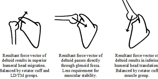
[image:6.612.35.583.89.326.2]Superior humeral head translation on the glenoid fossa, by the order of a few millimetres, dur-ing the initial stage of shoulder elevation has been well documented [3,36,37]. It occurs due to the lack of osseous congruity between the articular surfaces. Poppen and Walker, examining the resultant force vector during shoulder elevation, proposed that the superior subluxing force exerted by deltoid peaks at 60oof abduction before decreasing thereafter [14]. Other
Table 1. Mean signal amplitude during shoulder elevation and depression in the different plane of shoulder motion.
Muscles Flexion Scapular Plane Elevation Abduction Extension
Elevation Depression p valueb
Elevation Depression p valueb
Elevation Depression p valueb
Elevation Depression p valueb Mean Amplitude (%)a Mean Amplitude (%)a Mean Amplitude (%)a Mean Amplitude (%)a Mean Amplitude (%)a Mean Amplitude (%)a Mean Amplitude (%)a Mean Amplitude (%)a
AD 59±2 24±2 <0.001 69±4 25±2 <0.001 71±5 30±3 <0.001 69±6 35±4 <0.001
MD 52±2 20±2 <0.001 72±5 24±2 <0.001 78±5 32±3 <0.001 98±8 52±4 <0.001
PD 51±2 22±2 <0.001 68±5 27±2 <0.001 81±7 38±4 <0.001 153±19 90±11 0.001
UT 62±2 28±2 <0.001 75±6 32±2 <0.001 74±5 35±2 <0.001 98±12 49±7 <0.001 MT 55±3 29±3 <0.001 63±4 29±2 <0.001 82±13 43±9 <0.001 104±10 61±8 0.001
LT 59±4 29±3 <0.001 57±4 27±3 <0.001 68±10 32±6 <0.001 78±12 44±6 0.004
RM 63±7 40±11 0.002 77±18 34±4 0.046 90±23 35±3 0.057 124±41 46±5 0.129
SA 58±2 27±2 <0.001 60±3 26±3 <0.001 57±3 26±3 <0.001 52±5 27±5 <0.001 TM 61±2 33±3 <0.001 64±4 33±3 <0.001 62±3 36±4 <0.001 69±4 39±4 <0.001
LD 58±2 31±3 <0.001 67±3 33±4 <0.001 63±4 35±4 <0.001 72±8 45±8 <0.001
PM 54±3 35±3 <0.001 39±5 24±3 <0.001 37±4 24±4 <0.001 29±3 23±3 0.001
SSP 57±4 41±5 <0.001 78±11 52±9 <0.001 88±14 64±11 0.001 90±19 61±14 0.008 ISP 57±3 28±3 <0.001 64±13 32±7 0.005 74±15 34±7 0.013 72±12 47±15 0.140
SUBS 58±2 28±2 <0.001 56±5 25±2 <0.001 59±5 26±3 <0.001 57±9 33±6 0.010
AD–anterior deltoid; MD–middle deltoid, PD–posterior deltoid; UT–upper trapezius; MT–middle trapezius; LT–lower trapezius; RM–rhomboid major; SA–serratus anterior; TM–teres major; LD–latissimus dorsi; PM–pectoralis major; SSP–supraspinatus; ISP–infraspinatus; SUBS–subscapularis
aValues are means
±SEM
bpaired t-test comparing amplitude during each phase
muscles are required to have a stabilising effect to balance this superior subluxation and pre-vent the humeral head from impinging on the acromion.
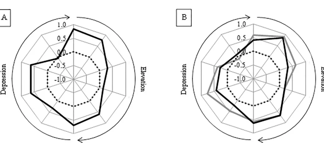
The anatomical arrangement of the rotator cuff muscles is such that contraction exerts a compressive force between the humeral head and glenoid fossa which ‘stiffens’ the joint [8]. The dynamic inter-muscular coordination between the deltoid and rotator cuff muscle groups, as indicated by the moving PCC, showed a high correlation during the initial (PCC = 0.79) stage of shoulder elevation (Fig 6). This synchronous activity pattern shows that the muscles are working in a coordinated fashion with rotator cuff activation balancing the superior sub-luxing force of the deltoid. Additionally, the coordination between the deltoid and LD/TM groups is particularly high (PCC = 0.89) during the second stage of elevation (Fig 7B). The LD and TM both exert an adducting force that can balance the superior destabilising force of the deltoid, in a similar fashion to the rotator cuff [38]. Consequently, this study suggests a stabilis-ing role for the rotator cuff, LD and TM durstabilis-ing the initial phase of shoulder elevation.
[image:7.612.118.569.74.370.2]In the mid-range of elevation the resultant force vector from the deltoid moves from exert-ing a superior subluxexert-ing force to pass through the glenoid in a more collinear fashion. At this time, the deltoid is generating its own compressive force across the GH joint which, as a conse-quence, is self-stabilising. Therefore, the requirement for other muscles to contribute to the stability during this phase is less. This suggestion is supported by the moving PCC results. Del-toid-rotator cuff coordination is significantly lower (PCC = 0.34) during the mid-range of ele-vation than at other times, and the moving PCC between the deltoid and LD/TM muscle group is also low, as illustrated inFig 7B.
Fig 2. Mean signal amplitude during shoulder elevation across the different planes of shoulder movement. P values are given for significant comparisons.
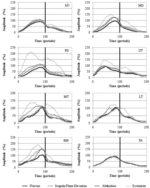
Fig 3. Ensemble average activation curves for the AD, MD, PD, UT, MT, LT, RM and SA. Mean EMG signal amplitude is presented as a function of time (periods). Elevation 0–100; Depression 100–200.
As shoulder elevation continues to progress, the humeral head translates inferiorly on the glenoid fossa [3,36,37], as the force vector exerted by the deltoid comes to pass below the axis of the glenoid. Accordingly, the requirement for other muscle groups to contribute to stability again increases. This is reflected by the high coordination between the deltoid and rotator cuff muscle groups at the end of elevation (PCC = 0.74). Coordination between the LD/TM and
Fig 4. Ensemble average activation curves for the PM, TM, LD, SSP, ISP and SUBS. Mean EMG signal amplitude is presented as a function of time (periods). Elevation 0–100; Depression 100–200.
deltoid muscle groups is understandably low as (because of their line of action) synchronised activation would perpetuate inferior humeral head migration.Fig 8is a schematic representa-tion of the proposed coordinarepresenta-tion between muscle groups at the GH joint during shoulder elevation.
[image:10.612.129.572.71.442.2]Considering the level of muscle activation, the PD was significantly more active during extension compared to movement in all other planes; this is consistent with its accepted func-tion and was expected given its anatomical orientafunc-tion and line of acfunc-tion [16]. Interestingly, the activity of the MD increased as the plane of shoulder elevation moved from flexion to extension. Similar findings were described in a recent study by Reed, although extension was not considered; in Reed’s study, MD activity was highest in the coronal plane, then decreased in the scapula plane before being lowest during elevation in a plane 30oanterior to the scapula plane [19]. In this work, the activity of PM was significantly higher during flexion compared to movement in the other planes, and its peak activity occurred significantly earlier during flex-ion compared to the other movements. This again was anticipated given its primary accepted role as a shoulder flexor [39]. There were no significant differences in the level of muscle acti-vation between the different movement planes for the AD, TM, LD, SSP, ISP or SUBS; this is in line with previous work [19].
Fig 5. Box plots illustrating time of peak muscle amplitude for the PD and PM during shoulder elevation. Boxes show the interquartile range and the thick white line the median.
Fig 6. Dynamic coordination between the deltoid and rotator cuff muscle groups during the different planes of shoulder elevation as illustrated by the moving PCC.
https://doi.org/10.1371/journal.pone.0211800.g006
Fig 7. A—Moving PCC indicating the dynamic coordination between the UT/SA and MT/LT muscle groups. B—Moving PCC indicating the dynamic coordination between the TM and deltoid (black line) and LD and deltoid (grey line) muscle groups.
[image:11.612.51.572.443.673.2]Scapulothoracic muscles
Scapula motion positions the glenoid effectively in space creating a basis for arm movement. The UT and SA elevate and upwardly rotate the scapula whilst the MT and LT counter this by depressing and downwardly rotating it. There is a high coordination between these functional muscle groups during the initial phase of shoulder elevation (Fig 7(A)). This is proposed to reflect the establishment of a force couple which enables controlled scapula motion and the early glenoid positioning.
UT activity was significantly higher during extension compared to flexion. The UT inserts on the lateral third of the clavicle and contraction results in clavicular elevation. The clavicle retracts, elevates and rotates posteriorly on its axis during shoulder elevation. A kinematic study found greater clavicular elevation and retraction during shoulder elevation in the coro-nal as compared to the sagittal plane [40]. Increasing UT muscle activity as the plane of eleva-tion moves posteriorly from flexion was therefore expected given the known kinematic patterns of the clavicle.
[image:12.612.46.572.82.346.2]MT activity was greatest in extension and decreased as the plane of shoulder elevation moved anteriorly. This is consistent with the results of other studies [19,41]. The scapula, dur-ing shoulder elevation, upwardly and internally rotates and tilts posteriorly. The scapulohum-eral rhythm is known to decrease as the plane of shoulder elevation moves in an anterior arc from abduction to flexion. Thus the scapula remains in a relatively more adducted position during coronal as compared to sagittal plane elevation [42]. The MT attaches to the spine of the scapula and is responsible for scapula adduction. The MT activity seen in this study there-fore reflects known kinematic patterns of the scapula. There were no significant differences in the activation of LT, SA, or RM between the movement planes.
Fig 8. Schematic representation of muscular coordination at the GH joint during elevation. The arrow within each figure indicates gives a representation of the resultant force vector.
Limitations of this study
The potential for cross talk is a limitation of all EMG studies that employ surface electrodes. This is particularly pertinent to the shoulder due to its compact anatomy. Further, during dynamic movement there is also the potential for surface electrodes to move in relation to the underlying muscle belly. Nevertheless EMG has extensively been used within the literature to record muscle activation during dynamic shoulder movements. In this study the careful place-ment of appropriately sized electrodes according to accepted anatomical criteria limited the impact of any potential cross talk. Normalisation was performed in the time domain to correct for any inter-individual variation in the speed at which the movement protocol was under-taken. This could be improved by the use of concurrent motion capture, which would allow normalisation to GH elevation angle. Furthermore, normalisation to the mean EMG ampli-tude precludes any conclusions about absolute muscle activity levels. However, the primary aim of this work was to investigate inter-muscular coordination, for which normalisation in the time domain and to the mean amplitude is appropriate. Including additional planes of shoulder elevation between abduction and extension would also provide a more complete pic-ture in this arc of movement. Finally, the conclusions drawn here remain an oversimplifica-tion. Muscles are not homogeneous, but rather have distinct segments that can act as different functional units [43]. However, evaluating coordination between all of these distinct segments would be unrealistically complicated at this stage.
Conclusion
The coordination between muscle groups is dynamic and changes during a task as the require-ments for stability alter. In broad outline, the destabilising force of the deltoid, during the ini-tial stage of shoulder elevation, is balanced by the coordinated activity of the rotator cuff, LD and TM. Stability requirements are lower during the mid-range of elevation and this is reflected in the reduced coordination between these muscle groups. At the end-range of move-ment the demand for muscular stability again increases and here this is provided by the rotator cuff. It is suggested that this fuller analysis of shoulder function in health will improve under-standing of the alterations associated with pathology, and assist development of more targeted, evidenced-based, treatment strategies.
Supporting information
S1 Table. Mean EMG amplitude during flexion. Mean amplitude data for flexion
disaggre-gated by sex (DOCX)
S2 Table. Mean EMG amplitude during scapula plane elevation. Mean amplitude data for
scapula plane elevation disaggregated by sex (DOCX)
S3 Table. Mean EMG amplitude during abduction. Mean amplitude data for abduction
dis-aggregated by sex (DOCX)
S4 Table. Mean EMG amplitude during extension. Mean amplitude data for extension
S5 Table. Individual muscle flexion PCC. PCC comparing EMG between individual muscles
during flexion. (DOCX)
S6 Table. Individual muscle scapula plane elevation PCC. PCC comparing EMG between
individual muscles during scapula plane elevation. (DOCX)
S7 Table. Individual muscle abduction PCC. PCC comparing EMG between individual
mus-cles during abduction. (DOCX)
S8 Table. Individual muscle extension PCC. PCC comparing EMG between individual
mus-cles during extension. (DOCX)
S1 Data. Processed EMG data for the individual subjects during the different planes of movement.
(XLSX)
Author Contributions
Conceptualization: David H. Hawkes, Simon P. Frostick.
Data curation: Anthony J. Howard.
Formal analysis: David H. Hawkes.
Investigation: David H. Hawkes, Omid A. Khaiyat, Anthony J. Howard.
Methodology: David H. Hawkes, Simon P. Frostick.
Project administration: David H. Hawkes.
Resources: Graham J. Kemp, Simon P. Frostick.
Supervision: Omid A. Khaiyat, Graham J. Kemp, Simon P. Frostick.
Writing – original draft: David H. Hawkes.
Writing – review & editing: Omid A. Khaiyat, Graham J. Kemp.
References
1. Cameron KL, Mauntel TC, Owens BD. The Epidemiology of Glenohumeral Joint Instability: Incidence, Burden, and Long-term Consequences. Sports Med Arthrosc. 2017; 25(3):144–9.https://doi.org/10. 1097/JSA.0000000000000155PMID:28777217
2. Luime JJ, Koes BW, Hendriksen IJ, Burdorf A, Verhagen AP, Miedema HS, et al. Prevalence and inci-dence of shoulder pain in the general population; a systematic review. Scand J Rheumatol. 2004; 33 (2):73–81. PMID:15163107
3. Bey MJ, Kline SK, Zauel R, Lock TR, Kolowich PA. Measuring dynamic in-vivo glenohumeral joint kine-matics: technique and preliminary results. J Biomech. 2008; 41(3):711–4.https://doi.org/10.1016/j. jbiomech.2007.09.029PMID:17996874
4. Dal Maso F, Raison M, Lundberg A, Arndt A, Allard P, Begon M. Glenohumeral translations during range-of-motion movements, activities of daily living, and sports activities in healthy participants. Clin Biomech (Bristol, Avon). 2015; 30(9):1002–7.
6. Inman VT, Saunders JB, Abbott LC. Observations of the function of the shoulder joint. 1944. Clin Orthop Relat Res. 1996; 330:3–12.
7. Burkhart SS. Arthroscopic treatment of massive rotator cuff tears. Clinical results and biomechanical rationale. Clin Orthop Relat Res. 1991; 267:45–56.
8. David G, Magarey ME, Jones MA, Dvir Z, Turker KS, Sharpe M. EMG and strength correlates of selected shoulder muscles during rotations of the glenohumeral joint. Clin Biomech (Bristol, Avon). 2000; 15(2):95–102.
9. Hawkes DH, Alizadehkhaiyat O, Kemp GJ, Fisher AC, Roebuck MM, Frostick SP. Shoulder muscle acti-vation and coordination in patients with a massive rotator cuff tear: An electromyographic study. J Orthop Res. 2011; 30(7):1140–6.https://doi.org/10.1002/jor.22051PMID:22213234
10. Veeger HE, van der Helm FC. Shoulder function: the perfect compromise between mobility and stability. J Biomech. 2007; 40(10):2119–29.https://doi.org/10.1016/j.jbiomech.2006.10.016PMID:17222853 11. Illyes A, Kiss RM. Kinematic and muscle activity characteristics of multidirectional shoulder joint
instabil-ity during elevation. Knee Surg Sports Traumatol Arthrosc. 2006; 14(7):673–85.https://doi.org/10. 1007/s00167-005-0012-7PMID:16362361
12. de Morais Faria CD, Teixeira-Salmela LF, de Paula Goulart FR, de Souza Moraes GF. Scapular muscu-lar activity with shoulder impingement syndrome during lowering of the arms. Clin J Sport Med. 2008; 18(2):130–6.https://doi.org/10.1097/JSM.0b013e318160c05dPMID:18332687
13. Ricci FP, Santiago PR, Zampar AC, Pinola LN, Fonseca MC. Upper extremity coordination strategies depending on task demand during a basic daily activity. Gait Posture. 2015; 42(4):472–8.https://doi. org/10.1016/j.gaitpost.2015.07.061PMID:26282047
14. Poppen NK, Walker PS. Forces at the glenohumeral joint in abduction. Clin Orthop Relat Res. 1978; 135:165–70.
15. Diedrichsen J, Shadmehr R, Ivry RB. The coordination of movement: optimal feedback control and beyond. Trends Cogn Sci. 2010; 14(1):31–9.https://doi.org/10.1016/j.tics.2009.11.004PMID:
20005767
16. Kronberg M, Nemeth G, Brostrom LA. Muscle activity and coordination in the normal shoulder. An electromyographic study. Clin Orthop Relat Res. 1990; 257:76–85.
17. Alpert SW, Pink MM, Jobe FW, McMahon PJ, Mathiyakom W. Electromyographic analysis of deltoid and rotator cuff function under varying loads and speeds. J Shoulder Elbow Surg. 2000; 9(1):47–58. PMID:10717862
18. Wickham J, Pizzari T, Stansfeld K, Burnside A, Watson L. Quantifying ’normal’ shoulder muscle activity during abduction. J Electromyogr Kinesiol. 2010; 20(2):212–22.https://doi.org/10.1016/j.jelekin.2009. 06.004PMID:19625195
19. Reed D, Cathers I, Halaki M, Ginn KA. Does changing the plane of abduction influence shoulder muscle recruitment patterns in healthy individuals? Man Ther. 2016; 21:63–8.https://doi.org/10.1016/j.math. 2015.04.014PMID:25920341
20. Cram JR, Kasman GS, Holtz J. Introduction to surface electromyography. Gaithersburg, Md.: Aspen Publishers; 1998.
21. Steenbrink F, de Groot JH, Veeger HE, Meskers CG, van de Sande MA, Rozing PM. Pathological mus-cle activation patterns in patients with massive rotator cuff tears, with and without subacromial anaes-thetics. Man Ther. 2006; 11(3):231–7.https://doi.org/10.1016/j.math.2006.07.004PMID:16890886 22. Prakash KM, Fook-Chong SM, Leoh TH, Dan YF, Nurjannah S, Tan YE, et al. The lower subscapular
nerve conduction studies and utilisation in brachial plexopathy evaluation. J Neurol Sci. 2006; 247 (1):77–80.https://doi.org/10.1016/j.jns.2006.03.014PMID:16647087
23. Noraxon. Kinesiological Fine Wire EMG. A practical introduction to fine wire EMG applications. Avali-able from:https://www.velamed.com/wp-content/uploads/2017/08/Fine_Wire_EMG.pdf
24. Kadaba MP, Cole A, Wootten ME, McCann P, Reid M, Mulford G, et al. Intramuscular wire electromyog-raphy of the subscapularis. J Orthop Res. 1992; 10(3):394–7.https://doi.org/10.1002/jor.1100100312
PMID:1569502
25. ISEK. Standards for reporting EMG data. J Electromyogr Kines. 1996; 6(4):III–IV.
26. Alizadehkhaiyat O, Hawkes DH, Kemp GJ, Frostick SP. Electromyographic Analysis of the Shoulder Girdle Musculature During External Rotation Exercises. Orthop J Sports Med. 2015; 3
(11):2325967115613988.https://doi.org/10.1177/2325967115613988PMID:26740950
28. Morris AD, Kemp GJ, Lees A, Frostick SP. A study of the reproducibility of three different normalisation methods in intramuscular dual fine wire electromyography of the shoulder. J Electromyogr Kinesiol. 1998; 8(5):317–22. PMID:9785252
29. Burden AM, Trew M, Baltzopoulos V. Normalisation of gait EMGs: a re-examination. J Electromyogr Kinesiol. 2003; 13(6):519–32. PMID:14573367
30. Winter DA, Yack HJ. EMG profiles during normal human walking: stride-to-stride and inter-subject vari-ability. Electroencephalogr Clin Neurophysiol. 1987; 67(5):402–11. PMID:2444408
31. Wattanaprakornkul D, Cathers I, Halaki M, Ginn KA. The rotator cuff muscles have a direction specific recruitment pattern during shoulder flexion and extension exercises. J Sci Med Sport. 2011; 14(5):376– 82.https://doi.org/10.1016/j.jsams.2011.01.001PMID:21333595
32. Wattanaprakornkul D, Halaki M, Cathers I, Ginn KA. Direction-specific recruitment of rotator cuff mus-cles during bench press and row. J Electromyogr Kinesiol. 2011; 21(6):1041–9.https://doi.org/10.1016/ j.jelekin.2011.09.002PMID:21978788
33. Hawkes DH, Alizadehkhaiyat O, Fisher AC, Kemp GJ, Roebuck MM, Frostick SP. Normal shoulder muscular activation and co-ordination during a shoulder elevation task based on activities of daily living: an electromyographic study. J Orthop Res. 2012; 30(1):53–60.https://doi.org/10.1002/jor.21482PMID:
21674607
34. Wren TA, Do KP, Rethlefsen SA, Healy B. Cross-correlation as a method for comparing dynamic elec-tromyography signals during gait. J Biomec. 2006; 39(14):2714–8.
35. Beard DJ, Rees JL, Cook JA, Rombach I, Cooper C, Merritt N, et al. Arthroscopic subacromial decom-pression for subacromial shoulder pain (CSAW): a multicentre, pragmatic, parallel group, placebo-con-trolled, three-group, randomised surgical trial. Lancet. 2018; 391(10118):329–38.https://doi.org/10. 1016/S0140-6736(17)32457-1PMID:29169668
36. Graichen H, Stammberger T, Bonel H, Karl-Hans E, Reiser M, Eckstein F. Glenohumeral translation during active and passive elevation of the shoulder—a 3D open-MRI study. J Biomech. 2000; 33 (5):609–13. PMID:10708782
37. Kelkar R, Wang VM, Flatow EL, Newton PM, Ateshian GA, Bigliani LU, et al. Glenohumeral mechanics: A study of articular geometry, contact, and kinematics. J Shoulder Elbow Surg. 2001; 10(1):73–84.
https://doi.org/10.1067/mse.2001.111959PMID:11182740
38. Steenbrink F, de Groot JH, Veeger HE, van der Helm FC, Rozing PM. Glenohumeral stability in simu-lated rotator cuff tears. J Biomech. 2009; 42(11):1740–5.https://doi.org/10.1016/j.jbiomech.2009.04. 011PMID:19450803
39. Suenaga N, Minami A, Fujisawa H. Electromyographic analysis of internal rotational motion of the shoulder in various arm positions. J Shoulder Elbow Surg. 2003; 12(5):501–5.https://doi.org/10.1016/ S1058274603001691PMID:14564277
40. Ludewig PM, Phadke V, Braman JP, Hassett DR, Cieminski CJ, LaPrade RF. Motion of the shoulder complex during multiplanar humeral elevation. Bone Joint J. 2009; 91(2):378–89.
41. Ishigaki T, Ishida T, Samukawa M, Saito H, Hirokawa M, Ezawa Y, et al. Comparing trapezius muscle activity in the different planes of shoulder elevation. J Phys Ther Sci. 2015; 27(5):1495–7.https://doi. org/10.1589/jpts.27.1495PMID:26157248
42. Giphart JE, Brunkhorst JP, Horn NH, Shelburne KB, Torry MR, Millett PJ. Effect of plane of arm eleva-tion on glenohumeral kinematics: a normative biplane fluoroscopy study. J Bone Joint Surg Am. 2013; 95(3):238–45.https://doi.org/10.2106/JBJS.J.01875PMID:23389787