0022-538X/85/080347-05$02.00/0
Copyright C) 1985, AmericanSociety forMicrobiology
Identification
and
Characterization
of the Epstein-Barr Virus
Receptor
on
Human
B
Lymphocytes and Its
Relationship
to
the
C3d
Complement Receptor
(CR2)t
GLEN R. NEMEROW,* ROBERT WOLFERT, MARY ELLEN McNAUGHTON, AND NEILR. COOPER
Department of Immunology, Scripps Clinic & Research Foundation, LaJolla, California 92037 Received 18 January 1985/Accepted 9 April 1985
Inpursuing studies ontheearlyeventsin theinfection of human B cellsby Epstein-Barrvirus(EBV), we examined the host cell attachment phase with a panel ofB-cell-specific monoclonal antibodies. One of the monoclonal antibodies, OKB7, directly blocked the attachment of purified EBV to B lymphocytes in the absence of a second anti-immunoglobulin antibody and thereby prevented EBV infection of tonsil and peripheral bloodBcells.Althoughearlier studies have shownaclose association of the EBV andcomplement receptor (CR2), ananti-CR2 monoclonal antibody, anti-B2, did notdirectly block the bindingof EBV toB cells. Acomparisonof the structuresrecognizedbythese monoclonalantibodiesonvariouscelltypes and their functionalandphysiochemical propertieswasundertaken. Flowcytometricanalysisrevealed that the molecules detected by OKB7and anti-B2werecoexpressedtothesameextentonBcellsbutwerenotexpressedonT-cell lines. OKB7 and anti-B2 both immunoprecipitated a 145,000-molecular-weight membrane protein with an isoelectric point of 8.2 from membrane extractsofRaji lymphoblastoid cells. OKB7 and, toalesser extent, anti-B2 directly blocked the attachment of C3d,g-coated fluorescent microspheres and sheep erythrocytes bearingC3d toB cells, indicating thattheseantibodies also reactwith CR2. These studies indicate that the EBV-CR2 receptor is a single membrane glycoprotein which possesses multiple antigenic and functional epitopes.
Epstein-Barr virus (EBV), an oncogenic herpesvirus of humans, selectively infects B lymphocytes (7, 12, 25) and probably nasoepithelial cells (31), suggesting that discrete membrane receptors areutilizedbythisvirusto entercells. Suchreceptorsundoubtedly participateintheendocytosisof EBVin normal Bcells(25,26). Earlier studies demonstrated that the EBV receptor was closely associated with the complement C3d receptor (CR2), a 140,000- to 145,000-molecular-weight membrane glycoprotein (13, 18, 28, 34, 35). Recently, evidencefor theidentityof the EBVreceptor
with CR2 was obtained by using an anti-CR2 monoclonal antibody, HB-5,whichdoesnotdirectlyblock EBVbinding (5). In the present studies, we examined the membrane structure recognized by OKB7, a B-lymphocyte-specific monoclonalantibody whichdirectly blocks EBV attachment andinfection, and therelationship of this structure toCR2.
MATERIALS AND METHODS
Cell lines, lymphocytes, and EBV isolation. EBV-transformed B-celllines Raji and B95-8 were maintained in RPMI 1640and 10%fetal bovine serumat 37°C. Peripheral blood Blymphocyteswereisolated fromnormaladultdonors by negative selection with neuraminidase-treated sheep erythrocytes as previously described (8). Tonsils were ob-tainedatChildren'sHospital,SanDiego, Calif., from normal children of 2 to 11 years ofageundergoingtonsillectomy.
EBV was grown in phorbol-myristate-acetate-stimulated B95-8 cells and purified as previously described (24). Virus wasintrinsically labeled with [35S]methionine by the method of Edson andThorley-Lawson (2). Viruspuritywasassessed by sodium dodecyl sulfate-polyacrylamide gel
electrophore-*Correspondingauthor.
t Publication no. 3809 IMMof the Department ofImmunology,
Scripps Clinic & Research Foundation.
sis(SDS-PAGE) and autoradiography and by negative-stain-ingtransmission electron microscopy (24).
Monoclonal and polyvalent antibodies and CR2 ligands. Monoclonal antibodies OKB2 and OKB7 were obtained from Ortho Diagnostics, Inc., Raritan, N.J.; Bl, anti-B2, and anti-B4 antibodieswere from CoulterImmunology, Hialeah, Fla.; the monocyte- and macrophage-specific monoclonal antibodyanti-MO2waspurchasedfromCoulter. Affinity-purified anti-mouse immunoglobulinM and anti-im-munoglobulin G and M antibodies were purchased from CappelLaboratories, Cochranville,Pa. The latterantibodies
were cross-linked to CNBr-activated Sepharose 6B (Phar-macia Fine Chemicals, Piscataway, N.J. [Div. Pharmacia, Inc.])asrecommendedbythemanufacturer. Highly purified C3d,g, the physiologic fragment of C3 which bindsto CR2 (1, 29), wasgenerously provided by Hans Muller-Eberhard and Michael J. Pangburn. Coumarin (green) fluorescent microspheres (1.0 ,um) (Covalent Technology Corp., Red-woodCity, Calif.)werecoated with purifiedC3d,gorbovine serumalbumin (BSA) exactlyasrecommendedbythe
manu-facturer.Antibody-sensitized sheep erythrocytes bearing the complement components C2 and C4 (EAC4,2) were coated with10,000to20,000 molecules of C3d by sequential incuba-tion with purifiedC3,factors H andI,and5,ugoftrypsinper
ml aspreviouslydescribed (30).
Radiolabeling, SDS-PAGE, and IEF analysis of CR2. Raji lymphoblastoidcells weresurface labeled with 1251 by using lodo-Beads (Pierce Chemical Co., Rockford, Ill.).Thecells were lysed in 1% Nonidet P-40, and the lysates were precleared by absorption with an equal volume ofpacked, fixed Staphylococcus aureus (Pansorbin; Calbiochem-Behring, La Jolla, Calif.) for 3 h at 4°C. The precleared
extractswerereacted with 2,ug of anti-B2antibody coupled
to anti-mouse immunoglobulin-M-Sepharose or with an
equivalentamountofOKB7antibody coupledtoanti-mouse
347
on November 10, 2019 by guest
http://jvi.asm.org/
348 NEMEROW ET AL.
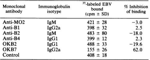
TABLE 1. Effectofmonoclonal antibodies on binding of EBV to Bcellsa
Monoclonal Immunoglobulin 35-labeledEBV % Inhibition
antibody isotype (cpm SD) of binding
Anti-MO2 IgM 421 ± 28 -3.0
Anti-Bl IgG2a 398 ± 32 2.5
Anti-B2 IgM 483 + 80 -18.0
Anti-B4 IgGl 399 ± 12 2.3
OKB2 IgGl 488 ± 33 -19.6
OKB7 IgG2a 155 ± 26 62.0
Control 408 ± 18
a Experiment was carried out with106peripheralblood Bcells and 1 ,ug of
eachmonoclonal antibody.
immunoglobulin G and M-Sepharose. The
immunoprecipi-tateswerewashedand then eluted fromtheSepharosebeads
with 2% SDS for SDS-PAGE (16) or with 8 M urea for
isoelectricfocusing (IEF) (4).
EBV binding to and infection of B cells in the presence of monoclonalantibodies. Inpreliminary studies,thebinding of
35S-labeled
EBV tountreatedRajilymphoblastoid cells wasexamined first. Increasingamountsof 35S-labeled EBV was
added to 107 Raji cells in 1 ml of phosphate-buffered saline-BSA withconstantmixingat4°C for60min.The cells were then washed twice inphosphate-buffered saline-BSA,
and the pellets were lysed and counted. Forvirus binding-inhibitionassays, peripheral blood Bcells
(106)
ortonsil orRaji cells
(10G)
were pretreated withvarious amounts ofthedifferent monoclonal antibodies in phosphate-buffered saline-BSAorwithbuffer alone for60 min at4°C.Thecells
were washed and reacted with 4,000 cpm (approximately 107
virions) of purified
35S-labeled
EBV and incubated for anadditional60min at4°C. Thecells wereagain washed, lysed with scintillation fluid (Cytoscint; Westchem, San Diego, Calif.), andcounted. Control cells were pretreated with an
irrelevant monoclonal antibody (anti-MO2) or with buffer alone. Forviralinfectivity assays, 6 x
105
peripheral blood Bcellswereincubated withthemonoclonalantibodies for60 min at 4°C. The monoclonal antibodies were diluted in RPMI-10% fetal calf serumand dialyzed against the samemedium to remove azide before exposure to B cells. The
cells were then washed with RPMI medium, plated into 96-well sterile trays at 2 x 10⁵ cells per well, and then
exposed to unlabeled EBV at amultiplicity of infection of 1.0, as determined by DNA content (27) and electron mi-croscopy (24),for14days. Cell transformation was assessed
by
incorporation
of[3H]thymidine
andby formation ofcellcolonies aspreviously described (24).
Binding of C3d,g and C3d to B cells in the presence of monoclonal antibodies. Binding of C3d or C3d,g to B cells was examinedby two separate assays. In the first assay, the
ability ofmonoclonal antibodies to influenceC3d,g binding
to B cells was studied by using
C3d,g-coated
fluorescent microspheres. B cells which had been reacted with various amounts of the monoclonal antibodies at 4°C for 60 min in 200Rl
of phosphate-buffered saline-BSA weresubsequently incubated for45min at37°Cwith 10,u1
ofa10%suspension of C3d,g-fluorescent microspheres or withmicrospheres
which had been coated with BSA as a control. Cells were
examined by combination phase-contrast and fluorescent microscopy. Cells with four or more microspheres were considered positive for C3d,g
binding.
In the second CR2 assay,Bcells werepretreatedwith monoclonalantibodiesat4°C,
washed,andthen reactedat aratio of1:50with EAC3dcells for45minat37°C. Formation ofrosettes wasexamined
bylight microscopy, and B cells with four or more EAC3d cells were considered positive. Controls included B cells incubated with EAorEAC4,2 cells.
RESULTS
Effect of B-cell-specific monoclonal antibodies on EBV at-tachment and infection. The specificity of35S-labeled EBV binding to B cells was first examined byincubating increas-ing amounts of 35S-labeled EBV with Raji cells and
determin-ing binddetermin-ing.Anuptakecurveindicative ofsaturablebinding
was observed; at saturable binding, 85 to 90% binding of labeled viruswas achieved. Such binding was inhibited by a 10-fold excess of cold EBV (data not shown), indicating the specificity of EBV binding. A panel of monoclonal
antibod-ies specific for B cells was then screened for the ability to directly inhibit the binding of radiolabeled EBV to isolated B lymphocytes in the absence of a second antibody. In these experiments, an amountof each monoclonal antibody (1 to 2 pug) which gave maximalfluorescent staining of the cells was used together with an amount of 35S-labeled EBV which resulted in thesaturation of50% of the EBV receptors on 107 untreated tonsil or Raji B cells. Only OKB7 inhibited the binding of labeled EBV toperipheral blood B cells (Table 1). There was no correlation ofvirus inhibition with the anti-body isotype (Table 1) or with the percentage of B cells recognized by antibodies as determined by fluorescence-activated cell sorter analysis. The percentage of peripheral blood B cells labeled by Bl, OKB7, and B2 anti-bodies, as determined by FACS analysis, was similar to the previously reported values (21-23). In addition, antibodies anti-B2 and OKB7 stained similar numbers oftonsil B cells and isolated peripheral blood B cellsanddidsowith similar intensities.
Dose-response studies revealed that 50% inhibition of the binding ofapproximately
107
particles of EBV(4,000 cpm)to107
tonsil B cells occurred with a dose of 300 ngofantibodyOKB7, whereas 800 ng wasrequiredto achieve50% inhibi-tion of binding to
107
Raji cells (Fig. 1A). The anti-CR2 monoclonal antibody, anti-B2 (10), failed to inhibit EBVbinding atall doses. Consistent with theability of antibody
OKB7 to inhibit thebinding of EBV to tonsil B lymphocytes and Raji lymphoblastoid cells directly, this antibody also
directly blocked EBV infection ofperipheral blood B
lym-phocytes in adose-dependentmanner(Fig. 1B)asmeasured either by colony outgrowth in soft agarose or by [3H]thymidine incorporation at 14 days of culture. A 50%
inhibitionofinfectionoccurred with a dose of300 to 500 ng ofantibody OKB7 per 6 x 10⁶peripheral blood B
lympho-cytes.
Reactivity ofOKB7 andanti-B2monoclonal antibodieswith CR2.Studies were nextcarriedout todetermine thepossible
CR2 specificity of monoclonal antibody OKB7. Antibody
OKB7efficiently inhibited bothEAC3d andC3d,g bindingto
tonsil Bcells andRaji cells,respectively, in the absenceofa second anti-immunoglobulin antibody, indicating that this
antibody blocks CR2 function (Table 2). Anti-B2 antibody
showed a limitedability toinhibitCR2 functionasindicated
by theblocking ofEAC3d andC3d,g microsphere bindingto Bcells, afinding consistent withprevious reports (10). Two other monoclonal antibody controls, anti-MO2and
anti-Bl,
failed to alter CR2 function significantly. Inhibition was observedat37°C(Table 2)aswellas at4°C (datanotshown).
This indicates that modulation by antibody redistribution of
receptors was notresponsible for the observed inhibition. SDS-PAGE and IEF analysis of theantigen recognized by
J. VIROL.
on November 10, 2019 by guest
http://jvi.asm.org/
[image:2.612.64.306.91.189.2]antibodies anti-B2 and OKB7. Biochemical studies were carried out to define the antigens recognized by the OKB7
and anti-B2 monoclonalantibodies. Antibodies OKB7 (Fig.
2A, lane 1) and anti-B2 (lane 2) both immunoprecipitated a
145,000-molecular-weight protein from detergent lysates of labeled Raji cells. An irrelevantmonoclonal antibody,
anti-M02(lane 3),failedto do so.Immunoprecipitates werealso subjected to IEF. Both antibodies anti-B2 (Fig. 2B, lane 1) and OKB7 (lane 2) detected a protein with an isoelectric
point of 8.2, a value consistent with the isoelectric pointof
CR2 reported by Iida et al. (10). The molecular weight estimate fortheprotein recognized by antibody OKB7 was somewhat different than thatoriginally reported (21).
DISCUSSION
Infection of B lymphocytes is initiated by the specific binding ofEBV to areceptorexpressed onthesurface of B
lymphocytes and B-lymphoblastoid cell lines (12-14, 25). Several lines of evidence have suggested that the EBV receptoris eitheraC3 receptor orisclosely associatedwith aC3receptor(3, 9, 13, 15, 20, 28,34,35). Directanalysis of
the EBV receptorand its relationshipto CR2 has recently beenpossible through theuseof monoclonal antibodies. The
140,000- to 145,000-molecular-weight B-cell membrane
pro-tein isrecognizedby monoclonal antibodies termed anti-B2 (10, 23) and HB-5 (5, 32, 33), which
immunoprecipitate
a
17.5-o
12.5-co
7.5-<'
2.5-A
0
.
..I
__
21
15
9.
3-W___,_ _-_ ---.- - --_
0.25 0.5 0.75 1.0 0.75 1.5 2.25 3.0
B
0
D:
.-(A
w
MW
X10-3
Daltons
200-
116-
92-
68-
45-I I
I
C.) *e
cm
CD
MonoclonalAntibody(Mg)
FIG. 1. Effect of monoclonal antibodies on 35S-labeled EBV
binding and infection. (A) Normal tonsil B (left)orRaji(right) cells (107) were reacted with various amounts of OKB7 (- *) or
anti-B2 (---) antibody before the addition of 4,000 cpm of
3"S-labeledEBV. XAxes,monoclonalantibody (,ug). (B)Bcells(6
x 105)werereacted with variousamountsof OKB7(-)oranti-B2
(A) antibody, washed, and then cultured for 14 days in thepresence
of EBV. Infectivity was measured by the stimulation of DNA
synthesis([3H]thymidine incorporation)andcolony formation.
TABLE 2. Inhibition of CR2 function by monoclonal antibodies Antibody Amt(,ug)
t%
Positive EAC3d % PositiveC3d,gAntibodyAmt (~~g) rosetteSab rosetteSb,c
None 73 (5) 93 (4)
Anti-MO2 2.0 67 (3) 88(5)
Anti-Bl 2.0 74 (5) 90 (5)
Anti-B2 0.5 40(2) 78 (6)
Anti-B2 1.0 43 (8) 57(4)
Anti-B2 2.0 40 (3) 25 (3)
OKB7 0.2 55 (4) 50(4)
OKB7 1.0 45(6) 20(4)
OKB7 2.0 30(2) 4(1)
a Positive rosettes were tonsil B cells having four or more bound EAC3d
cells.
bNumbers inparentheses indicate standard deviation from two
experi-ments. Atleast 100 cells were counted for eachantibodyconcentration.
c Positive rosettes were Raji cells having four or more fluorescent C3d,g microspheres.
polypeptide chain of this molecular weightfrom normal B
lymphocytes and B-lymphoblastoid cell lines and, together
with ananti-immunoglobulin secondantibody,block rosette
formation between particles bearing C3d and B-lymphoid
cellsbearingthereceptor(10, 33). Antibodyanti-B2 also has some ability to block such rosette function directly in the
absence ofasecondantibody (10). AntibodyHB-5, although unabletoblock EBV
binding
directly, possessed
thisability
in the presence ofa second
anti-immunoglobulin antibody
(5). Recently, good evidence for the identity of the EBV receptor with CR2 wasobtained by using theseantibodies,
since pretreatmentof
B-lymphoblastoid
cellswithantibody
HB-5 followed by
anti-immunoglobulin
blocked virus bind-ing, andHB-5-CR2complexes obtainedby
immunoprecipi-A
1
2
3
__
B
1
2
pH
.
.
-8.30
-8.15
-8.10
-8.00
-7.90
-7.82
-7.65
-7.50
FIG. 2. Immunoprecipitation of Raji B cells with monoclonal antibodies. Surface-labeledRaji cells were lysedwith1% Nonidet P-40andimmunoprecipitatedwithOKB7(lanes1Aand2B),anti-B2 (lanes 2A and 1B) antibody, or M02 (lane 3A) and analyzed by SDS-PAGE(A)orIEF(B). MW, Molecularweight.
on November 10, 2019 by guest
http://jvi.asm.org/
[image:3.612.313.553.88.214.2] [image:3.612.55.293.367.645.2] [image:3.612.313.553.449.679.2]350 NEMEROW ET AL.
tation from B-lymphoblastoidcellextracts andimmobilized onS. aureusparticles possessedtheabilitytobind EBV(5). Thestudies with OKB7 monoclonalantibodydescribed here
providedirect evidence that a single B-cell membrane pro-tein functions as a dual receptor for EBV and C3d. This
antibody reacts with a 145,000-molecular-weight protein with an isoelectricpoint of 8.2, values previouslyidentified ascharacteristic ofCR2, and directly blocks CR2 functionas well asEBVbindingandinfection in the absence of a second antibody.
These studies thusindicate that EBV utilizes anormal cell constituent to achieve specific B-cell tropism. Other
ex-amplesofviruses whichuse hostproteinstoinfect cells are rabiesvirus,whichmay utilize theacetylcholinereceptor on neural cells(18, 19),andlactatedehydrogenase virus,which may bindto Iaantigens (11)on macrophages.
These B-cell-specific monoclonal antibodies and
cor-responding ligands such asEBV andC3d,g, which all react withacommon proteinonlymphocytesbutinduce different
biological
functions, will provide very useful probes fordissecting the early stages of EBV infection as well as the biochemical events involved in B-cell activation and dif-ferentiation.
ACKNOWLEDGMENTS
This study was supported by Public Health Service grants Al 17354 andCA 14692 from the National Institutes of Health and a
LeukemiaSociety of AmericaSpecial FellowshipAward. LITERATURE CITED
1. Davies, A. E., R. A. Harrison, and P. J. Lachmann. 1984. Physiologicinactivation offluidphaseC3b: isolation and
struc-tural analysis of C3c, C3d,g (a2D) and C3g. J. Immunol. 132:1960-1966.
2. Edson, C. M., and D. A. Thorley-Lawson. 1981. Epstein-Barr virus membrane antigen: characterization, distribution, and straindifferences. J. Virol. 39:172-184.
3. Einhorn,L.,M.Steinitz,E.Yefenof,I.Ernberg,T.Bakacs,and G. Klein. 1978. Epstein-Barr virus (EBV) receptors,
comple-ment receptors, and EBV infectibility of differentlymphocyte fractions of human peripheral blood. II. Epstein-Barr virus studies. Cell. Immunol. 35:43-58.
4. Ferreira, A., and D. Eichinger. 1981. A simplified
two-dimensional electrophoretic technique. J. Immunol. Methods 43:291-299.
5. Fingeroth, J. D., J. J. Weis, T. F. Tedder, J. L. Strominger,
P. A.Biro,andD. T. Fearon. 1984.Epstein-Barrvirusreceptor of humanBlymphocytes is the C3dreceptorCR2. Proc.Natl. Acad. Sci. U.S.A. 81:4510-4514.
6. Gerber, P., and B. H.Hoyer. 1972. Induction of cellularDNA
synthesis in human leukocytes by Epstein-Barr virus. Nature (London)231:46-47.
7. Greaves, M.F., G.Brown,and A. B. Rickinson.1975.
Epstein-Barrvirusbindingsiteson lymphocytesubpopulationsand the
originoflymphoblastsin culturedlymphoidcell lines and in the bloodofpatientswith infectious mononucleosis. Clin.Immunol.
Immunopathol.3:514-524.
8. Huddlestone,J.R.,and M. B.A. Oldstone. 1978. T suppressor
(TG)
lymphocytes
fluctuate inparallel
withchanges
in the clinicalcourseofpatientswith multiple sclerosis.J. Immunol. 123:1615-1618.9. Hutt-Fletcher,L. M.,E.Fowler, J. D. Lambris, R.J. Feighny, J.G. Simmons, andG. D. Ross. 1983. Studies of the
Epstein-Barr virus receptor found on Raji cells. II. A comparison of lymphocyte binding sites for Epstein-Barr virus and C3d. J. Immunol. 130:1309-1312.
10. Iida, K.,L.Nadler,and V.Nussenzweig.1983. Identification of the membrane receptor for the complement fragment C3d by
meansofamonoclonalantibody. J.Exp. Med. 158:1021-1033.
11. Inada, T., and C. A. Mims. 1984. Mouse Ia antigens are receptors for lactate dehydrogenase virus. Nature (London) 309:59-61.
12. Jondal, M., and G. Klein. 1973. Surface markers onhuman B and Tlymphocytes. II. Presence of Epstein-Barrreceptors on B lymphocytes. J. Exp. Med. 138:1365-1378.
13. Jondal, M., G. Klein, M. B. A. Oldstone, V. Bokisch, and E. Yefenof.1976.Surface markerson human BandTlymphocytes. VIII.Association between complement and Epstein-Barr recep-tors onhumanlymphoid cells. Scand. J. Immunol. 5:401-410. 14. Jonsson, V., A. Wells, and G. Klein. 1982. Receptors for the
complement C3d component and the Epstein-Barr virus are quantitatively co-expressedon aseries ofBcelllinesand their derived somatic cellhybrids. Cell. Immunol. 72:265-276. 15. Klein, G.,E.Yefenof,K.Falk, and A. Westman.1978.
Relation-ship between Epstein-Barr virus (EBV) production and the loss of the EBVreceptor/complement receptorcomplex inaseries ofsublines derived fromthe sameoriginal Burkitt's lymphoma. Int.J.Cancer 21:552-560.
16. Laemmli,U. K. 1970.Cleavage of structural proteins during the assembly ofthe head ofbacteriophage T4. Nature (London) 227:680-685.
17. Lambris,J. D., N. J. Dobson, and G. D. Ross. 1981. Isolation of lymphocyte membrane complementreceptor type two(theC3d receptor) and preparation of receptor-specific antibody. Proc. Natl.Acad. Sci. U.S.A. 78:1828-1832.
18. Lentz,T.L., T. G. Burrage, A. L. Smith, J. Crick, and G. H. Tignor. 1982. Is theacetylcholinereceptor arabies virus recep-tor?Science215:182-184.
19. Lentz,T.L., P. T. Wilson, E. Hawrot, and D. W. Speicher. 1984. Aminoacidsequencesimilarity between rabies virus glycopro-tein and snake venom curaremimetic neurotoxins. Science 226:847-848.
20. Magrath, I.,C.Freeman,M.Santaella,J. Gadek, M. Frank, R. Spiegel,and L. Novikovs.1981.Induction of complement recep-torexpression in cell lines derived from human undifferentiated lymphomas. II. Characterization of the induced complement receptors and demonstration ofthe simultaneous induction of EBV receptor. J. Immunol. 127:1039-1043.
21. Mittler, R. S., M. A. Talle, K. Carpenter, P. E. Rao,and G. Goldstein.1983.Generationandcharacterization of monoclonal antibodies reactive with human B lymphocytes. J. Immunol. 131:1754-1761.
22. Nadler,L. M., K. C. Anderson, G. Marti,M. Bates, E. Park, J. F.Daley,andS. F. Schlossman. 1983.B4, ahuman lympho-cyte associated antigen expressed on normal, mitogen-activated, and malignant B lymphocytes. J. Immunol. 131:244-250.
23. Nadler, L. M., P.Stashenko, R.Hardy, A.Van Agthoven, C. Terhorst, and S. F. Schlossman. 1981. Characterization of a
humanBcellspecific antigen(B2) distinct from Bi.J.Immunol. 126:1941-1947.
24. Nemerow, G.R.,and N. R.Cooper. 1981. Isolation of Epstein-Barrvirus and studies of its neutralizationbyhuman IgGand
complement.J. Immunol. 127:272-278.
25. Nemerow, G. R., andN. R. Cooper. 1984. Earlyevents in the infection ofhuman B lymphocytes by Epstein-Barrvirus: the internalizationprocess.Virology 132:186-198.
26. Nemerow, G. R., and N. R. Cooper. 1984. Infection of B
lymphocytes by a human herpesvirus, Epstein-Barr virus, is blocked by calmodulin antagonists. Proc. Natl. Acad. Sci. U.S.A. 81:4955-4959.
27. Nemerow, G. R., F. C. Jensen, and N. R. Cooper. 1982. Neutralization of Epstein-Barr virus by nonimmune human
serum.Roleofcross-reacting antibodytoherpes simplexvirus
andcomplement.J. Clin. Invest. 70:1081-1091.
28. Rosenthal,K.S.,S.Yanovich,M.Inbar,andJ.L.Strominger. 1978. Translocation of a hydrocarbon fluorescent probe
be-tweenEpstein-Barrvirusandlymphoidcells:anassay forearly
events in viral infection. Proc. Natl. Acad. Sci. U.S.A.
75:5076-5080.
29. Ross,G.D.,S. L.Newman, J.D.Lambris, J.E.Devery-Pocius, J. A. Cain, and P. J. Lachmann. 1983. Generation of three J. VIROL.
on November 10, 2019 by guest
http://jvi.asm.org/
different fragmentsof boundC3withpurifiedfactor Ior serum II. Locationof binding sites in theC3 fragments forfactors B
and H, complementreceptors andbovine conglutinin. J. Exp. Med. 158:334-352.
30. Schreiber, R. D., M. K. Pangburn, A. B. Bjornson, M. A. Brothers, and H. J. Muller-Eberhard. 1982. The role of C3 fragmentsin endocytosis andextracellular cytotoxic reactions by polymorphonuclear leukocytes. Clin. Immunol. Im-munopathol. 23:335-357.
31. Sixbey, J. W., E. H. Vesterinen, J. G. Nedrud, N. Raab-Traub, L. A. Walton, and J. S. Pagano. 1983. Replication of Epstein-Barr virus in human epithelial cells infected in vitro. Nature (London) 306:480-483.
32. Tedder, T. F., L. T.Clement,and M. D.Cooper.1984. Expres-sion of C3d receptors during human B cell differentiation:
immunofluorescence analysis with the HB-5 monoclonal anti-body. J. Immunol. 133:678-683.
33. Weis, J. J.,T.F.Tedder,and D. T.Fearon.1984. Identification ofa145,000M,membraneproteinastheC3dreceptor(CR2)of human B lymphocytes. Proc. Natl. Acad. Sci. U.S.A. 81:881-885.
34. Yefenof, E., and G. Klein. 1977. Membrane receptor stripping confirms the association between EBVreceptors and
comple-ment receptorsonthesurface of human Blymphoma lines. Int. J. Cancer20:347-352.
35. Yefenof, E., G. Klein, M. Jondal, and M. B. A. Oldstone. 1976. Surfacemarkersonhuman B and Tlymphocytes.IX.Twocolor
immunofluorescence studies on the association between EBV
receptorsandcomplementreceptorsonthesurface oflymphoid
cell lines. Int. J. Cancer17:693-700.