0022-538X/06/$08.00⫹0 doi:10.1128/JVI.80.5.2216–2224.2006
Copyright © 2006, American Society for Microbiology. All Rights Reserved.
Heptad Repeat 2 in Herpes Simplex Virus 1 gH Interacts with Heptad
Repeat 1 and Is Critical for Virus Entry and Fusion
Tatiana Gianni,
1Angela Piccoli,
2Carlo Bertucci,
2and Gabriella Campadelli-Fiume
1*
Department of Experimental Pathology, Section on Microbiology and Virology, Via San Giacomo, 12,1and Department ofPharmaceutical Sciences, Via Belmeloro, 6,2Alma Mater Studiorum—University of Bologna, 40126 Bologna, Italy
Received 13 October 2005/Accepted 6 December 2005
Herpes simplex virus 1 (HSV-1) entry into cells and cell-cell fusion mediated by HSV-1 glycoproteins require four glycoproteins, gD, gB, gH, gL. Of these, gH is the only one that so far exhibits structural-functional features typical of viral fusion glycoproteins, i.e., a candidate fusion peptide and, downstream of it, a heptad repeat (HR) segment able to form a coiled coil, named HR-1. Here, we show that gH carries a functional HR-2 capable of physical interaction with HR-1. Specifically, mutational analysis of gH aimed at increasing or decreasing the ability of HR-2 to form a coiled coil resulted in an increase or decrease of fusion activity, respectively. HSV infection was modified accordingly. A mimetic peptide with the HR-2 sequence inhibited HSV-1 infection in a specific and dose-dependent manner. Circular dichroism spectroscopy showed that both HR-2 and HR-1 mimetic peptides adopt mainly random conformation in aqueous solution, while a decrease in
peptide environmental polarity determines a conformational change, with a significant increase of the␣-helical
conformation content, in particular, for the HR-1 peptide. Furthermore, HR-1 and HR-2 mimetic peptides formed a stable complex, as revealed in nondenaturing electrophoresis and by circular dichroism. The mixture of HR-1 and HR-2 peptides reversed the inhibition of HSV infection exerted by the single peptides. Complex formation between HR-1 and HR-2 was independent of the presence of adjacent gH sequences and of additional glycoproteins involved in entry and fusion. Altogether, HR-2 adds to the features typical of class 1 fusion glycoproteins exhibited by HSV-1 gH.
Viral fusion glycoproteins are responsible for execution of fusion between the viral envelope and cell membranes. Key structural elements of class 1 viral fusion glycoproteins (e.g., the influenza virus hemagglutinin, the paramyxovirus F pro-tein, human immunodeficiency virus gp41, Ebola virus, and severe acute respiratory syndrome coronavirus [CoV] glyco-proteins) are the fusion peptide and heptad repeat (HR) se-quences (12, 23). The fusion peptide is a hydrophobic␣-helix able to penetrate the membrane of the target cell and desta-bilize it. Through it, the fusion glycoproteins form a bridge between the virion envelope and the cell membrane, an event that initiates fusion pore formation (12, 23). Following recep-tor binding, or induced by low pH, the trimeric fusion glyco-proteins undergo a series of conformational changes and switch from the fusion-inactive to the fusion-active state. Crit-ical for these changes are the HRs, usually two, termed HR-1 and HR-2, located downstream of the fusion peptide and up-stream of the transmembrane segment, respectively. HR-1 forms a three␣-helical structure termed a coiled coil, which is further modified to form a trimer of hairpins, also designated a six-helix bundle. In this structure, three HR-1 helices form a central coiled coil surrounded by three HR-2 helices in an oblique antiparallel orientation (23).
Entry of herpes simplex virus (HSV) into the cell requires the concerted action of several glycoproteins, four of which, gD, gB, gH, and gL, play essential roles for the fusion of the virion envelope to the cell membranes (5, 7, 14, 27, 46, 49). The
number of glycoproteins involved in this process differentiates the fusion induced by HSV, and by herpesviruses in general, from the fusion induced by most viruses, which require one or, at most, two glycoproteins.
gD serves two functions. It acts as the receptor binding glycoprotein able to interact with two alternative protein re-ceptors, herpesvirus entry mediator and nectin 1 (10, 17, 28, 35, 52). It also triggers fusion, as inferred by the findings that soluble forms of gD encompassing at least residues 1 to 285, but not shorter forms, were sufficient to rescue the infectivity of a noninfectious gD-null HSV mutant. Structurally, the gD ectodomain is organized in two regions. The N terminus (amino acids [aa] 1 to 260 or 1 to 250), with an immunoglob-ulin (Ig)-folded core, carries the receptor-binding sites (8, 25, 56). The C terminus (aa 260 to 310) functions as the pro-fusion domain, required for infectivity and fusion but not for receptor binding (9). Biochemical and structural studies indicate that, at or around aa 260, gD folds back over the N terminus and that the receptor-unbound gD adopts a “closed” conformation, where the pro-fusion domain interacts with the N terminus. By contrast, the receptor-bound gD adopts an “opened” confor-mation, where the N and C termini are released from recip-rocal interactions and enabled to trigger fusion (15, 26).
The trio of gB, gH, and gL is required for downstream events that culminate in virus fusion with cell membranes. While the roles of gB and gL are unclear, accumulating evi-dence points to gH as the executor of fusion. Specifically, gH exhibits a hydrophobic ␣-helix (␣H-1) (aa 377 to 397) with attributes of an internal fusion peptide, located in a loop made of two cysteines (6, 19). Mutational disruption of␣H-1 abol-ished virus infectivity and cell-cell fusion. Its substitution with the fusion peptide of human immunodeficiency virus gp41 or
* Corresponding author. Mailing address: Department of Experi-mental Pathology, Section on Microbiology and Virology, University of Bologna, Via San Giacomo, 12, 40126 Bologna, Italy. Phone: 39 051 2094733. Fax: 39 051 2094735. E-mail: [email protected].
2216
on November 8, 2019 by guest
http://jvi.asm.org/
Downloaded from
on November 8, 2019 by guest
http://jvi.asm.org/
Downloaded from
on November 8, 2019 by guest
http://jvi.asm.org/
of vesicular stomatitis virus G partially rescued the infection and cell-cell fusion, whereas its substitution with the candidate fusion peptide of varicella-zoster virus fully rescued HSV in-fectivity and cell-cell fusion (19). Additional sequences poten-tially able to interact with membranes may be present in HSV type 1 (HSV-1) gH. They include a region located membrane-proximally, at aa 626 to 644 and␣H-2. The aa 624–644 region exhibits a tendency to partition at the membrane interface. A mimetic peptide with this sequence adopts an␣-helix confor-mation upon lowering the polarity of the solvent, is capable of interacting with nude lipid vesicles, and enhances the fusion activity of a synthetic peptide that carries the fusion peptide sequence (16). Further yet,␣H-2, located at aa residues 512 to 531, exhibits the ability to interact with membranes, although at lower extent than␣H-1 (19).
Bioinformatic search of HR motifs in HSV-1 gH by means of the Lupas advanced algorithm identified two HR motifs downstream of the fusion peptide, designated HR-1 and HR-2 (20). HR-1 exhibited a high predicted propensity to form a coiled coil. The simultaneous substitution of two amino acids in HR-1, predicted to abolish the coiled coil, abolished the ability of gH to complement the infectivity of a gH-null HSV mutant. When coexpressed with gB, gD, and gL, the mutant gH was unable to promote cell-cell fusion. A 25-amino-acid-long synthetic peptide with the sequence of HR-1 (pep.gH-HR125) specifically inhibited HSV replication if present at the
time of virus entry into the cell (20).
The purpose of this work was to ascertain whether the pre-dicted HR-2, located at aa residues 556 to 585, serves as a functional HR and whether it can physically interact with HR-1.
MATERIALS AND METHODS
Cells and viruses.The J1.1–2 cell line (J cells), a derivative of BHKtk⫺cells with high resistance to HSV infection, and their derivative J-nectin 1, stably expressing human nectin1, were described previously (10). BHK, COS, F6, and J-nectin 1 cells were grown in Dulbecco’s modified Eagle’s (DME) medium supplemented with 5% to 10% fetal calf serum (FCS). F6 cells are a stably transformed Vero cell line that expresses HSV-1 gH under the control of the
HSV-1 gD promoter (14). In the gH deletion mutant (⌬gH HSV) SCgHZ, the
gH gene was replaced with alacZgene. SCgHZ was grown and titrated in the
complementing F6 cells (14). R8102, a recombinant HSV-1 carrying a LacZ gene
under the␣27 promoter, and a pseudorabies virus (PrV) carrying a LacZ gene
were described previously (3, 32).
Bioinformatic analysis.The bioinformatic search for coiled coils was con-ducted by means of the optimized Lupas algorithm (30, 31), with window widths of 14, 21, and 28 and the MTIDK (myosins, tropomyosins, intermediate filaments type I to V, desmosomal proteins, and kinesins) matrix (optimized for a better resolution between the scores of globular and coiled-coil proteins). Weighted and unweighted scans were run in parallel, and the outputs were compared.
Plasmids.Expression plasmids for HSV-1 gD, gB, gH, and gL were described previously (2). Plasmid pcagt7 contains the T7 RNA polymerase gene under control of the CAG promoter, and the pt7emcluc plasmid expresses firefly luciferase under the T7 promoter (39, 45).
Constructs.gHHR2-up, carrying the A573L, H574A, and K577L substitutions in
HR-2, was derived by site-directed mutagenesis with oligonucleotide 5⬘-CGG
ACC GCG CTG GCC CGG CTC GAC GCC CAG CTG ATA TCC TTT TGG
CTT CCG GAC CAC-3⬘, containing a EcoRV site that introduced TL578–579IS
substitutions for easiness of screening, as described previously (32). gHHR2-down1,
carrying the L566I and L570A substitutions in HR-2, was derived by site-directed
mutagenesis with oligonucleotide 5⬘-GCA GCG ACC AAC GCC GAT ATC
CGG ACC GCG GCG GGC CGG GCC GAC CAC CAG-3⬘, containing an
EcoRV site that introduced the A572G substitution for screening. gHHR2-down2,
carrying the LARA570–573AVPQ substitutions in HR-2, was derived by
site-directed mutagenesis with oligonucleotide 5⬘-GCC GAC CTC CGG ACC GCG
GCG GTA CCG CAG GAC CAC CAG AAA ACC CTC-3⬘, containing a silent
Asp718 site for screening.
Indirect immunofluorescence assay (IFA).COS or BHK cells were grown on glass coverslips, transfected with the indicated plasmids by means of Polyfect (QIAGEN), and fixed 48 h later with 4% paraformaldehyde for 10 min at room temperature. Samples were incubated with monoclonal antibodies (MAbs) 52S and 53S to gH (43, 47) and fluorescein isothiocyanate-conjugated anti-mouse IgG (Jackson Immunoresearch) and observed with a Zeiss microscope. Micro-graphs were taken with a Kodak DC290 digital camera, as described previously (19).
CELISA.Cell enzyme-linked immunosorbent assay (CELISA) was performed as described previously (45). Briefly, subconfluent cultures of COS cells in 24-well plates were transfected with 250 ng of gL plasmid and plasmids encoding wild-type (wt) gH or mutant gH. After 8 h, the cells were replaced in 96-well plates and, after incubation for 18 h, were reacted with MAb 52S. Subsequently, cells were fixed with 4% formaldehyde in phosphate-buffered saline, followed by
biotinylated anti-mouse IgG conjugate, streptavidin–-galactosidase conjugate,
and ONPG (o-nitrophenyl--D-galactopyranoside) substrate. The optical density
at 405 nm was read.
Cell-cell fusion assay.The luciferase-based cell-cell fusion assay was per-formed as detailed previously (33, 45) using the luciferase assay system from
Promega (Florence, Italy). The-galactosidase (-Gal) fusion assay in BHK cells
was described previously (1). All assays were run three times and in triplicate.
Infectivity complementation assay.The infectivity complementation assay was
performed as detailed previously (9). Briefly, cells in T25flasks were transfected
with the appropriate gH plasmid. Four hours later, cells were infected with a
gH⫺/⫹stock of SCgHZ (7 PFU/cell). Unpenetrated virions were inactivated by
acidic wash for 1 min with 40 mM citric acid, 10 mM KCl, 135 mM NaCl, pH 3 (4). Progeny virus was titrated in F6 cells.
Inhibition of infection by mimetic peptides.Lyophilized peptides,
pep.gH-HR225, pep.gH-HR214, pep-gHwt25(herein designated pep.gH-HR125),
pep-gHscr25(herein designated pep.gH-HR1scr25), and pep-gD265–289(synthesized by
Primm, San Raffaele Biomedical Science Park, Milan, Italy, or by the Peptide Synthesis Facility, University of Wisconsin Biotechnology Center, Madison) were dissolved in DME medium without serum at 3 mM concentration; the solution was adjusted to neutral pH by addition of 1 M Tris-HCl, pH 10. The experiments were performed in 96-well plates with extracellular R8102 or PrV LacZ virions at an input multiplicity of infection of 7 PFU/cell. For cell coexposure, the cells were exposed to increasing concentrations of the peptides and the viral inoculum for 90 min at 37°C. After removal of the inoculum and rinsing with DME containing 1% FCS, the cells were incubated with the peptides for 16 h. For cell preexposure, cells were incubated with a single concentration of peptides (500
M) for 30 min at 4°C, rinsed twice with phosphate-buffered saline, and infected
with R8102 for 90 min at 37°C. Following infection, unpenetrated virus was inactivated by means of an acid wash as above, and the cells were incubated for a further 16 h in the absence of the peptide. For attachment, the cells were
infected with R8102 (14 PFU/cell) in the presence or absence of 500M
pep-tides for 90 min at 4°C. Unbound virus was inactivated by acidic wash. In all
cases, infection was measured as-Gal activity using ONPG as a substrate (35).
To measure the effect of peptide mixtures on virus infection, a mixture of 500M
(each) pep.gH-HR125and pep.gH-HR225, pep.gH-HR125and pep-gD265–289, or
pep.gH-HR225and pep-gD265–289or 500M pep-gD265–289was incubated with
the viral inoculum and cells for 90 min at 37°C. After removal of the inoculum and rinsing of the cells with DME containing 1% FCS, the cells were overlaid with medium containing the same peptide mixtures for 16 h. Infection was
quantified as-Gal activity using ONPG.
CD.For circular dichroism (CD) spectroscopy, a CD Jasco J-810
spectropo-larimeter (Jasco, Japan) was used. Individual peptides (pep.gH-HR125[HR-1],
pep.gH-HR225[HR-2], or pep-gD265–289[pep-gD]), and mixtures (HR-1/HR-2,
HR-1/pep-gD, and HR-2/pep-gD) were analyzed at 50M concentrations in 10
mM phosphate buffer, pH 7.4, and in the same buffer containing up to 15% (2.5, 5, 7.5, 10, 12.5, and 15%) trifluoroethanol (TFE). The CD spectra were recorded in the spectral range of 185 to 260 nm using a 1-mm cell at room temperature.
Spectra were recorded at 0.5-nm intervals. Stock solutions (200M) were
pre-pared by dissolving the lyophilized peptides in 10 mM phosphate buffer, pH 7.4.
Native polyacrylamide gel electrophoresis (PAGE).Pep.gH-HR125(15 nmol)
was mixed with increasing amounts of pep.gH-HR225, ranging from 3.75 to 15
nmol, for 15 min at 25°C. The peptides were subjected to electrophoresis in 12% polyacrylamide gels containing Tris-glycine, pH 8, without boiling, in the absence
of sodium dodecyl sulfate and-mercaptoethanol. The migration positions were
detected by Coomassie blue staining.
on November 8, 2019 by guest
http://jvi.asm.org/
RESULTS
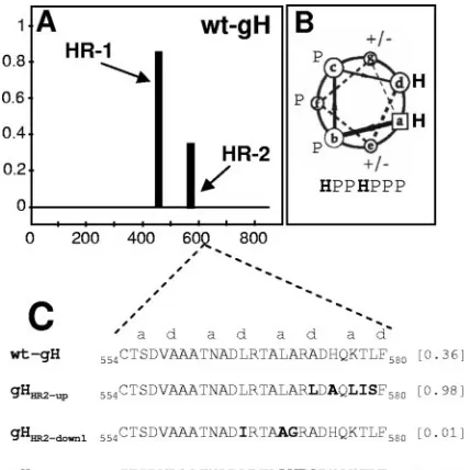
Mutational analysis of HR-2. Probability plots for HRs in
HSV-1 gH were generated by means of the Lupas algorithm (31, 30), with window widths of 14, 21, and 28, and the MTIDK matrix (optimized for a better resolution between the scores of globular and coiled-coil proteins). Weighted and unweighted scans were run in parallel, and the outputs were compared. This identified HR-2, located at aa residues 556 to 585 (Fig. 1 A). Its predicted probability to form a coiled coil was 0.4, i.e., below the suggested cutoff value of 0.5 (30). Three gH mutants in HR-2 were generated, named gHHR2-up, gHHR2-down1, and
gHHR2-down2, whose propensities to form a coiled coil were
either increased (0.98) or decreased (0.01 and 0.0), respec-tively. The relevant substitutions and their locations in the helix are reported in Fig. 1C. gHHR2-upcarried the A573L and
K577L substitutions (at thedandapositions) aimed at increasing the hydrophobicity. gHHR2-down1 carried three substitutions.
Two of them, L566I and L570A (at thed and a positions), predicted a decrease in hydrophobicity. gHHR2-down2 carried
three substitutions in addition to the L570A substitution. The A573Q substitution (positiond) was designed to decrease the hydrophobicity, whereas the A571V and R572P substitutions (bandcpositions) affected the solvent-exposed surface of the helix. In particular, the R-P exchange was chosen because it was one of the least conservative substitutions and, at the same time, disrupted the ␣-helix. The mutant forms of gH were tested in three functional assays, i.e., (i) for proper folding and ability to traffic to the cell surface, (ii) for ability to induce cell fusion when cotransfected with plasmids encoding gD, gB, and
gL, and (iii) for ability to complement the infectivity of a gH deletion HSV mutant (infectivity complementation assay).
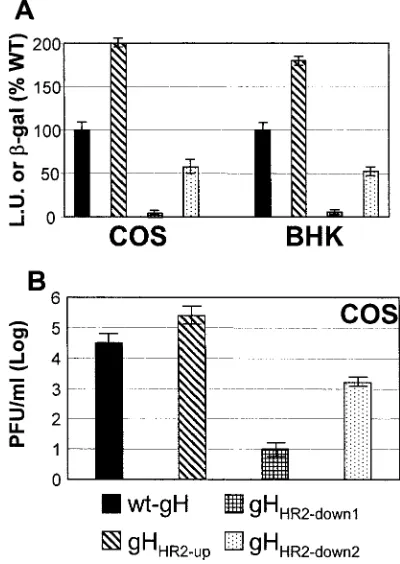
It is well established that gH dimerizes with its chaperone gL to achieve proper folding and be trafficked to the plasma mem-brane (46). The ability of mutant forms of gH to form a het-erodimer with gL and to be trafficked to the plasma membrane was determined by IFA of cells cotransfected with the mutant or wt gH plasmid and a gL expression plasmid. MAb 53S recognizes a discontinuous epitope and strictly requires gL for reactivity. MAb 52S recognizes a gL-independent discontinu-ous epitope with critical residues at position 536 to 537 (43, 47). All three gH mutants maintained the reactivity to MAb 52S, suggesting no major defect in proper folding (data not shown). The IFA reactivity of paraformaldehyde-fixed cells to MAb 53S indicates no major defect in trafficking to the plasma membranes and in heterodimer formation with gL (Fig. 2A to D). Cell surface expression measured by CELISA was essen-tially the same as that of cells expressing the wt gH-gL het-erodimer (Fig. 2E). Thus, the three mutants were capable of heterodimer formation with gL and of cell surface expression. The cell-cell fusion in COS cells was quantified by means of a T7 promoter-driven reporter luciferase gene (45); the cell-cell fusion in BHK cell-cells was quantified by cotransfection of a reporter LacZ gene, as detailed previously (1). As shown in Fig. 3, gHHR2-upexhibited a greatly increased fusion activity in
COS cells, whereas both gHHR2-down1and gHHR2-down2
exhib-ited a decreased fusion activity (Fig. 3A). The results in BHK cells fully agreed with those in COS cells. Thus, increasing the propensity to form a coiled coil resulted in an actual increase in fusion activity. The converse also held true.
In the infectivity complementation assay, cells were trans-fected with mutant or wt gH and superintrans-fected with the gH deletion mutant virus SCgHZ (14). The transgenic gH com-plements the deletion in the virus, and the complemented virions are or are not infectious, depending on whether gH is or is not functional. The results shown in Fig. 3B show that virions complemented with gHHR2-upexhibited an about
10-fold increase in infectivity, whereas virions complemented with gHHR2-down1and gHHR2-down2exhibited an about 1,000-fold or
10-fold decrease in infectivity, respectively.
A mimetic HR-2 peptide specifically inhibits HSV-1
infec-tion. Lopper and Compton and our laboratory showed that
synthetic peptides with the sequence of human cytomegalovi-rus gB and gH HRs and of HSV-1 gH HR-1 inhibit vicytomegalovi-rus infections (20, 29). Here, we tested the effect of two synthetic peptides with the HR-2 sequence. pep.gH-HR214and
pep.gH-HR225are 14 and 25 residues long, respectively (Fig. 4A). The
negative controls were a scrambled peptide from HSV-1 gH HR-1 (pep.gH-HR1scr25, aa 444 to 468) and pep-gD265–289.
Cells were infected with the recombinant R8102, which carries a LacZ gene under the␣-27 promoter. -Gal activity was a direct measure of the extent of virus infection. Figure 4B shows that pep.gH-HR225, but not pep.gH-HR214, reduced virus
in-fection in a dose-dependent manner. A 40% reduction was observed at a 1 mM peptide concentration. The requirement for such a high concentration is not surprising and was re-ported previously with mimetic peptides to HSV-1 gH and human cytomegalovirus as well as to other viruses (20, 29, 53). Irrespective of this, the inhibition was specific, based on the two criteria. First, pep.gH-HR214, the scrambled peptide
FIG. 1. (A) Bioinformatic prediction of HR motifs in HSV-1 gH performed by means of Lupas algorithm (30, 31), as reported in ref-erence 20. The abscissa shows the number of gH residues. The ordi-nate shows the predicted probability scores to form a coiled coil. (B) Helical wheel representation of heptad repeat with identification of the relative positions of hydrophobic (H) and polar (P) residues. (C) Sequence of HR-2 in wt gH and in the mutants. The values in brackets are predicted probabilities to form coiled coils.
on November 8, 2019 by guest
http://jvi.asm.org/
[image:3.585.57.271.68.282.2]pep.gH-HR1scr25, and pep-gD265–289were ineffective. Second,
pep.gH-HR214and pep.gH-HR225did not reduce PrV
infec-tion, ruling out that the reduction was due to a toxic effect (Fig. 4C). Of note, the extent of inhibition attained with pep.gH-HR225was lower than that attained with a 25-mer mimetic
peptide to HR-1, pep.gH-HR125(20).
To verify the step in HSV entry inhibited by pep.gH-HR225,
cells were exposed to pep.gH-HR225only during the interval of
virus attachment to cells at 4°C. Alternatively, they were ex-posed to the peptide only during the interval of virus entry or prior to virus exposure (preexposure). Figure 4D shows that pep.gH-HR225inhibited R8102 infection only if present during
the interval of virus entry.
Complex formation between HR-1 and HR-2 mimetic
pep-tides.To explore the possibility that HR-1 and HR-2 mimetic
peptides interact physically with each other, a fixed amount (15 nmol) of pep.gH-HR125was mixed with increasing amounts of
pep.gH-HR225for 15 min at 25°C. The peptides were then
subjected to nondenaturing PAGE and detected by Coomassie blue staining. As shown in Fig. 5, the single peptides exhibited the same migration position, in accordance with their equiva-lentMr. A mixture of the two peptides resulted in a
slower-migrating complex and concomitant disappearance of the bands corresponding to the single peptides. At a 1:1 molar ratio, the single-peptide species were not detectable, the pep-tides were in complex, suggesting that the stoichiometry of the complex was 1:1.
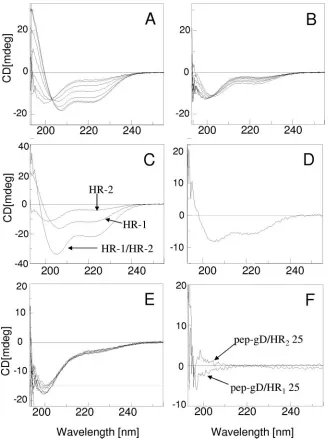
Secondary structures of HR-1 and HR-2 synthetic peptides
singly or in combination.The secondary structure of the HR-1
and HR-2 peptides was analyzed by CD spectroscopy in phos-phate buffer and in this buffer containing up to 15% TFE, both for the single peptides and for the 1:1 mixtures. The CD spec-trum in buffer solution indicated mainly a random coil confor-mation for both pep.gH-HR125and pep.gH-HR225(Fig. 6A
[image:4.585.319.519.71.353.2]and B). The effect of polarity on peptide conformation was analyzed in aqueous mixtures of TFE. This organic solvent
FIG. 2. Cell surface expression of gHHR2-up, gHHR2-down1, and
gHHR2-down2 mutants and wt gH. (A to D) Paraformaldehyde-fixed
COS cells transfected with wt gH or mutant gH plasmids and a gL-encoding plasmid and reacted with MAb 53S. Positive reactivity de-notes gH-gL heterodimer formation and no major defect in trafficking to the plasma membrane. (E) Quantification of cell surface expression (Expr.) of the gH mutants in COS cells cotransfected with a gL plasmid by CELISA. Each assay was performed in triplicate. Bars represent mean percentages relative to wt gH⫾standard deviations.
FIG. 3. Cell-cell fusion (A) and infectivity complementation (B) of mutant forms of gH. (A) Cell-cell fusion of COS or BHK cells trans-fected with the gH mutant plasmids plus plasmids encoding gL, gB, or gD. Fusion was quantified by means of a T7 promoter-driven reporter luciferase gene and expressed as luciferase units (L.U.) (according to reference 45) in COS cells or quantified as-Gal activity (1) in BHK cells. Each assay was performed in triplicate. Bars represent means⫾ standard deviations (SD). (B) COS cells were transfected with mutant or wt gH plasmids and superinfected with the gH deletion mutant virus SCgHZ 4 h later. Virus was harvested 24 h after transfection and titrated in F6 cells in triplicate. Bars represent means of triplicate assays⫾SD.
on November 8, 2019 by guest
http://jvi.asm.org/
[image:4.585.60.268.75.445.2]should mimic the decrease in the environmental polarity oc-curring when the peptide is transferred from water to mem-brane interfaces. Indeed, even if the oligopeptide or protein amino acid sequence usually gives the propensity to a prevail-ing secondary structure (34), the conformation is significantly affected by the environment, i.e., the nature of the solvent (54). A significant change in the conformation was observed for pep.gH-HR125upon increasing the percentage of TFE in the
solution (Fig. 6A). Two negative CD bands were present at about 222 and 208 nm; this behavior indicates the adoption of an ␣-helix structure. By contrast, only a minor change was observed in the CD spectrum of pep.gH-HR225mimetic
pep-tide in the same experimental conditions (Fig. 6B). This be-havior suggests a much lower propensity for the HR-2 mimetic peptide to adopt an␣-helix conformation, relative to the HR-1 mimetic peptide. Of note, also mimetic peptides of HSV-1 gH containing the fusion peptide or the aa 626 to 644 membrane to the interface region were shown to adopt an␣-helical con-formation in TFE-containing buffers but not in aqueous buffers (16).
Next, we analyzed the properties of the mixture of HR-1 and HR-2 mimetic peptides. The CD spectrum of the mixture of pep.gH-HR125and pep.gH-HR225in a 1:1 molar ratio gave an
enhancement of the spectrum intensity (Fig. 6C) compared to that of the single peptides. This change is indicative of het-erodimer formation with a significant increase of the␣-helix conformation content (Fig. 6D). The effect was specific, as supported by the same experiment carried out using an unre-lated peptide, pep-gD265–289, which is not expected to alter its
conformation. Indeed the peptide pep-gD265–289 exhibited a
very low propensity to adopt an␣-helix conformation, as indi-cated by the minor changes observed in the CD spectrum by increasing the TFE content (Fig. 6E). Furthermore, no signif-icant changes of the CD spectrum, and then of the secondary structure, were observed when the CD spectra of the pep-gD265–289/pep.gH-HR125or pep-gD265–289/pep.gH-HR225
mix-tures were compared to the sum of the single components. This behavior is shown in Fig. 6F, where the negligible differential CD spectra are reported.
[image:5.585.309.531.66.173.2]The simultaneous presence of HR-1 and HR-2 mimetic pep-tides reverts the inhibition exerted by the single peppep-tides.
FIG. 4. Inhibition of HSV-1 infection by synthetic peptides. (A) Se-quence and coordinates of synthetic peptides to HSV-1 gH HR-2, HSV-1 gD, and gH HR-1. (B, C) Effect of peptides on infection by the HSV-1 recombinant R8102 carrying LacZ under the␣-27 promoter (B) or of a PrV recombinant carrying LacZ (C). J-nectin1 cells were exposed to the indicated peptides from 30 min prior to infection until harvest. Infection was quantified as-Gal activity. The abscissas show M peptide concentrations. (D) Effect on R8102 infection of pep.gH-HR225administered to cells prior to infection (cell pre-exp), during
virus attachment (4°C for 90 min) (attachment), or during the time interval of virus infection (90 min at 37°C) (entry).
FIG. 5. Complex formation between pep.gH-HR125and
pep.gH-HR225as seen in nondenaturing PAGE. Fifteen nanomoles of
pep.gH-HR125was mixed with the indicated increasing nanomole quantities of
pep.gH-HR225for 15 min at 25°C. The complexes were subjected to
nondenaturing PAGE. At a 1:1 molar ratio (lane 4), the single peptides species (white arrows) were not detectable, and only the slower-mi-grating complex (black arrow) was detected.
on November 8, 2019 by guest
http://jvi.asm.org/
[image:5.585.56.279.74.618.2]Nondenaturing PAGE and CD spectra revealed a complex formation between pep.gH-HR125 and pep.gH-HR225. We
reasoned that complex formation between the two peptides should revert the inhibition of infection exerted by the single peptides. An experiment similar to that described for Fig. 4B was performed, except that a mixture of 500 M (each) pep.gH-HR125 and pep.gH-HR225 was used instead of the
single peptides. As shown in Fig. 7, no inhibition was observed when virus infection was carried out in the presence of a mixture of the two peptides.
DISCUSSION
[image:6.585.129.457.67.506.2]We provide two lines of evidence to support the conclusion that gH HR-2 (aa 556 to 585) constitutes a functional domain for HSV-1 entry into the cell and for fusion. First, a rational mutational analysis designed to either increase or decrease the probability to form a coiled coil indeed resulted in increased or decreased virus infection and cell-cell fusion activity. Second, a mimetic peptide with the sequence of HR-2 inhibited virus infection in a specific manner, although at high concentrations.
FIG. 6. CD spectra of HR-1 and HR-2 mimetic peptides singly and in combination. (A, B) CD spectra of pep.gH-HR125 (HR-1) and
pep.gH-HR225.(HR-2). The spectra were carried out in phosphate buffer (10 mM, pH 7.4) at different percentages of TFE (0, 2.5, 5, 7.5, 10, 12.5,
15); the CD intensities at 222 and 208 nm increase upon increasing the percentage of TFE. (C) CD spectra of single HR-1 and HR-2 mimetic peptides and of their 1:1 mixture. (D) Differential CD spectrum [(HR-1/HR-2 mixture)⫺single (HR-1⫹HR-2) CD contribution]. The solvent was phosphate buffer (10 mM, pH 7.4) with 10% TFE; cell size, 1 mm. (E) CD spectra of pep-gD265–289carried out in the same experimental
conditions as for panels A and B. (F) Differential CD spectra {[(HR-1/pep-gD265–289mixture)⫺single (HR-1⫹pep-gD265–289) CD contribution]
and [(HR-2/pep-gD265–289mixture)⫺single (HR-2⫹pep-gD265–289) CD contribution]}. The solvent was phosphate buffer (10 mM, pH 7.4) with
10% TFE; cell size, 1 mm.
on November 8, 2019 by guest
http://jvi.asm.org/
Examples of functional HRs with probability scores below the suggested cutoff value are infrequent but do exist, as is the case for Newcastle disease virus F glycoprotein (36). Of note, HR-2 is bracketed by two crucial cysteines (cysteines 5 and 6) for gH structure (6).
Two key properties of HSV gH HR-1 and HR-2 were (i) the ability to adopt an␣-helical conformation, an important at-tribute of HR sequences, and (ii) the ability to interact with each other and form a stable complex. (i) Specifically, CD spectroscopy revealed that the HR-1 mimetic peptide adopts mainly a random conformation in aqueous solution; the␣ -he-lical structure content increases by decreasing the polarity of the solvent, as obtained by increasing the concentration of TFE in the buffer solution. The secondary structure of HR-2 mi-metic peptide was much less affected by the nature of the solvent. (ii) Concerning the ability of HR-1 and HR-2 peptides to interact with each other, three lines of evidence support this conclusion. First, a complex between HR-1 and HR-2 synthetic peptides was revealed in nondenaturing PAGE. Second, the mixture of HR-1 and HR-2 peptides exhibited an ␣-helical content higher than that of the two single peptides, as shown by the increase of the CD intensities at 222 and 208 nm. Third, a mixture of HR-1 and HR-2 mimetic peptides reversed the inhibition of infection exerted by the single peptides. Our re-sults clearly show that HR-1 and HR-2 peptides interact with each other. Interestingly, complex formation is an intrinsic property of the HR-1 and HR-2 peptides, independent of the presence of adjacent gH sequences and of the additional viral glycoproteins required for fusion.
Cumulatively, previous and current analyses identify in HSV-1 gH the following structural elements: a candidate fu-sion peptide, located at aa residues 377 to 397, and two HRs with propensity to form a complex and adopt a coiled coil conformation (19, 20). HR-1 and HR-2 are located
down-stream of the candidate fusion peptide and updown-stream of the transmembrane segment, in a position canonical for class 1 viral fusion glycoproteins. As mentioned in the introduction, HSV-1 gH may contain further segments able to interact with lipid membranes (16; T. Gianni, C. Bergamini, R. Fato, G. Lenaz, and G. Campadelli-Fiume, unpublished observation), as observed also in a number of viral fusion glycoproteins, e.g., Sendai virus and paramyxovirus F glycoproteins (18, 41, 42). Altogether, these attributes make gH the leading candidate executor of HSV fusion.
Pertinent to the significance of gH as the candidate fusion glycoprotein are the following considerations. The role played by gB in HSV-mediated fusion is still unclear. Viral fusion glycoproteins are dead ends in the fusion process and do not undergo renaturation. The properties of gH make it likely that gH acts as the last link in the chain of events that starts with the formation of the gD-receptor complex and culminates in fu-sion execution. It follows that gB should act at an intermediate step between gD and gH, a hypothesis that remains to be verified. The alternative possibility is that gB also serves as a fusogenic glycoprotein. If this is the case, it seems reasonable to postulate that gB and gH act in concert to execute fusion; by contrast, the possibility that each of them is fully competent to execute fusion independent of one another appears less tena-ble. We favor the view that gB acts at an intermediate step between gD and gH, given that bioinformatic and molecular biology analyses of gB so far failed to highlight domains typical of viral fusion glycoproteins.
A further consideration centers on whether the role of gH is conserved in theHerpesviridaefamily. Bioinformatic analyses coupled with known properties of gH favor this view. Thus, all the examined isoforms of gH carry at least one positionally conserved predicted hydrophobic ␣-helix and generally two predicted HRs (19, 20). In some cases, they were shown to be functional or could be exchanged with one another (19, 29, 40). Consistent with this view are several attributes of gH. Thus, antibodies to gH exhibit potent neutralizing activity, gH dele-tion mutant viruses are completely debilitated, and together with gL and gB, gH is an essential component of the cell-cell fusion assays, which have been developed for different herpes-viruses (11, 14, 21, 24, 37, 40, 43, 44, 50). The two exceptions are an Epstein-Barr virus gB mutant and the human cytomeg-alovirus gH-gL heterodimer, each capable of inducing the for-mation of small syncytia in the absence of additional glycop-roteins (24, 40).
[image:7.585.56.270.69.273.2]Class 1 viral fusion glycoproteins form a broad group of proteins that not only share structural elements but also carry distinctive features. One such feature is the distance between HR-1 and HR-2, that ranges from a few amino acids (5 to 26) to about 250 aa (e.g., influenza virus hemagglutinin, paramyxo-virus simian paramyxo-virus 5 F) (12, 42). A major difference is the topological relation that exists between the fusion glycoprotein and the viral domain responsible for receptor binding activity. In some cases, the receptor binding activity lies in a specific domain of the fusion glycoprotein itself (e.g., severe acute respiratory syndrome CoV, vesicular stomatitis virus glycopro-tein). In other cases, the receptor binding activity lies in a separate glycoprotein that cross talks with the fusion glycop-rotein (e.g., HN for paramyxovirus, gp120 for human immu-nodeficiency virus, GP1 of Ebola virus, glycoproteins of some
FIG. 7. Lack of inhibition of R8102 entry by mixture of HR-1 and HR-2 mimetic peptides. J-nectin1 cells were exposed to the indicated peptides (250M each, except pep-gD265–289in right-hand bar, which
is 500M) from 30 min prior to infection until harvest. Infection was determined in triplicate assays as-Gal activity. Bars represent means of results from triplicate assays⫾standard deviations.
on November 8, 2019 by guest
http://jvi.asm.org/
retroviruses, and avian and mammalian CoVs) (12, 22, 36, 55). HSV, and herpesviruses in general, differ from both these groups in that they carry a multipartite fusion machinery made of a receptor binding glycoprotein (gD in HSV, gp42 in Ep-stein-Barr virus, gB in human cytomegalovirus), gB and gL, whose roles in fusion remain to be determined, and gH, with its structural elements (7, 13, 38, 48, 51). Taking into account what is currently known, we propose that gH be considered the candidate executor of fusion with attributes of class one fusion glycoproteins, that herpesvirus fusion be considered a group in itself, classified as a multipartite fusion system, and that HSV be the paradigm for it.
ACKNOWLEDGMENTS
We thank T. Minson (Cambridge University), B. Roizman (Univer-sity of Chicago), P. G. Spear (Northwestern Univer(Univer-sity), G. H. Cohen and R. Eisenberg (University of Pennsylvania), and T. Mettenleiter (Friedrich-Loeffler-Institut, Insel Riems, Germany) for gifts of viruses and antibodies. We are indebted to Elisabetta Romagnoli for invalu-able assistance.
The work was supported by FIRB autonomous and coordinated project, Cofin-MIUR 40%, University of Bologna 60%, Fondo Pallotti.
REFERENCES
1.Avitabile, E., G. Lombardi, and G. Campadelli-Fiume.2003. Herpes simplex virus glycoprotein K, but not its syncytial allele, inhibits cell-cell fusion mediated by the four fusogenic glycoproteins, gD, gB, gH and gL. J. Virol.
77:6836–6844.
2.Avitabile, E., G. Lombardi, T. Gianni, M. Capri, and G. Campadelli-Fiume.
2004. Coexpression of UL20p and gK inhibits cell-cell fusion mediated by herpes simplex virus glycoproteins gD, gH-gL, and wt-gB or an endocytosis-defective gB mutant, and downmodulates their cell surface expression. J.
Vi-rol.78:8015–8025.
3.Babic, N., B. G. Klupp, B. Makoschey, A. Karger, A. Flamand, and T. C. Mettenleiter.1996. Glycoprotein gH of pseudorabies virus is essential for penetration and propagation in cell culture and in the nervous system of
mice. J. Gen. Virol.77:2277–2285.
4.Brunetti, C. R., R. L. Burke, B. Hoflack, T. Ludwig, K. S. Dingwell, and D. C. Johnson.1995. Role of mannose-6-phosphate receptors in herpes simplex
virus entry into cells and cell-to-cell transmission. J. Virol.69:3517–3528.
5.Cai, W. H., B. Gu, and S. Person.1988. Role of glycoprotein B of herpes
simplex virus type 1 in viral entry and cell fusion. J. Virol.62:2596–2604.
[Erratum,62:4438.]
6.Cairns, T. M., D. J. Landsburg, J. C. Whitbeck, R. J. Eisenberg, and G. H. Cohen.2005. Contribution of cysteine residues to the structure and function
of herpes simplex virus gH/gL. Virology332:550–562.
7.Campadelli-Fiume, G., F. Cocchi, L. Menotti, and M. Lopez.2000. The novel receptors that mediate the entry of herpes simplex viruses and animal
al-phaherpesviruses into cells. Rev. Med. Virol.10:305–319.
8.Carfi, A., S. H. Willis, J. C. Whitbeck, C. Krummenacher, G. H. Cohen, R. J. Eisenberg, and D. C. Wiley. 2001. Herpes simplex virus glycoprotein D
bound to the human receptor HveA. Mol. Cell8:169–179.
9.Cocchi, F., D. Fusco, L. Menotti, T. Gianni, R. J. Eisenberg, G. H. Cohen, and G. Campadelli-Fiume.2004. The soluble ectodomain of herpes simplex virus gD contains a membrane-proximal pro-fusion domain and suffices to
mediate virus entry. Proc. Natl. Acad. Sci. USA101:7445–7450.
10.Cocchi, F., L. Menotti, P. Mirandola, M. Lopez, and G. Campadelli-Fiume.
1998. The ectodomain of a novel member of the immunoglobulin superfam-ily related to the poliovirus receptor has the attributes of a bona fide receptor
for herpes simplex viruses 1 and 2 in human cells. J. Virol.72:9992–10002.
11.Cole, N. L., and C. Grose.2003. Membrane fusion mediated by herpesvirus
glycoproteins: the paradigm of varicella-zoster virus. Rev. Med. Virol.13:
207–222.
12.Colman, P. M., and M. C. Lawrence.2003. The structural biology of type I
viral membrane fusion. Nat. Rev. Mol. Cell Biol.4:309–319.
13.Feire, A. L., H. Koss, and T. Compton.2004. Cellular integrins function as entry receptors for human cytomegalovirus via a highly conserved
disinte-grin-like domain. Proc. Natl. Acad. Sci. USA101:15470–15475.
14.Forrester, A., H. Farrell, G. Wilkinson, J. Kaye, N. Davis Poynter, and T. Minson.1992. Construction and properties of a mutant of herpes simplex
virus type 1 with glycoprotein H coding sequences deleted. J. Virol.66:341–
348.
15.Fusco, D., C. Forghieri, and G. Campadelli-Fiume.2005. The pro-fusion domain of herpes simplex virus glycoprotein D (gD) interacts with the gD N terminus and is displaced by soluble forms of viral receptors. Proc. Natl.
Acad. Sci. USA102:9323–9328.
16.Galdiero, S., A. Falanga, M. Vitiello, H. Browne, C. Pedone, and M. Galdiero.2005. Fusogenic domains in herpes simplex virus type 1
glycopro-tein H. J. Biol. Chem.280:28632–28643.
17.Geraghty, R. J., C. Krummenacher, G. H. Cohen, R. J. Eisenberg, and P. G. Spear.1998. Entry of alphaherpesviruses mediated by poliovirus
receptor-related protein 1 and poliovirus receptor. Science280:1618–1620.
18.Ghosh, J. K., S. G. Peisajovich, and Y. Shai.2000. Sendai virus internal fusion peptide: structural and functional characterization and a plausible
mode of viral entry inhibition. Biochemistry39:11581–11592.
19.Gianni, T., P. L. Martelli, R. Casadio, and G. Campadelli-Fiume.2005. The ectodomain of herpes simpex virus glycoprotein H contains a membrane alpha-helix with attributes of an internal fusion peptide, positionally
con-served in theHerpesviridaefamily. J. Virol.79:2931–2940.
20.Gianni, T., L. Menotti, and G. Campadelli-Fiume.2005. A heptad repeat in
herpes simplex virus 1 gH, located downstream of the␣-helix with attributes
of a fusion peptide, is critical for virus entry and fusion. J. Virol.79:7042–
7049.
21.Gompels, U., and A. Minson.1986. The properties and sequence of
glyco-protein H of herpes simplex virus type 1. Virology153:230–247.
22.Ito, H., S. Watanabe, A. Takada, and Y. Kawaoka.2001. Ebola virus glyco-protein: proteolytic processing, acylation, cell tropism, and detection of
neu-tralizing antibodies. J. Virol.75:1576–1580.
23.Jardetzky, T. S., and R. A. Lamb.2004. Virology: a class act. Nature427:
307–308.
24.Kinzler, E. R., and T. Compton.2005. Characterization of human
cytomeg-alovirus glycoprotein-induced cell-cell fusion. J. Virol.79:7827–7837.
25.Krummenacher, C., A. V. Nicola, J. C. Whitbeck, H. Lou, W. Hou, J. D. Lambris, R. J. Geraghty, P. G. Spear, G. H. Cohen, and R. J. Eisenberg.
1998. Herpes simplex virus glycoprotein D can bind to poliovirus receptor-related protein 1 or herpesvirus entry mediator, two structurally unreceptor-related
mediators of virus entry. J. Virol.72:7064–7074.
26.Krummenacher, C., V. M. Supekar, J. C. Whitbeck, E. Lazear, S. A. Con-nolly, R. J. Eisenberg, G. H. Cohen, D. C. Wiley, and A. Carfi.2005. Struc-ture of unliganded HSV gD reveals a mechanism for receptor-mediated
activation of virus entry. EMBO J.24:4144–4153.
27.Ligas, M. W., and D. C. Johnson.1988. A herpes simplex virus mutant in which glycoprotein D sequences are replaced by beta-galactosidase
se-quences binds to but is unable to penetrate into cells. J. Virol.62:1486–1494.
28.Lopez, M., F. Cocchi, L. Menotti, E. Avitabile, P. Dubreuil, and G. Cam-padelli-Fiume.2000. Nectin2␣(PRR2␣or HveB) and nectin2␦are low-efficiency mediators for entry of herpes simplex virus mutants carrying the
Leu25Pro substitution in glycoprotein D. J. Virol.74:1267–1274.
29.Lopper, M., and T. Compton.2004. Coiled-coil domains in glycoproteins B and H are involved in human cytomegalovirus membrane fusion. J. Virol.
78:8333–8341.
30.Lupas, A.1997. Predicting coiled-coil regions in proteins. Curr. Opin. Struct.
Biol.7:388–393.
31.Lupas, A., M. Van Dyke, and J. Stock.1991. Predicting coiled coils from
protein sequences. Science252:1162–1164.
32.Menotti, L., R. Casadio, C. Bertucci, M. Lopez, and G. Campadelli-Fiume.
2002. Substitution in the murine nectin1 receptor of a single conserved amino acid at a position distal from herpes simplex virus gD binding site
confers high affinity binding to gD. J. Virol.76:5463–5471.
33.Milne, R. S., S. L. Hanna, A. H. Rux, S. H. Willis, G. H. Cohen, and R. J. Eisenberg.2003. Function of herpes simplex virus type 1 gD mutants with
different receptor-binding affinities in virus entry and fusion. J. Virol.77:
8962–8972.
34.Minor, D. L., Jr., and P. S. Kim.1994. Measurement of the
beta-sheet-forming propensities of amino acids. Nature367:660–663.
35.Montgomery, R. I., M. S. Warner, B. J. Lum, and P. G. Spear.1996. Herpes simplex virus-1 entry into cells mediated by a novel member of the TNF/
NGF receptor family. Cell87:427–436.
36.Morrison, T. G.2003. Structure and function of a paramyxovirus fusion
protein. Biochim. Biophys. Acta1614:73–84.
37.Muggeridge, M. I.2000. Characterization of cell-cell fusion mediated by herpes simplex virus 2 glycoproteins gB, gD, gH and gL in transfected cells.
J. Gen. Virol.81:2017–2027.
38.Mullen, M. M., K. M. Haan, R. Longnecker, and T. S. Jardetzky.2002. Structure of the Epstein-Barr virus gp42 protein bound to the MHC class II
receptor HLA-DR1. Mol. Cell9:375–385.
39.Okuma, K., M. Nakamura, S. Nakano, Y. Niho, and Y. Matsuura.1999. Host range of human T-cell leukemia virus type I analyzed by a cell
fusion-dependent reporter gene activation assay. Virology254:235–244.
40.Omerovic, J., L. Lev, and R. Longnecker.2005. The amino terminus of Epstein-Barr virus glycoprotein gH is important for fusion with epithelial
and B cells. J. Virol.79:12408–12415.
41.Peisajovich, S. G., O. Samuel, and Y. Shai.2000. Paramyxovirus F1 protein has two fusion peptides: implications for the mechanism of membrane
fu-sion. J. Mol. Biol.296:1353–1365.
42.Peisajovich, S. G., and Y. Shai.2003. Viral fusion proteins: multiple regions
contribute to membrane fusion. Biochim. Biophys. Acta1614:122–129.
43.Peng, T., M. Ponce de Leon, M. J. Novotny, H. Jiang, J. D. Lambris, G.
on November 8, 2019 by guest
http://jvi.asm.org/
Dubin, P. G. Spear, G. H. Cohen, and R. J. Eisenberg.1998. Structural and antigenic analysis of a truncated form of the herpes simplex virus
glycopro-tein gH-gL complex. J. Virol.72:6092–6103.
44.Pertel, P. E.2002. Human herpesvirus 8 glycoprotein B (gB), gH, and gL can
mediate cell fusion. J. Virol.76:4390–4400.
45.Pertel, P. E., A. Fridberg, M. L. Parish, and P. G. Spear.2001. Cell fusion induced by herpes simplex virus glycoproteins gB, gD, and gH-gL requires a
gD receptor but not necessarily heparan sulfate. Virology279:313–324.
46.Roop, C., L. Hutchinson, and D. C. Johnson.1993. A mutant herpes simplex virus type 1 unable to express glycoprotein L cannot enter cells, and its
particles lack glycoprotein H. J. Virol.67:2285–2297.
47.Showalter, S. D., M. Zweig, and B. Hampar.1981. Monoclonal antibodies to herpes simplex virus type 1 proteins, including the immediate-early protein
ICP 4. Infect Immun.34:684–692.
48.Spear, P. G.2004. Herpes simplex virus: receptors and ligands for cell entry.
Cell. Microbiol.6:401–410.
49.Spear, P. G., and R. Longnecker.2003. Herpesvirus entry: an update. J.
Vi-rol.77:10179–10185.
50.Turner, A., B. Bruun, T. Minson, and H. Browne.1998. Glycoproteins gB, gD, and gHgL of herpes simplex virus type 1 are necessary and sufficient to
mediate membrane fusion in a Cos cell transfection system. J. Virol.72:873–
875.
51.Wang, X., W. J. Kenyon, Q. Li, J. Mullberg, and L. M. Hutt-Fletcher.1998. Epstein-Barr virus uses different complexes of glycoproteins gH and gL to
infect B lymphocytes and epithelial cells. J. Virol.72:5552–5558.
52.Warner, M. S., R. J. Geraghty, W. M. Martinez, R. I. Montgomery, J. C. Whitbeck, R. Xu, R. J. Eisenberg, G. H. Cohen, and P. G. Spear.1998. A cell surface protein with herpesvirus entry activity (HveB) confers susceptibility to infection by mutants of herpes simplex virus type 1, herpes simplex virus
type 2, and pseudorabies virus. Virology246:179–189.
53.Watanabe, S., A. Takada, T. Watanabe, H. Ito, H. Kida, and Y. Kawaoka.
2000. Functional importance of the coiled-coil of the Ebola virus
glycopro-tein. J. Virol.74:10194–10201.
54.Waterhous, D. V., and W. C. Johnson, Jr.1994. Importance of environment
in determining secondary structure in proteins. Biochemistry33:2121–2128.
55.Xu, Y., Z. Bai, L. Qin, X. Li, G. Gao, and Z. Rao.2004. Crystallization and preliminary crystallographic analysis of the fusion core of the spike protein of the murine coronavirus mouse hepatitis virus (MHV). Acta Crystallogr. D
60:2013–2015.
56.Yoon, M., A. Zago, D. Shukla, and P. G. Spear.2003. Mutations in the N termini of herpes simplex virus type 1 and 2 gDs alter functional interactions
with the entry/fusion receptors HVEM, nectin-2, and 3-O-sulfated heparan
sulfate but not with nectin-1. J. Virol.77:9221–9231.
on November 8, 2019 by guest
http://jvi.asm.org/
0022-538X/07/$08.00⫹0 doi:10.1128/JVI.02596-06
AUTHOR’S CORRECTION
Heptad Repeat 2 in Herpes Simplex Virus 1 gH Interacts with Heptad Repeat 1
and Is Critical for Virus Entry and Fusion
Tatiana Gianni, Angela Piccoli, Carlo Bertucci, and Gabriella Campadelli-Fiume
Department of Experimental Pathology, Section on Microbiology and Virology, Via San Giacomo,12,1 Department of Pharmaceutical Sciences, Via Belmeloro, 6, Alma Mater Studiorum—University of Bologna, 40126 Bologna, Italy
Volume 80, no. 5, p. 2216–2224, 2006. Page 2220: We recently have become aware that data in Fig. 5 were manipulated and misrepresented the actual data, in that, for aesthetic reasons, it underrepresented minor contaminant species present in the synthetic peptides’ preparations. The correct figure and its legend should appear as shown below. The original figure and the present figure were obtained with different peptide batches. The peptide batches shown in the figure below contain a predominant fast-migrating band (likely the monomer) and, in addition, a fainter band (likely an oligomer).
The legend to Fig. 5 should read “Complex formation between pep.gH-HR125and pep.gH-HR225as seen in nondenaturing
PAGE. Sixty nanomoles of pep.gH-HR225was mixed with the indicated increasing nanomoles of pep.gH-HR225for 15 min at
25°C. The complexes were subjected to nondenaturing PAGE. Both pep.gH-HR125and pep.gH-HR225exhibit a predominant
fast-migrating band (likely the monomer) and a fainter band (likely an oligomer) (white arrows). At a 1:1 molar ratio (lane 4), the species corresponding to the single peptides were not detectable, and only the slower-migrating complex (black arrow) was detected.”
The authors apologize for this fact. None of the results or conclusions in the original article are affected.