T
T
o
o
a
a
s
s
s
s
e
e
s
s
s
s
t
t
h
h
e
e
r
r
o
o
l
l
e
e
o
o
f
f
s
s
e
e
c
c
o
o
n
n
d
d
r
r
e
e
s
s
e
e
c
c
t
t
i
i
o
o
n
n
i
i
n
n
T
T
1
1
G
G
3
3
b
b
l
l
a
a
d
d
d
d
e
e
r
r
t
t
u
u
m
m
o
o
r
r
s
s
A thesis submitted for the degree of MCh,
Branch IV, Genitourinary surgery,
to Dr.M.G.R Medical University,
Acknowledgements
I thank God for giving me the opportunity and strength to complete
this study.
I wish to express my deep gratitude to my guide, Dr Antony Devasia,
for his encouragement and help during this period.
I sincerely thank Dr. Nitin Kekre for his invaluable inputs and
suggestions.
I am extremely thankful to all my seniors and colleagues for the help
and co-operation they extended during the course of the study.
I would like to thank Mr. Prasanna Samuel for his help in the
statistical analysis.
I sincerely thank all the patients who participated in this study.
Title of the Abstract : To assess the role of second resection in T1G3 bladder tumors Department : Department of Urology
Name of the candidate : Katumalla Francis Sridhar
Degree and subject : MCh, Branch IV, Genitourinary Surgery Name of the guide : Dr. Antony Devasia
Aim: To assess the role of second resection in T1G3 bladder tumors
Material and Methods: This is a prospective study conducted in the Department of Urology, Christian
Medical College, Vellore from April 2009 to December 2010. All T1G3 lesions were included in this study
and variables like size, multifocality, characteristics of the lesions were analysed. Prevalence of second
resection was calculated along with 95% CI. Chi square test was used to assess association between the
categorical variables and tumor positivity in second resection. Thirty seven patients were diagnosed to
have T1G3 disease, of which 27 had a second resection done. Hence, these patients were taken as the
study group and the sample size.
Results: Fifty five percent of the lesions were solitary papillary and 09% were multiple papillary.
All our resections had muscularis propria sampled at the end of the resection and separately sent for HPE
which were tumor free. Thirty three percent of patients had residual disease at second resection and
3.7% were upstaged. Eighty five percent of the patients with solitary papillary lesions did not have any
residual disease. When size also was considered, none of the patients with tumor size less than 3cms
and solitary papillary lesions had a residual disease in second resection.
Conclusion: Patients with T1G3 tumors do not represent a homogenous group. Second TUR is
recommended in patients with high grade T1 urothelial bladder carcinoma as it identifies residual
<3cm, none had tumor in the second TUR and therefore the need for a second TUR is questionable in
Contents
S. No.
Title
Page No.
1. Introduction
1
2.
Review of Literature
12
3.
Aims of the study
34
4.
Materials and Methods
36
5.
Results
40
6.
Discussion
54
7. Conclusion
59
8.
Bibliography
61
9.
Annexure
I. Informed
consent
II. Proforma
III. Master
Chart
I
INTRODUCTION
Transitional cell carcinoma (TCC) of the urinary bladder has a broad
spectrum of disease course and treatment. The diagnosis and accurate staging
of the tumor is an important initial step to determine the most appropriate
management strategy.
Epidemiology:
TCC of the urinary bladder is the ninth most common cancer in worldwide
cancer incidence. It is the 7th most common cancer in men and ranks 17th in
women 1. After prostate cancer, it is the second most common urological
malignancy. The incidence of bladder cancer varies significantly all over the
world, with Egypt, Eastern Europe, and North America having the highest
incidence rates, and Asian countries the lowest rates 2. More than 90% of new cases occur in people in the 6th decade, but the disease may occur in younger population as well 3. The mean age of the patients diagnosed with bladder cancer is 69 years for men and 71 for women and the estimated ratio between men and
women is 3.8/1 4. Risk factors:
Cigarette smoking and occupational exposure to urothelial carcinogens
non-smokers 7. The incidence of urinary bladder cancer is directly related to the duration of smoking and the number of cigarettes smoked per day 8.
Sixteen to 24% of all the bladder cancers are due to occupational
exposure to urothelial carcinogens and is the second most important risk factor 9.
Aromatic amines used in the chemical, rubber, and dye industries (eg, benzidine,
2-naphthylamine, 4-aminobiphenyl, o-toluidine, and 4-chloro-o-toluidine) and
polycyclic aromatic hydrocarbons (PAHs) used in the aluminum, coal, and roofing
industries are all known to be associated with the development of bladder cancer.
An increased risk of bladder cancer has also been reported in painters,
varnishers, and hairdressers 2.
Chronic urinary tract infections, cyclophosphamide use, and exposure to
radiotherapy are the other known causes which are associated with urinary
bladder malignancy 10. Long term irritation of the bladder by indwelling catheters
or stones, is related to development of squamous cell carcinoma of the bladder.
Schistosomiasis is endemic in Egypt and the Middle East and is considered to be
a definite cause of bladder cancer 11.
Inadequate consumption of fruits, vegetables, and certain vitamins may
also play a role in the development of bladder cancer. Although it has been
suggested that coffee consumption and artificial sweeteners may be associated
with an increased risk of bladder cancer, results from epidemiologic studies
investigating these agents have been inconclusive 2. There is an increased risk of
family case control study found an almost 2-fold increased risk among
first-degree relatives of patients with urothelial cell carcinoma 12. Pathology:
The urinary tract extends from the renal pelvis to the urethra and is
covered with transitional epithelium, also called the urothelium. Normal
urothelium is three to seven layers thick. The external layer consists of large
umbrella cells. The urothelium rests on the basal membrane of the lamina propria
which consists of the subepithelial tissue and muscularis mucosa, followed by the
muscularis propria (detrusor) and then the fat and the large venous plexus.
Transitional cell carcinoma bladder is the most common primary
pathologic subtype of bladder cancer and is observed in >90% of tumours. Other
subtypes are squamous cell carcinoma and adenocarcinoma and are less
common and occur in approximately 5% and 1% of bladder cancers,
respectively2.
Classic TCC grows exophytically into the lumen of the urinary bladder and
resembles swaying seaweed on cystoscopy. TCC can also grow as a sessile
tumor in the bladder wall. A special variant of TCC is carcinoma in situ (CIS), a
flat tumor, often multifocal and seen as velvety, red edematous patches on
cystoscopy. There are three different kinds of CIS:
2. Secondary CIS is when CIS occurs with previously diagnosed TCC
bladder.
3. Concomitant CIS implicates co-existing papillary or nodular tumor and
CIS.
Staging and Grading:
Stage and grade are significant prognostic factors for recurrence,
progression, and survival. They are critical for the appropriate treatment and
management of TCC bladder. The most widely used and universally accepted
staging system is the tumour-node-metastases (TNM) system (Table 1).
The new classification for grading of non-invasive urothelial tumours was
proposed by the World Health Organization (WHO) and the International Society
of Urological Pathology (ISUP) and published by the WHO in 2004 (Table 2) 13. It differentiates between papillary urothelial neoplasm of low malignant potential
(PUNLMP) and low-grade and high-grade urothelial carcinomas. The PUNLMP
are lesions that do not have cytological features of malignancy but show normal
urothelial cells in a papillary configuration. They have a negligible risk for
progression, but still have a tendency to recur. The intermediate grade (grade 2),
which was the subject of controversy in the 1973 WHO classification, has been
Table 1:TNM staging of bladder cancer.
T: Primary tumour
TX Primary tumour cannot be assessed
T0 No evidence of primary tumour
Ta Non-invasive papillary carcinoma
Tis Carcinoma in situ: ‘‘flat tumour’’
T1 Tumour invades subepithelial connective tissue
T2 Tumour invades muscle
T2a: Tumour invades superficial muscle (inner half)
T2b: Tumour invades deep muscle (outer half)
T3 Tumour invades perivesical tissue:
T3a: Microscopically
T3b: Macroscopically
T4 Tumour invades any of the following: prostate, uterus, vagina, pelvic wall,
abdominal wall
T4a: Tumour invades prostate, uterus, or vagina
N: Lymph nodes
NX Regional lymph nodes cannot be assessed
N0 No regional lymph node metastasis
N1 Metastasis in a single lymph node ≤2 cm in greatest dimension
N2 Metastasis in a single lymph node >2 cm but not >5 cm in greatest
dimension, or multiple lymph nodes, none >5 cm in greatest dimension
N3 Metastasis in a lymph node >5 cm in greatest dimension
M: Distant metastasis
MX Distant metastasis cannot be assessed
M0 No distant metastasis
Table 2 – World Health Organization (WHO) grading of urinary tumours in
1973 and 2004
WHO 1973
Urothelial papilloma
Grade 1: well differentiated
Grade 2: moderately differentiated
Grade 3: poorly differentiated
WHO 2004
Urothelial papilloma
PUNLMP
Low-grade papillary urothelial carcinoma
High-grade papillary urothelial carcinoma
Non Muscle Invasive Bladder Cancer (NMIBC):
Haematuria is the most common finding in non-muscle-invasive bladder
tumours. Bladder irritation, dysuria, or urgency may be symptoms of CIS.
Approximately 75–80% of bladder tumours present as non–muscle invasive
disease and the remainder present as muscle-invasive disease. In NMIBC,
approximately 70% present as Ta lesions, 20% as T1 lesions and 10% present
as CIS or Tis lesions 10. NMIBC represents a heterogeneous group of tumours with completely different oncologic outcomes. This heterogeneity in bladder
tumors complicates the ability to compare the efficacy of different treatment
modalities and thereby establish unified treatment recommendations. Therefore,
risk stratification is imperative for classifying patients with similar risks of
recurrence and progression, and it helps to determine the appropriate
management strategies for each risk category 2. T1G3:
T1G3 bladder tumor, a high grade lesion invading the lamina propria and
yet to involve the muscularis propria, is highly malignant in the non-muscle
invasive group 15. T1G3 tumours have a high propensity to recur and progress to
muscle invasion and are associated with a significant risk of metastasis and
death. Eventhough T1G3 is classified as non muscle invasive tumor, about 15 –
20% progress to higher stage and nearly 75% develop recurrence 16. 5% to 30%
of patients with T1G3 ultimately die of bladder cancer within 5 – 10 years 15,17.
The biological characteristics of these tumors have been proved to be same as
many of these tumors can be treated successfully with bladder preservation
approaches. The dilemma facing the urologist is how best to treat these tumors in
a timely manner so that the chances of bladder preservation and cancer control
are maximized while the risks of overtreatment with radical therapy are
minimized.
Factors predicting the poor prognosis of T1G3 tumors are multiple tumors,
high grade lesions, larger size, sessile pattern, associated CIS and early
recurrence of tumor after primary transurethral resection of bladder tumor
(TURBT) 17,18. A number of molecular and genetic prognostic factors have also been studied, but none of these have been validated prospectively 19. Besides these, the duration of disease also influences the disease specific survival 20.
TURBT and intravesical therapy is now accepted as the reference
standard for T1G3 tumors. The primary transurethral resection might leave
residual disease, more so in multiple tumors, thereby increasing the risk of early
tumor recurrence and stage progression even after intravesical therapy 21. Second resection of T1G3 tumors may help to eradicate the residual disease and
also to identify the early recurrence 21,22. Keeping in mind the limitations of the current staging modalities, these tumors are understaged and a patient may
receive inadequate treatment to a potentially curable disease 23. Therefore a re-resection is routinely advised in all T1G3 tumors.
For all cases of newly diagnosed T1G3 TCC, a secondary TUR 4–6 wk
after the primary TUR is strongly recommended. The advantages of a second
re-resection, the cost of the treatment and the delay in intravesical therapy
questions whether re-resection is necessary in all T1G3 lesions 24. Even after a satisfactory primary resection and a good muscle in the resected specimen, is it
R
REVIEW OF LITERATURE
T1G3 bladder cancer is associated with an aggressive course among
superficial bladder tumors. They have a high propensity to recur and also to
upstage to T2 disease. They are associated with a significant risk of metastasis
and death. Literature shows long-term death rates as high as 34% 25. An extremely variable 5-year progression rate ranging from 20 to 75% exists,
probably due to the different risk subgroups in this spectrum of T1G3 26. Identifying these subgroups and the associated risk factors will help plan an
effective management. The goal of the treatment of T1G3 bladder cancer is to
decrease the mortality, ensure reduced morbidity and achieve a good quality of
life. The dilemma facing the urologist is how best to treat these tumors in a timely
manner so that the chances of bladder preservation and cancer control are
maximized, while the risks of overtreatment with radical therapy are minimized 27.
To assess the role of conservative management in pT1G3 bladder cancer,
a number of issues need to be considered:
− Is it really a pT1G3 tumor?
− The variability of pT1G3 bladder cancer
To confirm the stage and grade as pT1G3
The management protocol for patients with different stages of the disease
(pTa, pT1 and pT2 or greater tumors) varies substantially 28. Pathologists hold a
key position in making a diagnosis and in commenting on the accurate stage &
grade of a patient’s tumor. They must be presented with suitable material along
with relevant clinical information: the case is then best reviewed in the setting of a
multidisciplinary uro-oncology meeting 29. The pathologist must receive muscle-bearing specimens to determine whether muscle invasion has occurred. In 40%
of 58 patients with apparently pT1 disease reviewed by Herr, muscle had not
been included in the resection specimens from the referring hospital 30. The uniformity of the pathologist’s report, assuming adequate specimens, also needs
to be accounted for. Evidence suggests that not only do experienced
uro-pathologists vary in their diagnoses, they also may be inconsistent regarding the
same specimen 31.Results in one study revealed that general pathologists tend to overstage tumors, but undergrade them 32. A close liaison between the urologist and the pathologist, preferably in a multidisciplinary team meeting is hence
important.
The variability of pT1G3 bladder cancer
Data on the untreated natural history of pT1G3 tumors is sparse, as these
tumors generally require more than just a TURBT 33,34. Heney and colleagues
reported a progression rate of 48% at around 3 years in 27 patients with pT1G3
invade the muscle.It was noticed that the risk of progression and death from
disease for patients with pT1G3 tumors is up to 10-fold greater than that for
patients with other pTa and pT1 tumors 29. As long-term data on the use of intravesical Bacillus Calmette-Gue´rin (BCG) has become available over the last
twenty years, the idea of the so-called ‘‘rule of threes’’ has emerged, viz., of all
the patients treated with BCG, approximately one third survive with their bladder
intact, around a third require radical cystectomy, and around a third die of their
disease 36. This difference in outcome varies markedly from the very low progression rates seen with pTa tumors 35, to the relentlessly aggressive course of muscle-invasive disease. The dilemma, therefore, is to treat adequately, but
not to over-treat. The outcome for patients with pT1G3 tumors also seems to be
strongly influenced by the presence or absence of carcinoma in situ (CIS) 37. The presence of associated CIS must therefore be carefully considered in the case for
conservative treatment of a patient with a pT1G3 tumor.
The quality of clinical data
Patients may present with primary or recurrent tumors, with and without
CIS, and with or without other factors that may influence the outcome, such as
the presence of tumor in the prostatic urethra or the upper tracts 38. Even with the information obtained from the systematic reviews and meta-analyses, at present,
PROGNOSTIC FACTORS
The important prognostic factors which help in predicting the outcome of
the T1G3 tumors following the initial TURBT include early recurrence or new
occurrence, multiplicity of the tumor lesions, size of the lesion, the presence of
associated carcinoma in situ along with the T1 tumors, urothelial carcinoma
involving the prostatic urethra or the ducts and the depth of the lamina propria
involved 39. T category and the grade of the tumor do not significantly influence the recurrence rate. Recurrence rate depends mainly on the multiplicity of tumors
(single tumor, 51% recurrence at 5 years; multiple tumors, 91%), the previous
recurrence rate, or the recurrence at 3 months 40. In contrast, the grade and, to a lesser extent, the stage of disease is the most important prognostic factors for
disease progression. The size of the tumor, the presence of tumor-associated
CIS and the involvement of the prostatic urethra also carry a worse prognosis 41. Increasing tumor size portends an increased risk for undetected infiltration
of the lamina propria. The depth of the lamina propria involved is also a
significant prognostic factor. The more the lamina propria appears infiltrated, the
earlier are the chances of dissemination. Multifocality of the tumor carries a
worse prognosis. The reasons for this are multiple. Multifocal tumors tend to be
understaged, and the chances of complete resection decrease with the number
of tumors 42. Multifocality indicates the susceptibility of the entire urothelium to develop tumours (field change effect). There is thus a strong need for the close
follow-up of the urethra and the upper urinary tract after cystectomy and of the
It has been seen in recent reports that patients with TaG3 tumors are also
at high risk of life-long progression, not very different from that of patients with
T1G3 tumors. Herr 43 followed 125 such patients for 15 years and observed progression in 39% and cancer related death in 26%. Similar data was reported
by Lebret et al 44 where 32 patients with TaG3 tumors followed for 4 years had a 25% progression rate with 12% cancer-related deaths.
Apart from these unfavourable prognostic factors, the duration of the T1G3
disease also influences disease-specific survival. In a large study 45, on follow up for 8 years, 50% of the G3 patients either died from bladder cancer or developed
muscle-invasive disease. After 15 years, more than half the patients with T1
disease developed a muscle-invasive tumor and a third died from metastatic
carcinoma 46.
Molecular and genetic aspects such as altered expression of p53 have
been implicated in the aggressive biological behavior of the bladder tumors. p53
mutations have been found in many cancers, including TCC. P53 overexpression
is an independent predictive factor of recurrence for T1G3 bladder cancers.
Retinoblastoma (Rb) gene was also evaluated as a possible predictive factor for
the disease progression. Abnormal expression of p53 was found to be
significantly responsible for the tumor progression 10.
As patients with T1G3 tumors have a life-long risk of progression some
surgeons advocate cystectomy at the time of diagnosis 47,48. The ideal course
would be to find a compromise, i.e. to be as conservative as possible and at the
factors discussed, treatment may be improved by risk stratification and patient
selection. Identifying the precise subset of patients with T1 bladder tumor who
are at risk of disease progression is a major goal.
PREDICTING RECURRENCE AND PROGRESSION
This heterogeneity in bladder tumours complicates the ability to compare
the efficacy of different treatment modalities and thereby establish unified
treatment recommendations. Risk stratification is therefore imperative for
classifying patients with similar risks of recurrence and progression. It also helps
determine the appropriate management strategies for each risk category. To
date, the European Organisation for Research and Treatment of Cancer
(EORTC) risk tables are considered the most reliable tools for estimating
progression and/or recurrence of NMIBC. The EORTC scoring system combines
data on previous tumour recurrence rate, number of tumours, tumour diameter, T
category and WHO grade, and the presence or absence of concomitant CIS to
Table 3: European organization for research and treatment of cancer
weighting used to calculate recurrence and progression scores 49
FACTOR RECURRENCE PROGRESSION
Number of tumors
Single 0 0
2 – 7 3 3
>8 6 3
Tumor diameter
<3 0 0
≥3 3 3
Prior recurrence rate
Primary 0 0
≤1 recurrence per year 2 2
>1 recurrence per year 4 2
Category
Ta 0 0
T1 1 4
Carcinoma insitu
No 0 0
Yes 1 4
Grade (1973 WHO)
G1 0 0
G2 1 0
G3 2 5
The main limitation of the EORTC risk tables is that the risk groups were
based on patients who were mostly treated with older intravesical chemotherapy
regimens 49. The use of a single, immediate chemotherapeutic instillation, induction and maintenance BCG, and repeat transurethral resection of the
bladder tumour (TURBT) were therefore not considered in the development of
these tables. Improvements in chemotherapy administration, along with the
increased use of BCG, may reduce the predictability of these tables. Notably,
very few cases of CIS were included in the studies used as the basis for this
scoring system and, hence, the EORTC tables may not accurately predict
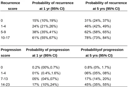
Table 4: Probability of recurrence and progression according to score 49
Recurrence Probability of recurrence Probability of recurrence
score at 1 yr (95% CI) at 5 yrs (95% CI)
0 15% (10%,19%) 31% (24%, 37%)
1-4 24% (21%,26%) 46% (42%, 49%)
5-9 38% (35%,41%) 62% (58%, 65%)
10-17 61% (55%,67%) 78% (73%, 84%)
Progression Probability of progression Probabilityof progression
score at 1 yr (95% CI) at 5 yrs (95% CI)
0 0.2% (00%,0.7%) 0.8% (0%, 1.7%)
1-4 01% (0.4%,1.6%) 06% (05%, 08%)
7-13 05% (04%,07%) 17% (14%, 20%)
14-23 17% (10%,24%) 45% (35%, 55%)
TREATMENT OPTIONS IN T1G3 BLADDER CANCER:
Transurethral Resection of Bladder Tumor
Transurethral resection of the bladder tumor (TURBT) is the first step in
the initial treatment of bladder cancer. A TURBT is both diagnostic and
therapeutic, and the procedure provides critical staging information. In the setting
of a TURBT, the configuration (flat, sessile, or papillary), location (trigone, base,
dome, or lateral walls), size (centimeters), and number of tumors should be
noted. Tumors have to be completely resected and muscularis propria must be
evaluation, and accurate clinical staging. Larger tumours must be resected
separately in fractions, which include the exophytic part of the tumour, the
underlying bladder wall with the detrusor muscle, and the edges of the resection
area. The specimens from different fractions have to be referred to the
pathologist in separate containers. Cauterization has to be avoided as much as
possible during the resection to prevent tissue destruction. The pathologic report
should specify the grade of the lesion & the depth of tumour invasion into the
bladder wall. It must also and give information on whether the lamina propria and
muscle are present in the specimen. A complete and correct TUR is essential for
the prognosis of the patient.
Role of random biopsies
In the 1970s, the bladder cancer was believed to be a multifocal tumor
with co-existing CIS distant from the primary tumour 40. Biopsies from the normal
looking mucosa were also recommended 50. The role of random biopsies was questioned and in the recently published EAU guidelines: It is not routinely
recommended.
Fujimoto et al. 51 and Van der Meijden et al52 assessed the usefulness of
random bladder biopsies of normal bladder mucosa after TURBT. Cancer was
identified in only few biopsies and was thought to be un-warranted. If needed,
multiple random biopsies were needed only in patients with multiple papillary
tumors or in those with positive cytology results. Conversely, May et al 53 found
that random bladder biopsies altered therapy in 7% of 1033 consecutive patients.
not in the resection of the primary tumor. Importantly, however, these
investigators excluded patients with small, primary, solitary bladder tumors. It was
concluded that the patients with low- risk–appearing tumors and negative
cytology results need not undergo random biopsy.
Role of Immediate Adjuvant Intravesical Chemotherapy
Clear consensus exists on the role of prophylactic chemotherapy in the
first 6 hours after TUR. In an effort to reduce this risk, many trials have
investigated the use of prophylactic or adjuvant intravesical therapy at the time of
TURBT. Tolley et al 54 performed a multicenter, randomized trial of 502 patients
with either Ta or T1 urothelial carcinoma. Those who received intravesical
mitomycin C (MMC) within 24 hours of TURBT had a statistically significant
decreased risk of tumor recurrence versus those who received placebo. Sylvester
et al.55 performed a meta-analysis on this and concluded that adjuvant
intravesical therapy is the treatment of choice in patients with a single, low-grade
superficial bladder tumor and should be the initial treatment (before subsequent
intravesical BCG) in those with higher-risk bladder tumors. Multicenter,
randomized, prospective studies demonstrate that the risk of recurrence can be
reduced by half at 2 years and by 15% at 5 years with a single dose of adjuvant
intravesical chemotherapy 54. Routine use of prophylactic chemotherapy after TUR can thus be recommended. Contraindications to its use include very deep
resections, bladder perforation, and prior documented allergic reaction. Although
a wide variety of chemotherapeutic agents were used in the pooled trials
centres use MMC (40 mg in 40 cc of saline) as the prophylactic
chemotherapeutic agent of choice.
A Second TURBT
T1G3 TCC of the bladder is associated with a significant risk of tumor
progression and recurrence when transurethral resection (TUR) is the only
treatment. Adjuvant immediate post TUR intravesical chemotherapy or
maintenance immunotherapy can reduce this risk; however, long-term results of
more than 15 years of follow-up indicate that almost 50% of the patients may end
up in a radical surgery for the disease, or even die due to the cancer. The
alternative to TUR is cystectomy at either the initial presentation or time of first
recurrence. Although the results of this treatment strategy are encouraging, an
unknown percentage of patients will lose their bladder and go on to experience all
possible complications of urinary diversion unnecessarily- this option does seem
to be overkill.
The concern of conservative treatment lies in the quality of TUR. With the
available literature it is evident that a ‘perfect TUR’ cannot be performed on
every patient, i.e. macroscopical clearance of the tumor from the bladder,
separate resection of the tumor base, inclusion of the deep muscle in the
specimen and separate biopsies of the borders of the resection area. Even in
cases of a so-called ‘correct TUR’, a significant proportion of residual tumors is
left behind and will be the source of local recurrence or progression. In addition,
TUR specimens may be difficult to diagnose accurately, especially with respect to
Literature demonstrates that the routinely performed second TUR detects
residual tumors of similar or higher stage in a significant percent of patients. The
clinical implications of these findings can be quite significant, as the absence or
presence of tumor may determine whether patients undergo conservative or
aggressive treatment. Moreover, results of various retrospective studies support
this suggestion 10,19. The TUR after incomplete resection resulting from factors such as multiplicity, size, and location must be called repeat resection. If a
second intervention is done to provide additional pathologic information for the
muscularis propria, it has to be called re-staging TUR. The term second TUR is to
be used only if the procedure is done after a complete and correct TUR. For all
cases of newly diagnosed T1G3 TCC, a second TUR after the primary TUR is
strongly recommended. In cases when a second TUR in a T1 is considered
necessary to secure the completeness of the initial resection, the question arises:
When should it be done? Several authors have shown that a second TUR can be
performed safely a week after the first TUR 56, although proposed time frames
range from 7 days to 3 months 56.The argument that this strategy should be
based on the result of the second TUR indicates that this procedure should be
done as early as possible. Thus, there is no reason to wait three months unless a
second resection is not performed (as for cases with an intact mucosa and
negative urine cytology) 56. The advantages of performing a second TUR are manifold: It provides more accurate staging, detects and potentially clears the
entire residual tumor and is helpful in prognostication. Second TUR reduces
recurrence and also increases the recurrence-free interval and the
Second TUR is of particular importance as the probability of understaging
a T1G3 tumour ranges from 20–70%, depending on the presence of muscularis
propria in the sample 27. Herr 30 retrospectively evaluated the difference in the pathologic diagnoses between an initial TURBT and a second TURBT in 150
patients. The results of the second TURBT altered the treatment in 33% of the
patients. Importantly, he noted the inability to correctly diagnose T1 tumors
without muscle in the specimen. Of 23 patients with T1 lesions without muscle in
the primary resection, 11 (49%) were upstaged to T2 lesions after review of the
second TURBT specimen. Dutta et al. 57 similarly reported a 64% risk of
understaging T1 lesions when muscle was absent versus 30% when muscle was
present in the TURBT specimens. Kla¨n et al. demonstrated that patients who
initially had a fractionated TUR had a reduced rate of residual tumor (36.7%)
compared to patients in whom a separate resection biopsy of the tumor bed was
not performed (56.3%) 56. If muscle is absent from the initial TUR, a repeat
resection is essential because of the high rate of understaging 27. Even with
muscularis propria sampling at the first resection, several reports have proven
occult T2 disease in up to 10% of second resections 27. The high incidence of understaging at initial resection has been confirmed by analyzing the cystectomy
specimens. In one study, 78 patients with non muscle invasive bladder cancer,
who underwent cystectomy, 37% of the specimens showed muscle involvement
48. As a second TUR often upstages T1 lesions and/ or provides additional
Divrik et al 58 report that doing a second TUR in patients with T1 tumors
reduces the risk of bladder cancer recurrence and progression. A total of 210
patients were randomized to one of two groups. An initial TUR was performed in
both groups, followed by a second TUR in one group only. This was done 2–6
weeks after the initial resection. Residual tumor was detected in 35 of 105
patients in the restaged group, and 8 patients were upstaged to T2. Recurrence,
progression, and disease-free survival were followed in all patients, and
significantly higher levels of recurrence and progression were seen in the patients
who had not undergone a second TUR. Tumor recurrence occurred in 37 of 93
patients in the group who underwent a second TUR, and 70 of 98 patients in the
other group, while progression was seen in 6.5% of patients in the second TUR
group, compared with 23.5% of patients who did not undergo a second TUR.
There were also more cancer- related deaths among the patients who did not
undergo a second TUR - 11 deaths in 35 patients in this group, compared with 5
deaths in 30 patients in the group with a second TUR. Overall survival was 67.7%
and 64.3% in the second TUR and without a second TUR groups respectively.
In addition to the diagnostic benefit, repeat TUR also has the ability to
detect and potentially clear residual TCC. Since at least 27% of patients harbour
residual tumour (with the highest reported rates of 62%) 27, repeat resections may
have a therapeutic benefit. Zurkirchen et al. 59 retrospectively analyzed patients
who underwent second TURBT within 6 weeks of their initial resection and found
that 37% of patients with initial T1 bladder tumors had residual tumors on second
resection. Grimm et al.60 similarly analyzed retrospectively 83 patients who
analysis, both tumor stage and grade were identified as predictive for residual
tumor on second TURBT. After 5 years, there was a significant decrease in
disease-free survival between those who underwent a second TURBT and those
who did not (63% and 40%, respectively). Brauers et al. 23 evaluated 42 patients’
withT1 bladder tumors and found that 24% of patients were upstaged to T2 or
CIS on second TURBT. Schips et al. 24 prospectively evaluated the findings at
first and second TURBT for patients with high-grade T1 bladder tumors and
reported residual disease in nearly 50% of patients. Both multifocality and tumor
grade increased the risk of finding residual tumor on second TURBT.
Early repeat TURBT can be justified for the purposes of identifying
understaged T2 tumors that would benefit from prompt surgery (cystectomy). In a
series of 189 patients who underwent cystectomy within 3 months of diagnosis of
muscle- invasive disease, there was a significantly better 5-year progression-free
survival compared with those in whom cystectomy was performed in more than
3 months after diagnosis (55% and 34%, respectively) 19. In summary, these studies show that the risk of upstaging on second TURBT is >30% if muscle is
present in the specimen and even higher if muscle is not present 57. Further, the risk of residual tumor on second TURBT is also significant. The risk for even
solitary, papillary-appearing tumors is 24% to 27% 59.
An appreciated advantage of repeat TUR is prognostication. Although
upstaging of T1G3 lesions to pT2 disease or higher automatically selects patients
for radical therapy, Herr and colleagues showed that evidence of T1 disease on
contrast, of 260 T1 patients who had no lamina propria invasion on a second
TUR, only 19% progressed at 5 yr. Based on these results, residual T1 TCC on
second TUR was deemed a negative prognostic indicator and a potential
indication for immediate cystectomy in T1G3 patients. Due to these significant
advantages, it is recommended that a second TURBT be considered for all
patients with high-grade T1 urothelial TCC.
Additional Intravesical chemotherapy
Intravesical chemotherapy prevents recurrence but not progression, as
confirmed by a meta-analysis comparing intravesical chemotherapy to TUR alone
61. Two other meta analyses also demonstrated the efficacy of intravesical
chemotherapy in decreasing the risk of tumour recurrence 62. It is still
controversial as to how long and how frequently intravesical chemotherapy
instillations need to be given. From a systematic review of the literature of
randomised clinical trials, comparing different schedules of intravesical
chemotherapy instillations, one can only conclude that the ideal duration and
intensity of the schedule remains undefined, because of conflicting data 63.
Adjuvant intravesical bacillus Calmette-Gue´rin immunotherapy
Adjuvant intravesical bacillus Calmette-Gue’rin´ (BCG) immunotherapy is
the treatment of choice in T1G3 TCC. Standard induction therapy is of 6 weekly
instillations after a second resection for a diagnosed T1G3 disease. The
therapeutic benefit of BCG for T1G3 bladder cancers has been definitively
There is evidence to support the role of maintenance BCG therapy for
T1G3 bladder cancer. In a prospective, randomised Southwest Oncology Group
(SWOG) trial, Lamm et al evaluated the efficacy of induction plus maintenance
BCG to induction BCG only in 384 patients with Ta or T1 disease at high risk of
recurrence/ progression 27. The maintenance protocol consisted of three weekly BCG instillations at 3 and 6 months post-TUR and semiannually thereafter, for 3
yr. Both 5-yr recurrence free survival (RFS) (60% vs. 41%, p < 0.001) and
progression- free survival [PFS], (76% vs. 70%, p = 0.04) were improved with
maintenance BCG.
MMC is a reasonable alternative with less side effects, but used only in
situations where adjuvant BCG is contraindicated (allergy, intolerance to BCG, or
immunosuppressed states) 27. Patients experiencing severe side-effects from full-dose BCG may be considered for dose reduction, which may, however,
compromise efficacy. Dose reductions of one-half to one-third BCG colony-
forming units with improved side effect profiles without compromising on the
efficacy and safety have been studied 67,68. Recent reports by Martinez-Pineiro et al 69 and Yoneyama et al 70 have suggested worse outcomes with low-dose regimens in T1G3 patients, with trends towards reduced recurrence free
survival. In T1G3, TCC dose reduced BCG regimens must be used with caution.
Additional investigation of the role of reduced dose BCG is required.
Role of immediate or early cystectomy
The increased recurrence and progression rates associated with
A number of advantages exist to this approach: a definitive opportunity for cure,
and appropriate treatment of understaged lesions, to name a few. Though a
second TUR refines local cancer staging, a high percentage of patients will still
be understaged even after second TUR. Additionally, cystectomy enables
lymphadenectomy. As up to 18% of T1 patients have positive lymph nodes,
cystectomy can be both diagnostic and therapeutic for nodal metastases 27. Lastly, cystectomy obviates the need for repeated intravesical therapies and
simplifies follow-up.
Cystectomy for T1G3 TCC, however, also has a number of potential
drawbacks. The perioperative mortality and morbidity rate following cystectomy
(at 1–6% and 30% respectively) is not trivial 27. Cystectomy may have a harmful impact on quality of life secondary to long-term changes in sexual,
gastrointestinal, and genitourinary function. Finally, RC may be considered
overtreatment for many patients as a conservative bladder-sparing approach with
BCG is effective in around half the cases. An approach using reliable risk
stratification is thus required to decide which patients to offer early cystectomy vs.
where conservative treatment.
Other Treatment Modalities
The role of radiation therapy (RT) with or without chemotherapy for the
treatment of T1G3 TCC is limited. Weiss et al retrospectively evaluated the
impact of radiochemotherapy in 141 patients with T1G3 bladder cancer 71. Of the
84 patients with T1G3 disease, 89% did not have tumour on restaging TUR six
showed that the 10-yr risk of progression for T1G3 patients was 29%. Vis a vis.
BCG, however, the results of RT have not been as encouraging. In the largest
randomized trial, Harland et al compared RT to conservative therapy (observation
or intravesical BCG/MMC) in T1G3 TCC and found no difference in progression
free survival 72. Due to cost, inconvenience, potential for toxicity, and a lack of demonstrable benefit over conventional intravesical therapies, RT cannot be
recommended for routine use as a bladder-preservation strategy.
Is A Second TUR A Must In All T1G3 Bladder Cancers?
Despite the existence of literature supporting a second TUR, there are a
few investigators who still question this. Even after a satisfactory primary
resection and presence of adequate muscle in the resected specimen, do we
need to proceed with a second resection? Is there a sub group which can avoid a
second TUR? The morbidity associated with invasive re-resection, the cost of the
treatment and the delay in intravesical therapy makes one wonder if re-resection
is required in all T1G3 lesions
T1G3 tumors are famed for their high progression and recurrence rates. A
vast majority of this is due to incomplete primary resection and due to absence of
muscle in the TURT specimen. Dutta et al. 57 reported a 64% risk of
understaging T1 lesions when muscle was absent compared with 30% when
muscle was present in the TURBT specimens.
Tumor architecture, (papillary or sessile, solitary or multifocality of the
lesions) during the primary TURT are important prognostic factors for recurrence
prognostic factor over multiple papillary and sessile lesions 73. Schips et al. 24
prospectively evaluated the findings at first and second TURBT for patients with
high-grade T1 bladder tumors and also found residual disease in nearly 50% of
patients.
Both multifocality and tumor grade increased the risk of finding residual
tumor on second TURBT. Although 76% of patients with a solitary T1 lesion at
first TURBT had a negative second TURBT, only 53% of those with multifocal T1
lesions had a negative repeat TURBT. Moreover, approximately 3/4ths of with
papillary-appearing T1 lesions at first resection had a negative repeat TURBT
compared with only 47% of those with solid-appearing T1 lesions. In another
study, of 17 patients with a solitary T1G3 lesion who underwent a radical
cystectomy after the primary TURBT, only one patient had a tumor in the
resected specimen. The others were probably over treated 16. It has also been felt that re-resection may not be required in all T1G3 lesions, and the primary
TURBT can identify the subset of aggressive T1G3 tumors 74. Invasion of lamina propria superficial to the muscularis mucosa (T1a) is considered a good
prognostic factor as against the lamina propria deeper to muscularis mucosa.
Involvement of muscularis mucosa in the lamina propria is considered T1b, and
T1c is beyond the muscularis mucosa of lamina propria 75. Orsola et al 76
commented on the European guidelines for NMIBC and suggested a second
TUR in all T1 tumors.
In our institution we analysed a few characters of these T1G3 tumors and
attempted to answer the question: Is a second TUR necessary in all T1G3 tumors
A
AIM OF THE STUDY
M
MATERIAL AND METHODS
This is a prospective study conducted in the Department of Urology,
Christian Medical College, Vellore from April 2009 to December 2010. All the new
patients who were diagnosed to have a space occupying lesion in the bladder
and were planned for a transurethral resection of bladder tumor were assessed.
The patient was explained in detail about the present study, the disease (Bladder
cancer and T1G3 disease), and the management needed in the language best
understood by the patient. Informed consent was taken for the study. Patient
details, along with the clinical history and physical and systemic examinations
were noted. Relevant blood and urine investigations and the radiological
assessment were performed. Preanaesthetic evaluation was done for all the
patients prior to the surgery.
The procedure was conducted in the operation room under general or
regional anesthesia. A few of the patients needed obturator block to avoid
obturator jerk and the complications associated with it. Detailed cystoscopic
evaluation was carried out. The bladder lesions were meticulously assessed and
all the features including the site, size, multiplicity, relation to the ureteric orifices
and the appearance of the rest of the bladder mucosa were recorded.
Endoscopic, video assisted transurethral resection of the bladder tumor was
performed or supervised by a consultant. Karl Stroz / Wolf 26 fr active cutting
resectoscopes were used for the procedure. All the visible lesions were resected
and the tissue sent for histopathological examination. In all the subjects, after
complete resection of the tumor macroscopically during the primary resection,
examination. The pathologists’ report of the inclusion of the muscularis propria in
the specimen is the indication for a complete resection of the lesion.
A dedicated uropathologist assessed all the specimens sent and the presence of
the malignancy along with the stage, grade, and the involvement of the muscularis
propria was specifically mentioned. A second TUR was routinely performed in all the
pT1G3 bladder tumors within 4 – 6 weeks. Resection was performed at the previous
resection scar sites as documented by the earlier surgery records. and also at the
doubtful areas. Presence of any new residual or recurrent disease is specifically looked
for and the doubtful areas resected and sent for histopathological examination. The
resected specimen was assessed for any residual tumor, its stage, grade and upstaging
of the disease if any.
Statistical analysis:
Sample size: Sample Size was calculated using the following formula
4 p q N = ---
d 2
• Outcome = Presence/absence of re-resection
• prevalence (p) of re-resection = 20%.
• q = 100-p : 100-20 = 80 %.
The required sample size to estimate a prevalence of 20% with a precision
of 10% and with 95% CI was 70. This prospective study from April 2009 to
December 2010, aims to include this sample size in the study, but being a
prospective and time constrained thesis, all the patients in this time period were
included for this thesis. A total of 37 patients were diagnosed to have T1G3
disease and of which only 27 had a second resection done. Hence, these 27
patients were taken as the study group and the sample size.
All the T1G3 lesions were included in this study and different variables like
the size, multifocality, characteristics of the lesions would be analysed.
Descriptive statistics was calculated for all study variables. Prevalence of
re-resection was calculated along with 95% CI. Chi square test was used to assess
R
bladd and in the resec exam differ Figu 0 5 10 15 20 25 30 35 40 45 There w
der lesion f
17 women
e fifth and
ction of b
mination. T
rent stages
ure 1: Age g 2
2nd decade
ere a total
from April 2
(13.2%) in
the sixth d
bladder tum
Transitional
s and grade
group of p 9
e 3rd decade
RE
of 129 ne
2009 to De
this group
decades. (F
mor and
cell carci
es (Table 1)
patients 20
4th decade
ESULTS
ew patients
cember 20
. More than
Fig 1). All o
the specim
inoma was
).
34
5th decade
s who were
10. There w
n fifty perce
of them und
mens sent
s diagnose
39
6th decade
e diagnose
were 112 m
ent of the p
derwent a t
t for histo
ed in 126
21
7th decade
d to have
men (86.8%
patients wer
transurethra
opathologica
patients a
4
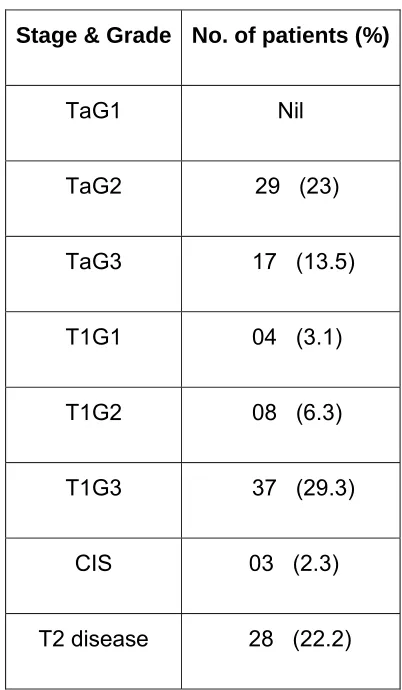
[image:46.612.112.519.350.592.2]Table 1: Stage & Grade of TCC
Association of smoking was looked at in these patients with bladder
lesions. None of the women had any association with tobacco. Out of the 129
patients with bladder lesions, 69 (53.48%) were regular smokers with variable
cigarette pack years.
Of the 129 patients who underwent TURBT, 37 patients had T1G3 TCC
bladder. Thirty of them were men and 7 were women. The mean age of the T1G3
patients was 57.3 years (37 – 75). Of these 37 patients with T1G3 lesions, 10
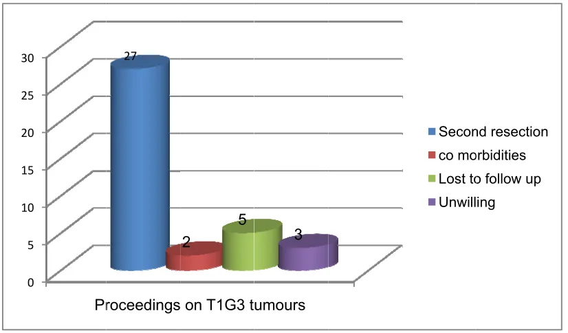
patients did not undergo a second resection. Two patients had multiple Stage & Grade No. of patients (%)
TaG1 Nil
TaG2 29 (23)
TaG3 17 (13.5)
T1G1 04 (3.1)
T1G2 08 (6.3)
T1G3 37 (29.3)
CIS 03 (2.3)
como and 5 and t Fig 2 analy varia the c were patie the 0 5 10 15 20 25 30 orbidities fo
5 were lost
these patie
2: Proceed
The 27
yzed in de
ables like th
characterist
Of the 2
e women. T
ents along w
lesions ha 0 5 0 5 0 5 0 Pr
or a second
to follow u
ents are ana
dings on T1
patients w
tail. The p
he site, size
tics of the p
7 patients
The mean
with T1G3 h
ave been roceedings
27
d procedure
p. Out of 3
alyzed as th
1G3 tumou
who underw
rimary rese
e of the les
primary lesio
who under
age was 5
had associa
assessed on T1G3 t
2
5
e, 3 were n
7, 27 patien
he study gro
urs
went a seco
ection and
sion, assoc
on were stu
rwent secon
56.18 years
ated CIS (s
during the umours
3
not willing f
nts had a s
oup. ond resect the secon ciated CIS, udied. nd resectio
s (37 – 75
secondary C
e primary
for a secon
econd rese
tion in the
d resection
depth of i
on, 22 were
years). Th CIS). Chara resection. Secon co mo Lost to Unwill nd procedur
ection (Fig 2
group wer
n along wit
nvasion an
e men and
ree of thes
acteristics o
They wer
nd resection orbidities
[image:48.612.110.523.160.402.2]classified as solitary papillary, multiple papillary, sessile and multiple sessile
lesions (Table 2).
Characteristics of the lesion No. of patients (%)
Solitary papillary 15 (55.5)
Multiple papillary 09 (33.3)
Solitary sessile 03 (11.1)
[image:49.612.136.475.166.358.2]Multiple sessile Nil
Table 2: Characteristics of the primary lesion.
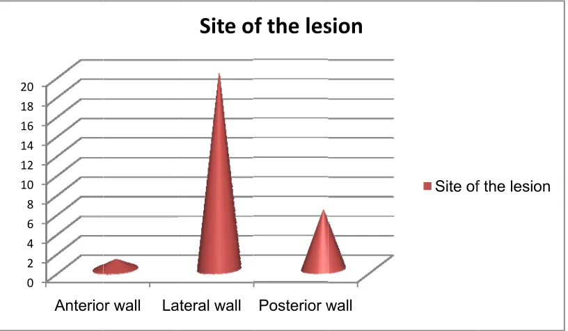
Most of these tumors were located on the lateral wall (Fig 3). When there
were multiple tumors, the site of the largest tumor was considered. Twelve of
these lesions were ≥3cm in size, and fifteen were < 3cm in size (Table 3)
Size of the lesion No. of patients (%)
≥ 3cm 12 (44.4)
< 3cm 15 (56.6)
Figu proto prima resec out. A lesio spec tumo
the s 0 2 4 6 8 10 12 14 16 18 20
ure 4: Site o
Out of t
ocol that 40
ary resecti
ction were
At the com
n resected cimens had or. The seco study group 0 2 4 6 8 0 2 4 6 8 0 Anterio
of the lesio
hese 27 p
0mg of mi
on if it is
thought to
mpletion of t
separately
the deep
ond resecti
p, the mea or wall La
on
atients, 24
tomycin dil
not contra
o be muscle
the primary
y, and sent
muscle (de
on is usual
an time for ateral wall
Site
o
4 had intra
luted in 40
aindicated.
e invasive;
y resections
t for histopa
etrusor) ide
lly planned
the secon Posterior w
of
the
les
vesical mit
0ml of salin
Three pati
hence no
s, all of the
athology. A
entified and
within a sp
nd resection wall
sion
tomycin ins
ne is instill
ients after
instillation
em had the
All the histo
d was not in
pan of 4 to
n is 6week Site of
stilled. It’s
ed after th
the primar
was carrie
base of th
opathologica
nvolved wit
8 weeks. I
ks 5 days ( f the lesion
[image:50.612.113.521.110.348.2]week perio resec visib the a over tumo Figu 0 2 4 6 8 10 12 14 16 18 20
ks to 12 we
od.
During t
cted sites. T
le lesion se
area of the
the bladde
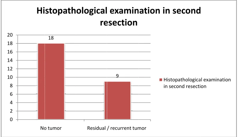
or in 9 patie
ure 5: Histo No
Hi
eeks). Non
the second The majorit een during previous re er. Histopat ents (33.3% opathologic 18
o tumor
stopath
ne of the pa
d resection
ty of the sc
the second
esection. Th
hological e
%) (Figure 5
cal examin Residual /
ological
re
atients had
n, the scar
cars were h
d resection
he other on
examination
)
nation of th 9
/ recurrent tum
examin
esection
any compl
r was iden
ealthy. Six
. Five of th
ne had mult
n of the sec
he second mor
nation
in
His in s
laints in the
ntified at t
patients (2
hem were o
iple papilla
cond resect
resection.
n
second
stopathologica second resecti
e intervenin
he previou
22.2%) had
on or aroun
ry lesions a
tion reveale
.
d
al examination
[image:51.612.110.521.352.589.2]There is no strong evidence to classify the tumor in the second resection
as residual or recurrent tumor. Among the 9 patients with tumor in the second
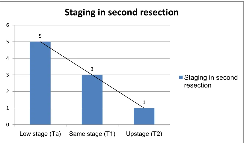
[image:52.612.112.519.229.468.2]resection, only one patient (3.7%) had upstaging to muscle invasive disease
(Figure 6).
Figure 6: Tumor staging in second resection.
Many variables like the primary characteristics of the lesion, the size of the
lesion, muscle in the primary resection specimen and associated CIS assessed
and correlated with the result of the second resection. Only 3 patients had a
secondary CIS in the primary resected specimen among the study group. On the
second resection, two were positive for tumor and one did not have tumor in the
second resection. All the primary resections had uninvolved deep muscle in the
specimen. 5
3
1
0 1 2 3 4 5 6
Low stage (Ta) Same stage (T1) Upstage (T2)
Staging
in
second
resection
than resec sess signi of tu follow relati to so more 5.0, C sess when 0 2 4 6 8 10 12 14 When th 85% (13/1 ction. Five ile lesions ficant asso umor positi
wed by the
ive risk was
olitary lesio
e in the ses
CI-95%, 1.
ile and mu
n compared 0 2 4 6 8 0 2 4 Solitary 2 he primary
15) of solita
of 9 multi
had tumo
ociation bet
vity in the
e multiple p
s 4.17 time
ons (RR 4.1
ssile group
1-22.8). Th
ultiple papill
d to solitary
papillary M
13
characteris
ary papillar
iple papilla
or in the s
tween lesio
second re
papillary an
es more in
17, CI-95%
when com
he chances
lary groups
y papillary le
Multiple papi
5 4
stics of the
ry lesions d
ry lesions
second res
on type and
esection w
nd least in
the multiple
%, 1.01-17.1
mpared with
of tumor p
s were high
esions.
illary S
4
e lesions w
did not hav
(71.4%) an section (Fig d positivity were maxim the solitary e papillary
18). The re
h the solitar
ositivity in t
h and were
Sessile
2 1
were consid
ve tumor in
nd 2 of the
gure 7). T
(p=0.04). T
mum in ses
y papillary
group whe
elative risk w
ry papillary
the second
e statisticall
dered, mor
the secon
e 3 (66.6%
here was
The chance
ssile tumor
lesions. Th
en compare
was 5 time
Figu char the s of wh lesio (Figu posit high in th <3cm Figu 0 2 4 6 8 10 12 14
ure 7: H
racteristics
Tumor s
second rese
hich, 8 had
ns (6.6%)
ure 8). Ther
tivity (p=0.0
with a lesio
e tumors w
m. (RR 10,
ure 8: Siz
. 0 2 4 6 8 0 2 4 istopatholo s.
ize from th
ection. The
tumor pos
of < 3 cm
re was a sig
001). The c
on of more
with size m
CI-95%, 1.4
e of the ≥ 3cm
8
4
ogy in s
e primary r
re were a t
sitivity in the
m in size h
gnificant as
chances of
than 3 cm
ore than 3
44-69.3).
primary
second re
resection w
total of 12 le
e second re
ad tumor p
ssociation b
tumor posi
in size. Th
cm when c
lesion an < 3cm
1
esection a
was analyze
esions with
esection (66
positivity in
between the
itivity in the
he relative r
compared w
d tumor m
14
according
ed with the
h more than
6.6%). Only
n the secon
e size of th
e second re
risk was 10
with the les
positivity
to lesio
e positivity o
n 3cm in siz
y 1 of the 1
nd resectio
e lesion an
esection wa
0 times mor
sions of siz
[image:54.612.110.520.410.653.2]prima in th resec papil grou posit Figu posi resec and o 0. 1. 2. 3. 4. These tw
ary lesion t
e second
ction, 8 ha
llary lesion
p and 2 f
tivity in the
ure 9: Char
tive secon
Out of t
ction, 13 p
one in the s 0 .5 1 .5 2 .5 3 .5 4 .5 5 solitary wo variables together we resection.
d ≥3cm pri
(Figure 9
from the s
second res racteristics nd resectio the eightee atients wer sessile grou y papillary 2 0
s i.e. the siz
ere analyze
Of the 9
imary lesio
9). There w
sessile grou
section
s and the
on
en patients
re in solitar
up. (Figure
Multiple pap
4 1
ze of the tu
ed with the
patients w
ns, and 4
were two le
up who ha
size of the
s who did
ry papillary
10)
pillary
umor and th
presence o
ith tumor p
of them we
esions from
ad >3cm l
e primary
not have
group, 4 i
Sessile 2 0 he characte or absence positivity in ere among
m the solita
esion who
lesion and
tumor in
n the multi
eristics of th
of the tumo
the secon
the multip
ary papillar
o had tumo
d the tumo
[image:55.612.111.521.265.505.2]Figu rese solita had t the s lesio resec a tum in siz 0 2 4 6 8 10 12 14
ure 10: Cha
ction
In the st
ary papillary
tumor in the
second rese
ns, 5 had
ction. The r
mor in the s
ze and two 0 2 4 6 8 0 2 4 solitary aracteristic udy group,
y lesions. E
e second re
ection (Figu
lesions >3
remaining 4
second res
of them ha
papillary
2 11
cs & size o
of the 27
Eleven of th
esection. F
ure 11). Of
3cm in size
4 patients w
section (Fig
ad tumor in
Multiple pap
1 3
of the prim
patients w
hem were o
our had >3
f the 9 patie
e and 4 of
with <3cm s
ure 12). Al
the second
pillary
ary lesion
ho had sec
of <3cm in
3cm lesion o
ents with m
f which had
size primary
l the 3 ses
d resection.
Sessile
10
and negat
cond resec
size and n
of which 2 h
ultiple papi
d tumor in
y lesions, o
sile lesions
tive secon
ction, 15 ha
none of them
had tumor
llary primar
the secon
only one ha
[image:56.612.113.520.108.354.2]Figu seco Figu seco 0 2 4 6 8 10 12 0. 1. 2. 3.
ure 11: So
ond resect
ure 12: Mu
ond resect 0 2 4 6 8 0 2 0 .5 1 .5 2 .5 3 .5 4 olitary pap ion ultiple pap ion ≥ 3cm 2 2 ≥ 3cm 4 1 pillary les pillary les sions, prim sions, prim < 3 cm
0 1
< 3 cm 1 mary lesio mary lesio 11 m 3
on size an
[image:57.612.112.503.74.305.2] [image:57.612.111.519.389.632.2]Out of the 9 (33.3%) patients with positive second resection, 5 (18.5%)
had low stage disease. Two were solitary papillary and two were multiple
papillary lesions and one was a sessile lesion. Four of them had lesions <3cm in
size. Three patients (11.1%) had the same stage (T1G3) and 1 patient (3.7%)
had upstaging to T2 disease. Same or higher stage disease in the second TUR is
D
DISCUSSION
Bladder cancer is known to occur in the elderly age group, and mostly in
the 6th and 7th decade of life. In our study, most of the patients were in the fifth and sixth decade. Eventhough smoking is attributed as a risk factor which
significantly increases the risk of bladder cancer, nearly half the patients with
TCC in our study were nonsmokers.
TURBT is the initial step in the management of bladder cancer. A
technically complete primary resection is warranted for accurate pathological
staging and grading of the tumor. After complete resection of all the visible
tumors, deep muscle is resected separately for histopathology. If the resected
specimen has no muscle, there is a potential for understaging T1 tumors. The
important factor is the complete primary resection of the bladder tumor. Presence
of the uninvolved muscularis propria in the resected specimen is the only
identification for a complete resection 77. Retrospective studies have shown that residual disease can be seen in upto 68% cases 78. These high rates may also have been due to the fact that no muscle was present in many of the primary
TUR specimens. Forty nine percent of T1 lesions without muscle in the resected
specimen were understaged when compared to only 14% with muscle in the
resected specimen 30. Understaging was reported in 64% of T1 tumors when
muscle was absent in the specimen versus 30% when it was present 57. All our
resections had muscularis propria sampled at the end of the resection and
separately sent for HPE which were tumor free. This can explain why only 3.7%
Tumor architecture, papillary or sessile, and multifocality of these lesions,
and the size of the lesion are important prognostic factors for recurrence and
progression of the disease. A solitary papillary lesion is considered to be a good
prognostic factor as against multiple papillary and sessile lesions 73. In our series, among the 15 patients with solitary papillary lesions, two had tumor positive in the
second resection and both of them were >3cm lesions. All the 11 solitary papillary
lesions which were <3cm were tumor free in the second resection and 2 of the
>3cm lesions were also free of tumor in the second resection. Perhaps this is the
subgroup, solitary papillary with <3cm in size lesions, that are least likely to
benefit from a second resection. Multiple papillary lesions and the sessile lesions
had significant positivity in the second resection. More than fifty percent of the
multiple papillary lesions and 66.6% of the sessile lesions had tumor positivity in
the second resection. The relative risk is 4.17 times more in the multiple papillary
group when compared to solitary lesions (RR 4.17, CI-95%, 1.01-17.