Copyright © 1997, American Society for Microbiology
Two Classes of Human Papillomavirus Type 16 E1 Mutants Suggest
Pleiotropic Conformational Constraints Affecting E1 Multimerization,
E2 Interaction, and Interaction with Cellular Proteins
TOSHIHARU YASUGI,1MARC VIDAL,2HIROYUKI SAKAI,1PETER M. HOWLEY,1ANDJOHN D. BENSON1*
Department of Pathology, Harvard Medical School, Boston, Massachusetts 02115,1and
Massachusetts General Hospital Cancer Center, Charlestown, Massachusetts 021292
Received 5 March 1997/Accepted 25 April 1997
Random mutagenesis of human papillomavirus type 16 (HPV16) E1 was used to generate E1 missense mutants defective for interaction with either hUBC9 or 16E1-BP, two cDNAs encoding proteins that have been identified by their ability to interact with HPV16 E1 in two-hybrid assays. hUBC9, the human counterpart of
Saccharomyces cerevisiae UBC9, is a ubiquitin-conjugating enzyme known to be involved in cell cycle
progres-sion. 16E1-BP encodes a protein of no known function but does contain an ATPase signature motif. Eight hUBC9 or 16E1-BP interaction-defective HPV16 E1 missense mutants were identified and characterized for origin-dependent transient DNA replication, ATPase activity, and various protein-protein interaction pheno-types. Six of these mutant E1 proteins were significantly impaired for replication. Among these, two classes of replication-defective HPV16 E1 missense mutants were observed. One class, represented by the S330R repli-cation-defective mutant (containing an S-to-R change at position 330), remained competent for all protein-protein interactions tested, with the exception of hUBC9 association. Furthermore, this mutant, unlike the other replication-defective HPV16 E1 missense mutants, had a strong dominant negative replication phenotype in transient-replication assays. The other class, represented by five of the missense mutants, was defective for multiple protein-protein interactions, usually including, but not limited to, the interaction defect for which each mutant was originally selected. In many cases, a single missense mutation in one region of HPV16 E1 had pleiotropic effects, even upon activities thought to be associated with other domains of HPV16 E1. This suggests that E1 proteins are not modular but may instead be composed of multiple structurally and/or functionally interdependent domains.
The papillomavirus E1 protein is essential for viral DNA replication. As an ATP-dependent DNA helicase, E1 is thought to act in the initiation of viral DNA replication, by binding at the viral replication origin and beginning unwinding of the viral DNA (20, 21, 46, 49, 55). Although E1 proteins can act alone to support low levels of papillomavirus DNA repli-cation (10, 15, 35, 55, 56), efficient in vivo origin-dependent papillomavirus replication requires interaction of E1 with the viral E2 protein (50). E2 stimulates binding of E1 to the viral replication origin, resulting in efficient viral DNA replication (6, 29, 33, 40, 43, 45, 48, 50). Association of E1 with other proteins is also important for these events. For example, bo-vine papillomavirus (BPV) type 1 E1 interacts with the cellular DNA polymerase alpha primase enzyme (9, 37). Other cellular proteins are also required for papillomavirus replication (32, 35), some of which may interact directly with the viral E1 protein.
In order to understand other functions of E1 in papilloma-virus DNA replication, we have used the yeast two-hybrid system to identify additional cellular proteins that interact with E1. This screen identified two human papillomavirus type 16 (HPV16) E1-interacting cellular proteins, one of which (hUBC9) is a human homolog of the Saccharomyces cerevisiae UBC9 ubiquitin-conjugating enzyme (57). The other clone,
referred to as 16E1-BP, encodes a protein of unknown func-tion. This protein contains an ATP binding consensus motif. We sought to test the significance of HPV16 E1 interaction with hUBC9 or 16E1-BP by using the reverse two-hybrid sys-tem (51, 52) to generate HPV16 E1 missense mutants defec-tive in these interactions. Thus, by assessing the replication activity of such mutant E1 proteins, it might be possible to evaluate the role of these protein-protein interactions in E1 replication activity.
Eight HPV16 E1 missense mutants were identified and char-acterized for their ability to support origin-dependent transient DNA replication, ATPase activity, E1 multimerization, and HPV16 E2 interaction. All eight mutations occurred at sites encoding amino acids that are conserved among the papillo-mavirus E1 proteins. Four mutations changed amino acid res-idues that are conserved between papillomavirus E1 proteins and the polyomavirus and simian virus 40 (SV40) large T antigens (LgTs). Most of these mutations resulted in signifi-cant replication defects. One class of HPV16 E1 mutants had multiple functional deficiencies in addition to the protein in-teraction defect for which they were originally selected. For example, the G482D mutant (containing a G-to-D change at position 482) was originally selected for its inability to interact with 16E1-BP but was also defective for all other functions examined. In contrast, another mutant, S330R, was replication defective but only impaired for hUBC9 interaction, suggesting that hUBC9 interaction, or a closely related HPV16 E1 func-tion, is required for its DNA replication activity. Interestingly, the S330R mutant also had a strong dominant negative phe-notype in transient-replication assays.
* Corresponding author. Mailing address: Department of Pathology, Harvard Medical School, 200 Longwood Ave., Boston, MA 02115. Phone: (617) 432-2892. Fax: (617) 432-0727. E-mail: jbenson@warren .med.harvard.edu.
5942
on November 9, 2019 by guest
http://jvi.asm.org/
MATERIALS AND METHODS
Isolation of cDNAs that encode HPV16 E1-interacting proteins.A yeast two-hybrid screen using full-length HPV16 E1 (pPC97-16E1) was performed as previously described (51, 52). The activated human T-cell library used in this screen was selected primarily on the basis of its availability, although we also believe that the actively dividing status of these cells might favor the likelihood that general factors involved in DNA replication would be expressed and rep-resented. The pPC97-derived plasmids in this system express fusion proteins containing the DNA binding domain of the yeast Gal4 protein (Gal4DBD). pPC86-derived plasmids express fusion proteins containing the activation do-main of the yeast Gal4 protein (Gal4AD). Isolation and characterization of the hUBC9 cDNA have been described elsewhere (57). The pPC86 plasmid encod-ing the HPV16 E1-interactencod-ing protein 16E1-BP was isolated durencod-ing the same two-hybrid screen. The plasmid containing the 16E1-BP cDNA was rescued by transformation of competent Escherichia coli DH5a bacteria with total yeast DNA. The sequences of both strands of the 16E1-BP clone were determined by dideoxynucleotide sequencing with appropriate synthetic oligonucleotide prim-ers using Sequenase 2.0 (United States Biochemical).
Random mutagenesis of HPV16 E1 and isolation of E1 mutants.The yeast expression plasmid pPC97Trp was constructed by replacing the ApaI-SacI frag-ment of pPC86 with the ApaI-SacI fragfrag-ment of pPC97. This places Gal4DBD and the adjacent multiple cloning site from pPC97 into a single-copy yeast vector containing a TRP1 selectable marker. An insert fragment containing amino acids (aa) 144 to 649 of HPV16 E1 with a silent mutation at codon 355 (TTT was mutated to TTC, creating a BstBI restriction site) was created by PCR-directed mutagenesis. This SalI-NotI fragment was cloned into pPC97Trp, yielding pPC97Trp-16E1(aa144-649). The yeast expression plasmid pPC86Leu, contain-ing Gal4AD and a LEU2 marker, was constructed from pPC97 and pPC86 in a manner similar to that of pPC97Trp by replacing the ApaI-SacI fragment of pPC97 with that of pPC86. The inserts of the pPC86-derived plasmids containing hUBC9 or 16E1-BP were cloned into pPC86Leu, yielding pPC86Leu-hUBC9 and pPC86Leu-16E1-BP, respectively.
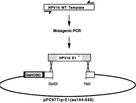
Random mutagenesis of HPV16 E1 was performed as previously described (51, 52; see also Fig. 2). Briefly, mutagenic PCR using oligonucleotide primers GCGTAGTACAGCAGCAGC (nucleotides [nt] 1729 to 1816) and AGGCCA CCTAGAATCTGTAC (nt 2575 to 2594) was performed utilizing concentrations of 200mM for three deoxynucleoside triphosphates, 25mM for one deoxynucleo-side triphosphate, and 200mM dITP. (Nucleotide numbering corresponds to that of GenBank accession no. K02718.) pPC97Trp-16E1(aa144-649) was digested with BstBI and NsiI, and this gapped vector fragment was gel purified. MaV103 harboring pPC86Leu-hUBC9 or pPC86Leu-16E1-BP was transformed with ap-proximately 0.5mg of gapped pPC97Trp-16E1(aa144-649) vector fragment along with 2mg of mutagenized PCR products. Colonies were selected on leucine-tryptophan dropout plates containing uracil and 0.1% 5-fluoro-orotic acid (5-FOA). 5-FOA-resistant yeast clones were then examined for expression of the Gal4DBD-E1 fusion protein by Western blot (immunoblot) analysis. The inserts of rescued plasmids that appeared to express full-length HPV16 E1 were se-quenced for determination of missense mutations. E1 mutants were named according to the targeted amino acid and its position within the HPV16 E1 open reading frame. For example, the mutant with a substitution of arginine for serine at amino acid 330 was designated S330R.
Two-hybrid binding activity assay.Yeast expression plasmids expressing mu-tant Gal4DBD-E1 full-length fusion proteins were generated by inserting a NarI restriction fragment from pPC97-16E1 that extended from within Gal4DBD through aa 170 of HPV16 E1 into pPC97Trp-16E1(aa144-647) plasmids harbor-ing mutant E1 genes. pPC86Leu-16E2(aa1-251) was constructed by insertharbor-ing a PCR-derived SalI-BglII HPV16 DNA fragment encoding E2 (nt 2755 to 3507). Fusion proteins expressed in MaV103 from pPC97Trp and pPC86Leu were examined for interaction by determining viability on histidine dropout plates containing various concentrations of 3-aminotriazole (3-AT).
Transient-replication assay.An SphI-XbaI fragment containing the HPV16 origin (nt 7463 to 100) was generated by PCR and cloned into the corresponding sites of the pCAT-Basic vector (Promega), yielding pSp16oriCAT. Transient replication of pSp16oriCAT was analyzed essentially as previously described (40). Mammalian expression vectors containing the mutated HPV16 E1 genes were constructed by exchanging the NarI-XmaI fragment containing a mutation with the analogous fragment of pCMV-E116(12, 40). CV-1 cells (CCL-70) were
used for transient-replication assays. Plasmid DNAs were introduced into cells by calcium phosphate coprecipitation (24). At 70 h after transfection, low-molecu-lar-weight DNA was extracted by the method of Hirt (19), with modifications described elsewhere (40). DNA samples were digested with DpnI to distinguish between replicated and input DNA. Sample DNAs were also digested with XbaI to linearize replicated pSp16oriCAT. The digested samples were separated by 1% agarose gel electrophoresis and analyzed by Southern blotting. A Phosphor-Imager (Bio-Rad Laboratories) was used for quantitation of hybridized signals. Western blot analysis.Total yeast lysates were prepared by adding 1 pellet volume of 23sodium dodecyl sulfate-polyacrylamide gel electrophoresis (SDS-PAGE) sample buffer directly to cell pellets and boiling the samples for 2 min. Expression of Gal4DBD fusion proteins was confirmed by Western blotting using a mouse monoclonal antibody (Santa Cruz Biotechnology). Total cell lysates of CV-1 cells were prepared by adding 1 ml of chilled lysis buffer (0.1% Nonidet
P-40, 250 mM NaCl, 20 mM sodium phosphate [pH 7.0], 30 mM sodium vana-date, 1 mM phenylmethylsulfonyl fluoride, 100 kIU of aprotinin, 1mg of leu-peptin perml, 1mg of pepstatin perml, and 5 mM dithiothreitol) to one 6-cm2
plate seeded with 1.53105cells 1 day prior to transfection. The cells were
scraped, and the lysates were incubated for 10 min on ice and then centrifuged for 5 min at 4°C at 12,000 rpm in an Eppendorf microcentrifuge. Supernatants were used for further analysis by SDS–10% PAGE, and proteins were trans-ferred to Immobilon-P membranes (Millipore Corp.). Expression of HPV16 E1 was detected by using anti-HPV16 E1 polyclonal rabbit antiserum (58) and visualized by chemiluminescence as recommended by the manufacturer (ECL detection kit; Amersham).
ATPase activity assay.The HPV16 E1 protein and E1 amino acid substitution mutants from pPC97Trp-derived plasmids were cloned as SalI-NotI fragments into pGEX4T-2 for expression and purification as glutathione S-transferase (GST) fusion proteins. The TOPP3 bacterial strain (Stratagene) was used for expression of GST-E1 fusion proteins, and these proteins were purified by standard procedures using glutathione-Sepharose 4B beads (Pharmacia) (23). The beads were washed three times in ice-cold reaction buffer (50 mM Tris-HCl [pH 8.0], 100 mM NaCl, 10 mM MgCl2, 1mM ATP, 1 mM dithiothreitol), and
GST fusion proteins were eluted with reaction buffer containing 10 mM gluta-thione. Approximately 100 ng of each GST-E1 fusion protein was used for ATPase activity assays. ATPase activity was detected by release of [a-32P]ADP
from [a-32P]ATP. The reaction mixtures were in 20ml of reaction buffer and
contained 0.1mCi of [a32-P]ATP. After incubation at 37°C for 1 h, 1ml of the
reaction mix was spotted onto a polyethyleneimine-cellulose thin-layer chroma-tography plate (Sigma), allowed to dry, and developed with 0.8 M acetic acid and 0.8 M LiCl2. Data were quantitated with a phosphorimager. The presented
values are averages from three independent assays in which the ATPase activity of an equivalent amount of GST protein was subtracted and the ATPase activity of wild-type GST-E1 was designated as 100%.
Nucleotide sequence accession number.The 16E1-BP sequence described in this paper has been submitted to GenBank under accession no. U96131.
RESULTS
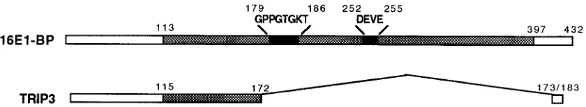
Use of the two-hybrid system to identify HPV16 E1-inter-acting proteins.We have previously used the two-hybrid sys-tem to identify cDNAs encoding HPV16 E1-interacting pro-teins. A description of one such protein, a human homolog of the yeast UBC9 ubiquitin-conjugating enzyme, has been pub-lished previously (57). This screen also identified a second HPV16 E1-interacting clone, which we call 16E1-BP. In a screen of approximately 1.53106yeast transformants, hUBC9 arose independently five times, whereas 16E1-BP arose only once. The amino acid sequence of the protein encoded by the 16E1-BP cDNA is shown in Fig. 1A. 16E1-BP appears to be a form of TRIP13, a protein previously shown to bind thyroid hormone receptor in yeast two-hybrid assays (26) (Fig. 1B). The 16E1-BP clone described here encodes a 432-aa protein that includes a Gly-X4-Gly consensus ATP binding motif at aa 179 through 186. The coding sequence described for the TRIP13 clone (GenBank accession no. L40384) contains cod-ing sequence for 183 aa but does not appear to include the 59 end of that mRNA, since the L40384 sequence does not in-clude an initiation methionine. TRIP13 and 16E1-BP diverge at a position equivalent to 16E1-BP residue 172. The TRIP13 coding sequence extends 11 aa past this point of divergence and then terminates. Thus, a significant portion of the 16E1-BP coding sequence, including the putative ATP binding motif, is not represented in TRIP13. We considered the pos-sibility that 16E1-BP and TRIP13 might represent alternatively spliced mRNAs. However, Northern (RNA) blot analysis of HeLa, HaCaT, B-cell, and T-cell polyadenylated RNAs de-tected only one mRNA species. The size of the RNA species we observed was consistent with that of 16E1-BP and not TRIP13 (data not shown). Since the TRIP13 clone was origi-nally identified in a HeLa cell cDNA library two-hybrid screen, the explanation for our inability to detect an mRNA species representing TRIP13, especially in HeLa cells, is unclear. It is clear, however, that 16E1-BP is the predominant RNA species. Moreover, a large region of 16E1-BP encompassing the ATP binding domain bears homology to a Caenorabditis elegans
VOL. 71, 1997 CHARACTERIZATION OF HPV16 E1 MISSENSE MUTANTS 5943
on November 9, 2019 by guest
http://jvi.asm.org/
cDNA (GenBank accession no. Z48334) and to a YTA-like yeast protein of unknown function (42) (GenBank accession no. Z36055, nt 594 through 2204). This conserved domain includes the amino acid sequence GPPGTGKT, which repre-sents a specialized form of the ATP A box and is one hallmark of YTA1 family members (42).
Generation of interaction-defective HPV16 E1 missense mu-tants.Full-length HPV16 E1 was previously cloned into pPC97 for use as bait in a two-hybrid screen of a human activated T-cell cDNA library (57). The particular yeast two-hybrid sys-tem used here employs three reporter genes: HIS3,b
-galacto-sidase, and URA3 (51, 52). Transcriptional activation as a result
of intermolecular interaction between the Gal4DBD and Gal4AD fusion proteins results in activated expression of these reporters. Induced expression ofb-galactosidase and HIS3 is detected by blue color on X-Gal (5-bromo-4-chloro-3-indolyl-b-D-galactopyranoside) plates and resistance to higher levels of 3-AT, respectively. The URA3 marker allows both positive and negative selection for two-hybrid interactions. Interaction in-duces URA3 expression and results in growth on minimal me-dium lacking uracil. Alternatively, since expression of the
URA3 gene in the presence of 5-FOA is toxic in yeast (8),
induced URA3 expression due to two-hybrid interaction results in toxicity on 5-FOA plates. Plating on 5-FOA allows selection of yeast survivors in which interaction between pPC97- and pPC86-encoded fusion proteins has been lost. In this study, we have used this URA3 counterselectable marker system as a means of screening for random mutations in HPV16 E1 that abrogate interaction with hUBC9 or 16E1-BP.
Unlike BPV E1, HPV16 E1 expressed as a DNA-binding fusion protein exhibits strong intrinsic transactivation activity in yeast. This activity maps to the amino terminus of HPV16 E1. aa 1 to 190 of HPV16 E1 are sufficient for transactivation
in yeast when expressed as a DNA-binding fusion protein (58), whereas expression of aa 144 to 649 produces a fusion protein without intrinsic transactivation activity. Accordingly, whereas it is possible to score interactions with full-length Gal4DBD-HPV16 E1 by using 3-AT resistance (HIS3 expression) as a positive selection, intrinsic transactivation of the URA3 gene by this bait protein results in constitutive toxicity on 5-FOA plates. For this reason, it was necessary to use HPV16 E1 aa 144 to 649 fused to Gal4DBD as a target for random mutagen-esis to screen for interaction-defective missense mutations, since this region of HPV16 E1 does not have intrinsic trans-activation activity in yeast but does exhibit specific protein-protein interaction in two-hybrid assays (58).
The mutagenesis strategy is summarized in Fig. 2. Mutagen-esis was carried out by conducting PCR amplification of an HPV16 E1 template using conditions that favor random nu-cleotide misincorporation into the PCR product. The mu-tagenized PCR product mixture generated in this manner was used with the pPC97Trp-16E1(aa144-649) BstBI-NsiI fragment to transform yeast expressing hUBC9 or 16E1-BP from the pPC86Leu vector. This procedure relies upon recombination between the linearized vector and the PCR products to yield yeast colonies that coexpress a potentially mutated E1 protein from pPC97, along with hUBC9 or 16E1-BP from pPC86. Plating on 5-FOA–uracil allows for selection of survivors in which interaction has been lost due to one or more mutations in HPV16 E1. Expression of intact Gal4DBD-E1(aa149-649) protein in yeast from 5-FOA-resistant colonies was confirmed by Western blotting using a monoclonal antibody against Gal4DBD (data not shown). Plasmids encoding full-length mutated E1 bait proteins were rescued from yeast, used to transform E. coli, propagated, and sequenced. Four hUBC9-defective mutants (W439R, S330R, Y412F, and W439L) and FIG. 1. (A) Amino acid sequence encoded by the 16E1-BP full-length cDNA clone. The consensus ATP binding motif is indicated (boxed). (B) Comparison of the 16E1-BP and TRIP3 clones. A region conserved between 16E1-BP, TRIP3, and a number of other related proteins from other organisms (see text) (shaded bars) and ATP binding motif sequences that are highly conserved within this family of proteins (black bars) are indicated. The remaining sequences, except for the short regions described in the text, are unique to and shared by 16E1-BP and TRIP13. The region of 16E1-BP that includes the ATP binding motifs is not present in TRIP3.
on November 9, 2019 by guest
http://jvi.asm.org/
[image:3.612.98.518.254.331.2]four 16E1-BP interaction-defective mutants (G482D, D438G, L494Q, and G496R) were generated in this manner.
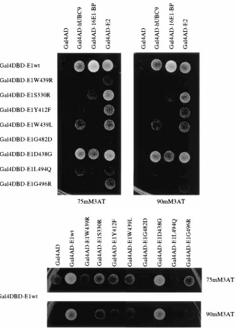
HPV16 E1 mutant protein-protein interaction phenotypes. Fragments of pPC97Trp-16E1(aa144-649) that contained mis-sense mutations in HPV16 E1 were recloned into pPC97-16E1 in order to assess their interaction phenotypes in the context of the full-length E1 protein. These mutants were tested for hUBC9, 16E1-BP, HPV16 E2, or HPV16 E1 interaction. Fig-ure 3 shows the result of a two-hybrid interaction experiment in which eight amino acid substitution mutants were tested for interaction with hUBC9, 16E1-BP, HPV16 E2, and HPV16 E1. This assay was performed by using the HIS3 marker as a reporter under two conditions of stringency: 75 and 90 mM 3-AT. Since the concentration of 3-AT to which yeast are resistant is proportional to HIS3 expression, these concentra-tions represent increasingly stringent assay condiconcentra-tions. Expres-sion of only Gal4DBD as a negative control led to no detected interaction and activation in these assays (data not shown). Of the mutants derived from the hUBC9 screen, W439R, S330R, and Y412F displayed no growth at either 3-AT concentration when coexpressed with pPC86-hUBC9, confirming their inabil-ity to interact. Compared to wild-type HPV16 E1, W439L exhibited decreased but detectable interaction with hUBC9 in these two-hybrid assays. Thus, in the context of full-length E1, mutation of tryptophan 439 to leucine had only a slight effect on E2 and hUBC9 interaction, whereas mutation of this resi-due to a charged amino acid (arginine) severely abrogated all interaction phenotypes. Full-length HPV16 E1 mutants G482D, D438G, L494Q, and G496R were retested for loss of interaction with 16E1-BP. The D438G mutant displayed only a minimal decrease in 16E1-BP binding in the context of full-length HPV16 E1, whereas the G482D, L494Q, and G496R mutants retained their inability to interact with 16E1-BP when expressed in the context of full-length E1.
Multiple protein-protein interactions appear to be involved in the replication activity of E1 proteins. We and others have documented the necessity of E1-E2 interaction for efficient
papillomavirus DNA replication (6, 33, 40, 43, 45, 48, 50). The ability of E1 to form multimers is also believed to be an im-portant aspect of its replication activity (14, 30, 31, 44). We tested HPV16 E1 mutants defective for hUBC9 and 16E1-BP interaction for their abilities to interact with HPV16 E2 and HPV16 E1. We also tested the ability of hUBC9 interaction-defective HPV16 E1 mutants to interact with 16E1-BP and the ability of 16E1-BP interaction-defective mutants to interact with hUBC9. By documenting correlations between different E1 mutant phenotypes in this manner, we sought to establish functional relationships between the various activities of the multifunctional E1 protein.
For analysis of HPV16 E1-E2 interaction, plasmids express-ing wild-type and mutant HPV16 E1 bait proteins were used to cotransform yeast with an HPV16 E2 prey expression vector. Interactions were scored by 3-AT resistance. Of the HPV16 E1 mutants tested, only the G482D mutation completely abolished E1-E2 interaction, although a number of mutations resulted in decreased E1-E2 interactions (W439R, L494Q, and G496R). HPV16 E1-E2 interaction is clearly independent of hUBC9 and 16E1-BP interaction activity, since S330R interacts well with E2 but not with hUBC9. Similarly, Y412F interacts well with E2 but fails to exhibit binding to either hUBC9 or 16E1-BP.
E1 multimerization was examined by expressing mutant forms of HPV16 E1 from pPC86, along with wild-type Gal4DBD-HPV16 E1 expressed from pPC97. Coexpression of wild-type HPV16 E1 from both pPC97 and pPC86 resulted in activation of the HIS3 reporter gene (Fig. 3, bottom panel), whereas no interaction between wild-type HPV16 E1 and Gal4AD was detected. The W439R and D438G mutations completely abolished E1 interaction activity. L494Q had very low E1 interaction activity that was detectable only at 3-AT concentrations lower than those shown in Fig. 3 (data not shown). Y412F, G496R, and W439L had lower E1-E1 action activity than wild-type E1 but retained significant inter-action activity. The ability of W439L, but not W439R, to in-teract with E1 suggests the likelihood that hydrophobic interactions are involved in E1-E1 association mediated by this region. HPV16 E1 multimerization activity appears to be sep-arable from hUBC9 interaction, since S330R interacts nor-mally with E1 but not with hUBC9. Comparison of the E1-E1 and E1-E2 interaction activities of HPV16 E1 mutants does, however, reveal a correlation between these phenotypes. The W439R, G482D, and L494Q mutations dramatically decrease both interaction activities. Y412F and W439L have intermediate interaction phenotypes, whereas S330R is vir-tually wild type for both interactions. These results imply that either the E2 interaction domain of HPV16 E1 is func-tionally inseparable from the E1-E1 interaction domain or HPV16 E1 multimerization activity is a prerequisite for interaction with E2.
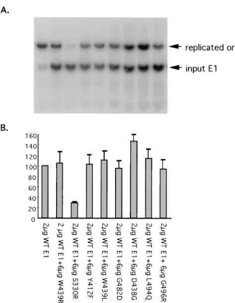
Replication phenotypes of HPV16 E1 missense mutants. Wild-type HPV16 E1 and HPV16 E1 missense mutants were subcloned into the pCMV4 vector for expression in mamma-lian cells. E1 protein expression was confirmed by Western blotting using anti-HPV16 E1 polyclonal antiserum. All HPV16 E1 amino acid substitution mutants were expressed at levels comparable to that of the wild type (data not shown). Transient-replication phenotypes were determined by cotrans-fecting CV-1 cells with HPV16 E1- and E2-expressing plas-mids, along with a plasmid containing the HPV16 origin of replication (ori). Replicated DNA was detected and quanti-tated by Southern blotting as described previously (40). The blot is shown in Fig. 4A, and the phosphorimager-quantitated data are shown in Fig. 4B. D438G and L494Q displayed rep-lication activities approximately 45 and 35% of that of wild-FIG. 2. Random mutagenesis of HPV16 E1. Wild-type (WT) HPV16 E1 was
used as a PCR template to generate a product that included the coding sequence of the HPV16 E1 protein. This PCR was carried out with deoxyribonucleotide concentrations that favor random misincorporation. MaV103 was cotransformed with these randomly mutagenized products (HPV16 E1*) and the gapped pPC97Trp-16E1(aa144-649) vector, which had been gel isolated following diges-tion with BstBI and NsiI. Recombinadiges-tion in yeast between the randomly mutated HPV16 E1* PCR products and the gapped vector results in reconstitution of pPC97-16E1(aa144-649), which expresses an HPV16E1(aa144-649)-Gal4DBD fusion protein containing a randomly acquired mutation.
VOL. 71, 1997 CHARACTERIZATION OF HPV16 E1 MISSENSE MUTANTS 5945
on November 9, 2019 by guest
http://jvi.asm.org/
[image:4.612.62.295.71.247.2]type E1, respectively. W439L demonstrated low but measur-able replication activity. All other HPV16 E1 missense mutants were unable to support detectable transient replication of the HPV16 ori-containing plasmid.
The relatively high replication activity phenotype of the D438G mutant is consistent with its ability to participate in protein-protein interactions at nearly wild-type levels. L494Q, which bore only a partial replication defect, was not completely defective for any single interaction but was instead partially impaired for all interactions. This suggests the possibility that the sum of its partial activities is sufficient to support its sub-optimal transient-replication activity. Similarly, W439L has
[image:5.612.139.481.69.544.2]multiple partial interaction defects but could support low levels of transient replication. The apparent inability of W439L to interact with 16E1-BP but ability to support low levels of tran-sient replication suggests that 16E1-BP interaction may not be necessary for the replication function of HPV16 E1. However, these results are consistent with a requirement of hUBC9 interaction for HPV16 E1 replication activity, since the S330R mutant could not support transient replication but could inter-act normally with all tested proteins except hUBC9. Thus, hUBC9 interaction, or an E1 activity that correlates with the ability of HPV16 E1 to interact with hUBC9, appears to be essential for E1 replication activity.
FIG. 3. Assessment of HPV16 E1 mutant protein interaction phenotypes. Various Gal4DBD-E1 mutant proteins were coexpressed in yeast with prey constructs representing various E1-interacting proteins, including hUBC9, 16E1-BP, and HPV16 E2 (upper panel) and HPV16 E1 (lower panel) expressed as activation domain fusion proteins. Interaction results in activation of the HIS3 gene, which leads to 3-AT resistance. Resistance to two concentrations of 3-AT (75 and 90 mM) was tested. The Gal4DBD prey vector was used as a negative interaction control in these experiments.
on November 9, 2019 by guest
http://jvi.asm.org/
ATPase activity of HPV16 E1 missense mutants.Since E1 is an ATP-dependent DNA helicase enzyme, mutations affecting the ATP hydrolysis activity of E1 would be predicted to have significant effects on replication activity. In order to assess the ATP hydrolysis activity of the HPV16 E1 missense mutants, both wild-type E1 and the various missense mutants were cloned, expressed, and purified from E. coli as GST fusion proteins. These purified fusion proteins were then tested for ATP hydrolysis activity in vitro. Hydrolyzed ATP was sepa-rated from unhydrolyzed forms by thin-layer chromatography, and the respective forms were quantitated by phosphorimager analysis (Fig. 5). Most of the amino acid substitution mutations in HPV16 E1 reduced ATPase activity by approximately 50%. G482D, however, bore a more dramatic ATPase defect, with activity approximately 20% of that of the wild type. The glycine at HPV16 E1 aa 482 is part of a Gly-X4-Gly ATP binding consensus motif (53) and is highly conserved among the pap-illomavirus E1 proteins (11). S330R was the only missense mutant with ATPase activity virtually indistinguishable from that of wild-type HPV16 E1. The remaining mutations that have a moderate effect on HPV16 E1 ATPase activity lie
out-side the predicted ATP binding domain. These mutations may influence HPV16 E1 ATPase activity by causing structural alteration of the ATP binding domain or may reflect the exis-tence of E1 domains involved in ATPase activity that lie out-side the consensus motif.
S330R is a dominant negative HPV16 E1 mutant. Replica-tion-defective HPV16 E1 missense mutants were also tested for dominant negative replication phenotypes. In these exper-iments, an HPV16 E2-expressing plasmid was used for trans-fection along with an HPV16 origin-containing plasmid. A 2-mg sample of the wild-type E1 expression plasmid and a threefold excess of mutant E1 plasmid were also used for transfection. Transient replication of the origin-containing plasmid was detected and quantitated as described above. The blot is shown in Fig. 6A, and the phosphorimager-quantitated data are shown in Fig. 6B. These experiments revealed that the S330R mutant has a dominant negative effect on transient replication. No other E1 missense mutants tested had any effect on wild-type E1 replication activity when coexpressed in transient-replication assays.
DISCUSSION
We have identified a human homolog of the yeast UBC9 ubiquitin-conjugating enzyme as a protein that interacts with HPV16 E1 in the two-hybrid system (57). In the same screen, we identified a second HPV16 E1-interacting protein, which we call 16E1-BP. 16E1-BP has no known function. The signif-icance of the interaction between hUBC9 or 16E1-BP and HPV16 E1 is unclear. Overexpression of neither hUBC9 nor 16E1-BP has a consistent effect upon HPV replication in tran-sient-replication assays and has no effect on intracellular E1 levels in such assays (data not shown). A form of hUBC9 in which the critical active-site cysteine was mutated, and which might be predicted to have dominant negative properties (57), also had no clear effect on transient viral DNA replication (data not shown). As an alternative means of examining the FIG. 4. Transient-replication activity of HPV16 E1 amino acid substitution
mutants. (A) CV-1 cells were cotransfected with 6mg of pCMV4 expressing HPV16 E1 wild-type or mutant proteins, 2mg of HPV16 E2 expressed from the same vector, and 2mg of the HPV origin-containing replication reporter plasmid pSp16oriCAT. Unreplicated input origin DNA was digested with DpnI and then with XbaI to linearize pSp16oriCAT (upper panel). The probe used in Southern blot analysis comprised a radiolabelled mixture of the chloramphenicol acetyl-transferase (CAT), HPV16 E1, and HPV16 E2 gene fragments. Southern blot analysis of BamHI/PstI-digested DNA (lower panel) was used to ensure that origin vector transfection efficiency was consistent throughout the experiment. (B) Phos-phorimager quantitation of Southern blot replication analysis. The data shown are averages of three experiments in which replication levels supported by mu-tant HPV16 E1 proteins were tested. Hybridization signals of the HPV16 E1 and E2 bands were used to normalize for transfection efficiency. In each experiment, the replication supported by wild-type HPV16 E1 was assigned a value of 100%.
FIG. 5. ATPase activities of HPV16 E1 mutants. Wild-type and mutant HPV16 E1 proteins were expressed in E. coli as GST fusion proteins and purified by using glutathione beads. Equivalent amounts of each purified fusion protein were tested for ATPase activity by monitoring the release of [a-32P]ADP from
[a-32P]ATP by thin-layer chromatography. The values presented are normalized
quantitative phosphorimager data from three experiments, in which the ATPase activity of the wild-type HPV16 E1 fusion protein was assigned a value of 100%.
VOL. 71, 1997 CHARACTERIZATION OF HPV16 E1 MISSENSE MUTANTS 5947
on November 9, 2019 by guest
http://jvi.asm.org/
possible importance of hUBC9 and 16E1-BP interaction with HPV16 E1, we used a negative selection feature of the yeast two-hybrid system (51, 52) to generate a number of random HPV16 E1 missense mutants defective for these interactions. We next sought to determine any correlation between the
ability of HPV16 E1 mutants to support transient viral DNA replication and their ability to participate in various protein-protein interactions. Eight HPV16 E1 missense mutants were examined for various activities, including binding to hUBC9 and 16E1-BP, as well as other protein-protein interactions, ATPase activity, and transient DNA replication activity.
The properties of these mutants are summarized in Table 1. Five mutations, the W439R, S330R, Y412F, G482D, and G496R mutations, completely abolished HPV16 E1-mediated replication. Of the correlations examined between protein-protein interactions and replication activity, the best is be-tween replication and hUBC9 interaction. The level of tran-sient replication supported by various E1 mutants correlated with their ability to interact with hUBC9. In fact, the S330R replication-defective mutant retained considerable ability to participate in all interactions tested, with the sole exception of hUBC9 interaction. This supports the importance of this in-teraction in E1 replication activity. However, it is possible that the ability of HPV16 E1 to interact with hUBC9 merely cor-relates with a different essential E1 activity. It is unclear whether 16E1-BP interaction activity is required for HPV16 E1-dependent replication. W439L could replicate weakly but displayed no detectable interaction with 16E1-BP in two-hy-brid assays. Since it is possible that low levels of interaction between W439L and 16E1-BP that are undetectable in these experiments could occur, and that this amount of interaction could explain the small amount of replication supported by W439L, we hesitate to rule out the possibility that 16E1-BP binding is an essential activity of HPV16 E1. However, 16E1-BP interaction, or an activity that correlates with this E1 phenotype, appears to be required for optimal HPV16 E1 replication activity.
hUBC9 interaction may explain several known properties of HPV16 E1. For example, it has been observed that expression of HPV16 E1 in fibroblasts delays cell cycle progression (1, 2). UBC9-deficient yeast also experience perturbation of cell cycle progression (5, 47). Yeast UBC9 has been recently implicated in degradation of G2cyclins (7). Mammalian hUBC9 has been shown to interact with several cell cycle progression, DNA repair, and cell division proteins, including c-Jun (16); Rad51 (25); centromere components Cbf2p, Cbf3p, and Ctf13p (22); the Wilms tumor protein WT-1 (54); and the E1A viral onco-protein (17). Thus, any effect of HPV16 E1 on hUBC9 activity, by altering either hUBC9 enzymatic activity or substrate selec-tion, might profoundly influence mammalian cell cycle pro-gression. For example, HPV16 E1 could inhibit the ability of hUBC9 to recognize or ubiquitinate critical cell cycle check-FIG. 6. Dominant negative effect of amino acid substitution mutants. (A)
HPV16 E1 amino acid substitution mutants were tested for a dominant pheno-type by cotransfecting CV-1 cells with 2mg of pSp16oriCAT along with 2mg of HPV16 E2 and wild-type E1. In addition, cells were transfected with 6mg of pCMV4 expressing HPV16 E1 mutant proteins. A 6-mg sample of pCMV4 was cotransfected as a negative control. DNA samples were digested with XbaI and
DpnI, and the Southern blot was probed with a mixed radiolabelled DNA probe
[image:7.612.61.298.66.371.2]including an HPV16 origin fragment (nt 7463 to 100) and the HPV16 E1 gene fragment. (B) Quantitative analysis of dominant negative phenotypes. Amounts of replicated DNA were quantitated by phosphorimager analysis, using hybrid-ized E1 signal for transfection efficiency normalization. Levels of transient rep-lication by wild-type E1 and E2 were assigned a value of 100%.
TABLE 1. Summary of HPV16 E1 amino acid substitution mutant phenotypes
Protein Replicationa Dominant negative
phenotype
Growth on 3-ATb
ATPase (%) HPV16 E1 HPV16 E2 hUBC9 16E1-BP
Wild type 111 2 111 111 111 111 100
W439R 2 2 2 1 2 2 51
S330R 2 11 11 111 2 11 94
Y412F 2 2 1 11 2 2 45
W439L 1 2 1 11 11 2 46
G482D 2 2 2 2 2 2 20
D438G 11 2 111 111 111 11 57
L494Q 11 2 1/2 1 1 1 54
G496R 2 2 1 1 2 2 49
a2, no replication;1,11, and111, low, moderate, and high levels of replication, respectively.
b111, growth on 3-AT indistinguishable from that of the wild type;11, growth slower than that of the wild type on 75 to 110 mM 3-AT;1, no visible growth on
110 mM 3-AT and decreased growth at lower 3-AT concentrations;2, growth on 3-AT not observed.
on November 9, 2019 by guest
http://jvi.asm.org/
[image:7.612.61.557.584.701.2]point proteins or could cause inappropriate degradation of proteins required for cell cycle progression. Either scenario would be consistent with previously described effects of HPV16 E1 on cell division and would likely be advantageous to vege-tative papillomavirus DNA replication. Papillomavirus DNA replication occurs during S phase but is not limited to one occurrence per cellular replication cycle (36). Accordingly, a delay in cell cycle progression would allow greater opportunity for viral DNA replication. Romanczuk and Howley have also observed that disruption of HPV16 E1 dramatically increases keratinocyte immortalization efficiency (39), but the mecha-nism of this HPV16 E1 effect is unknown. It is possible that alteration of hUBC9 activity by HPV16 E1 might affect the outcome of such keratinocyte immortalization assays by influ-encing the cell cycle progression of these cells. Further exper-iments will be necessary to determine what effects, if any, HPV16 E1 may have on hUBC9 activity.
We have previously identified the amino-terminal 190 aa of HPV16 E1 as a domain necessary and sufficient for transcrip-tional activation in yeast when fused to the lexA DNA binding domain (58). Comparable activity is seen when this region of HPV16 E1 is fused to the DNA binding domain of Gal4. We do not observe transactivation by such proteins in mammalian cells (unpublished data). Therefore, it is unclear whether this
[image:8.612.81.526.71.430.2]transactivation activity is biologically significant. Intrinsic transactivation in yeast by the HPV16 E1 amino terminus does not appear to be a conserved function, since this is not a feature of the BPV E1 protein (3). BPV E1 has been shown to suppress E2 transactivation of the BPV long control region in bovine fibroblasts (41) but has been shown to have dose- and reporter-dependent transcriptional effects in different cell types (27, 34). When we tested full-length HPV16 E1 mutants expressed as Gal4DBD fusion proteins for intrinsic transacti-vation activity in yeast, only S330R and D438G retained wild-type levels of intrinsic transactivation (data not shown). The remaining amino acid substitution mutants were expressed equally but displayed greatly reduced levels of intrinsic trans-activation activity. These mutations do not lie within the pre-viously defined HPV16 E1 transactivation domain. Thus, we regard the presence or absence of this transactivation activity to possibly be a general indication of the overall structural integrity of the HPV16 E1 protein: missense mutations which abrogate transactivation may do so by causing pervasive struc-tural changes that affect other domains of the E1 protein. Alternatively, the integrity of domains distinct from the char-acterized amino-terminal activation domain that modulate its transactivation activity may be altered in the transactivation-defective HPV16 E1 mutants.
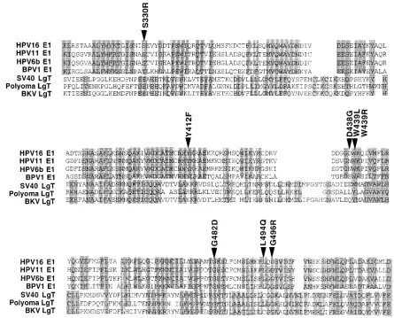
FIG. 7. Amino acid substitution mutations occur in residues conserved among the papillomavirus E1 and papovavirus LgT replication proteins. Alignment of regions conserved among the papillomavirus E1 proteins, as well as the LgT replication proteins of SV40, mouse polyomavirus, and BK virus, is shown. The positions and identities of missense mutations examined in this study are indicated (arrowheads).
VOL. 71, 1997 CHARACTERIZATION OF HPV16 E1 MISSENSE MUTANTS 5949
on November 9, 2019 by guest
http://jvi.asm.org/
S330R is a distinctive mutant for several reasons. First, S330R is wild type for all phenotypes tested, with the excep-tions of transient DNA replication and hUBC9 interaction. As noted above, this suggests but does not prove the possibility that hUBC9 interaction is important in HPV16 E1-mediated replication. The other distinctive trait of the S330R mutation is its dominant negative replication phenotype. S330R, when co-transfected with wild-type E1 and E2, inhibits transient HPV16 ori DNA replication. Certain preconditions must be met in order for a mutant protein to act in a negative transdominant manner. The protein must have at least two functional do-mains. In addition, these domains must be separable, allowing mutations in one domain that destroy that domain’s function while the other domain(s) retains its function. Thus, mutant proteins can be selected for by loss of one particular function, but retention of other activities may allow the mutant protein to titrate out other proteins or critical substrates (18).
The very existence of a transdominant replication-negative HPV16 E1 mutant indicates that these are likely properties of HPV16 E1. Recent evidence that papillomavirus E1 proteins form multiple homo- and heteromeric replicative complexes (14, 30, 31, 44), in addition to a number of other protein-protein complexes, suggests that such a mode of action might apply to the S330R dominant negative mutant. The integrity of these functions in S330R may allow this mutant HPV16 E1 protein to be incorporated into E1WT-E2 and/or E1WT oli-gomeric complexes. In this manner, the formation of higher-order complexes may be a prerequisite for the S330R domi-nant negative phenotype.
Several of the randomly derived HPV16 E1 mutations tested here occurred at amino acids that are well conserved among the papillomaviruses. In fact, the G482D, W439R, W439L, and G496R mutations occur at residues that are conserved be-tween the papillomavirus E1 proteins and SV40 LgT (Fig. 7). The G482D mutation, which affects all E1 phenotypes tested, is positioned at a well-conserved position within the putative ATP binding domain of E1. DNA replication and all protein-protein interactions and ATPase activity are disrupted by this mutation. It is possible that ATP binding or hydrolysis is re-quired for all of these E1 activities. However, it seems equally or more likely that the G482D mutation causes wholesale functional disruption of many HPV16 E1 domains by distort-ing the enzyme’s structure. In fact, nucleotide binddistort-ing itself might play an important role in the formation of active HPV16 E1 proteins: associated nucleotide might itself constitute a structural feature of the active HPV16 E1 protein.
Replication-defective dominant negative SV40 LgT mutants have been described, but their phenotypes have not been ex-plained in detail. However, all existing dominant negative LgT mutations reside in the ATP binding domain (4, 13). Two such LgT mutants, 5061 and 5062, contain short linker disruptions of the Gly-X4-Gly-Lys motif, the conserved ATP binding motif that is also altered in the HPV16 E1 G482D mutant. G482D ostensibly represents an analogous mutation in HPV16 E1, but this mutant does not exhibit a dominant negative phenotype. Previously studied mutations in this region of HPV16 E1 have documented the importance of prolines within the ATP bind-ing domain (38). S330R, the dominant negative mutant iden-tified and characterized here, lies outside the ATP binding motif domain of HPV16 E1. Thus, it appears that the HPV16 E1 S330R mutant exerts its dominant negative phenotype by a mechanism distinct from that of the known LgT dominant negative mutants.
Previous targeted mutagenesis studies of papillomavirus E1 have identified the nuclear localization, DNA binding, and ATP binding domains of BPV E1 (28). Generation and
char-acterization of the HPV16 E1 mutants described here consti-tutes the first effort to blindly scan a papillomavirus E1 protein for mutations that disrupt its function. Although the mutants tested here were produced by selection for loss of interaction with hUBC9 or 16E1-BP, many were also negative for several other E1 phenotypes. The observation that amino acid substi-tution mutations affecting one function of HPV16 E1 could also affect activities thought to reside in other functional do-mains suggests that E1 proteins are not composed of distinct, modular, functionally separable domains. To some extent, these results could be explained by multiple closely overlap-ping activities within the E1 protein or by a complex allosteric relationship between various E1 functional domains. Further biochemical analysis of these HPV16 E1 mutants, along with similar mutational analysis of other papillomavirus E1 pro-teins, will allow a greater understanding of the E1 protein’s role in the seminal events of papillomavirus DNA replication.
ACKNOWLEDGMENTS
This work was supported in part by a Sponsored Research Agree-ment to Harvard University from the Terumo Corporation of Japan. H.S. was supported by a JSPS fellowship. M.V. is the recipient of an American Cancer Society of Massachusetts award and thanks Ed Har-low for support.
REFERENCES
1. Belyavskyi, M., M. Westerman, L. DiMichele, and V. G. Wilson. 1996. Per-turbation of the host cell cycle and DNA replication by the bovine papillo-mavirus replication protein E1. Virology 219:206–219.
2. Belyavskyi, M., J. Miller, E. Belyavaskaya, and V. Wilson. 1996. BPV E1 protein alters the kinetics of cell cycle entry of serum starved mouse fibro-blasts. Cytometry 21:257–264.
3. Benson, J. D., and P. M. Howley. 1995. Amino-terminal domains of the bovine papillomavirus type 1 E1 and E2 proteins participate in complex formation. J. Virol. 69:4364–4372.
4. Bentivoglio, C. M., J. Zhu, and C. N. Cole. 1992. Mechanisms of interference with simian virus 40 (SV40) DNA replication by trans-dominant mutants of SV40 large T antigen. J. Virol. 66:4209–4219.
5. Betting, J., and W. Seufert. 1996. A yeast Ubc9 mutant protein with tem-perature-sensitive in vivo function is subject to conditional proteolysis by a ubiquitin- and proteasome-dependent pathway. J. Biol. Chem. 271:25790– 25796.
6. Blitz, I. L., and L. A. Laimins. 1991. The 68-kilodalton E1 protein of bovine papillomavirus is a DNA-binding phosphoprotein which associates with the E2 transcriptional activator in vitro. J. Virol. 65:649–656.
7. Blondel, M., and C. Mann. 1996. G2 cyclins are required for the degradation of G1 cyclins in yeast. Nature 384:279–282.
8. Boeke, J., F. Lacroute, and G. Fink. 1984. A positive selection for mutants lacking orotidine-59 phosphate decarboxylase activity in yeast. Mol. Gen. Genet. 197:345–346.
9. Bonne-Andrea, C., S. Santucci, P. Clertant, and F. Tillier. 1995. Bovine papillomavirus E1 protein binds specifically DNA polymerase alpha but not replication protein A. J. Virol. 69:2341–2350.
10. Bonne-Andrea, C., S. Santucci, and P. Clertant. 1995. Bovine papillomavirus E1 protein can, by itself, efficiently drive multiple rounds of DNA synthesis in vitro. J. Virol. 69:3201–3205.
11. Clertant, P., and I. Seif. 1984. A common function for polyoma virus large-T and papillomavirus E1 proteins? Nature (London) 311:276–279. 12. Delvecchio, A. M., H. Romanczuk, P. M. Howley, and C. C. Baker. 1992.
Transient replication of human papillomavirus DNAs. J. Virol. 66:5949– 5958.
13. Farber, J. M., K. C. Peden, and D. Nathans. 1987. trans-Dominant defective mutants of simian virus 40 T antigen. J. Virol. 61:436–445.
14. Gillette, T. G., M. Lusky, and J. A. Boroweic. 1994. Induction of structural changes in the bovine papillomavirus type 1 origin of replication by the viral E1 and E2 proteins. Proc. Natl. Acad. Sci. USA 91:8846–8850.
15. Gopalakarishan, V., and S. A. Khan. 1994. E1 protein of human papilloma-virus type 1a is sufficient for initiation of viral DNA replication. Proc. Natl. Acad. Sci. USA 91:9597–9601.
16. Go¨ttlicher, M., S. Heck, V. Doucas, E. Wade, M. Kullmann, A. C. B. Cato, R. M. Evans, and P. Herrlich.1996. Interaction of the Ubc9 human homolog with c-Jun and with the glucocorticoid receptor. Steroids 61:257–262. 17. Hateboer, G., E. M. Jijmans, J. B. D. Nooj, S. Schlenker, S. Jentsch, and R.
Bernards.1996. mUBC9, a novel adenovirus E1A-interacting protein that complements a yeast cell cycle defect. J. Biol. Chem. 271:25906–25911. 18. Herskowitz, I. 1987. Functional inactivation of genes by dominant negative
on November 9, 2019 by guest
http://jvi.asm.org/
mutations. Nature (London) 329:219–222.
19. Hirt, B. 1967. Selective extraction of polyoma DNA from infected mouse cell cultures. J. Mol. Biol. 26:365–369.
20. Hughes, F. J., and M. A. Romanos. 1993. E1 protein of human papilloma-virus is a DNA helicase/ATPase. Nucleic Acids Res. 21:5817–5823. 21. Jenkins, O., D. Earnshaw, G. Sarginson, A. Delvecchio, J. Tsai, H.
Kal-lender, B. Amegadzie, and M. Browne.1996. Characterization of the helicase and ATPase activity of human papillomavirus type 6b E1 protein. J. Gen. Virol. 77:1805–1809.
22. Jiang, W., and Y. Koltin. 1996. Two-hybrid interaction of a human UBC9 homolog with centromere proteins of Saccharomyces cerevisiae. Mol. Gen. Genet. 251:153–160.
23. Kaelin, W. G., W. Krek, W. R. Sellers, J. A. DeCaprio, F. Ajchenbaum, C. S. Fuchs, T. Chittenden, Y. Li, P. J. Farnham, and M. A. Blanar.1992. Ex-pression cloning of a cDNA encoding a retinoblastoma-binding protein with E2F-like properties. Cell 70:351–364.
24. Kingston, R. E., C. A. Chen, and H. Okayama. 1987. Introduction of DNA into mammalian cells, p. 9.1.1–9.1.9. In F. M. Ausubel, R. Brent, R. E. Kingston, D. D. Moore, J. G. Seidman, J. A. Smith, and K. Struhl (ed.), Current protocols in molecular biology. John Wiley & Sons, Inc., New York, N.Y.
25. Kovalenko, O. V., A. W. Plug, T. Haaf, D. K. Gonda, T. Ashley, D. C. Ward, C. M. Radding, and E. I. Golub.1996. Mammalian ubiquitin-conjugating enzyme Ubc9 interacts with Rad51 recombination protein and localizes in synaptonemal complexes. Proc. Natl. Acad. Sci. USA 93:2958–2963. 26. Lee, J. W., H.-S. Choi, J. Gyuris, R. Brent, and D. D. Moore. 1995. Two
classes of proteins dependent on either the presence or absence of thyroid hormone for interaction with the thyroid hormone receptor. Mol. Endocri-nol. 9:243–254.
27. LeMoal, M. A., M. Yaniv, and F. Thierry. 1994. The bovine papillomavirus type 1 (BPV1) replication protein E1 modulates transcriptional activation by interacting with BPV1 E2. J. Virol. 68:1085–1093.
28. Lentz, M. R., D. Pak, I. Mohr, and M. R. Botchan. 1993. The E1 replication protein of bovine papillomavirus type 1 contains an extended nuclear local-ization signal that includes a p34cdc2 phosphorylation site. J. Virol. 67:1414– 1423.
29. Lusky, M., and E. Fontane. 1991. Formation of the complex of bovine papillomavirus E1 and E2 proteins is modulated by E2 phosphorylation and depends upon sequences within the carboxyl terminus of E1. Proc. Natl. Acad. Sci. USA 88:6363–6367.
30. Lusky, M., J. Hurwitz, and Y.-S. Seo. 1993. Cooperative assembly of the bovine papilloma virus E1 and E2 proteins on the replication origin requires an intact E2 binding site. J. Biol. Chem. 268:15795–15803.
31. Lusky, M., J. Hurwitz, and Y.-S. Seo. 1994. The bovine papillomavirus E2 protein modulates the assembly of but is not stably maintained in a replica-tion-competent multimeric E1-replication origin complex. Proc. Natl. Acad. Sci. USA 91:8895–8899.
32. Melendy, T., J. Sedman, and A. Stenlund. 1995. Cellular factors required for papillomavirus DNA replication. J. Virol. 69:7857–7867.
33. Mohr, I., J. R. Clark, S. Sun, E. J. Androphy, P. MacPherson, and M. R. Botchan.1990. Targeting the E1 replication protein to the papillomavirus origin of replication by complex formation with the E2 transactivator. Sci-ence 250:1694–1699.
34. Monini, P. T., L. deLellis, P. Borgatti, M. Hassan-Omran, and E. Cassai. 1993. Activation of eukaryotic transcriptional promoters by the bovine pap-illomavirus E1-replication factor. Intervirology 36:245–252.
35. Mu¨ller, F., Y.-S. Seo, and J. Hurwitz. 1994. Replication of bovine papillo-mavirus type 1 origin-containing DNA in crude extracts and with purified proteins. J. Biol. Chem. 269:17086–17094.
36. Nallaseth, F. S., and M. L. DePamphillis. 1994. Papillomavirus contains cis-acting sequences that can suppress but not regulate origins of DNA replication. J. Virol. 68:3051–3064.
37. Park, P., W. Copeland, L. Yang, T. Wang, M. R. Botchan, and I. J. Mohr. 1994. The cellular DNA polymerasea-primase is required for papillomavirus DNA replication and associates with the viral E1 helicase. Proc. Natl. Acad. Sci. USA 91:8700–8704.
38. Raj, K., and M. A. Stanley. 1995. The ATP-binding and ATPase activities of human papillomavirus type 16 E1 are significantly weakened by the absence
of prolines in the ATP-binding domain. J. Gen. Virol. 76:2949–2956. 39. Romanczuk, H. R., and P. M. Howley. 1992. Disruption of either the E1 or
the E2 regulatory gene of human papillomavirus type 16 increases viral immortalization capacity. Proc. Natl. Acad. Sci. USA 89:3159–3163. 40. Sakai, H., T. Yasugi, J. D. Benson, J. J. Dowhanick, and P. M. Howley. 1996.
Targeted mutagenesis of the human papillomavirus type 16 E2 transactiva-tion domain reveals separable transcriptransactiva-tional activatransactiva-tion and DNA replica-tion funcreplica-tions. J. Virol. 70:1602–1611.
41. Sandler, A. B., S. B. VandePol, and B. A. Spalholz. 1993. Repression of bovine papillomavirus type 1 transcription by the E1 replication protein. J. Virol. 67:5079–5087.
42. Schnall, R., G. Mannhaupt, R. Stucka, R. Tauer, S. Ehnle, C. Schwarzlose, I. Vetter, and H. Feldmann.1994. Identification of a set of putative ATPases with high similarity to constituents of the 26S protease complex. Yeast 10:1141–1155.
43. Sedman, J., and A. Stenlund. 1995. Cooperative interaction between the initiator E1 and the transcriptional activator E2 is required for replication of bovine papillomavirus in vivo and in vitro. EMBO J. 14:6218–6228. 44. Sedman, J., and A. Stenlund. 1996. The initiator protein E1 binds to the
bovine papillomavirus origin of replication as a trimeric ring-like structure. EMBO J. 15:5085–5092.
45. Seo, Y.-S., F. Mu¨ller, M. Lusky, E. Gibbs, H.-Y. Kim, B. Phillips, and J. Hurwitz.1993. Bovine papilloma virus (BPV)-encoded E2 protein enhances binding of E1 protein to the BPV replication origin. Proc. Natl. Acad. Sci. USA 90:2865–2869.
46. Seo, Y.-S., F. Mu¨ller, M. Lusky, and J. Hurwitz. 1993. Bovine papilloma virus (BPV)-encoded E1 protein contains multiple activities required for BPV DNA replication. Proc. Natl. Acad. Sci. USA 90:702–706.
47. Seufert, W., B. Futcher, and S. Jentsch. 1995. Role of ubiquitin-conjugating enzyme in degradation of S- and M-phase cyclins. Nature 373:78–81. 48. Sverdrup, F., and S. A. Khan. 1994. Replication of human papillomavirus
(HPV) DNAs supported by the HPV type 18 E1 and E2 proteins. J. Virol. 68:505–509.
49. Thorner, L. K., D. A. Lim, and M. R. Botchan. 1993. DNA-binding domain of bovine papillomavirus type 1 E1 helicase: structural and functional as-pects. J. Virol. 67:6000–6014.
50. Ustav, M., and A. Stenlund. 1991. Transient replication of BPV-1 requires two viral polypeptides encoded by the E1 and E2 open reading frames. EMBO J. 10:449–457.
51. Vidal, M., P. Braun, E. Chen, J. D. Boeke, and E. Harlow. 1996. Genetic characterization of a mammalian protein-protein interaction domain by us-ing a yeast reverse two-hybrid system. Proc. Natl. Acad. Sci. USA 93:10315– 10320.
52. Vidal, M., R. K. Brachmann, A. Fattaly, E. Harlow, and J. D. Boeke. 1996. Reverse two-hybrid and one-hybrid systems to detect dissociation of protein-protein and DNA-protein-protein interactions. Proc. Natl. Acad. Sci. USA 93:10321– 10326.
53. Walker, J. E., M. Saraste, M. J. Runswick, and N. J. Gray. 1982. Distantly related sequences in the alpha- and beta-subunits of ATP synthase, myosin, kinases and other ATP-requiring enzymes and a common nucleotide binding fold. EMBO J. 1:945–951.
54. Wang, Z.-Y., Q.-Q. Qui, W. Seufert, T. Taguchi, J. R. Testa, S. A. Whitmore, D. F. Callen, D. Welsh, T. Shenk, and T. F. Deuel.1996. Molecular cloning of the cDNA and chromosome localization of the gene for human ubiquitin-conjugating enzyme 9. J. Biol. Chem. 271:24811–24816.
55. Yang, L., I. Mohr, E. Fouts, D. A. Lim, M. Nohaile, and M. Botchan. 1993. The E1 protein of bovine papillomavirus 1 is an ATP-dependent DNA helicase. Proc. Natl. Acad. Sci. USA 90:5086–5090.
56. Yang, L., R. Li, I. J. Mohr, R. Clark, and M. R. Botchan. 1991. Activation of BPV-1 replication in vitro by the transcription factor E2. Nature (London) 353:628–632.
57. Yasugi, T., and P. M. Howley. 1996. Identification of the structural and functional human homolog of the yeast ubiquitin conjugating enzyme UBC9. Nucleic Acids Res. 24:2005–2010.
58. Yasugi, T., J. D. Benson, H. Sakai, M. Vidal, and P. M. Howley. 1997. Mapping and characterization of the interaction domains of the human papillomavirus type 16 E1 and E2 proteins. J. Virol. 71:891–899.
VOL. 71, 1997 CHARACTERIZATION OF HPV16 E1 MISSENSE MUTANTS 5951