0022-538X/98/$04.0010
Copyright © 1998, American Society for Microbiology. All Rights Reserved.
Development of a Self-Inactivating Lentivirus Vector
HIROYUKI MIYOSHI, ULRIKE BLO¨ MER,† MASAYO TAKAHASHI,‡
FRED H. GAGE,ANDINDER M. VERMA*
Laboratory of Genetics, The Salk Institute for Biological Studies, La Jolla, California 92037
Received 30 April 1998/Accepted 13 July 1998
We have constructed a new series of lentivirus vectors based on human immunodeficiency virus type 1 (HIV-1) that can transduce nondividing cells. The U3 region of the 5* long terminal repeat (LTR) in vector constructs was replaced with the cytomegalovirus (CMV) promoter, resulting in Tat-independent transcription but still maintaining high levels of expression. A self-inactivating (SIN) vector was constructed by deleting 133 bp in the U3 region of the 3*LTR, including the TATA box and binding sites for transcription factors Sp1 and NF-kB. The deletion is transferred to the 5*LTR after reverse transcription and integration in infected cells, resulting in the transcriptional inactivation of the LTR in the proviruses. SIN viruses can be generated with no significant decreases in titer. Injection of viruses into the rat brain showed that a SIN vector containing the green fluorescent protein gene under the control of the internal CMV promoter transduced neurons as efficiently as a wild-type vector. Interestingly, a wild-type vector without an internal promoter also successfully transduced neurons in the brain, indicating that the HIV-1 LTR promoter is transcriptionally active in neurons even in the absence of Tat. Furthermore, injection of viruses into the subretinal space of the rat eye showed that wild-type vector transduced predominantly retinal pigment epithelium and photoreceptor cells, while SIN vector was able to transduce other types of retinal cells, including bipolar, Mu¨ller, horizontal, and amacrine cells. This finding suggests that the HIV-1 LTR can negatively influence the internal CMV promoter in some cell types. SIN HIV vectors should be safer for gene therapy, and they also have broader applicability as a means of high-level gene transfer and expression in nondividing cells.
Gene therapy approaches rely on efficient transfer of genes to the desired target cells (for reviews, see references 12, 32, 34, 35, and 48). A wide variety of viral and nonviral vectors have been developed and evaluated for their efficiency of transduction, sustained expression of the transgene, and safety. Among them, retrovirus vectors derived from oncoretroviruses such as murine leukemia virus (MLV) have been the most widely used for gene therapy applications. However, a major problem with these retrovirus vectors is the requirement for proliferation of the target cells for integration, limiting their use for gene transfer into nondividing cells such as hepato-cytes, myoblasts, neurons, and hematopoietic stem cells. In contrast, lentiviruses such as human immunodeficiency virus type 1 (HIV-1) can infect nondividing cells (7, 30, 49).
We have recently developed a lentivirus vector based on HIV-1 that can transduce nondividing cells in vitro and in vivo (38). These HIV vectors are pseudotyped with the vesicular stomatitis virus G glycoprotein (VSV-G); hence they can trans-duce a broad range of tissues and can be concentrated to high titers. We have shown that HIV vectors can stably integrate into the host cell genome and obtained long-term expression of transgenes in brain, liver, muscle, and retina (4, 24, 33, 38, 39). No cellular immune response can be detected at the site of injection. Furthermore, second injection of the HIV vector
into the animals is possible, indicating the lack of any potent humoral immune response to the vector (24).
Although HIV vectors promise great utility for gene therapy, there is concern about their safety since HIV-1 is the etiologic agent of AIDS. The major safety concern is the generation of replication-competent virus during the production of vectors. In this regard, we have minimized the possibility for generating replication-competent virus through recombination by using a three-plasmid expression system which consists of packaging, envelope, and vector constructs (38, 39). Furthermore, recent studies have demonstrated the possibility of eliminating all accessory genes (vif, vpr, vpu, and nef) from a packaging con-struct without losing the ability to transduce nondividing cells (24, 26, 43, 56).
Another safety concern about HIV vectors, as for MLV-based vectors, is the possibility of insertional activation of cellular oncogenes by random integration of the vector provi-rus into the host genome. To overcome this problem, we have constructed a self-inactivating (SIN) vector in which the viral enhancer and promoter sequences have been deleted. In this report, we show that SIN vectors can be generated and trans-duce nondividing cells in vivo with an efficacy similar to that of wild-type vectors. The transcriptional inactivation of the long terminal repeat (LTR) in the SIN provirus should prevent mobilization by replication-competent virus. This should also enable the regulated expression of genes from internal pro-moters by eliminating any cis-acting effects of the LTR. A further modification has been made in the vector construct in which the U3 region of the 59LTR has been replaced with the cytomegalovirus (CMV) promoter, resulting in Tat-indepen-dent transcription with no decreases in viral titer. SIN vectors combined with this hybrid 59LTR further reduce the possibil-ity of recombination to generate replication-competent virus because there is no complete U3 sequence in the virus
pro-* Corresponding author. Mailing address: Laboratory of Genetics, The Salk Institute for Biological Studies, P.O. Box 85800, San Diego, CA 92186-5800. Phone: (619) 453-4100, ext. 1462. Fax: (619) 558-7454. E-mail: [email protected].
† Present address: Department for Neurosurgery, Medical School Hannover, 30625 Hannover, Germany.
‡ Present address: Department of Ophthalmology and Visual Sci-ences, Graduate School of Medicine, Kyoto University, Sakyo-ku, Kyoto 606, Japan.
8150
on November 9, 2019 by guest
http://jvi.asm.org/
duction system. These modifications add additional safety fea-tures to the HIV-1-based lentivirus vector system.
MATERIALS AND METHODS
Plasmid constructions and preparation of viral stocks.Plasmid pHR9-MCS was constructed by replacing a 420-bp BamHI-XhoI fragment of pHR9(38) with a fragment of multiple cloning sites containing BamHI, Eco47III, BstXI, SacII,
PstI, EcoRI, ClaI, XbaI, HpaI, and XhoI sites. To restructure the HIV-1 LTR and
generate the SIN LTR, PCR amplification products of pHR9-MCS, an
MluI-BspEI (2454 to2145) and a BspEI-ApaI (2144 to 1180) fragment or an
MluI-BspEI (2454 to2145) and a BspEI-ApaI (28 to1180) fragment, were cloned into pcDNA3.1/Zeo(1) (Invitrogen) between the MluI and ApaI sites to generate pHIV-LTR or pHIV-SIN, respectively (numbers in parentheses indi-cate positions relative to the transcriptional start site). To construct HIV vectors pLL and pLS, a 2.3-kb BspEI fragment of pHR9-MCS was cloned into the BspEI sites of pHIV-LTR and pHIV-SIN, respectively. pLS has a 133-bp deletion (2141 to29) in the 39LTR. pHIV-DU3L and pHIV-DU3S were constructed by replacing a MluI-KasI (2454 to1183) fragment of pLL and pLS, respectively, with a MluI-KasI PCR amplification product (213 to1183) to delete the U3 region of the 59LTR. To generate the hybrid 59LTR with the CMV promoter, pCL and pCS were constructed by inserting a PvuI-SacI fragment containing the CMV promoter from pcDNA3.1/Zeo(1) into the PvuI and MluI sites of
pHIV-DU3L and pHIV-DU3S, respectively. The hybrid 59LTR maintains the 23-bp distance between the TATA box of CMV promoter and the transcription start site in HIV-1 LTR. pLL-CG, pLS-CG, pCL-CG, and pCS-CG were constructed by inserting a 1.3-kb BamHI-XhoI fragment of pKS-CMV-GFP (33) containing the CMV-green fluorescent protein (GFP) expression cassette into the same sites of pLL, pLS, pCL, and pCS, respectively. pLL-G, pCL-G, and pCS-G were constructed by deleting the CMV promoter from pLL-CG, pCL-CG, and pCS-CG, respectively.
The VSV-G-pseudotyped HIV vectors were generated by transient cotrans-fection of a vector construct (15 mg) with the VSV-G-expressing construct pMD.G (5mg) and the packaging construct pCMVDR8.2 (10mg) into 293T cells as previously described (39). High-titer stocks of HIV vectors were prepared by ultracentrifugation. The titers of viruses were determined by infection of 293T cells or HeLa-CD4-LTR-b-gal cells (27), seeded in six-well plates at 105cells/well the day before infection, with serial dilutions of the vector stocks. After overnight incubation, the cell culture medium was changed; 48 h later, the number of GFP-positive cells was scored to quantify the titer. The vector stocks were assayed for p24 antigen levels by using an HIV-1 p24 enzyme-linked immunosorbent assay (ELISA) kit (DuPont NEN). After concentration by ultracentrifugation, titers of 13109to 23109transducing units (TU)/ml were usually obtained. Absence of replication-competent virus was determined by the marker rescue assay and mea-suring p24 antigen level by ELISA as described elsewhere (39).
Southern and Northern blot analyses.Genomic DNA was isolated from cells infected with virus by the procedure of Wu et al. (51). DNA (10mg) was digested with the restriction enzymes BspEI and BamHI, separated on a 0.7% agarose gel, and transferred to a Hybond-N1membrane (Amersham) in 203SSC (13SSC is 0.15 M NaCl plus 0.015 M sodium citrate). Total cellular RNA was isolated from virus-infected cells by using an RNAqueous phenol-free total-RNA isola-tion kit (Ambion). RNA (10mg) was separated on a 1% agarose-formaldehyde gel and transferred to a Hybond-N1membrane in 25 mM potassium phosphate (pH 6.5). Membranes were UV irradiated with a UV cross-linker (Hoefer), hybridized with32P-labeled probes generated by a Megaprime DNA labeling kit (Amersham) in Rapid-hyb buffer (Amersham) for 2 h at 65°C, washed twice in 0.13SSC–0.1% sodium dodecyl sulfate for 15 min at 65°C, and exposed to an X-Omat AR X-ray film (Kodak). The BN and GFP probes were isolated from pCL-CG as 0.43-kb BssHII-NotI and 0.75-kb Eco47III-XhoI fragments, respec-tively.
In vivo experiments.High-titer stocks of HIV vectors (13109to 23109 TU/ml) were used to inject the brain and eye. The titers of CL-G and CS-G vectors were normalized for HIV-1 p24 antigen levels. Adult female Fischer 344 rats were anesthetized by intramuscular injection of ketamine-acepromazine-xylazine. Three microliters of vector stock (33106TU) was injected into the striatum and the hippocampus bilaterally (n54 for each vector) with a 5-ml Hamilton syringe. Rats were sacrificed at 2 and 6 weeks postinjection.
Fischer 344 rat pups (age 2 to 5 days) were anesthetized by chilling on ice for 5 min. The eyeball was exposed by an incision in the eyelid, parallel to the edge of the open eyelid. Subretinal injections were performed under an operating microscope. A 0.5-ml volume of vector stock (106TU) was injected into the subretinal space (n58 for each vector), using a glass capillary connected with 5-ml Hamilton syringe. Rats were sacrificed at 6 and 12 weeks postinjection.
Rats were intracardially perfused with cold fixing solution (4% paraformalde-hyde and 0.2% glutaraldeparaformalde-hyde in 0.1 M phosphate buffer). The tissues were removed, further fixed in the fixing solution, and cryoprotected in 30% sucrose at 4°C overnight. The tissues were then frozen in Tissue-Tek O.C.T. compound (Sakura Finetek) on dry ice and sectioned on a sliding microtome (50-mm-thick brain sections) or a cryostat (20-mm-thick eye sections). Immunofluorescence staining was performed as previously described (4). The sections were analyzed by confocal laser scanning microscopy (Bio-Rad). The signals were collected, digitally color enhanced, and superimposed.
RESULTS
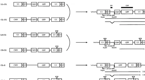
Construction and generation of modified HIV vectors.In the life cycle of retroviruses, the U3 region of the 39 LTR is duplicated to form the corresponding region of the 59 LTR during the process of reverse transcription and viral DNA synthesis in infected cells. This mechanism of viral replication allowed us to make two principal modifications to the HIV vector, as shown in Fig. 1. First, the U3 region of the 59LTR was replaced with the CMV promoter. Second, 133 bp in the U3 region of the 39LTR, which contains the TATA box and binding sites for transcription factors Sp1 and NF-kB, were deleted. This deletion will be transferred to the 59LTR after reverse transcription. Consequently, the transcriptional unit from the LTRs in a provirus is eliminated. This type of vector is called a SIN vector (54).
A series of vectors termed LL-CG, LS-CG, CL-CG, and CS-CG (Fig. 1) were constructed to determine whether these modifications would alter the titer of virus generated from the vectors. All vectors contain the GFP gene as a reporter with an internal CMV promoter. CL-CG and CS-CG vectors contain the CMV promoter in place of the U3 region of the 59LTR. The virus generated from CL-CG and CS-CG should contain the same RNA genome as the virus from LL-CG and LS-CG, respectively (Fig. 1). We also constructed CL-G and CS-G vectors without an internal promoter, in which the GFP gene is under the control of the 59LTR promoter.
VSV-G-pseudotyped vectors were generated by transient cotransfection of each vector construct with a VSV-G expres-sion construct and a packaging construct into 293T cells. The vector virus was harvested 62 h after transfection and used to infect 293T cells to determine the viral titer by quantitation of the number of GFP-positive cells. The LL-CG vector yielded a mean titer of 63105TU/ml. As shown in Table 1, replacement
of the U3 region of the 59 LTR with the CMV promoter did not reduce the viral titers. The titers of SIN vectors LS-CG and CS-CG were approximately the same as those of their wild-type counterparts, LL-CG and CL-CG, respectively. These re-sults indicate that the small deletion in the U3 region in SIN vectors did not significantly affect titers. Lower titers of CL-G may reflect the relative inefficiency of the LTR promoter be-cause of the absence of the trans activator Tat in the infected cells. A few cells were scored positive for GFP with the CS-G vector, probably due to integration events near host genome promoters since there is no promoter in the provirus. To mea-sure viral production with CL-G and CS-G vectors, the levels of HIV-1 p24 antigen in the supernatants were measured, as these correlate with the amount of virions. The levels of p24 obtained with the CL-G and CS-G vectors were roughly equivalent to those obtained with other vectors, suggesting ap-proximately similar levels of virus production with all vectors (Table 1). However, we cannot rule out the possibility of gen-erating virions lacking viral genome RNA with this assay.
Expression from the hybrid 5*LTR with the CMV promoter is Tat independent.It has been shown that Tat trans activation of the HIV-1 LTR requires not only the Tat-responsive region (TAR) in the R region of the LTR but also the TATA box and binding sites for Sp1 and NF-kB (2, 3, 17, 22, 31, 36, 41, 55). To verify that the hybrid CMV-LTR promoter is Tat independent, the promoter activity of the CL-G vector construct was com-pared with that of construct LL-G, in which the internal CMV promoter was deleted from LL-CG and therefore the GFP gene was expressed under the control of wild-type 59 LTR. These constructs were transfected into 293T or HeLa cells with either a Tat expression plasmid or a control plasmid, and levels of GFP in cell extracts were measured 48 h later by
fluores-VOL. 72, 1998 DEVELOPMENT OF A SELF-INACTIVATING LENTIVIRUS VECTOR 8151
on November 9, 2019 by guest
http://jvi.asm.org/
cence spectroscopy (40). Table 2 shows that the GFP expres-sion in cells transfected with LL-G was increased three- to sixfold in the presence of Tat. The low level of trans activation by Tat in 293T cells is probably caused by the higher level of expression from the LTR due to trans activation by the ade-novirus E1A gene product expressed in 293T cells (37). On the other hand, CL-G was not responsive to Tat, though levels of expression were similar to those obtained with LL-G in the presence of Tat. Thus, the replacement of U3 region of the 59 LTR with the CMV promoter resulted in loss of Tat respon-siveness, but the basal activity was comparable to that of wild-type LTR in the presence of Tat in 293T and HeLa cells.
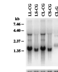
Characterization of proviral structure and transcription. The structures of proviruses were characterized by Southern blot analysis. HeLa-CD4-LTR-b-gal cells (27) were infected with each vector and cultured for 36 days with 11 passages. Genomic DNA was isolated from infected cells, digested with the restriction enzymes BspEI and BamHI, and hybridized with a BN probe (Fig. 2). The predicted structures of proviruses are shown in Fig. 1. BspEI cleaves once in each LTR, immediately upstream of the deleted region in SIN vectors, and BamHI cleaves once downstream of the 59LTR. Therefore, a 1.69-kb
BspEI-BamHI fragment should be generated from provirus
containing wild-type 59 LTR when hybridized with the BN probe. If the 133-bp deletion present in the 39 LTR of SIN vector is transferred to the 59LTR of the integrated provirus, a 1.56-kb BspEI-BamHI fragment should be generated in place of the 1.69-kb fragment. As shown in Fig. 2, the expected fragments were generated from each provirus. Analysis of in-fected 293T cells yielded identical results (data not shown).
We next analyzed the transcriptional activities of proviruses. Total cellular RNA was isolated from HeLa-CD4-LTR-b-gal cells infected with each virus described above, and Northern blot analysis was performed with the GFP gene as a probe. As shown in Fig. 3, expression of the expected transcripts depicted in Fig. 1 was observed for each of the proviruses. In the pro-virus derived from the LL-CG or CL-CG vector, three tran-scripts were produced: two viral trantran-scripts initiated in the 59 LTR, a 3.60-kb full-length form and a 2.44-kb spliced form; and a 1.48-kb transcript initiated in the internal CMV pro-moter. All transcripts terminated at the polyadenylation signal in the R region of the 39 LTR. The full-length and spliced forms were confirmed by using the BN probe, which detects only the full-length form (data not shown; see Fig. 1 for struc-tures). The level of transcripts expressed from the internal CMV promoter is higher than that of transcripts initiated in FIG. 1. Structures of HIV vector constructs and corresponding proviruses. (Left) HIV vector constructs. Each vector construct is cotransfected with the packaging and VSV-G expression constructs into 293T cells. Viral transcription initiates at the U3/R border in the 59LTR and terminates at the R/U5 border in the 39LTR. The viral RNA is packaged into virions. Virus is harvested and used to infect target cells. Triangles represent deleted U3 region. (Right) Structures of integrated proviruses. In infected cells, the U3 region of the 39LTR is used as a template for the synthesis of the U3 region in both LTRs during the process of reverse transcription of the viral RNA into double-stranded DNA. As a result, the U3 region of the 39LTR is duplicated and transferred to the 59 LTR in the integrated provirus. The
BspEI-BamHI restriction fragments expected in Southern blot analysis (Fig. 2) and the RNA transcripts expected in Northern blot analysis (Fig. 3) are shown below
[image:3.612.52.546.69.342.2]each proviral structure with their sizes. The locations of the BN and GFP probes used in Southern blot and Northern blot analyses are also indicated.
TABLE 1. Titers of HIV vectors
Vector Titer on 293T cells(TU/ml)a Relative p24levelsb
LL-CG 5.73105 1.00
CL-CG 9.33105 1.42
LS-CG 5.03105 0.56
CS-CG 8.23105 1.39
CL-G 2.13104 1.51
CS-G ,10 1.26
aAverage of at least three independent experiments. bRelative to the p24 level of the LL-CG vector.
on November 9, 2019 by guest
http://jvi.asm.org/
the 59 LTR. In the case of SIN vectors LS-CG and CS-CG, transcripts expressed from the 59LTR were undetectable and a 1.35-kb transcript expressed from the internal promoter was detected. This transcript is shorter than the corresponding transcript from the LL-CG or CL-CG provirus because of the 133-bp deletion in the U3 region of the 39LTR. The provirus derived from the CL-G vector produced detectable levels of the full-length and spliced viral transcripts. As expected, no RNA transcripts were detected in the cells infected with the CS-G vector. The same results were obtained with 293T cells infected with each virus (data not shown).
Rescue of virus from cells infected with SIN vectors. To determine if there was any transcriptional activity from the 59 LTR of the SIN provirus, we used a more sensitive virus rescue assay. The VSV-G-expressing construct and the packaging construct were transfected into 293T cells previously infected with SIN vectors. If RNA was initiated in the 59 LTR of the SIN provirus, it would be packaged into viral particles and released into the medium. Such virus would then be detected by infection of virgin 293T cells. The results showed that 3 to 8 TU of virus per ml was rescued from cells previously infected with the LS-CG or CS-CG vector, whereas 53104to 83104
TU/ml was rescued from cells infected with the LL-CG or CL-CG vector (Table 3). It is unlikely that the rescued virus arose through recombination during transfection and regener-ated a functional 59 LTR because there is no complete U3 sequence, especially in the case of CS-CG. In fact, the virus preparations used for infection were shown to be free of
rep-lication-competent virus by the marker rescue assay (39). Vi-ruses were able to be rescued presumably because SIN vectors integrated near an active cellular promoter and expressed the proviral genome, as seen with the CS-G vector (Table 1). On the basis of Northern blot analysis and virus rescue, it is ap-parent that effective transcription from the 59LTR of the SIN provirus was inactivated.
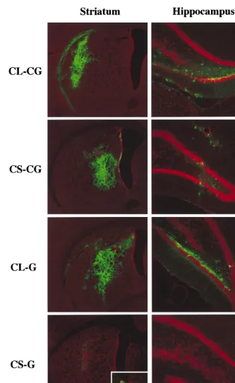
In vivo delivery of modified vectors.To transduce terminally differentiated neurons in vivo, high-titer stocks (13109to 23
109TU/ml) of the CS-CG and CL-CG vectors were injected
into the striatum and hippocampus of adult rat brains. The CL-G and CS-G vectors, normalized for equal amounts of p24 antigen, were also injected to detect the expression from the 59 LTR of wild-type and SIN vectors. At 2 and 6 weeks postin-jection, the rats were sacrificed and the brains were cryosec-tioned and analyzed for GFP expression by fluorescence mi-croscopy. As shown in Fig. 4, the CS-CG vector was found to transduce at efficiencies comparable to those of the CL-CG vector. Interestingly, the CL-G vector gave similar transduc-tion efficiencies as well. This observatransduc-tion is consistent with previous studies showing high levels of expression from the HIV-1 LTR through constitutively active NF-kB in neurons (10, 25, 42). In brains injected with the CS-G vector, very few GFP-positive cells could be detected, and only at higher mag-nification. This was most likely due to proviral integration near a cellular promoter and expression of GFP, as seen in infection in vitro. There were no significant differences in the GFP
FIG. 2. Southern blot analysis of the integrated proviral structure. Genomic DNA isolated from HeLa-CD4-LTR-b-gal cells infected with HIV vectors was digested with BspEI and BamHI. The blot was hybridized with the BN probe. The vector used for infection is indicated above each lane. Control, uninfected HeLa-CD4-LTR-b-gal cells. The expected sizes of fragments are shown in Fig. 1. Size markers are indicated on the left.
[image:4.612.371.486.70.213.2]FIG. 3. Northern blot analysis of the proviral expression. Total cellular RNA was isolated from HeLa-CD4-LTR-b-gal cells infected with HIV vectors. The blot was hybridized with the GFP probe and rehybridized with the human glyceraldehyde-3-phosphate dehydrogenase (G3PDH) probe (Clontech). The vector used for infection is indicated above each lane. Control, uninfected HeLa-CD4-LTR-b-gal cells. The expected size of each transcript is shown in Fig. 1. Size markers are indicated on the left.
TABLE 2. Transcriptional activities of LL-G and CL-G constructsa
Construct Tat Relative GFP expression
293T HeLa
pLL-G 2 1.0 1.0
1 3.0 6.2
pCL-G 2 4.9 7.3
1 3.9 9.4
a293T or HeLa cells were seeded at a concentration 2.53105/six-well dish and transfected on the following day with 0.5mg of vector construct together with 0.5mg of the Tat expression plasmid pSV2 tat72 (16) or the control plasmid pBluescript II (Stratagene), using Lipofectamine (GibcoBRL). Cells were har-vested 48 h after transfection, and the levels of GFP were measured by fluores-cence spectroscopy. The results are expressed relative to the GFP level of pLL-G in the absence of Tat and are means of triplicate transfections.
TABLE 3. Titers of viruses rescued by transient transfection of packaging and VSV-G constructsa
Vector Titer on 293T cells (TU/ml)
Expt 1 Expt 2
LL-CG 73104 53104
CL-CG 83104 73104
LS-CG 3 8
CS-CG 3 3
aThe VSV-G expression construct pMD.G (5mg) and packaging construct
pCMVDR8.2 (15mg) were transfected into 293T cells previously infected with each vector. Virus stocks were prepared 48 h after transfection and used to infect 293T cells. Titers were measured by counting the number of GFP-positive cells 3 days after infection.
VOL. 72, 1998 DEVELOPMENT OF A SELF-INACTIVATING LENTIVIRUS VECTOR 8153
on November 9, 2019 by guest
http://jvi.asm.org/
[image:4.612.51.290.83.154.2]transduction frequencies of the vectors between 2 and 6 weeks postinjection (data not shown). The nature of transduced cells was determined by immunofluorescence staining with three markers: NeuN for neurons, glial fibrillary acidic protein for astrocytes, and RIP for oligodendrocytes. The results showed that the majority of cells transduced with each vector were terminally differentiated neurons expressing NeuN, consistent
with our previous observation (4, 38, 39). Additional immuno-fluorescence staining showed that cells transduced with the CS-CG vector were frequently labeled with choline acetyl-transferase, which is a marker of cholinergic neurons (data not shown). In contrast, the CL-CG vector-transduced cells were rarely labeled with this marker.
[image:5.612.134.467.67.609.2]The CL-CG and CS-CG vectors were also injected into the
FIG. 4. Expression of GFP in the striatum and hippocampus of adult rat brains 6 weeks after injection of HIV vectors. Sections were counterstained with propidium iodide. The vectors used for injection are indicated on the left. Inset, higher magnification of a GFP-positive cell.
on November 9, 2019 by guest
http://jvi.asm.org/
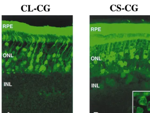
subretinal space of rat eyes, and transduction was evaluated 6 and 12 weeks later. Retinal cell types can be readily identified by their positions in the different retinal layers and by their distinctive morphologies. In the eyes injected with the wild-type vector CL-CG, GFP was predominantly expressed in ret-inal pigment epithelium and photoreceptor cells (Fig. 5A). On the other hand, the SIN vector CS-CG efficiently transduced not only retinal pigment epithelium and photoreceptor cells but also other retinal cells in inner nuclear layer, including bipolar, Mu¨ller, horizontal, and amacrine cells (Fig. 5B). This result together with that for the brain suggests that transcrip-tion from the HIV-1 LTR may have inhibitory effects on tran-scription from the internal promoter in some cell types. There were no significant differences in the patterns and transduction frequencies of the vectors between 6 and 12 weeks postinjec-tion (data not shown).
DISCUSSION
HIV-1, unlike other retroviruses such as MLV, is character-ized by a complex genome that encodes two regulatory pro-teins (Tat and Rev) and four accessory propro-teins (Vif, Vpr, Vpu, and Nef) in addition to the common gag, pol, and env gene products. One of the accessory proteins, Tat, is an essen-tial nuclear protein that augments levels of viral RNA by in-creasing transcriptional initiation and/or elongation (reviewed in reference 23). To manifest its function, Tat binds to a nas-cent RNA stem-loop structure of TAR, located at the 59end of all viral transcripts. Tat trans activation has also been reported to require the TATA box and additional sequences in the 59 LTR, including binding sites for Sp1 and NF-kB (2, 3, 17, 22, 31, 36, 41, 55). This is consistent with our result that replace-ment of the U3 region of the 59LTR with the CMV promoter resulted in Tat-independent transcription (Table 2). In the absence of Tat, this hybrid CMV-LTR promoter can drive high levels of expression comparable to those of the wild-type LTR in the presence of Tat. This allows production of HIV vectors in a system devoid of Tat. Indeed, Kim et al. (26) have recently reported that an HIV vector containing a similar promoter swap could generate virus with no significant decrease in titer in the absence of Tat. Recent studies have demonstrated the possibility of eliminating all accessory genes from a packaging
construct without losing the ability to transduce nondividing cells (24, 26, 43, 56), although some accessory proteins ap-peared to be required for maximum transduction of specific tissues (e.g., Vpr for macrophages and Vpr and/or Vif for liver) (24, 56). Taken together, these observations suggest the pos-sibility of eliminating all regulatory and accessory genes except
rev from the HIV vector production system. Rev, together with
the Rev response element in the vector, is required for efficient export of full-length vector transcripts to the cytoplasm. How-ever, it may be possible to substitute Rev function by replace-ment with a protein serving a similar function (28).
The transcription of HIV-1 is directed by regulatory se-quences in the 59 LTR. The core element in the U3 region, containing a canonical TATA box and three Sp1 binding sites, is essential for basal promoter activity and viral replication (2, 3, 17, 22, 41, 55). Immediately upstream of the Sp1 binding sites, tandem binding sites for NF-kB constitute an activation-dependent enhancer element (31, 36). Elimination of all Sp1 and NF-kB binding sites results in essentially total inactivation of viral replication (29, 44). Although a number of enhancer elements that modulate transcriptional activity have been iden-tified upstream of the NF-kB binding sites, none of them can compensate for the loss of Sp1 and NF-kB binding sites. In the SIN HIV vector described in this study, as expected, the dele-tion of the TATA box and binding sites for Sp1 and NF-kB resulted in transcriptional inactivation of the LTR in the pro-virus in infected cells in vitro and in vivo. A notable aspect of the expression from the HIV-1 LTR is its low basal activity in the absence of Tat. In addition, Tat is not packaged into viri-ons. Therefore, in most cells infected with HIV vectors, low levels of expression from the LTR would be expected because of the absence of Tat. However, we have demonstrated high-level expression from the LTR in the brain (Fig. 4), consistent with the finding that transgenic mice expressed an HIV-1 LTR-driven reporter gene at high levels in the brain (10). It has been reported that strong HIV-1 LTR promoter activity in neurons is mainly due to the presence of constitutively active NF-kB (25, 42). One safety concern about HIV vectors is the activa-tion of proto-oncogenes through the LTR resulting from ran-dom integration of provirus into the host genome. In this regard, the SIN vector should reduce the potential of inser-tional activation. The transcripinser-tional inactivation of the LTR in the SIN provirus should also prevent mobilization by repli-cation-competent virus and minimize any risk of vector virus spread. It can also be argued that the rearranged viruses may arise through recombination during transfection and regener-ate a wild-type LTR. Since SIN vectors combined with the hybrid CMV-LTR promoter (e.g., CS-CG) contain a single U3 region with a deletion, there is no complete U3 sequence in the virus production system: recombination to regenerate a wild-type U3 is not possible with such vectors. Furthermore, this combined modification minimizes the risk of recombination to generate replication-competent virus, although we have not detected replication-competent virus so far.
[image:6.612.51.292.68.249.2]The application of MLV-based SIN vectors has been lim-ited, since deletion of the TATA box resulted in low titers (9, 20, 21, 45, 53, 54). This is probably because the region of the TATA box in the 39LTR plays an important role in processing of the 39 end of the viral RNA through secondary-structure interactions. Therefore, most SIN MLV vectors had a deletion only in the enhancer region, and transcriptional inactivation of the LTR was relatively inefficient (9, 20, 45, 54). In HIV-1, sequences within the U3 region, especially between the TATA box and the transcription initiation site, have been shown to be required for efficient 39 end processing (6, 8, 13, 18, 46, 47). These sequences direct the stable binding of cleavage and
FIG. 5. Expression of GFP in the retina of rat pups 12 weeks after injection of the CL-CG vector (A) and the CS-CG vector (B). Inset, higher magnification of GFP-positive inner nuclear layer (INL) cells. Scale bars, 10mm. RPE, retinal pigment epithelium; ONL, outer nuclear layer.
VOL. 72, 1998 DEVELOPMENT OF A SELF-INACTIVATING LENTIVIRUS VECTOR 8155
on November 9, 2019 by guest
http://jvi.asm.org/
polyadenylation specificity factor, the factor responsible for recognition of the AAUAAA hexamer, to the poly(A) site and enhance the efficiency of 39-end processing (19). However, Northern blot analysis of RNA from 293T cells transfected with the SIN vector construct showed no decreased levels of transcripts (data not shown) compared with the wild-type vec-tor, consistent with no significant reduction in viral titer (Table 1). This finding suggests that the deletion of U3 sequences in the SIN vector may not affect the efficiency of 39-end process-ing.
The results obtained from in vivo experiments revealed that the SIN vector can improve the expression of transgene in cholinergic neurons compared to the wild-type vector. Simi-larly, additional retinal cell types also showed high levels of transgene expression with the SIN vector (Fig. 5). It seems likely that the expression from the internal CMV promoter is negatively influenced by expression from the HIV-1 LTR in some cell types. This phenomenon, called transcriptional in-terference, has been reported for other retrovirus vectors (1, 5, 11, 14, 15, 45, 50, 52). Our results suggest that deletion of the U3 region of the LTR in HIV vectors may result in efficient expression from internal promoters in some tissues. Further-more, SIN vectors should be particularly useful to introduce tissue-specific or regulatable promoters, since the LTR can have no cis-acting influence on these internal promoters. SIN HIV vectors should not only increase the safety of HIV vector-mediated gene therapy but also have general utility for high-efficiency transduction of genes into nondividing cells.
ACKNOWLEDGMENTS
We are grateful to N. Somia and T. Kafri for critical reading of the manuscript. We thank members of the Verma and Gage laboratories for helpful suggestions.
H.M. was supported by a Uehara Memorial Foundation fellowship, and M.T. was supported by a Nippon Eye Bank Association fellowship. This work was supported by grants from the National Institutes of Health and Frances Berger Foundation. I.M.V. is an American Cancer Society Professor of Molecular Biology.
REFERENCES
1. Armentano, D., S.-F. Yu, P. W. Kantoff, T. V. Ruden, W. F. Anderson, and E.
Gilboa.1987. Effect of internal viral sequences on the utility of retroviral vectors. J. Virol. 61:1647–1650.
2. Berkhout, B., A. Gatignol, A. B. Rabson, and K.-T. Jeang. 1990. TAR-independent activation of the HIV-1 LTR: evidence that tat requires specific regions of the promoter. Cell 62:757–767.
3. Berkhout, B., and K.-T. Jeang. 1992. Functional roles for the TATA pro-moter and enhancers in basal and tat-induced expression of the human immunodeficiency virus type 1 long terminal repeat. J. Virol. 66:139–149. 4. Blo¨mer, U., L. Naldini, T. Kafri, D. Trono, I. M. Verma, and F. H. Gage.
1997. Highly efficient and sustained gene transfer in adult neurons with a lentivirus vector. J. Virol. 71:6641–6649.
5. Bowtell, D. D. L., S. Cory, G. R. Johnson, and T. J. Gonda. 1988. Comparison of expression in hemopoietic cells by retroviral vectors carrying two genes. J. Virol. 62:2464–2473.
6. Brown, P. H., L. S. Tiley, and B. R. Cullen. 1991. Efficient polyadenylation within the human immunodeficiency virus type 1 long terminal repeat re-quires flanking U3-specific sequences. J. Virol. 65:3340–3343.
7. Bukrinsky, M. I., N. Sharova, M. P. Dempsey, T. L. Stanwick, A. G.
Bukrin-skaya, S. Haggerty, and M. Stevenson.1992. Active nuclear import of human immunodeficiency virus type 1 preintegration complexes. Proc. Natl. Acad. Sci. USA 89:6580–6584.
8. Cherrington, J., and D. Ganem. 1992. Regulation of polyadenylation in human immunodeficiency virus (HIV): contribution of promoter proximity and upstream sequences. EMBO J. 11:1513–1524.
9. Cone, R. D., A. Weber-Benarous, D. Baorto, and R. C. Mulligan. 1987. Regulated expression of a complete humanb-globin gene encoded by a transmissible retrovirus vector. Mol. Cell. Biol. 7:887–897.
10. Corboy, J. R., J. M. Buzy, M. C. Zink, and J. E. Clements. 1992. Expression directed from HIV long terminal repeats in the central nervous system of transgenic mice. Science 258:1804–1808.
11. Correll, P. H., S. Colilla, and S. Karlsson. 1994. Retroviral vector design for long-term expression in murine hematopoietic cells in vivo. Blood 84:1812– 1822.
12. Crystal, R. G. 1995. Transfer of genes to humans: early lessons and obstacles to success. Science 270:404–410.
13. DeZazzo, J. D., J. E. Kilpatrick, and M. J. Imperiale. 1991. Involvement of long terminal repeat U3 sequences overlapping the transcription control region in human immunodeficiency virus type 1 mRNA 39end formation. Mol. Cell. Biol. 11:1624–1630.
14. Emerman, M., and H. M. Temin. 1984. Genes with promoters in retrovirus vectors can be independently suppressed by an epigenetic mechanism. Cell
39:459–467.
15. Emerman, M., and H. M. Temin. 1986. Quantitative analysis of gene sup-pression in integrated retrovirus vectors. Mol. Cell. Biol. 6:792–800. 16. Frankel, A. D., and C. O. Pabo. 1988. Cellular uptake of the Tat protein from
human immunodeficiency virus. Cell 55:1189–1193.
17. Garcia, J. A., F. K. Wu, R. Mitsuyasu, and R. B. Gaynor. 1987. Interactions of cellular proteins involved in the transcriptional regulation of the human immunodeficiency virus. EMBO J. 6:3761–3770.
18. Gilmartin, G. M., E. S. Fleming, and J. Oetjen. 1992. Activation of HIV-1 pre-mRNA 39processing in vitro requires both an upstream element and TAR. EMBO J. 11:4419–4428.
19. Gilmartin, G. M., E. S. Fleming, J. Oetjen, and B. R. Graveley. 1995. CPSF recognition of an HIV-1 mRNA 39-processing enhancer: multiple sequence contacts involved in poly(A) site definition. Genes Dev. 9:72–83. 20. Guild, B. C., M. H. Finer, D. E. Housman, and R. C. Mulligan. 1988.
Development of retrovirus vectors useful for expressing genes in cultured murine embryonal cells and hematopoietic cells in vivo. J. Virol. 62:3795– 3801.
21. Hawley, R. G., L. Covarrubias, T. Hawley, and B. Mintz. 1987. Handicapped retroviral vectors efficiently transduce foreign genes into hematopoietic stem cells. Proc. Natl. Acad. Sci. USA 84:2406–2410.
22. Jones, K. A., J. T. Kadonaga, P. A. Luciw, and R. Tjian. 1986. Activation of the AIDS retrovirus promoter by the cellular transcription factor, Sp1. Sci-ence 232:755–759.
23. Jones, K. A., and B. M. Peterlin. 1994. Control of RNA initiation and elongation at the HIV-1 promoter. Annu. Rev. Biochem. 63:717–743. 24. Kafri, T., U. Blo¨mer, D. A. Peterson, F. H. Gage, and I. M. Verma. 1997.
Sustained expression of genes delivered directly into liver and muscle by lentiviral vectors. Nat. Genet. 17:314–317.
25. Kaltschmidt, C., B. Kaltschmidt, H. Neumann, H. Wekerle, and P. A.
Bae-uerle.1994. Constitutive NF-kB activity in neurons. Mol. Cell. Biol. 14:3981– 3992.
26. Kim, V. N., K. Mitrophanous, S. M. Kingsman, and A. J. Kingsman. 1998. Minimal requirement for a lentivirus vector based on human immunodefi-ciency virus type 1. J. Virol. 72:811–816.
27. Kimpton, J., and M. Emerman. 1992. Detection of replication-competent and pseudotyped human immunodeficiency virus with a sensitive cell line on the basis of activation of an integratedb-galactosidase gene. J. Virol. 66: 2232–2239.
28. Krug, R. M. 1993. The regulation of export of mRNA from nucleus to cytoplasm. Curr. Opin. Cell Biol. 5:944–949.
29. Leonard, J., C. Parrott, A. J. Buckler-White, W. Turner, E. K. Ross, M. A.
Martin, and A. B. Rabson.1989. The NF-kB binding sites in the human immunodeficiency virus type 1 long terminal repeat are not required for virus infectivity. J. Virol. 63:4919–4924.
30. Lewis, P., M. Hensel, and M. Emerman. 1992. Human immunodeficiency virus infection of cells arrested in the cell cycle. EMBO J. 11:3053–3058. 31. Liu, J., N. D. Perkins, R. M. Schmid, and G. J. Nabel. 1992. Specific NF-kB
subunits act in concert with tat to stimulate human immunodeficiency virus type 1 transcription. J. Virol. 66:3883–3887.
32. Miller, A. D. 1992. Human gene therapy comes of age. Nature 357:455–460. 33. Miyoshi, H., M. Takahashi, F. H. Gage, and I. M. Verma. 1997. Stable and efficient gene transfer into the retina using an HIV-based lentiviral vector. Proc. Natl. Acad. Sci. USA 94:10319–10323.
34. Morgan, R. A., and W. F. Anderson. 1993. Human gene therapy. Annu. Rev. Biochem. 62:191–217.
35. Mulligan, R. C. 1993. The basic science of gene therapy. Science 260:926– 932.
36. Nable, G., and D. Baltimore. 1987. An inducible transcription factor acti-vates expression of human immunodeficiency virus in T cells. Nature 326: 711–713.
37. Nabel, G. J., S. A. Rice, D. M. Knipe, and D. Baltimore. 1988. Alternative mechanisms for activation of human immunodeficiency virus enhancer in T cells. Science 239:1299–1302.
38. Naldini, L., U. Blo¨mer, P. Gallay, D. Ory, R. Mulligan, F. H. Gage, I. M.
Verma, and D. Trono.1996. In vivo gene delivery and stable transduction of nondividing cells by a lentiviral vector. Science 272:263–267.
39. Naldini, L., U. Blo¨mer, F. H. Gage, D. Trono, and I. M. Verma. 1996. Efficient transfer, integration, and sustained long-term expression of the transgene in adult rat brains injected with a lentiviral vector. Proc. Natl. Acad. Sci. USA 93:11382–11388.
40. Niswender, K. D., S. M. Blackman, L. Rohde, M. A. Magnuson, and D. W.
Piston.1995. Quantitative imaging of green fluorescent protein in cultured cells: comparison of microscopic techniques, use in fusion proteins and
on November 9, 2019 by guest
http://jvi.asm.org/
detection limits. J. Microsc. 180:109–116.
41. Olsen, H. S., and C. A. Rosen. 1992. Contribution of the TATA motif to
tat-mediated transcriptional activation of human immunodeficiency virus
gene expression. J. Virol. 66:5594–5597.
42. Rattner, A., M. Korner, M. D. Walker, and Y. Citri. 1993. NF-kB activates the HIV promoter in neurons. EMBO J. 12:4261–4267.
43. Reiser, J., G. Harmison, S. Kluepfel-Stahl, R. O. Brady, S. Karlsson, and M.
Schubert.1996. Transduction of nondividing cells using pseudotyped defec-tive high-titer HIV type 1 particles. Proc. Natl. Acad. Sci. USA 93:15266– 15271.
44. Ross, E. K., A. J. Buckler-White, A. B. Rabson, G. Englund, and M. A.
Martin.1991. Contribution of NF-kB and Sp1 binding motifs to the repli-cative capacity of human immunodeficiency virus type 1: distinct patterns of viral growth are determined by T-cell type. J. Virol. 65:4350–4358. 45. Soriano, P., G. Friedrich, and P. Lawinger. 1991. Promoter interactions in
retrovirus vectors introduced into fibroblasts and embryonic stem cells. J. Vi-rol. 65:2314–2319.
46. Valsamakis, A., S. Zeichner, S. Carswell, and J. C. Alwine. 1991. The human immunodeficiency virus type 1 polyadenylation signal: a 39long terminal repeat element upstream of the AAUAAA necessary for efficient polyade-nylation. Proc. Natl. Acad. Sci. USA 88:2108–2112.
47. Valsamakis, A., N. Schek, and J. C. Alwine. 1992. Elements upstream of the AAUAAA within the human immunodeficiency virus polyadenylation signal are required for efficient polyadenylation in vitro. Mol. Cell. Biol. 12:3699– 3705.
48. Verma, I. M., and N. Somia. 1997. Gene therapy—promises, problems and prospects. Nature 389:239–242.
49. Weinberg, J. B., T. J. Matthews, B. R. Cullen, and M. H. Malim. 1991. Productive human immunodeficiency virus type 1 (HIV-1) infection of non-proliferating human monocytes. J. Exp. Med. 174:1477–1482.
50. Williams, D. A., S. H. Orkin, and R. C. Mulligan. 1986. Retrovirus-mediated transfer of human adenosine deaminase gene sequences into cells in culture and into murine hematopoietic cells in vivo. Proc. Natl. Acad. Sci. USA 83: 2566–2570.
51. Wu, Q., M. Chen, M. Buchwald, and R. A. Phillips. 1995. A simple, rapid method for isolation of high quality genomic DNA from animal tissues. Nucleic Acids Res. 23:5087–5088.
52. Xu, L., J.-K. Yee, J. A. Wolff, and T. Friedmann. 1989. Factors affecting long-term stability of Moloney murine leukemia virus-based vectors. Virol-ogy 171:331–341.
53. Yee, J.-K., J. C. Moores, D. J. Jolly, J. A. Wolff, J. G. Respess, and T.
Friedmann.1987. Gene expression from transcriptionally disabled retroviral vectors. Proc. Natl. Acad. Sci. USA 84:5197–5201.
54. Yu, S.-F., T. V. Ru¨den, P. W. Kantoff, C. Garber, M. Seiberg, U. Ru¨ther,
W. F. Anderson, E. F. Wagner, and E. Gilboa.1986. Self-inactivating retro-viral vectors designed for transfer of whole genes into mammalian cells. Proc. Natl. Acad. Sci. USA 83:3194–3198.
55. Zeichner, S. L., J. Y. H. Kim, and J. C. Alwine. 1991. Linker-scanning mutational analysis of the transcriptional activity of the human immunode-ficiency virus type 1 long terminal repeat. J. Virol. 65:2436–2444. 56. Zufferey, R., D. Nagy, R. J. Mandel, L. Naldini, and D. Trono. 1997. Multiply
attenuated lentiviral vector achieves efficient gene delivery in vivo. Nat. Biotechnol. 15:871–875.
VOL. 72, 1998 DEVELOPMENT OF A SELF-INACTIVATING LENTIVIRUS VECTOR 8157