A STUDY ON
SIRANGU
Dissertation submitted to
THE TAMILNADU DR. M.G.R MEDICAL UNIVERSITY
Chennai-32
For the partial fulfillment of the requirements to the Degree of
DOCTOR OF MEDICINE (SIDDHA)
(Branch IV - Kuzhanthai Maruthuvam)
DEPARTMENT OF KUZHANTHAI MARUTHUVAM
GOVERNMENT SIDDHA MEDICAL COLLEGE
PALAYAMKOTTAI – 627 002.
ACKNOWLEDGEMENT
First of all I wish to register my deep sense of gratitude to god the
Almighty and my parents for their abundant blessings and immeasurable
grace which enabled me to complete the dissertation work successfully.
I gratefully record my debt to the Vice chancellor, The TamilNadu
Dr. M.G.R. Medical University, Chennai and to the Special Commissioner,
Commissionerate of Indian System of Medicine and Homeopathy, and to the
Joint Director, Indian System of Medicine and Homeopathy, Chennai.
I owe my debt of gratitude to Dr. R.Devarajan M.D(S).,
The Principal and Dr. S.Soundarajan M.D(S)., The Vice Principal for
permitting me to do this dissertation work.
I place on record my deep sense of thankfulness to Dr. N.Chandra
Mohan Doss M.D(S)., Head of the Department of Kuzhanthai Maruthuvam,
Govt. Siddha Medical College, Palayamkottai for having imparted necessary
guidelines and scholarly comments which helped me in the successful
completion of this dissertation.
With a deep sense of gratitude I thank Dr. R.Patturayan M.D(S).,
Former HOD, Department of Kuzhanthai Maruthuvam, Govt. Siddha Medial
I am very happy to express my gratitude to Dr. D. K. Soundararajan
M.D(S)., Lecturer, PG Department of Kuzhanthai Maruthuvam, Govt.
Siddha Medical College, Palayamkottai for the valuable suggestions.
It gives me great pleasure to thank Dr. K.Shyamala M.D(S)., Assistant
Lecturer, P.G. Department of Kuzhanthai Maruthuvam, Govt. Siddha
Medical College, Palayamkottai for her auspicious support and great help in
this dissertation.
I express my special and sincere thanks to
Dr. Kathir Subramaniyam M.D. D.CH., HOD and Dr. Mathivanan
M.D., D.C.H., Assistant Professor, Department of Pediatrics, Tirunelveli
Medical College, Palayamkottai for their kind opinions in the dissertation
work.
My cordial thanks to Mr. M.Kalaivannan M.Sc., HOD and all the
technicians of the Department of Pharmacology for having helped me in
several ways in this dissertation study.
My sincere thanks goes to Prof. Mrs. N.Nagaprema M.Sc.,M.Phil.,
HOD and the technical experts of Department of Biochemistry, Govt.
Siddha Medical College, Palayamkottai for their help in Biochemical
My thanks goes to Dr. S. Bagirathi M.B.B.S., Department of Clinical
pathology, Govt Siddha Medical College, Palayamkottai for her guidance in
doing laboratory studies.
My thanks to Dr.V.S. Padma M.B.B.S., DMRD, and other staffs,
Department of Radiology, Govt Siddha Medical College, Palayamkottai for
her suggestions.
I express my thanks to our Librarian Mrs. Poonkodi M.A, B.Lib.Sc.,
and all library staffs, Govt. Siddha Medical College, Palayamkottai for their
help in literary collections.
I express my special thanks to Dr.R.Napoleon B.Sc.,M.D., Malar
Micro Diagnostic centre,Palayamkottai, for his help in doing Anti Microbial
Studies.
My special thanks to my friends and colleagues who gave me the
constant support and kind co-operation during my study.
My thanks goes to Broad Band Net cafe(BBNC) staffs for their kind
INTRODUCTION
Life is man’s most valuable possession and next in order of value
is health. Health is the chief basis for the development of the ethical,
economical, artistic and spiritual sides of man. The wealth of a country
depends not merely on its natural resources but also on the vitality of its
people.
The science of medicine is of fundamental importance to man’s
well being and his survival which might have originated with man and
developed gradually as civilization advanced.
From the very ancient days, there were many systems of medicines
to cure the diseases. Siddha system of medicine is one among the ancient
medical science which is propounded and practiced by eminent spiritual
scientists called “Siddhars”. They were the man of highly cultured
intellectual and spiritual combined with divine aspects.
Medicine is one which prevents physical illness, maintains perfect
mental health, saves one individual from further illness and prolongs the
longevity. This is quoted by great Siddhar Thirumoolar as,
‘kWg;gJly; Neha; kUe;njzyhFk;
kWg;gJs Neha; kUe;njd;rhYk;
kWg;gjpdp Neha; thuhjpUf;f
The basic emphasis of siddha system is to prevent diseases by
careful dieting and proper relaxation of the mind to achieve a totality of
health that assures not only longevity but also immortality. It is
explained by Thirumoolar in Thirumanthiram as,
‘clk;ghh; mopapy; capuhh; mopth;
jplk;gl nka;Qhdk; NruTk; khl;lhh;
clk;ig tsh;f;Fk; cghak; mwpe;Nj
clk;ig tsh;j;Njd; caph;tsh;j;NjNd”
Balavagadam is that branch of medical science of Siddhars that
deals with the disease of children their essential nature, especially on the
functional changes together with planetary influence,morbid diathesis etc
and the treatment. In Balavagadam diseases are classified according to
the stage of development.
Skin diseases are a common occurrence which amounts for a great
deal of misery, suffering, economic loss and mental stress.SIRANGU is
one of the serious contagious disease encountered in Pediatric practice in
densely populated countries like India. This work attempts to find an
effective and economic drug therapy for the disease Sirangu through
AIM AND OBJECTIVES
Though diseases of various systems affect the human race, the
most incapacitating disease of all is the disease of the skin, which is
considered to be largest organ of the body.
Scabies is worldwide health problem. Prevalence is more in
developing countries. It is known that overcrowding, poor socio
economic status, illiteracy, lack of personal hygiene in the rural
population leads to its higher incidence.
The Prime aim of the present study is to alleviate the sufferings of
sirangu patients by administering one of the efficacious medicine said in
the siddha literature. The study would involve trial and observation of
the action of CHIRUTHETKODUKILAI CHOORANAM (internal
Medicine) and SIRANGU ENNAI(External Medicine) for this disease.
In brief the objectives were,
¾ To collect literary evidences in both siddha and modern aspects.
¾ To establish a correlation with the modern concepts of the disease
Sirangu Noi with Scabies regarding etiology, classification,
symptoms and the diagnostic methods.
¾ To obtain an idea of the incidence of Sirangu with reference to
age, sex, socio-economic status, habits, family history and
¾ To do complete study of Sirangu under the topics of Mukkutram,
Udalkattukal, Envagai thervu etc. in order to evaluate the
pathology.
¾ To utilize the possible diagnostic tools in the confirmation of the
diagnosis and prognosis of the disease.
¾ To bring out the efficacy of trial drugs through pharmacological
analysis.
¾ To study the bio-chemical analysis and establish phytochemical
standards of the drugs
¾ To study the anti- microbial activity of the trial medicine and
establish its efficacy
¾ To highlight the influence of the factors like nature of land,
seasonal changes, personal hygiene and diet over the severity of
the diseases.
¾ To establish awareness among the patients through health
education and manage the disease by altering the personal
REVIEW OF LITERATURE
SIDDHA ASPECTS
Synonyms (NtW ngah;)
Sori, Pun
Definition (,ay;):
Kjypy; mhpg;Gz;lhfp> mt;tplj;jpy; Nth;f;FU Nghy; xd;W
my;yJ gy FUf;fs; Njhd;wp mit rpwpa ePh;f; nfhg;Gsq;fshf
khWtJk; rpy rkak; rPo;nfhg;Gsq;fshf khWtJkhd ,ay;GilaJ.
Sirangu is a disease characterized by itching followed by one or
more papules, which then change into vesicles and sometimes pustules
and are commonly seen in interdigital areas of fingers, wrist, folds of
axilla, penis and buttocks etc.
Aetiology (Neha; tUk; top)
According to Siddha maruthuvam sirappu, there are two kinds of
thoughts.
1. Due to excessive heat of the body the blood is altered to produce
sirangu.
2. A type of kirumi causes sirangu.
Due to excess heat of the body, a type of kirumi penetrates the
musle to produce sirangu and also the excessive heat impures the
blood results in pruritis and itching. It is well quoted in the text Guru
‘cl;bzNk mjpfk; tUkpe;jphpa Nghfj;jh
Y}DUfpaj;jpNy NtT nfhz;L
el;lzkha; nte;jnjhU kr;ir jd;dpy;
ehl;lkpl;l fpUkpaJaZFk; NghJ
kl;LlNd fpUkpnay;yhk; gwe;jq;Nfwp
tifAlNd khq;fp\j;ijj; Jisj;JNkTk;
jpl;lKld; tplfug;ghd; gwe;JNkNy
jpdTlNd guguj;Jr; nrhwpAz;lhNk”
‘tay; jdpNy G+ehfkz;izj;jhNd
tUe;jpaJ Gj;Jg;Nghy te;ijahFk;
gay;nkhop aPh;Njfj;jpy; fpUkpjhNd
gue;JUfp Fl;lk; Nghy; Gs;sp fhZk;
kay JTq;fpUkpAe;jhd; ele;J Gf;fpy;
NkdpaJ rurnud ntbj;Jg; Gz;zhk;
fay; ngUFk;Foy; kltPH; nrhy;yf; NfsPH;
fufuj;Jr; nrhwp ngUFq; fug;ghd; jhNd:”
- FUehb E}y;
It is stated that in Siddha maruthuam noi nadal, some types of
kirumi are responsible for skin disease like kuttam, sori, sirangu, padai,
karappan, hair falling and fever.
‘fpUkpahy; te;jNjhlk; ngUfTz;L
Nfl;fpyjpd; gphptjidf; fpukkhf
nghUkptUk; thAnty;yhq; fpUkpahNy
nrUkptUk; gTj;jpuq;fs; fpUkpahNy
Njfkjpy; nrhhpf; Fl;lq; fpUkpahNy
JUkptUQ; RNuhzpjq; fpUkpahNy
R+l;rKld; fphpirg;ghy; njhopy; nra;tPNu”
- FUehb E}y;
3. Dietary habits:
Intake of certain foods will produce sirangu. Some of them are,
Kambu:
Excessive use of kambu will produce sirangu. It is well noted from
the verse,
‘fk;G Fsph;r;rpnadf; fhrpdpapw; nrhy;Ythh;fhz;
gk;G nrhwp rpuq;if ghypf;Fk; - ntk;Gk;
clypd; nfhjpg;gfw;Wk; cl;gyj;ijAld; lhf;Fk;
mlyapw;fz; khNj awp”
Cholam:
Excessive use of cholam will produce sirangu. The verse is
follows,
‘Nrhsnkdg; Ngh;gilj;j NrhWfsp dhYlypy;
kPsr; nrhwprpuq;F tph;j;jpahFk; - ehSq;
fug;gDKz;lhFq; fdkUe;Jk; ghohk;
gug;guida fz;khNj ghh;”
Varagu:
Using varagu in excess will produce sirangu. It is mentioned in the
‘vhpfgj; NjhNl gyNeh naa;Jk; twl;rp
nrhwprpuq;F gpj;je; njhlUk; - epiwAq;
fufnkdg; G+hpj;j fr;RKiy khNj
tuf hprpr; Nrhw;why; tOj;J”
Kathiri:
Excessive use of this will produce sirangu. It is mentioned as,
‘fj;jhpf;fha; gpj;jq; fdd;wfge; jPH;j;JtpLk;
njhj;J nrhwprpuq;ifj; J}z;b tpLk;”
4. Changes in the external environment like bathing in certain
waterbodies produce sirangu. In text, Noi illa Neri,it is stated that
Allikulathu Neer Produce sirangu.
‘my;ypf; Fsj;jpdP h;f;ff; fpdpkQ; jg;Ngjp
nky;yr; nrhwprpuq;F ntg;GlNd - njhy;Yyfpy;
jhYjdp yl;ruKk; jhJel;l Kq;nfhLf;Fq;
Nfhy kyh;j;jpU Nt $W.”
Occurence of the periods:
In Balavadagam, age of the children was divided into various
paruvams. They are
Duration Name of the Paruvam
0 – 6 Months - Kaappu Paruvam
6 – 12 Months - Senkeerai Paruvam
1 – 1½ Years - Thaalattu Paruvam
2 – 2½ Years - Mutha Paruvam
2½ - 3 Years - Varugai Paruvam
3 – 3½ Years - Ambuli Paruvam
3½ - 4 Years - Sitril Paruvam
4 – 4½ Years - Siruparai Paruvam
4½ - 5 Years - Siruther Viduthal Paruvam
For Female Child Name of the Paruvam
3½ - 4 Years - Ammanai Paruvam
4 – 4½ Years - Neeraduthal Paruvam
4½ - 5 Years - Oonjal Paruvam
1 – 7 Years - Paethai Paruvam
8 – 11 Years - Pethumbai Paruvam
For Male Child Name of the Paruvam
1 – 5 Years - Pillai Paruvam
5 – 11 Years - Siru Paruvam
In addition, diseases are also divided into Karuvil thondrum noigal,
Pal unnum paruvathil thondrum noigal, Palum sorum unnum paruvathil
thondrum noigal and Soru unnum paruvathil thondrum noigal. Sirangu
Classification:
In siddha maruthuvam sirappu, there are two types of
classification. The two types are>
1. Siru Sirangu:
The lesions are small and the burrows also appear small
2. Perum sirangu:
The lesions are larger and the burrows also appear large which may
be secondarily infected. This is also called as Yaanai Sirangu.
Another classification shows four types. The four types are,
1. Adar Sirangu:
Sirangu occurs densely in clusters.
2. Ottu sirangu:
If the sirangu develops by close physical contact, it is called Ottu
sirangu.
3. Kilaitha Sirangu:
When the lesion develops adjacent to the primary one, it is called
Kilaitha sirangu.
4. Thutta Sirangu:
Guru Naadi Saathiram - 235 classified sirangu into four types and
the names of these types are not mentioned in the text, the verse for this is
as follows,
‘Gz;zpdpw; rd;dpiae;J nghUe;jpa tho; ePiue;J
fz;zpa ehrpNuhf ehnyhL %d;W nkd;dj;
jpz;zpa thjnkl;LQ; rpuq;fz ehyjhFk;
gz;zpa NuhfQ; R+o;e;J ghhpw;Fk; thwjhNk”.
- FUehb rhj;jpuk; - 235
Sirangu is classified into 6 types in T.V. Sambasivam pillai
dictionary. They are,
1. Aaanai Sirangu – Itching with red scaly patches
2. Sori Sirangu – Itching with scales
3. Namuttu Sirangu – Itching with pustules
4. Thotar Sirangu – Confluent itching
5. Perum Sirangu – Itching with wide vesiculation
6. Parangi Sirangu – Syphilitic itch
Pulipani Vaidhyam – 500 classified sirangu into two types. They are,
1. Siru Sirangu
The verse for this as follows,
‘Neuhf gj;jpae;jh dpr;rh gj;jpak;
epr;rakhiae;J ehNsO ehshk;
rPuhf jiyKOfp tUthahfpy;
rpWrpuq;Fk; ngUQ;rpuq;Fk; epy;yhNjhLk;
Nguhf NghfUl flhl;rj;jhNy
NgrpNdd; Gypg;ghzp NgrpNdNd.”
- Gypg;ghzp itj;jpak; - 500
Sites of Occurence (fhzg;gLk; ,lq;fs;):
According to Pararasa seharam, the commonly affected areas are
kai, viral idukkugal, puttam, arai idukkugal, marmasthanam, kongai etc.
Clincial Features (FwpFzq;fs;)
Pararasa seharam (Bala Roga Nithanam) describes the clinical
features of sirangu as follows.
1. The lesions occur over the webs of fingers, wrist, inguinal region,
buttocks etc
2. There is intense itching.
3. The lesions like vesicles filled with fluid.
4. The lesions may also occur over the waist, breast and near the eyes.
‘rpWtDWq; ifj;jyj;jpw; Gwq;if jd;dpw;
NrWkiw Kjyhd kWjhdj;jpy;
tpWtpnwdr; nrhwpe;J jz;zPh; fl;bg; gpd;G
kpFe;jjpd Tz;lhf;Fk; tpspk;G fpw;wha;
,WFkpil rpWFE}jy; tiu Neh; nfhq;if
apUtpopAk; gapdkiy apfD ey;yha;
fUTjy; NrUQ; rpuq;fpd; Fzkpnjd;W
fl;Liug;ghh; kiwAzh;e;j fhl;rpNahNu”
- guuhr Nrfuk; (ghyNuhf epjhdk;)
In the text siddha maruthavam sirappu, the clinical features of
sirangu are mentioned as,
1. Itching
2. Vesicles
3. Pus may be formed
4. Lesions commonly occur over the webs of fingers and toes,
wrist, folds of axillae, penis, buttocks and inner part and
thigh.
Places (jpiz)
It is important to know the geographical variation in relation to the
disease manifestations, their preventions and curative measures for the
ailments. In our system the thinai denotes land, and in each region some
condition can be compared with the well being and the occurence of the
disease in a specific area.
It is of five types,
Kurinchi:
Hilly regions – Inhabitants of this thinai are prone to fever
affecting the blood, enlargement of liver and spleen and increase of
kabha.
Mullai:
Plain forests and surroundings – shrubs are quiet common in this
land. Inhabitants of this thinai are prone to pitha and vatha diseases. Liver
enlargement is also common.
Marutham:
This is an ideal place for living. All the three dhosas are in proper
proportion in this thinai.
Majority of the patients covered in this study were from Marutha
nilam.
This deviates from our concept. This may be due to environmental
pollution, change of life style, and usage of pesticides etc.
Neithal:
The place in and around seashore is called as Neithal. Vatha
diseases are quiet common in this land. Inhabitants are also prone to
Palai:
This is not a suitable place for living. Inhabitants of this thinai are
prone to vatha, pitha and kabha diseases.
Seasons (fhyq;fs;)
In accordance with the position of sun, the year is divided into a
cycle of six seasons.
1. Kaar Kaalam - Aavani and Puratasi
2. Koothir Kaalam - Iyppasi and Karthigai
3. Munpani Kaalam - Margazhi and Thai
4. Pinpani kaalam - Masi and Panguni
5. Elavenil Kaalam - Chitirai and Vaigasi
6. Muthuvenil Kaalam - Aani and Aadi
According to climatic condition, normally changes will occur in the
land, water, plants and human beings in every season.
With reference to the seasonal changes humours vatham, pitham
and kabam also shows variations. This will modify the physiology and
make humans susceptible to certain disease.
Mukkutram Thannilai Valarchi Vaetru nilai Valarchi Thannilai adaithal
Vatham Muthuvenil kaalam Kaar kaalam Koothir kaalam
Pitham Kaar kaalam Koothir kaalam Munpani kaalam
Thus with the help of seasons we can study the periods at which
the disease sirangu will aggravate.
In case of sirangu, the prevalence of the disease is in Muthuvenil
kaalam due to elevation of vatha by the coding of Theraiyar,
‘thjkyhJ Nkdp nflhJ”.
Panchabootha theory:
The universe is made up of five fundamental principles called
panchaboothams.
They are,
¾ Prithivi
¾ Appu
¾ Theyu
¾ Vayu
¾ Aagayam
Prithivi:
Prithivi is represented as the primordial element in the formation of
the bone, skin, muscles, hair, blood vessels etc.
Appu:
The appu is represented as the blood, cholesterol, urine, seminal
fluid and marrow.
Theyu:
Theyu is represented as the excessive mental activities, fear,
Vayu:
The vayu is represented as walking, general posture, day to day
activities, limb movements and body languages.
Aagayam:
Aagayam is represented as excessive feeling of one’s behaviours
like anger, sexual lust, quarrel etc.
Mukkutra Theory:
The siddha system of medicine is based on the Thridhosa theory.
This include the three humours viz. Vatham, Pitham and kabam. This
three humours are essential constitutional factors of the human body and
they exist in 1: 1/2: 1/4 ratio respectively in the normal body. This normal
existence is responsible for the proper functioning of the body systems.
Any alteration in the above ratio can cause diseases.
‘cw;wNjhH clypd; $W
cWg;Gld; tputp epd;W
Kw;WNk Neha;fs; vy;yhk;
Kjpw; ngwj; Njhd;Wk; NghJ
gw;WNk thj gpj;j
rpNyw;gde; jd;dpy; xd;iwAk;
gw;wpNa Njhd;Wk; vd;W
gfHe;jdH KdptH jhNk.”
Relation of Panchaboothams and Mukkutram:
Vatham - Vayu & Aagayam
Pitham - Theyu
Kabam - Prithivi & Appu
Formation of Suvai by Panchaboothams:
Inippu - Prithivi + Appu
Pulippu - Prithivi + Theyu
Uppu - Appu + Theyu
Kaippu - Vayu + Aagayam
Kaarppu - Vayu + Theyu
Thuvarppu - Prithivi + Vayu
If there is alteration in Suvai through diet, there will be alteration in
mukkutram, which leads to diseases.
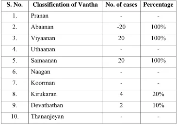
Tridhosas:
Vatham:
Vatham respresents vayu residing in the lower part of the body (i.e)
below the level of umbilicus.
Vatham is located in the abanan, face, idakalai, spermatic cord,
Classification:
Piranan (Uyir Kaal):
Controls knowledge, mind and five objects of sense. Responsible
for breathing and digestion.
Abanan (Keezh nokku kaal):
Responsible for passing urine, stools, sperm and menstrual flow.
Transfers the digested food to their respective places.
Uthanan (Mel nokku kaal):
Transport the digested food to different parts of the gut.
Responsible for vomiting, sneezing, cough etc.
Viyanan (Paravu kaal):
Spreads all over the body in all nerve endings causing contraction
and relaxation. Responsible for movements of all parts of the body.
Samanan (Nadu Kaal):
Neutralizes the above four vayus. Aids proper digestion.
Naagan:
Responsible for higher intellectual functions like learning,
thinking, singing etc., and also for opening and closing of eyelids.
Koorman:
Kirugaran:
Produces secretion from mouth and nose. Responsible for appetite,
sneezing and cough.
Devathathan:
Responsible for laziness, sleep and anger.
Thananjayan:
Responsible for swelling of the body after death and it escapes on
the third day by bursting the cranium.
In Sirangu, viyanan and samanan are affected.
Pitham:
Pitham represents “Theyu” residing in the middle part of the body.
It is located in pirana vayu, pingalai, bladder, moolakkini, heart,
umbilical region, stomach, abdomen, sweat, saliva, blood, eyes and skin.
Classification: Anar Pitham:
Responsible for proper digestion. Increases the appetite.
Ranjagam:
It gives colour to the blood
Saathagam:
It controls the entire body function
Aalosagam:
Responsible for vision
Prasagam:
Gives complexion to the skin
In sirangu Prasaga pitham is affected.
Kabam:
Kabam represents Appu, Prithivi and is situated in the upper part of
the body.
Kabam is located in samana vayu, sperm, head, tongue, uvula, fat,
bone marrow, blood, nose, chest, nerves, bones, large intestine, brain,
eyes and joints.
Classification:
Avalambagam:
Responsible for the proper functions of other four types of kabam.
Helps in respiration.
Kilaethagam:
Makes the food moist and soft to help digestion.
Pothagam:
Responsible for identifying taste in the tongue
Tharpagam:
Santhigam:
Responsible for the lubrication and aids free movements of the
joints.
Udal Vanmai:
Udal Vanmai is classified into three types as follows.
Iyearkai Vanmai:
Natural resistance of the body, by birth.
Seyarkai Vanmai:
Improving the health by the intake of nutritional food materials,
activities and medicines.
Kaala Vanmai:
This is the development of immunity according to age, season and
environment.
When udal vanmai is affected, Sirangu may occur.
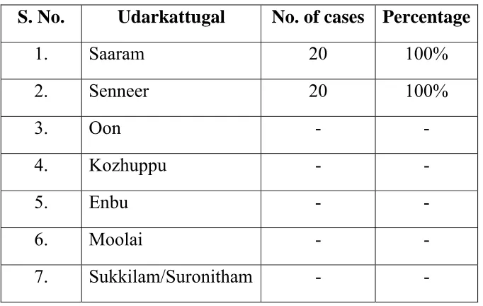
Udal Kattugal:
According to Siddha system, body is made up of seven important
constituents, called Udal Kattugal. They are necessary for proper
functioning of the body. They are
Saaram:
Saaram is responsible for the growth and development. It keeps the
Senneer:
Senneer is responsible for intellectual nourishment, strength and
helps in determining the colour and sound of the body.
Oon:
Oon gives shape to the body according to the requirement for
physical activity.
Kozhuppu:
Kozhuppu acts as a lubricant for different organs and helps in their
proper function.
Enbu:
Enbu supports the body system and is responsible for the posture,
movement and structure of the body.
Moolai:
Moolai occupies the bony spaces and gives strength to the bone.
Sukkilam / Suronitham:
Sukkilam/suronitham is responsible for reproduction.
In sirangu Saaram and Senner are affected.
Mukkutra Pathology:
In Siddha system the manifestation of all diseases are the results of
derangement of mukkutram due to various aetiological diet, activities,
Theraiyar stated in his Noikana mudal karam as,
‘thjkyhJ Nkdp nflhJ”
Therefore, in sirangu the principal factor, involves is vatham which
accompanies with pitham and kabham produces the clinical symptoms.
Viyanan one of the ten types of vatham spreads over the body
responsible for sensation and functioning of this skin. As skin having the
function of excretion through sweat, the derangement of viyanan
accompanying abanan affect the excretion of viyarvai (Sweat) which
results in itching.
As pitham resides in viyarvai its affection deranges the prasaga
pitham causing changes in lustre and complexion of skin.
Kabam which resides in senneer (Blood) affected causing
hypervitiation. This leads to the formation of pustules and vesicles.
Diagnosis (Piniyari Muraimai):
The diagnostic tool adopted to evaluate diseases in siddha medicine
is termed as Piniyari muraimai. It is based upon three main principles.
They are,
¾ Poriyalarithal
¾ Pulanaaltherthal
Pori is considered as the five senses of perception namely.
¾ Nose
¾ Tongue
¾ Eye
¾ Skin
¾ Ear
Pulan are five objects of senses. They are,
¾ Smell
¾ Taste
¾ Sight
¾ Sensation
¾ Sound
Physician’s Pori and pulan are used as the tools for examining the
Pori, pulan of the patient.
Vinaathal is obtaining the informations regarding the history of the
disease, its clinical features etc., from the patient or his immediate
relatives who are taking care of him, when the patients is a child or when
the patient is not in a position to speak.
The above principles correspond to the methodology of
interrogation, inspection, palpation of modern medicine in arriving at a
Siddhars have developed a unique method of diagnosing the
disease by Envagai thervugal
‘ehbg;ghprk; ehepwk; nkhoptpop
kyk; %j;jpukpit kUj;JtuhAjk;”.
- Neha; ehly; Neha; Kjy; ehly;
Hence the following makes the diagnosis,
1. Naadi
2. Sparisam
3. Naa
4. Niram
5. Mozhi
6. Vizhi
7. Malam
8. Moothiram
Naadi:
The three ‘Uyir thathukkal’ are formed by the combination of three
Naadi with three Vayu.
Idakalai + Abanan - Vatham
Pingalai + Piranan - Pitham
Naadi can be felt one inch below the wrist on the radial side by
means of palpation with tips of index, middle and ring fingers. The verse
is as follows,
‘fhpKfdbia tho;j;jpf; ifjdpy; ehb ghHf;fpy;
ngUtpuyq; Fyj;jpy; gpbj;jb eLNt njhl;lhy;
xU tpuNyhby; thjKaH eLtpuypw; gpj;jk;
jpUtpuy; %d;wpNyhby; Nrj;Jk ehbjhNd’
- Neha; ehly; Kjy; ghfk;
But it is stated in sathaga Nadi that the correct Naadi for children
cannot be felt.
The verse is as follows,
‘nfhz;blNt faNuhfp fhrNuhfp
Fwpg;ghf rpw;wpd;gk; nra;j NgHfs;
mz;blNt jhpj;jpuHfs; tpUj;jH ghyH
md;ghfj; jz;zPhpy; %o;fpNdhHfs;
nfhz;blNt ,tHfsJ cWg;gpd; jhJ
$wNt KbahJ vtHf;Ff; ,l;Lk;
gz;blNt apg;ghPl;ir ahHjhd; fhz;ghH
guhguj;jpd; kfpikapJ ghUghNu”
- Neha; ehly; Kjy; ghfk;.
So Naadi does not play a vital role in the diagnosis of sirangu in
Sparisam:
This reveals about the warmth, chillness, dry, weeping, soft, rough,
tenderness, fissures, pigmentation and changes in the skin.
In sirangu, thinavu, kurukkal etc., can be noticed at the affected
areas.
Naa:
The colour, salivation, ulceration, coating, movements, taste etc., is
noted for diagnosis.
Niram:
Colour of the skin is noted for diagnosis. In sirangu, the affected
site becomes brownish than the remaining normal site.
Mozhi:
Indicates the speech of the patient.
Vizhi:
The eye is noted for colour, lacrimation, and irritation.
Malam:
Quantity, colour, odour, froth, abnormal consistency including
indigestion, frequency, constipation etc., should be noted for diagnosis.
Moothiram:
Neerkuri:
Niram - Colour of the urine
Edai - Specific gravity of urine
Manam - Smell of urine
Nurai - Frothy nature of urine
Enjal - Quantity of urine
Neikuri:
Prior to the day of urine examination for Neikuri, the patient is
advised to take a balanced diet and the quantity of food must be
proportionate to his appetite. The patient must have a good sleep.
‘mUe;JkhwpujKk; mtpNuhjkjha;
m0fy; myHjy; mfhyT+z; jtpHe;jow;
Fw;wstUe;jp cwq;fp itfiw
Mbf;fyrj; jhtpNa fhJ nga;
njhUK$Hj;jf; fiyf;Fl;gL ePhpd;
epwf;Fwp nea;f;Fwp epUkpj;jy; flNd.”
- Neha; ehly; Kjy; ghfk;
Next day the urine is collected in a glass container in the early
morning. This specimen should be examined within one and half hours.
A drop of gingelly oil is dropped on the surface of urine collected
as above and the spreading of the oil should be observed. The verse is as
’epwf;Fwpf; Fiuj;j epUkhd ePhpw;
rpwf;f ntz;nza;NahH rpWJsp eLtpLj;
njd;Wwj; jpwe;njhyp Nafhjike;jjp
dpd;wjptiy Nghk; newptpopawpTk;
nrd;wJ GfYQ; nra;jpia AzNu”.
- Neha; ehly; Kjy; ghfk;
Vatha neer:
‘muntd ePz;bd0Nj thjk;”
When the drop of oil spreads like a serpent it indicates vatha neer.
Pitha neer:
‘Mop Nghy; gutpd; m0Nj gpj;jk;”
When the drop of oil spreads like a ring it indicates pitha neer.
Kapha neer:
‘Kj;njhj;J epw;fpd; nkhoptnjd; fgNk”
When the drop of oil remains as a pearl it indicates kaba neer.
Thontha neer:
When the drop of oil shows two shapes enclosed with in one
another it indicates thontha neer.
Mukkutra neer:
When the drop of oil drowns into the urine it indicates mukkutra
neer.
In majority of the cases the oil spreads like a pearl. In some cases
Besides Envagai thervugal, Uyir thathukkal, Ezhu Udal kattugal,
Paruvakalangal, Thinaigal also helps in making diagnosis.
Complications:
Pararasa seharam says that sirangu, if untreated can cause
Mahothara karappan. The features are,
¾ Itching gets reduced.
¾ Odema of the body
¾ Oliguria
¾ Impaired defecation
¾ Hoarsenes of voice
¾ Dyspnoea
¾ Loss of appetite
¾ Polydypsia
¾ These symptoms get exaggerated during night.
The verse is as follows,
‘rpuq;fpd; kNfhjuf; fug;ghd; nrAq; Fze;jhd;
wpdtlq;fp tPq;fp ky ryKk; tw;wp
cuk;gapY Kjh KwTw; Kl;lhfp
Athjp kpFe;jhp Fuyha; kplWk; tpf;fp
tuk;gapW %r;R kpF Rthj Kz;lha;
thA kpFe;J}z; kwe;J jhfkpQ;rp
,uq;FwNt fis Nrhf kpFjpAz;lh
apuhf; fhyj; jjpfhpf;F kpak;Gq; fhNy.”
Although sirangu is easily manageable it may become complicated.
This is mentioned as one of the kutramulla viyathigal in the Ankathi
patham. The verse is as follows.
‘%yKW fpue;jpAld; %yNuhf
Kjphpypq;fg; Gw;WlNd apypq;f #iy
#iy KW ePuopT fghy tPr;Rf;
nrhy;yW kNfhju fug;ghd; fhaNrw;gg;
rPyKW Ks;SWf;fp Ayw;W #iy
rpuq;fpd; Ke;jpkpHthj rd;dp fhrk;
Nfhy KW ntz;Fl;lk; nts;SNuhfq;
nfhba nfHg;g Nuhf Kjypuj;j Fd;kk;”.
- mq;fhjp ghjk;
Differential diagnosis:
Sirangu must be differentiated from Varal karappan and Ari
karappan.
Varal karappan:
Balavagadam describes Varal karappan as one of the eighteen
types of karappan.
The symptoms are sirangu like lesions all over the body, oozing of
fluid, itching and insomnia.
‘cr;rpKj Ys;sq;fh Yw;wstp nyt;tplKk;
er;Rr; rpWrpuq;F ez;zpNa - epr;ry;
ntbj;JePH NkTeik Nkth Jwf;fk;
Unlike sirangu there are lesions all over the body. There is no
severe disturbance of sleep in sirangu, whereas the sleep is disturbed in
Varal karappan.
Ari karappan:
The initial lesions occur over the penis and vulva. It ulcerates
further and eventually erodes the adjoining tissues, which becomes puttru
(malignancy).
The verse for this is as follows.
‘cw;w mhpfug;ghd; Xjf;Nfs; xz;nlhbaPH
kw;wypq;f Nahdpfspy; te;jpLk; Gw;Wlypy;
Mwhkw; Gz;zhpf;Fk; cUtopf;Fk;
khwhJ ,f;Fzj;jpd; thW”.
- ghythflk;
Only resembling feature is the early sirangu like lesions over the
MODERN ASPECT
ANATOMY The Skin:
The skin or integument covers the body and is continuous with the
membranes lining the body orifices. The skin has a surface area of about
1.5 to 2cm2 in adults and it contains glands, hair and nails.
It consists of a layer of dense connective tissue the dermis and an
external covering of epithelium termed the epidermis or cuticle.
Between the skin and underlying structures there is a layer of
subcutaneous fat.
Development:
The epidermis and its appendages are derived from the ectoderm,
the dermis is of mesodermal orgin.
About the fifth week of fetal development, the epidermis is formed
with two layers and the papillae of true skin about the sixth month.
The nails are formed at the third month and begin to project about
the sixth month.
The hairs appear between the third and fourth months. About the
fifth month the fetal hairs (Lanugo) appear first on the head and then on
the other parts. They drop after birth and give place to permanent hairs.
STRUCTURE
Epidermis (Cuticle, Scarf Skin)
It is the most superficial layer and is composed of stratified
epithelium which varies in thickness in different parts of the body. The
average thickness of the skin is about 1 to 2mm. It is thickest on the
palms of the hands and soles of the feet. There are no blood vessels or
nerve endings in the epidermis but its deeper layers are bathed in
interstitial fluid from the dermis.
The surface of the epidermis is ridged by projections of cells in the
dermis called the papillae. The pattern of ridges is different in every
individual and the impression made by them is the finger print.
The stratified squamous epithelium of the epidermis is composed
of several layers named according to various properties such as shape of
cells, texture, composition and position.
Beginning with the deepest, they are
1. Stratum Basale
2. Stratum Spinosum
3. Stratum Granulosum
4. Stration Lucidum
Stratum basale:
[Stratum Cyclindrium, Stratum Germinativum]
It is composed of columnar or cylindrical cells. The ends of the
cells are in contact with basement membrane and appear to anchor the
cells to the underlying dermis. The cells undergo divisions by mitosis
supplying new cells and the dead cells which are constantly being rubbed
off and replaced by kertain.
Stratum Spinosum:
It is composed of several layers of polygonal cells depending upon
the area of the body. The cells in this layer adhere to its neighbours at
particular points called desmosomes and these points are drawn into the
spines by shrinkage.
Stratum granulosum:
Composed of two to three rows of flat cells that lie parallel with the
surface. They are composed of keratohyalin, a substance that apparently
is transformed into keratin.
Stratum Lucidum:
It appears to be a homogenous translucent band much thinner than
other stratum. The cells in this layer contain droplets of eleidin.
Stratum corneum:
It is composed of squamous plate of scales fused together to make
the outer horny layer. These plates are the remains of the cells and
Pigmentation:
The colour of the skin is due to the presence of pigment in the cells
of the epidermis. The pigment is especially distinct in the cells of stratum
basale.
Melanin, a dark pigment secreted by melanocytes in the deep
germinative layer, is absorbed by surrounding epithelial cells. The
amount varies between different races and between members of the same
race. Melanin protects the skin from the harmful effects of sunlight.
Dermis: (Corium, Cutis Vera)
The dermis is tough, flexible and elastic. It is very thick in the
palms of the hands and soles of the feet. In the eyelids, scrotum and
penis it is exceedingly thin and delicate.
The dermis consists of felted connective tissue, composed of
collagen with a varying amount of elastic fibres and numerous blood
vessels, lymphatics and nerves. The collagen fibres bind water and give
skin its tensile strength. The connective tissue is arranged in two layers –
a deeper or reticular layer and superficial or papillary layer.
Blood Vessels:
Arterioles form a fine network with capillary branches supplying
sweat glands, sebaceous glands, hair follicles and the dermis. The
epidermis has no blood supply. It obtains nutrition and oxygen from
Lymphatic Vessels:
It form two networks, superficial and deep, which communicate
with each other and with those of the subcutaneous tissue by oblique
branches.
Nerve endings:
Nerve impulses generated in the nerve endings are conveyed to the
spinal cord by sensory (somatic cuteneous) nerves, then to the sensory
area of the cerebrum where the sensations are perceived.
GLANDS OF THE SKIN
Sebaceous Glands:
The glands are small, sacculated, glandular organs lodged in the
substance of the dermis. They are most numerous in the skin of the scalp,
face, axillae and groins. The glands pour their oily secretion sebum into
the hair follicles. In some areas face, lips, nipple, glans penis and labia
minora these glands open directly into the exterior.
Sebum contains free fatty acids, triglycerides, squalene, sterols,
waxes and paraffin. It has antibacterial and antifungal actions and it
keeps the skin smooth and oily.
Sweat glands (Sudoriferous glands)
3-4 Million sudoriferous glands release their secretions by
the hair follicles. They are divided into two main types, as eccrine and
apocrine based on their structure, location and type of secretion.
Eccrine Sweat Glands:
The eccrine gland is a tubular, coiled formed by single layer of
cuboidal or columnar cells. They are distributed throughout the body.
They secrete a clear watery sweat which is increased during emotional
conditions and in higher temperature. Sweat contains water, sodium
chloride, urea and lactic acid. Their main function is regulating body
temperature.
Apocrine Sweat Glands:
Apocrine glands are situated only in limited areas like axilla, pubis,
areola and umbilicus. They have the same structure as eccrine glands.
These glands start functioning only at the time of puberty. The sweat is
thick, milky and odorless. When microorganism grows in this secretion,
it develops a characteristic odour. It is increased only in emotional
condition.
Glands of eyelids, glands of external auditory and mammary
glands are the modified apocrine glands.
APPENDAGES OF THE SKIN Hairs:
These are formed by a down growth of epidermal cells into the
follicle there is cluster of cells called the bulb. The hair is formed by the
multiplication of cells of the bulb and when the cells die, become
keratinized. The part of the hair above the skin is the shaft and the
remainder, the root.
The colour of the hair is genetically determined and depends on the
amount of melanin present.
Arrectores Pilorum:
These are little bundles of involuntary muscle fibres attached to the
hair follicles. Contraction makes the hair stand erect and raises the skin
around the hair causing goose flesh. These muscles are activated in
response to fear and cold.
Nails:
The nails are derived from the same cells as epidermis and hair and
consist of a hard horny type of keratinized dead cell. They protect the
tips of the fingers and toes.
The root of the nail is embedded in the skin, covered by the cuticle,
and forms the hemispherical pale area called the lunula.
The body of the nail is exposed part which has emerged from the
germinative zone called the nail bed.
Finger nails grow more quickly than toe nails and growth is
PHYSIOLOGY OF SKIN
The Functions of the Skin are
1. Protective function
2. Role as a sense organ
3. Storage function
4. Synthesis of Vitamin –D
5. Regulation of body temperature
6. Regulation of water balance.
7. Excretory function
8. Absorptive function
9. Secretary function.
1. Protective Function:
Skin forms the covering of all the organs of the body and protects
the organs from,
a) Bacteria and toxic substances – The keratinized stratum
corneum is responsible for the protection. It offers resistance
to the skin against toxic chemicals like acids and alkalis. The
sebaceous glands secreting oily sebum contain bactericidal
chemicals that kill the surface bacteria.
b) Protection from mechanical blow-The Skin is not tightly
placed over the underlying organs. It becomes loose and
moves over the tissues in response to any blow.
2. Role as a Sense Organ:
Skin is the largest sense organ in the body. Cutaneous receptors
are stimulated by the sensations of touch, pain, pressure and temperature
and convey them to the brain through the afferent nerves.
3. Storage
Skin can store fat, water, chloride and sugar. It can also store
blood by the dilation of the blood vessels.
4. Synthesis of Vitamin – D.
Vitamin D3 is synthesized in the skin by the action of ultraviolet
rays on cholesterol. Vitamin D is essential for skeletal development.
5. Regulation of Body Temperature:
Excessive heat is lost from body through skin by radiation,
conduction, convection and evaporation. Sweat glands in the skin take
active part in heat loss by secreting sweat. The lipid content of sebum
prevents loss of heat from the body in cold environment.
6. Regulation of Water Balance:
Skin regulates water balance and electrolyte balance by excerting
water and salts through sweat.
7. Excretory function:
Skin can excrete small quantities of waste materials like urea, salts
and fatty substance through sweat. About 400 ml of water evaporates
8. Absorptive Function:
The absorption of water soluble substances through skin is
negligible. But certain fat soluble substances like fat soluble vitamins
(A,D,E and K) Oxygen, carbon dioxide gases and ointments, toxic
materials, organic solvents like acetone, carbon tetrachloride, salts of
heavy metals can also be absorbed.
9. Secretory Function:
Skin secretes sweat through sweat glands and sebum through
sebaceous glands. By secreting sweat, skin regulates body temperature
SCABIES
Definition:
Scabies is an intensely pruritic skin infestation caused by the host
specific mite, Sarcoptes scabiei var hominis, an obligate human parasite.
(Sar, koptein means to smite or cut) (Scabere means to scratch).
Etiology and epidemiology:
Scabies affects persons of all ages, races and socio-economic
groups.
Over crowding, poverty, poor hygiene and poor public education,
delayed diagnosis and treatment contribute to their prevalence. Incidences
are higher in children than others. Persons with poor sensory perception
due to conditions like leprosy, and immuno compromised persons due to
conditions such as status post transplantation, HIV disease and old age all
at particular risk for the crusted scabies.
Mode of Transmission:
The efficient means of transmission of scabies is via direct and
prolonged physical contact with an infected individual. Mites can survive
several days away from human skin, so fomites present in infested
Parasitology:
Class - Arachnida
Subclass - Acari
Order - Astigmata
Family - Sarcoptidae
Name - Sarcoptes scabiei var hominis.
Characters of Sarcoptes Scabiei:
The adult female has a hemispherical body marked by transverse
corrugations, brown spines and bristles on the dorsal surface. The male
mite is similar in configuration but smaller.
The female measures 0.4mm in length and the male mite are
approximately one half of her size.
The mite has four sets of legs. The anterior 2 pairs end as elongated
peduncles tipped with small suckers.
Lifecycle:
Acarus scabiei undergoes its lifecycle on the skin surface. The
male mite has a short life span. It dies shortly after copulation. The adult
female, after impregnation, burrows into the skin and forms a tunnel in
the horny layer (Stratum Corneum).
The mite burrows at the rate of 1-5mm per day. Two days after
fertilization, she starts laying eggs along her course in the burrow at the
The female mite exudes a keratolytic substance and burrows into
the stratum corneum. The tract is extended by 0.5-5mm/24 hour within
stratum granulosum and she deposits 1-3 oval eggs and numerous brown
fecal pellets (Scybala) daily.
The egg hatches in 3-4 days producing a larva that moves to the
skin surface. It then molts through various stages of oclopod nymph into
an adult mite. The entire life cycle can be completed in 10 to 14 days.
The average number of adult female mites on an individual
suffering from scabies is usually less than 100. Only in crusted scabies
there are large numbers of mites present.
Pathogenesis:
The mites move through the top layers of skin by secreting
proteases that degrade the stratum corneum.They feed on dissolved tissue
but do not ingest blood. Scybala (faeces) are left behind as they travel
through the epidermis creating linear lesions clinically recognized as
burrows.
Histological Findings:
The histologic features revealed by the excision of a burrow shows
mites, larvae, ova and faeces within the stratum corneum. Superficial and
deep dermal infiltrate show lymphocytes, histiocytes, mast cells and
eosinophils. Spongiosis and vesicle formation with exocytosis of
Crusted scabies demonstrates massive hyperkeratosis of the
stratum corneum with innumerable mites.
Nodular scabies reveals a dense, mixed superficial and deep dermal
inflammatory cell infiltrate.
Immunology:
Allergic sensitivity to the mite or its products appears to play an
important role in determining the development of lesions other than
burrows and in producing pruritis.
Immunological reactions mediated by antibodies of IgG, IgM and
especially IgE classes may be involved. None of these reactions may
have been shown to eliminate all mites but locally these reactions may
prevent the epidemic multiplication of scabies organism.
Clinical Features:
Clinical findings include both primary and secondary lesions.
Primary lesions are the first manifestation of the infestation and these
include,
1. Itching.
2. Burrows
3. Papules and Vesicles
Itching:
Itching is the primary symptom of scabies and it may be due to
type IV hypersensitivity to the mite or its products. Usually itching
appears after 4-6 weeks of initial infection, due to development of
sensitization to the mite or its products (Scabin in saliva) which takes
much time.
Itching is characteristically worse at night. This nocturnal
periodicity is usually due to the movement of the young acarus to reach
the pores favoured by the warmth of the bed. And also the mast cell
degranulation at night may be another reason for itching worsened at
night.
Burrows:
Burrows represent the intra epidermal tunnel created by the moving
female mite. They appear as serpiginous, greyish thread like elevations
ranging from 2-10mm long. The distribution of lesions is characteristic
include flexor aspects of interdigital spaces of the finger, wrists, elbows,
axillae, belt line, dorsal feet, buttocks, scrotum in men and nipple area in
women. In infants burrows are commonly located on the palms and soles.
Papules and Vesicles:
Erythematous, papules and vesicles 1-3mm sized are seen in
typical distribution. The vesicles are discrete lesions filled with clear
fluid. Papules are common on the shaft of the penis in men and on the
3. Nodules (Nodular Scabies):
Common in young children. They are pinkish brown or red nodules
ranging from 2-20mm, eczematous eruption primarily seen on the trunk.
The nodules may develop due to deeper penetration by the acarus or a
severe dermal reaction to the toxins of the acarus.
Secondary Lesions:
These are the result of scratching, secondary infection and the host
immune response against the mites.
Other findings include excoriations, widespread eczema,
honey-colored crusting, post-inflammatory hyper pigmentation, erythroderma,
prurigo nodules and frank pyoderma.
Crusted scabies (Norwegian Scabies):
It manifests with marked thickening and crusting of the skin in
which the mite population is enormous. Lesions are often hyperkeratosis,
crusted and cover large areas. Nail dystrophy and scalp lesions may be
prominent. Predominantly affected persons are those with
immunosuppresion, elderly and bedridden patients.
Scabies in Infants:
In infants the features differ from that of older children and adults.
Bullae and pustules are more common and the palms, soles, face and
scalp are often affected. Eruptions may also include wheals, papules and
Diagnosis:
It is based on the following
¾ History of nocturnal itching
¾ History of exposure or multiple cases in the family
¾ The characteristic lesions distributed at the sites of predilection
and the identifiable typical burrows.
¾ Light microscopic identification of mites, larvae, ova or scybala
from the burrow.
¾ Skin scrapping
Application of a drop of mineral oil on the selection site,
scrapping of it with No.15 blade and transfer them on the glass
slide, covered with coverslip and examined under microscope
40 × magnifications.
¾ Clinically inapparent infection can be detected by amplification of
sarcoptes DNA in epidermal scale by polymerase chain reaction.
Differential diagnosis:
Scabies is often confused with other papulo vesicular lesion like
papular utricaria, Canine scabies, dermatitis herpetiformis, folliculitis,
and eczematous diseases.
Nodular scabies is misdiagnosed as utricaria pigmentosa and
1. Canine Scabies:
The distribution of lesions is different but not transmitted between
humans.
2. Eczematous disease:
Eczematous lesions may mimic atopic dermatitis. Usually there is a
history of eczema in the patient or the family.
3. Dermatitis herpetiformis:
Similar in distribution but the vesicles and utricarial lesions are
more prominent.
4. Pediculosis:
It is also caused by a parasite and is excluded by the presence of
lice and nits.
Complication:
Complications of scabies generally result from vigorous rubbing
and scratching. Disruption of the lesion makes the patient at risk for
secondary bacterial invasion chiefly by streptococcus pyogenes and
staphylococcus. Super infection with S. Pyogenes can precipitate acute
post streptococcal glomerulonephritis. Common pyodermas include
impetigo and cellulitis which may rarely result in sepsis.
Acute post streptococcal Glomerulonephritis:
It is more common is tropical climates. Immune mechanism is
glomeruli. The symptoms are dark coloured urine, peri-orbital odema,
oliguria, general malaise, low grade fever and acute hypertension.
Impetigo:
This superficial pyoderma is due to group
A
β-haemolyticstreptococci. It develops as vesicular lesions on the arms and legs or
around mouth, nose and scalp which becomes pustules. Lymphangitis and
MATERIALS AND METHODS
The clinical study on sirangu was carried out in the Post Graduate
department of Kuzhanthai Maruthuvam in Government Siddha Medical
College at Palayamkottai.
Selection of cases:
The patients were selected according to the signs and symptoms of
sirangu as mentioned in siddha aspects. The patients were treated either
in OP and IP. 20 cases were admitted from both sexes of various ages
upto 12 and 50 and more OP cases were seen.
Evaluation of Clinical Parameters:
During admission, the patients were selected according to the
clinical features of sirangu like,
¾ Itching
¾ Burrows
¾ Inflammatory papules
¾ Pruritic papules
¾ Vesicles
¾ Pustules
These symptoms are present in the webs of the fingers, wrist,
ankles, buttocks, groin and the genital areas. Complete case history and
Study on Siddha Mode of Diagnosis:
A case sheet was prepared on the basis of siddha methodology (ie)
Poriyalarithal, Pulanaaltherthal, Vinavuthal and envagai thervugal.
Clinical Investigations:
All the patients were subjected to the following routine laboratory
investigations available at Government Siddha Medical College,
Palayamkottai.
Haematological Investigation:
Total WBC Count
Differential Count
Erythrocyte sedimentation rate
Haemoglobin.
Urine:
Albumin
Sugar
Deposit
Stools:
Ova
Cyst
Efficacy of the trial drugs was found by biochemical analysis,
carried out in the Department of the Bio-Chemistry, Government Siddha
Pharmacological analysis of the trial drug was carried out in the
Department of Pharmacology, Government Siddha Medical College,
Palayamkottai.
Anti-microbial study was done to reveal the anti-bacterial activity
of the trial drugs at Malar diagnostic centre, Palayamkottai.
Trial Drugs:
The patients were treated with chiruthetkodukilai chooranam with
hot water thrice a day after meals. Sirangu Ennai was used as external
application for twice a day.
All the patients admitted for the study were given uniform regular
hospital diet.
At the time of discharge all the 20 patients were advised to attend
LINE OF TREATMENT
The Line of Treatment for sirangu is as follows
¾ Bringing the three doshas into its equilibrium state
¾ Administration of internal medicine to arrest the disease
process.
¾ Application of the external medicine topically over the
lesions
¾ Pathiyam (ie) diet restriction to normalize the vitiated dhosas
and to maintain good drug action.
¾ Advising personal hygiene.
Bringing the three doshas into its equilibrium state
To bring the three doshas in equilibrium – Nilavagai choornam –
½
- 2 gm with hot water according to age and physical state.Administration of internal medicine:
All the twenty patients were given the trial drug chiruthetkodukilai
chooranam with hot water thrice daily and regularly and the progress was
noted.
Dosage:
2 - 4 Years - 250mg
4 - 7 years - 500mg
Also the dosage was adjusted according to the severity and
condition of the patient.
Anupanam in Siddha system:
‘mDghdj;jhNy atpo;jk; gypf;Fk;
,dpjhd Rf;F fd;dy; ,Q;rp - kpDKjfhy
NfhNkak; ghy; Kiyg;ghy; Nfhnea; Njd; ntw;wpiy ePh;
Mkpij ahuha;j;J nra;ayhk;”.
- Njiuah; ntz;gh
Siddha system considers anupanam as an important adjuvant of the
drug than the medicine itself. In this work, the author used hot water as
anupanam.
Application of External medicine:
All the twenty patients were treated with Sirangu ennai externally
twice a day, over the lesions.
Pathiyam:
Diet restrictions are important for good drug action and also to
normalize the vitiated dlaosas. For sirangu the patients were strictly
advised to avoid,
Agathi Keerai
Pagal Kaai
Poosani Kaai
Kaanam
Motchai
Palapazham.
Also the sirangu provoking food items, or factors like Kathirikaaai,
Kambu arisi, Cholam, Senchemba, Pudalangaai, Maangaai, Varagu
Satham, allikulathu neer, certain type of fish and dry fish were strictly
avoided.
Management:
The following measures were advised to follow for the early cure
and to avoid reoccurrence.
1. Patients were advised to take bath atleast once a day with luke
warm water.
2. They were advised to avoid bathing in public tanks.
3. They were advised to use Nalungu maa, Paasipayaru maa or
Kadalai paruppu maa instead of soap.
4. Patients were advised not to wear other clothes and to change
clothes daily.
5. All the family members affected by this disease were advised to
take medicines at the same period.
6. School going children were advised to take medicines at the
same period.
7. Infested bedding and clothes should be washed and dried in sun
Control of sirangu:
After recovery all the patients were advised with health education like,
1. Environmental modifications such as avoiding
overcrowding.
2. Maintaining personal hygiene for the prevention and control
of sirangu.
3. Improving patient’s general health by taking rich diet with
OBSERVATION AND RESULTS
Results were observed with respect to the following criteria.
1. Sex Distribution
2. Age Distribution
3. Kaalam
4. Paruva kaalam
5. Distribution of Lands (Thinai)
6. Religion Reference
7. Socio Economic status
8. Diet Reference
9. Etiology
10.Clinical features
11. Site of infection
12. Duration of illness
13. Tridhosha Theory
14. Ezhu udarkattugal
15. Envagai Thervugal
16. Neikuri
17. Gradation of total result
18. IP Case Report
19.Investigation Chart
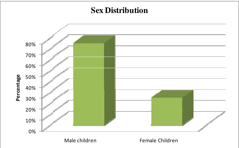
Table 1:Sex Distribution 1. Sex Distribution:
S. No. Sex No. of cases Percentage
1. Male children 15 75%
2. Female Children 5 25%
Among the 20 patients selected, 75% of patients were male
children and 25% of patients were female children.
0% 10% 20% 30% 40% 50% 60% 70% 80%
Male children Female Children
Pe
rc
e
n
ta
ge
Sex Distribution
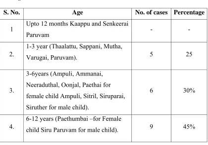
Table 2:Age Distribution 2. Age distribution:
S. No. Age No. of cases Percentage
1 Upto 12 months Kaappu and Senkeerai
Paruvam - -
2.
1-3 year (Thaalattu, Sappani, Mutha,
Varugai, Paruvam). 5 25
3.
3-6years (Ampuli, Ammanai, Neeraduthal, Oonjal, Paethai for female child Ampuli, Sitril, Siruparai, Siruther for male child).
6 30%
4.
6-12 years (Paethumbai –for Female
child Siru Paruvam for male child). 9 45%
The percentage was highest in the age group of 6 - 12 years, the
percentage was 45%, between the age group 3 – 6 years the percentage
was 30%. 25% belonged to the age group 1-3 years.
0% 5% 10% 15% 20% 25% 30% 35% 40% 45%
Upto 12 months
1‐3 year 3‐6years 12 years
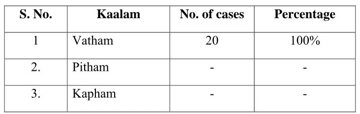
Table 3: Kaalam 3. Kalam
S. No. Kaalam No. of cases Percentage
1 Vatham 20 100%
2. Pitham - -
3. Kapham - -
In this study, all the 20 patiens were in Vatha Kaalam since all the
patients belong to children age group.
Table 4: Paruva Kaalam 4. Paruva Kaalam
S. No. Paruva kaalam No. of cases Percentage
1. Kaar kaalam (Aavani& Purattasi)
(Aug.16 - Oct.15) 9 45%
2. Koothir kaalam (Iyppasi & Karthigai)
(Oct.16 - Dec.15) 2 10%
3. Munpani kaalam (Markazhi & Thai)
(Dec.16 - Feb.15) - -
4. Pinpani kaalam (Masi & Panguni)
(Feb.16 - April 15) - -
5. Elavenil kaalam (Chithirai & Vaigasi)
(April – 16 - June 15) 2 10%
6. Muthuvenil kaalam (Aani & Aadi)
[image:67.612.97.520.339.700.2]Among the 20 cases selected, 45% of cases were admitted in Kaar
kaalam, 10% of cases were admitted in Koothir kaalam and Elavenil
[image:68.612.126.487.200.387.2]kaalam and 35% of cases were admitted in Muthuvenil Kaalam.
Table 5: Distribution of Lands (Thinai) 5. Thinai
S. No. Thinai No. of cases Percentage
1 Kurunji (Hill area) - -
2. Mullai (Forest area) - -
3. Marutham (Fertile area) 19 95%
4. Neithal (Coastal area) -1 -5%
5. Paalai (Desert area) - -
Among 20 cases admitted, 19 cases belonged to Marutham and 1
case belonged to Neithal.
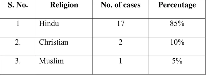
Table 6:Religion Distribution
6. Religion Distribution
S. No. Religion No. of cases Percentage
1 Hindu 17 85%
2. Christian 2 10%
3. Muslim 1 5%
Out of 20 cases 85% were Hindus, 10% were Christians and 5%
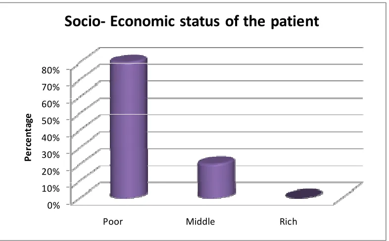
[image:68.612.136.479.522.647.2]Table 7: Socio- Economic status of the patient 7. Socio – Economic Status of the Patient
S. No. Socio-Economic Status No. of cases Percentage
1 Poor 16 80%
2. Middle 4 20%
3. Rich - -
Out of the 20 patients, 80% of cases were poor and 20% were
middle class people.
0% 10% 20% 30% 40% 50% 60% 70% 80%
Poor Middle Rich
Pe
rc
e
n
ta
ge
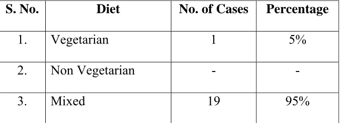
Table 8: Diet 8. Diet
S. No. Diet No. of Cases Percentage
1. Vegetarian 1 5%
2. Non Vegetarian - -
3. Mixed 19 95%
95% of cases have mixed diet and 5% of cases have vegetarian diet.
Table 9: Aetiological Factors
9. Aetiological Factors
S. No. Aetiological Factors No. of Cases Percentage
1. Poor hygiene 14 70%
2. Contact 10 50%
3. Over crowding 9 45%
4. Poverty 16 80%
Poor hygiene was noted in 70% of cases Poverty was noted in 80%
of cases, contact history was noted in 50% of cases. Overcrowding was
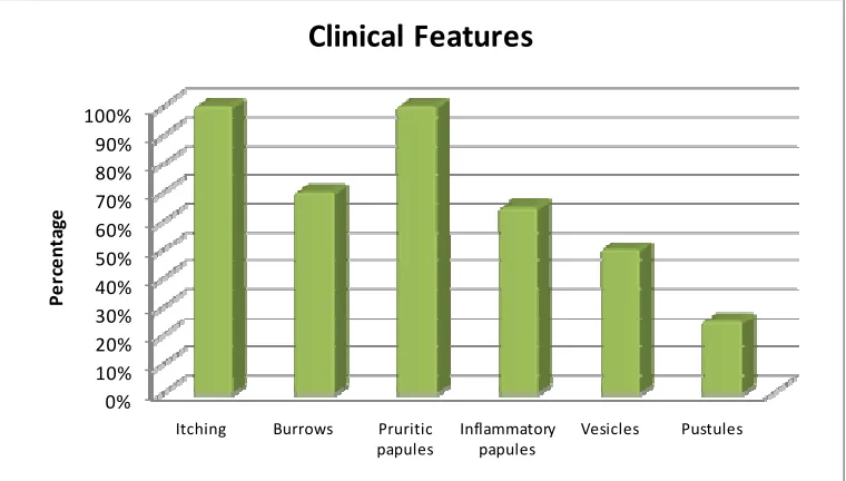
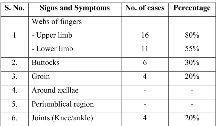
[image:70.612.136.477.359.519.2]Table 10: Clinical Features 10. Clinical Features
S. No. Signs and Symptoms No. of cases Percentage
1 Itching 20 100%
2. Burrows 14 70%
3. Pruritic papules 20 100%
4. Inflammatory papules 13 65%
5. Vesicles 10 50%
6. Pustules 5 25%
100% of cases had Itching and Pruritic papules. 70% of cases show
burrows 65% of cases had Inflammatory papules. 50% of cases had
vesicles. 25% of cases had Pustules.
0% 10% 20% 30% 40% 50% 60% 70% 80% 90% 100%
Itching Burrows Pruritic
papules Inflammatory papules Vesicles Pustules Pe rc e n ta ge