0022-538X/96/$04.0010
Copyrightq1996, American Society for Microbiology
Repression of Human Immunodeficiency Virus Type 1 Long
Terminal Repeat-Driven Gene Expression by Binding of the
Virus to Its Primary Cellular Receptor, the CD4 Molecule
PIERRE BE´ RUBE´,1BENOIˆT BARBEAU,1RE´ JEAN CANTIN,1RAFICK-PIERRE SE´ KALY,2
ANDMICHEL TREMBLAY1*
Centre de Recherche en Infectiologie, Centre Hospitalier de l’Universite´ Laval, and De´partement de Microbiologie, Faculte´ de Me´decine, Universite´ Laval, Ste-Foy, Que´bec, Canada G1V 4G2,1and Laboratoire d’Immunologie,
Institut de Recherches Cliniques de Montre´al, and De´partement de Microbiologie et Immunologie, Universite´ de Montre´al, Montre´al, Que´bec, Canada H2W 1R72
Received 10 November 1995/Accepted 21 March 1996
We have previously postulated that the binding of the human immunodeficiency virus type 1 (HIV-1) to cell surface CD4 induces signal transduction pathways that down-modulate production of progeny virions in acutely infected T cells (M. Tremblay, S. Meloche, S. Gratton, M. A. Wainberg, and R.-P. Se´kaly, EMBO J. 13:774–783, 1994). To evaluate the possibility that CD4 cross-linking might indeed affect viral gene expression, we have introduced a molecular construct made of the luciferase reporter gene placed under the control of the regulatory elements of HIV-1 in several CD4-positive T-cell lines. We found that cross-linking of CD4 with defective HIV-1 particles and heat-inactivated viruses inhibits long terminal repeat-dependent luciferase expression. Experiments revealed that the gp120-CD4 interaction was necessary to repress HIV-1 long terminal repeat-dependent luciferase activity. The cytoplasmic domain of CD4 was also found to be required for this effect to occur. The virus-mediated signal transduction was shown to be mediated via p56lck
-dependent and -independent pathways. These results indicate that the earliest event in the HIV-1 replicative cycle, namely, the binding of the virus to its cellular receptor, can lead to signal transduction culminating in down-modulation of viral gene expression. Thus we propose that defective viruses could regulate the pathogenesis of HIV disease as they constitute the vast majority of circulating HIV-1 particles.
The CD4 antigen is a glycoprotein of approximately 55,000 in molecular weight expressed primarily on the surface of a functionally distinct population of human T cells and, to a lesser extent, on cells of the monocyte/macrophage lineage (57, 65, 73). It has been proposed that CD4 functions as an adhe-sion structure that interacts with nonpolymorphic determi-nants located on the major histocompatibility complex (MHC) class II molecules, thereby stabilizing and facilitating the MHC class II–T-cell receptor (TCR) complex interaction (18, 23, 51, 72). Other studies have also indicated that CD4 can actively participate in transmembrane signal transduction. The initial evidence that the cell surface CD4 glycoprotein is playing an active role as a signal transduction molecule came from the demonstration that, without appropriate MHC class II recog-nition, anti-CD4 antibodies abrogated lectin-induced T-cell mitogenesis (3, 66, 74, 80). Results from these studies suggest that the CD4 molecule negatively affects T cells when this structure is ligated independent of the TCR. In contrast, the binding of the coreceptor molecule CD4 to the same MHC class II molecule as the TCR results in an optimal T-cell activation (19, 20, 36, 42, 44, 45). Greater activation of the T lymphocytes is observed when the TCR and the coreceptor CD4 bind to the same ligand, suggesting that CD4 actively participates in transmembrane signal transduction through the TCR. This has led to the concept of receptor cooperativity in T-cell signaling and activation (35).
Beside its role in antigen-specific T-cell activation, CD4 is also established as the major cellular receptor for the human immunodeficiency virus (HIV) (16, 39). Viral tropism is me-diated by the high-affinity interaction between CD4 and the major viral envelope glycoprotein gp120 (43, 52). There have been conflicting reports on the ability of HIV and/or gp120 to transduce signaling through the CD4-p56lckcomplex. Ligation of CD4 by gp120 has been demonstrated to generate stimula-tion or modificastimula-tion of protein kinase C activity, an increase in the intracellular calcium concentration, activation of protein tyrosine kinases, arachidonic acid metabolism, and interleukin 1 (IL-1) release (15, 21, 27, 41, 59, 79). Furthermore, binding of gp120 or HIV to CD4 has also been shown to abrogate anti-TCR-induced calcium mobilization, IL-2 production, pro-liferation, and antigen-specific T-cell responses (13, 17, 27, 48, 50, 55, 61). However, others have failed to observe these events (33, 38, 60). Such contradictory results may be associated with variations in technical approaches.
Recently, we and others have demonstrated that cross-link-ing of cell surface CD4 can result in inhibition of HIV type 1 (HIV-1) replication at a postbinding step (4, 10, 14, 29, 31, 56, 76). Cross-linking of CD4 was achieved in these experiments with anti-CD4 antibodies (4, 10, 14, 31, 56), recombinant HIV-1-like particles carrying viral envelope proteins (29), and, more importantly, whole HIV-1 particles (76). The demonstration that the binding of viruses to already infected cells results in a decrease of virus production is of great interest because it implies that HIV-1 could regulate its own replication.
In this study, we used a molecular construct made of a reporter gene placed under the control of the regulatory ele-ments of HIV-1. Whole heat-inactivated and defective HIV-1 particles were used to cross-link surface CD4. Cell lines
car-* Corresponding author. Mailing address: Centre de Recherche en Infectiologie (room 9500), Centre Hospitalier de l’Universite´ Laval, 2705 boul. Laurier, Ste-Foy, Que´bec, Canada G1V 4G2. Phone: (418) 654-2705. Fax: (418) 654-2715. Electronic mail address: michel.j. [email protected].
4009
on November 9, 2019 by guest
http://jvi.asm.org/
rying wild-type and mutated forms of CD4, as well as wild-type p56lck, were generated to investigate in depth the signaling pathway. Our results demonstrate that the binding of HIV-1 to the CD4 glycoprotein can markedly reduce long terminal re-peat (LTR)-driven reporter gene activity. Furthermore, we present evidence that the signal transduction pathway requires the cytoplasmic domain of CD4 and is partly mediated through the CD4-associated protein tyrosine kinase p56lck.
(This work was performed by P. Be´rube´ in partial fulfillment of the requirements for his graduate studies in the Department of Microbiology and Immunology, Faculty of Medicine, Laval University.)
MATERIALS AND METHODS
Cells and culture conditions.1G5 is a derivative of Jurkat E6-1 that contains a stably integrated HIV-1SF-2LTR-luciferase construct (1), C8166-45 is an
IL-2-independent human T-cell leukemia virus type 1-transformed T-cell line that does not express p56lck
(40), Jurkat-tatis a Jurkat E6-1 derivative stably express-ing the HIV-1 Tat protein (11), Sup-T1 cells express high levels of surface CD4 (70), WE17/10 is a human IL-2-dependent T-lymphoblastoid cell line (81), and A2.01 is a human leukemic CD4-negative cell line (22). These cell lines were provided by the AIDS Research and Reference Reagent Program, Division of AIDS, National Institute of Allergy and Infectious Diseases (Rockville, Md.). Cell lines were maintained in complete culture medium made of RPMI 1640 supplemented with 10% fetal calf serum, glutamine (2 mM), penicillin G (100 U/ml), and streptomycin (100mg/ml).
Flow cytometry analysis.Levels of surface CD4 molecule were detected by direct immunofluorescence with a cytofluorimeter. Briefly, 105cells were first
incubated with an experimentally determined saturating concentration of a flu-orescein-conjugated anti-human CD4 antibody (Leu-3a; Becton Dickinson) for 30 min on ice. After two washes with phosphate-buffered saline, samples were fixed with 1% (vol/vol) of paraformaldehyde and analyzed by a cytofluorimeter. Controls consisted of a commercial isotype-matched murine antibody (Sigma, St. Louis, Mo.).
Viruses.Stocks of infectious HIV-1IIIBand HIV-1IIIRFwere prepared from
acutely infected H9 cells. The IIIB and IIIRF strains of HIV-1 were kindly provided by R. C. Gallo through the AIDS Research and Reference Reagent Program as cell-free supernatant from infected H9 cells. The clinical isolate HIV-1334was grown on phytohemagglutinin-activated peripheral blood
mono-nuclear cells isolated from healthy donors and has been described previously (77). Defective HIV-1IIIBparticles were also used in our experiments and
orig-inate from cellular clone UHC-8 (9). Virus particles harvested from this cell line are devoid of both reverse transcriptase and integrase proteins (8). Virus stocks were quantitated by measuring the amount of the major viral core p24 protein by a commercial enzymatic assay (Organon Teknika, Durham, N.C.).
Generation of cells stably transfected with cDNA encoding wild-type, trun-cated, and mutated forms of CD4, as well as wild-type p56lck.Truncated (t-CD4)
and mutated (C4202A) CD4 constructs were generated as previously described (76). Briefly, a stop codon was introduced into the cDNA coding for human CD4, leading to deletion of 32 of 38 amino acids located in the cytoplasmic domain of CD4. The mutated CD4 was achieved by substituting cysteines at positions 420 and 422 with alanines by a PCR overlap extension procedure. The full-length human p56lck
cDNA originates from the pHK-28 plasmid (kindly provided by R. M. Perlmutter, Seattle, Wash.). The various molecular constructs were subcloned in the eukaryotic expression retroviral vector pMNC, which contains the
neo-mycin (G418) resistance gene and a cytomegalovirus promoter for eukaryotic gene expression (69). The amphotropic helper packaging cell line DAMP (63) was transfected with the different pMNC constructs by the calcium phosphate transfection technique. The resulting recombinant amphotropic retrovirus par-ticles containing the cDNA of interest driven by the cytomegalovirus promoter region and theneogene driven by the LTR of the Moloney murine leukemia virus were used to infect cells of interest. Stable cellular transfectants were cultured in complete culture medium supplemented with appropriate concen-trations of the selective agent G418 (Gibco-BRL, Gaithersburg, Md.).
Transfection, cell treatments, and luciferase assay.Cells (103106) were
washed once in TS (25 mM Tris-HCl [pH 7.4], 5 mM KCl, 0.6 mM Na2HPO4, 0.5
mM MgCl2, and 0.7 mM CaCl2) and resuspended in 1 ml of TS containing the
desired plasmid(s) and 500mg of DEAE-dextran per ml (final concentration). Plasmids pLTRX-LUC and pCMVtatwere provided by O. Schwartz (Unite´ d’Oncologie Virale, Institut Pasteur, Paris, France). The pLTRX-LUC plasmid contains a 722-bpXhoI (2644)-HindIII (178) fragment from HIV-1LAIplaced
in front of the luciferase reporter gene, and the pCMVtatvector contains the immediate early enhancer/promoter region of the human cytomegalovirus placed upstream of the viraltatgene (67). The pCMV-LUC vector has been provided by A. Darveau (Laval University, Ste-Foy, Quebec, Canada). The cell–TS–plas-mid(s)–DEAE-dextran mix was incubated for 15 min at room temperature. Thereafter, cells were diluted at a concentration of 106/ml by using complete
culture medium supplemented with 100mM chloroquine (Sigma). After 45 min of incubation at 378C, cells were centrifuged, washed once, and seeded in 96-well flat-bottomed plates at a density of 53104cells per well (100ml) in the presence
of 20 ng of phorbol myristate acetate (PMA) per ml (Sigma). Cells were inoc-ulated with heat-inactivated or defective HIV-1 particles. Heat-inactivated HIV-1 particles (HIV-1IIIB, HIV-1IIIRF, and HIV-1334) originated from frozen
virus stocks that were thawed and incubated at 568C for 30 min before their use, while fresh cell-free culture supernatant from UHC-8 cells filtered through a 0.45-mm-pore-size membrane was used as a source of defective HIV-1 particles. The amount of defective HIV-1 particles used to cross-link CD4 varied from one experiment to another because levels of p24 in fresh unfrozen clarified UHC-8 cell supernatant were unknown when the assays were initiated. In some experi-ments, recombinant soluble CD4 was also added to the culture medium. After 24 h of incubation at 378C, cells were centrifuged and resuspended in cell culture lysis reagent (Promega, Madison, Wis.). Cells were next incubated at room temperature for 30 min, and the lysates were clarified from insoluble material by centrifugation. Samples were then mixed with the luciferase assay buffer (Pro-mega) and counted for 50 s in a standard liquid scintillation counter (Beckman Instruments, Fullerton, Calif.) equipped with single-photon monitor software. This technique has been previously shown to be very sensitive and appropriate to detect HIV-1 LTR-driven luciferase activity in mammalian cell lysates (67).
RESULTS
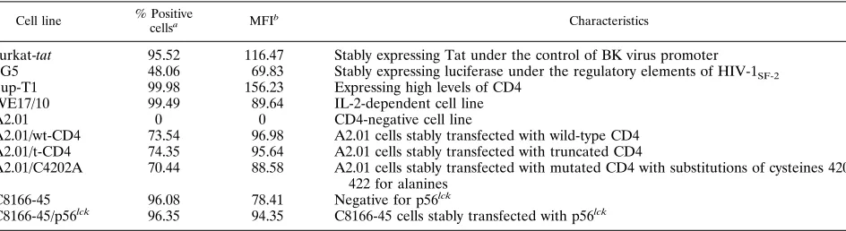
[image:2.612.62.534.83.212.2]Cross-linking of CD4 by different isolates of HIV-1 leads to a decrease in HIV-1 LTR-dependent gene activity.To investi-gate the modulatory effects of virus-mediated CD4 cross-link-ing on the HIV-1 regulatory elements, a molecular construct harboring the LTR region positioned upstream of the lucif-erase reporter gene (pLTRX-LUC) was introduced into sev-eral T-lymphoid cell lines by transient transfection. Character-istics of these cell lines are presented in Table 1. Surface CD4 expression determined by fluorescence-activated cell sorter
TABLE 1. Description of cell lines used in our studies
Cell line % Positive
cellsa MFIb Characteristics
Jurkat-tat 95.52 116.47 Stably expressing Tat under the control of BK virus promoter
1G5 48.06 69.83 Stably expressing luciferase under the regulatory elements of HIV-1SF-2
Sup-T1 99.98 156.23 Expressing high levels of CD4
WE17/10 99.49 89.64 IL-2-dependent cell line
A2.01 0 0 CD4-negative cell line
A2.01/wt-CD4 73.54 96.98 A2.01 cells stably transfected with wild-type CD4 A2.01/t-CD4 74.35 95.64 A2.01 cells stably transfected with truncated CD4
A2.01/C4202A 70.44 88.58 A2.01 cells stably transfected with mutated CD4 with substitutions of cysteines 420 and 422 for alanines
C8166-45 96.08 78.41 Negative for p56lck
C8166-45/p56lck 96.35 94.35 C8166-45 cells stably transfected with p56lck
aPercentage of CD41cells as measured by flow cytometry with the Leu-3A anti-CD4 antibody (Becton Dickinson).
bMFI, linear-scale mean fluorescence intensity of CD4 on studied cells.
on November 9, 2019 by guest
http://jvi.asm.org/
analysis with the anti-CD4 Leu-3A antibody is also indicated for each cell line.
We first sought to determine whether the binding of HIV-1 to cell surface CD4 could regulate HIV-1 LTR activity. Since we judged that it was critical for our assays to abolish subse-quent postbinding steps in the virus replicative cycle, experi-ments were carried out with defective HIV-1IIIB harvested
from the cellular clone UHC-8. Viral particles produced by this cell line have been demonstrated to be devoid of both reverse transcriptase and integrase proteins (8). Furthermore, two lab-oratory isolates (HIV-1IIIRFand HIV-1IIIB) and one clinical
isolate (HIV-1334) of HIV-1 were also tested. In this set of
experiments, frozen virus stocks were thawed and heat inacti-vated before incubation with transfected cells, as this physical treatment has been shown to abolish virus infectivity without disrupting binding (53). LTR-driven luciferase activity was monitored after a 24-h incubation period with viruses, based on preliminary experiments which demonstrated that optimal luciferase activity was obtained at this time point (data not shown). As shown in Fig. 1A, all HIV-1 strains tested in trans-fected Jurkat-tatcells inhibited LTR-driven expression. Inter-estingly, the degree of inhibition was stronger with defective particles (74%) than with other heat-inactivated strains of HIV-1 (ranging from 17 to 39%) despite the use of half as many viruses, as estimated by their p24 content. The inhibitory effect on luciferase gene expression induced by defective HIV-1IIIBparticles was next evaluated in other CD41T-lymphoid
cell lines and gave levels of inhibition ranging between 31 and 71% (Fig. 1B), thereby suggesting that this phenomenon is not cell type specific. The presence of surface CD4 was found to be important as the lowest inhibitory effect was seen in 1G5 (31%), a cell line expressing lower levels of CD4 on a lower percentage of cells (Table 1). More importantly, treatment of the CD4-negative A2.01 T-cell line with defective HIV-1IIIB
did not result in a decrease of LTR-driven luciferase gene expression, thereby supporting even further the critical role played by CD4 in this phenomenon. Data with the A2.01 cell line also suggest that the virus-mediated diminution of HIV-1 LTR-driven luciferase activity was not mediated by cellular soluble factor(s) that may affect cell proliferation and/or via-bility. Moreover, our results indicate that the strain origin of the LTR is not a determinant in the inhibitory process, as the LTR from the pLTRX-LUC construct is derived from HIV-1LAI, while the regulatory elements of the integrated
LTR-dependent luciferase vector in 1G5 cells originate from HIV-1SF-2.
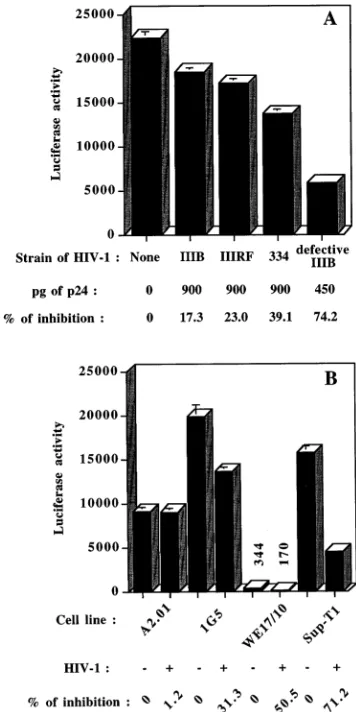
The necessity of the presence of HIV-1 for this effect to occur was next investigated by using Sup-T1 since maximal inhibition of LTR-dependent reporter gene activity was seen in these cells. A dose-dependent decrease of HIV-1 LTR-modu-lated gene expression was seen when using increasing concen-trations of defective HIV-1IIIB(Fig. 2A). Inhibition of
lucif-erase activity was found to be specific for the regulatory elements of HIV-1, as incubation of transfected Jurkat-tatand Sup-T1 cells with defective HIV-1IIIBparticles had no
inhibi-tory effect on luciferase activity when this reporter gene was placed under the control of the immediate early enhancer/ promoter region of the human cytomegalovirus (Fig. 2B). Fur-thermore, the observation that shutoff of transcription does not extend to all gene promoters is another indication that the virus-induced repression of HIV-1 LTR-driven luciferase ac-tivity is independent of soluble cellular factors.
Repression of LTR-dependent gene expression is indepen-dent of Tat and of PMA-induced translocation of NF-kB.To delineate the mechanism of virally induced repression of LTR-mediated gene activity, virus particles were incubated with
transfected Sup-T1 cells in the absence or presence of the transactivating viral Tat protein. As depicted in Table 2, sim-ilar repression of LTR-dependent luciferase activity was seen in the presence of the viral Tat protein (69%) or in a Tat-free system (71%). As expected, the addition of the vector encoding Tat was associated with an increase in reporter gene activity even though three times less LTRX-LUC was used in this transfection.
Next, to investigate the putative involvement of the tran-scription factor NF-kB, experiments were performed in the presence or absence of the phorbol ester PMA, a known in-ducer of NF-kB nuclear translocation (58). We detected com-parable levels of inhibition when Jurkat-tat cells were incu-bated with defective HIV-1IIIBin the presence or absence of
[image:3.612.345.523.70.426.2]PMA (Table 3).
FIG. 1. Inhibition of LTR-directed reporter gene expression is induced by different strains of HIV-1 and is observed in several T-lymphoid cell lines. (A) Jurkat-tatcells were transfected by DEAE-dextran with pLTRX-LUC at 15
mg/103106cells and were next incubated for 24 h at 378C with PMA and HIV-1
(900 pg of p24 from heat-inactivated frozen stocks of HIV-1IIIB, HIV-1IIIRF, and
HIV-1334; 450 pg of p24 from defective HIV-1IIIBharvested from fresh UHC-8
cell supernatant). (B) 1G5 cells were transfected by DEAE-dextran with 15mg of pCMVtat; Sup-T1, WE17/10, and A2.01 cells were cotransfected with 5mg of pLTRX-LUC and 10mg of pCMVtat. Cells were next incubated for 24 h at 378C with PMA and 400 pg of p24 from defective HIV-1IIIBharvested from fresh
UHC-8 cell supernatant. Luciferase activity was monitored as described in Ma-terials and Methods. Results shown are the mean values (in 103
cpm) of triplicate samples6standard deviations. The percentage of inhibition of luciferase activity was calculated with the formula % inhibition5100312(mean value for PMA-treated cells incubated with HIV-1/mean value for PMA-treated cells).
on November 9, 2019 by guest
http://jvi.asm.org/
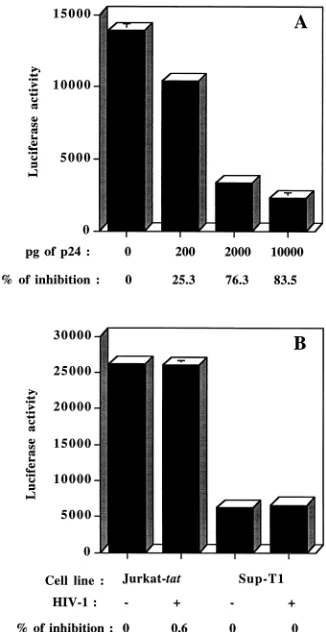
Interaction between gp120 and CD4 is required to achieve a virally mediated repression of HIV-1 LTR-driven gene activity. We next evaluated the role played by gp120-CD4 interactions in the present phenomenon by exploiting the reported weak association between the external envelope gp120 and the trans-membrane gp41 proteins, which frequently results in shedding of surface gp120 spikes from the virion (25). As shown in Fig. 3A, inhibition of LTR-driven luciferase activity was markedly decreased when Sup-T1 cells were incubated with defective HIV-1IIIBparticles denuded of their external envelope protein
gp120 by freeze-thaw cycle(s). These results may explain the
diminished ability demonstrated by HIV-1IIIRF, HIV-1IIIB, and
HIV-1334 to repress LTR-driven reporter gene expression
compared with defective particles originating from fresh UHC-8 culture supernatant (Fig. 1A). Treatment with 20mg of soluble CD4 per ml was sufficient to almost abrogate the HIV-1-mediated decrease of LTR-driven luciferase activity (Fig. 3B), thereby reinforcing the notion that the binding of HIV-1 to its primary cellular receptor is an event that is required to generate the virus-mediated decrease in HIV-1 LTR-driven reporter gene expression.
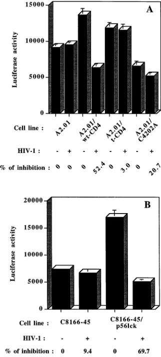
The signaling event leading to virus-mediated repression of HIV-1 LTR-driven gene activity requires the cytoplasmic do-main of CD4 and is transduced via p56lck
-dependent and -in-dependent pathways.We next investigated the importance of the cytoplasmic domain of CD4 by introducing three different CD4 constructs within the A2.01 CD4-negative cell line. No decrease in HIV-1 LTR-driven reporter gene activity was seen either for the parental CD4-negative cell line or for cells ex-pressing a CD4 molecule truncated at position 402 (A2.01/t-CD4), which can no longer interact with endogenous p56lck. However, introduction of a full-length CD4 glycoprotein (A2.01/wt-CD4) gave rise to a marked diminution of luciferase activity following CD4 cross-linking with defective HIV-1IIIB
particles (inhibition of 52%) (Fig. 4A). On the other hand, the expression of a CD4 construct with cysteines at positions 420 and 422 substituted for alanines (A2.01/C4202A), residues that have been reported to be responsible for the physical associa-tion of CD4 with p56lck (68, 78), restored only partially the virus-mediated inhibition of HIV-1 LTR-driven reporter gene expression (inhibition of 21%). On the basis of these results suggesting that the signaling pathway is mediated at least in part via p56lck, it was of interest to further investigate the role played by this CD4-associated tyrosyl kinase in this process. To more directly measure the importance of p56lck, we stably transfected the p56lck-negative C8166-45 cell line with a cDNA coding for the wild-type p56lckprotein. Incubation of the pa-rental p56lck-negative cell line with defective HIV-1
IIIB
re-sulted in a minimal diminution of LTR-driven luciferase activ-ity (9%), while the expression of p56lckwas associated with a marked enhancement of the observed inhibition (70%) (Fig. 4B). Altogether, these results indicate that the cytoplasmic domain of CD4 is necessary for the HIV-1-induced repression of LTR-mediated gene activity and suggest that the CD4-associated tyrosyl kinase p56lck is also one of the elements participating in the signaling pathway.
DISCUSSION
The aim of this study was to determine the effect of virus-mediated CD4 cross-linking on viral gene expression in an effort to elucidate the molecular basis for our previous obser-vations suggesting that the binding of HIV-1 particles to CD4 down-modulates virus expression in infected cells acutely in-fected with HIV-1 (76). In the present study, we used a
mo-FIG. 2. Virus-mediated repression of LTR-driven reporter gene activity is dose dependent on concentrations of HIV-1 added and is specific for the regu-latory elements of HIV-1. (A) Sup-T1 cells were cotransfected with 5mg of pLTRX-LUC and 10mg of pCMVtatbefore a 24-h incubation with PMA and increasing concentrations of defective HIV-1IIIBharvested from fresh UHC-8
cell supernatant. (B) Jurkat-tatwas transfected with pCMV-LUC, and Sup-T1 was cotransfected with pCMV-LUC and pCMVtat. Next, cells were incubated for 24 h at 378C with PMA and 475 pg of p24 from defective HIV-1IIIBharvested
[image:4.612.96.259.70.386.2]from fresh UHC-8 cell supernatant. Data shown are the means6standard deviations of triplicate samples (in 103cpm).
TABLE 2. HIV-1-induced repression of LTR-driven luciferase activity in the presence or absence of the viral Tat protein Cell line and conditionsa
Luciferase activityb
% of inhibition
Sup-T11pLTRX-LUC 1,577677 0
Sup-T11pLTRX-LUC1450 pg of p24 HIV-1 (UHC-8) 45460.6 71.18
Sup-T11pLTRX-LUC1pCMVtat 7,5346563 0
Sup-T11pLTRX-LUC1pCMVtat1450 pg of p24 HIV-1 (UHC-8) 2,3686483 68.56
a
Sup-T1 cells were transfected with 15mg of pLTRX-LUC alone or cotransfected with 5mg of pLTRX-LUC and 10mg of pCMVtatprior to incubation for 24 h at 378C with PMA in the presence or absence of defective HIV-1IIIB.
b
Results shown are the mean 103
cpm6standard deviations of triplicate samples.
on November 9, 2019 by guest
http://jvi.asm.org/
[image:4.612.58.556.643.702.2]lecular construct consisting of a vector containing the regula-tory elements of HIV-1 (LTR region) placed in front of the reporter luciferase gene. Others have also reported that cross-linking of CD4 glycoproteins can inhibit HIV-1 gene expres-sion. Indeed, it was shown that an anti-CD4 antibody (IOT4A/ 13B8-2), specific for the CDR3-like region of CD4, was able to inhibit HIV-1 replication at a late step in the virus replicative cycle by blocking provirus transcription (4, 5, 7). In in vitro tissue culture experiments, with peripheral blood mononuclear cells isolated from HIV-1-infected individuals stimulated with anti-CD3 antibodies, virus production from such latently
[image:5.612.355.516.267.623.2]in-fected cells was markedly diminished by treatment with recom-binant particles carrying HIV-1 envelope glycoproteins (29). Interestingly, inhibition was not detected after treatment with recombinant HIV-1 particles expressing Gag proteins, suggest-ing that CD4 bindsuggest-ing was required to achieve the observed inhibitory effect.
FIG. 3. Inhibition of LTR-dependent luciferase activity mediated by HIV-1 requires intact virus particles and is inhibited by soluble CD4. (A) Sup-T1 cells were first cotransfected with 5mg of pLTRX-LUC and 10mg of pCMVtatprior to 24 h of incubation with PMA and defective HIV-1IIIBcontained in UHC-8 cell
supernatant (200 pg of p24) either fresh or subjected to one or two freeze-thaw cycles. (B) Similar experiments were carried out with defective HIV-1IIIB
parti-cles harvested from fresh UHC-8 culture supernatant (50 pg of p24) in the presence of increasing concentrations of soluble CD4 (0, 0.2, and 2.0mg of soluble CD4 per ml). Data shown represent the means (in 103cpm) of triplicate
samples6standard deviations.
FIG. 4. Inhibition of LTR-driven gene expression by HIV-1 is dependent on the cytoplasmic domain of CD4 and partly on p56lck. The parental A2.01 cell line
(CD4 negative) and stable A2.01 transfectants expressing either the full-length version (A2.01/wt-CD4), the truncated version (A2.01/t-CD4), or a mutated form of CD4 (A2.01/C4202A) were cotransfected with 5mg of pLTRX-LUC and 10
mg of pCMVtat. These cells were next incubated for 24 h at 378C with PMA and defective HIV-1IIIBcontained in fresh UHC-8 cell supernatant (500 pg of p24).
(B) The parental p56lck-negative C8166-45 cell line and C8166-45 cells stably
transfected with cDNA encoding wild-type p56lckwere cotransfected with 5mg of
pLTRX-LUC and 10mg of pCMVtat. These cells were next incubated for 24 h with PMA and defective HIV-1IIIBcontained in fresh supernatant from UHC-8
(450 pg of p24). Data represent the means (in 103cpm) of triplicate samples6
[image:5.612.95.261.321.640.2]standard deviations.
TABLE 3. HIV-1-induced repression of LTR-driven luciferase activity in cells treated or not with the phorbol ester PMA Cell line and conditionsa
Luciferase activityb
% of inhibition
Jurkat-tat 18,5196266 0
Jurkat-tat1450 pg of p24 HIV-1 (UHC-8) 5,6886669 69.29
Jurkat-tat1PMA 144,37262,105 0
Jurkat-tat1PMA1450 pg of p24 HIV-1 (UHC-8) 41,2826139 71.41
a
Jurkat-tatcells were transfected with 15mg of pLTRX-LUC prior to incubation for 24 h at 378C in the presence or absence of PMA and defective HIV-1IIIB.
b
Results shown are the mean 103
cpm6standard deviations of triplicate samples.
on November 9, 2019 by guest
http://jvi.asm.org/
We first investigated whether the binding of the virus to its cell surface receptor could induce repression of HIV-1 LTR-driven luciferase gene expression. We found that a decrease of LTR-dependent reporter gene activity was detected when transfected cells were incubated with different strains of HIV-1. A more pronounced inhibition of LTR-driven lucif-erase activity was seen with defective HIV-1 particles. We have presented evidence indicating that this seems associated with the fact that fresh supernatant was the source of defective particles, while heat-inactivated virus stocks were submitted to a freeze-thaw cycle before their use. The demonstration that freeze-thaw cycles abrogate the ability of HIV-1 to inhibit LTR-mediated reporter gene activity supports this notion. Fur-thermore, the inhibition process was diminished in a dose-dependent fashion by treatment with soluble CD4 and was not seen in CD4-negative cells, indicating that the binding of HIV-1 particles to the cell surface CD4 glycoprotein is essen-tial for the repression to occur. The virus-mediated decrease of HIV-1 LTR-driven luciferase activity is not a restricted phe-nomenon, as it was seen in six different CD41T-lymphoid cell lines. The specificity for the regulatory elements of HIV-1 was demonstrated by the inability to achieve a similar inhibitory effect when cDNA coding for luciferase was placed under the control of the enhancer/promoter sequences of the cytomega-lovirus.
The cross-linking of CD4 with antibodies has been shown to lead to a rapid and strong autophosphorylation of p56lckand to an increase of its kinase activity on exogenous substrates (49). We believe that binding of HIV particles to CD4-expressing cells will also lead to multimerization of its receptor. Indeed, it is logical to assume that HIV, which has a mean diameter of 100 nm and possesses an average of 216 to 288 gp120 mole-cules per virion (26), will induce CD4 cross-linking at the contact site. It is thus likely that the observed inhibitory effect is dependent on multimerization of CD4 molecules and might thus be dependent on an interaction between the cytoplasmic region of CD4 and the protein tyrosine kinase p56lck. The present transfection assays do indeed indicate that the CD4-p56lckcomplex plays a critical role in virus-mediated repression of HIV-1 LTR-dependent reporter gene activity. However, our results revealed that, although weaker, another signaling path-way that is p56lckindependent is also participating in the virus-mediated repression of LTR-driven gene activity. This is based on experiments showing partial inhibition of LTR-mediated luciferase activity in cells expressing CD4 with substitutions of cysteines 420 and 422 for alanines and in a cell line that lacks endogenous p56lck. Further studies are needed to identify cel-lular signaling intermediates that couple HIV-1-mediated CD4 cross-linking with a decrease of LTR-driven gene expression. The observed repression of HIV-1 LTR-dependent gene activity generated by the virus binding event is most likely attributable to cellular protein(s) interacting with DNA or RNA located within the regulatory elements of HIV-1. Nu-merous studies have shown that the regulatory elements of HIV-1 respond to several cellular transcription factors (re-viewed in reference 24). The most widely studied binding do-main of LTR is the NF-kB element, which acts as an enhancer element in HIV-1 regulation (58). Most of our assays were performed in the presence of the phorbol ester PMA, which has been reported to lead to the dissociation of the IkB–NF-kB protein complex and to the translocation of NF-kB to the nucleus (46). Moreover, treatment with PMA alone has been reported to efficiently activate the regulatory elements of HIV-1, which are composed of two NF-kB binding sites (75). Experiments carried out in the absence of PMA gave compa-rable levels of inhibition of LTR-dependent reporter gene
expression in transfected cells incubated with HIV-1IIIB.
Therefore, it is likely that the PMA-induced increase in the amount of NF-kB binding to HIV-1 LTR is not modulated by cross-linking of CD4 with viral particles. However, our studies cannot permit us to clearly demonstrate that the inhibition is totally NF-kB independent. Indeed, basal levels of NF-kB could similarly be affected by the binding of HIV-1 to its cellular receptor. This is supported by a recent study by Jabado et al. showing that CD4 cross-linking mediated either with different anti-CD4 antibodies or with purified gp120 prior to T-cell stimulation caused a reduction in NF-kB binding to the regulatory elements of IL-2 (34). Experiments performed with an HIV-1 LTR mutant with deletions in the NF-kB binding sites would permit more direct measurement of the role played by NF-kB in the inhibition process. Transcription of all viral messages is also markedly upregulated by the viral activating protein Tat (2, 71). We have determined that the observed inhibition is Tat independent, as a similar HIV-1-mediated repression of LTR-driven luciferase activity was detected in transfected CD41T cells despite the absence of this transac-tivating viral protein. This indicates that our mode of inhibition of the HIV-1 LTR is different from the one recently described in some studies showing a Tat-dependent mode of inhibition of LTR activity via the binding of antibodies to the CDR3-like region of the CD4 molecule (4, 5, 7).
Benkirane et al. have recently published that cross-linking of CD4 with heat-inactivated HIV-1LAI induced activation of
LTR-driven chloramphenicol acetyltransferase expression via an increased nuclear translocation of NF-kB, suggesting that the binding of HIV-1 particles to CD4-expressing cells results in signal transduction that will positively modulate virus repli-cation (6). These observations are not in agreement with the present data and with previous findings from the same group and others which demonstrated that antibody-mediated cross-linking of CD4 can repress replication of HIV-1 at a postbind-ing step (4, 10, 14, 31, 56). Additional studies are required to elucidate such a discrepancy. However, the fact that we have detected a decrease in LTR-directed reporter gene activity mediated by cross-linking of CD4 with three different HIV-1 isolates in six different T-cell lines is a strong indication that our observation is not fortuitous and is a broad phenomenon. Furthermore, since HIV-1 is known to be modulated at the transcriptional level after signaling through the TCR (75), it is logical that TCR-independent CD4 cross-linking, which has been reported to generate suboptimal T-cell stimulation (3, 66, 74, 80), might negatively affect LTR-dependent gene expres-sion.
Several lines of evidence clearly suggest that the integral membrane CD4 protein is an active component of the signal-ing cascade operatsignal-ing in T cells. Thus, ligation of CD4 by the external envelope gp120 glycoprotein can induce pleiotropic effects such as down-modulation of cell surface CD4, induction of p56lckactivation, dissociation of p56lckfrom CD4, IL-6 pro-duction, and inhibition of anti-TCR-CD3-induced activation (12, 32, 37, 62). We now demonstrate that cross-linking of cell surface CD4 glycoprotein with HIV-1 particles can also lead to repression of HIV-1 LTR-driven gene expression.
Our data suggest for the first time that HIV-1 can down-modulate its own replication by mediating a cascade of bio-chemical events leading to the repression of HIV-1 LTR-de-pendent gene expression through its primary cellular receptor. This is consistent with the in vivo situation in which the fre-quency of cells carrying transcriptionally active HIV-1 proviral DNA was reported to be at least 1 to 2 orders of magnitude lower than the total amount of infected cells (30), indicating that the majority of infected cells exist in a state of
on November 9, 2019 by guest
http://jvi.asm.org/
tional latency. The demonstration that defective HIV-1 parti-cles can repress LTR-driven gene expression coupled with results indicating that the great majority of proviral DNA and circulating particles in HIV-1-infected individuals are defective (28, 47, 54, 64) suggests that noninfectious viruses may play a cardinal role in transcriptional latency. This may represent a strategy used by HIV-1 to favor virus transmission by prevent-ing a rapid destruction of the host. Our data also suggest a novel mechanism by which defective particles could limit or interfere with replication of infectious viruses. Understanding of the mechanism of repression and of the cellular factor(s) involved in the present phenomenon may yield insights into the replicative cycle of HIV-1 and will permit the design of new therapeutic strategies that are less toxic and more efficient for the treatment of AIDS. Experiments are now in progress to identify cellular transducing elements involved in the signaling cascade which is initiated by the virus binding event.
ACKNOWLEDGMENTS
P. Be´rube´ and B. Barbeau contributed equally to this work. We thank Guylaine Briand, Christine Fargas, Sylvie Perron, Richard Bernier, and Sophie Gratton for technical assistance and Maurice Dufour for expert assistance in flow cytometry studies. We are in-debted to Olivier Schwartz for the kind gift of pLTRX-LUC and pCMVtatand to Andre´ Darveau for the pCMV-LUC construct. We are also thankful to R. Sweet (SmithKline Beecham) for providing the soluble CD4.
R.C. is the recipient of a Ph.D. fellowship from the Fonds pour la Formation de Chercheurs et l’Aide a` la Recherche. R.-P.S. holds an MRC scientist award. M.T. is supported by a scholarship award from the Fonds de la Recherche en Sante´ du Que´bec. M.T. is funded by grants from the National Health Research and Development Program/ Medical Research Council of Canada (4305-AIDS and 6605-4527-AIDS) and the Canadian Foundation for AIDS Research.
REFERENCES
1.Aguilar-Cordova, E., J. Chinen, L. Donehower, D. E. Lewis, and J. W. Belmont.1994. A sensitive reporter cell line for HIV-1tatactivity, HIV-1 inhibitors, and T cell activation effects. AIDS Res. Hum. Retroviruses10: 295–301.
2.Arya, S. K., C. Guo, O. Joseph, and F. Wong-Staal.1985. Trans-activator gene of human T lymphotropic virus type III. Science229:69–74. 3.Bank, I., and L. Chess.1985. Perturbation of the T4 molecule transmits a
negative signal to T cells. J. Exp. Med.162:1294–1303.
4.Benkirane, M., P. Corbeau, V. Housset, and C. Devaux.1993. An antibody that binds the immunoglobulin CDR3-like region of the CD4 molecule inhibits provirus transcription in HIV-infected T cells. EMBO J.12:4909– 4921.
5.Benkirane, M., M. Hirn, D. Carrie`re, and C. Devaux.1995. Functional epitope analysis of the human CD4 molecule: antibodies that inhibit human immunodeficiency virus type 1 gene expression bind to the immunoglobulin CDR3-like region of CD4. J. Virol.69:6898–6903.
6.Benkirane, M., K.-T. Jeang, and C. Devaux.1994. The cytoplasmic domain of CD4 plays a critical role during the early stage of HIV infection in T-cells. EMBO J.13:5559–5569.
7.Benkirane, M., H. Schmid-Antomarchi, D. R. Littman, M. Hirn, B. Rossi, and C. Devaux.1995. The cytoplasmic tail of CD4 is required for inhibition of human immunodeficiency virus type 1 replication by antibodies that bind to the immunoglobulin CDR3-like region in domain 1 of CD4. J. Virol. 69:6904–6910.
8.Bernier, R., and M. Tremblay.1995. Homologous interference resulting from the presence of defective particles of human immunodeficiency virus type 1. J. Virol.69:291–300.
9.Boulerice, F., S. Bour, R. Geleziunas, A. Lvovich, and M. A. Wainberg.1990. High frequency of defective human immunodeficiency virus type 1 and heterogeneity of viral expression in clones of infected U937 cells. J. Virol. 64:1745–1755.
10.Burkly, L. C., D. Olson, R. Shapiro, G. Winkler, J. J. Rosa, D. W. Thomas, C. Williams, and P. Chisholm.1992. Inhibition of HIV infection by a novel CD4 domain 2-specific monoclonal antibody. Dissecting the basis for its inhibitory effect on HIV-induced cell fusion. J. Immunol.149:1779–1787. 11.Caputo, A., J. G. Sodroski, and W. A. Haseltine.1990. Constitutive
expres-sion of HIV-1 Tat protein in human Jurkat T cells using a BK virus vector. J. Acquired Immune Defic. Syndr.3:372–379.
12. Cefai, D., M. Ferrer, N. Serpente, T. Idziorek, A. Dautry-Varsat, P. Debre, and G. Mismuth.1992. Internalization of HIV glycoprotein gp120 is asso-ciated with down-modulation of membrane CD4 and p56lcktogether with
impairment of T cell activation. J. Immunol.149:285–294.
13. Chimurle, N., V. S. Kalyanaraman, N. Oyaizu, H. B. Slade, and S. Pahwa. 1990. Inhibition of functional properties of tetanus antigen-specific T-cell clones by envelope glycoprotein gp120 of human immunodeficiency virus. Blood75:152–159.
14. Corbeau, P., M. Benkirane, R. Weil, C. David, S. Emiliani, D. Olive, C. Mawas, A. Serre, and C. Devaux.1993. Ig CDR3-like region of the CD4 molecule is involved in HIV-induced syncytia formation but not in viral entry. J. Immunol.150:290–301.
15. Cruikshank, W. W., D. M. Center, S. W. Pyle, and H. Kornfeld.1990. Biologic activities of HIV-1 envelope glycoprotein: the effects of crosslink-ing. Biomed. Pharmacother.44:5–11.
16. Dalgleish, A. G., P. C. L. Beverly, P. R. Clapham, D. H. Crawford, M. F. Greaves, and R. A. Weiss.1984. The CD4(T4) antigen is an essential com-ponent of the receptor for the AIDS retrovirus. Nature (London)312:763– 767.
17. Diamond, D. C., B. P. Sleckman, T. Gregory, L. A. Lasky, J. L. Greenstein, and S. J. Burakoff.1988. Inhibition of CD41T cell function by the HIV envelope protein. J. Immunol.141:3715–3717.
18. Doyle, C., and J. Strominger.1987. Interaction between CD4 and class II MHC molecules mediates cell adhesion. Nature (London)330:256–259. 19. Eichmann, K., J. Jonsson, I. Falk, and F. Emmrich.1987. Effective
activa-tion of resting mouse T lymphocyte by crosslinking submitogenic concentra-tions of the T cell antigen receptor with either Lyt-2 or L3T4. Eur. J. Immunol.17:643–650.
20. Emmrich, F., L. Kantz, and K. Eichmann.1987. Cross-linking of the T cell receptor complex with the subset-specific differentiation antigen stimulates interleukin 2 receptor expression in human CD4 and CD8 T cells. Eur. J. Immunol.17:529–534.
21. Fields, A. P., D. P. Bednarik, A. Hess, and W. S. May.1988. Human immu-nodeficiency virus induces phosphorylation of its cell surface receptor. Na-ture (London)333:278–280.
22. Folks, T., D. M. Powell, M. M. Lightfoote, S. Benn, M. A. Martin, and A. S. Fauci.1986. Induction of HTLV-III/LAV from a non virus-producing T-cell line: implications for latency. Science231:600–602.
23. Gay, D. P., P. Maddon, R.-P. Se´kaly, A. Talle, M. Godfrey, E. Long, G. Goldstein, L. Chess, R. Axel, J. Kappler, and P. Marrack.1987. Functional interaction between human T-cell protein CD4 and the major histocompat-ibility complex HLA-DR antigen. Nature (London)328:626–629. 24. Gaynor, R.1992. Cellular transcription factors involved in the regulation of
HIV-1 gene expression. AIDS6:347–363.
25. Gelderblom, H. R., H. Reupke, and G. Pauli.1985. Loss of envelope antigens of HTLV-III/LAV, a factor in AIDS pathogenesis? Lancetii:1016–1017. 26. Gelderblom, H. S.1991. Assembly and morphology of HIV: potential effect
of structure and viral function. AIDS5:617–638.
27. Goldman, F., W. A. Jensen, G. L. Johnson, L. Heasly, and J. C. Cambier. 1994. gp120 ligation of CD4 induces activation and TCR desensitization independent of TCR tyrosine phosphorylation. J. Immunol.153:2905–2917. 28. Goodenow, M., T. Huet, W. Saurin, S. Kwok, J. Sninsky, and S. Wain-Hobson.1989. HIV-1 isolates are rapidly evolving quasispecies: evidence for viral mixtures and preferred nucleotide substitutions. J. Acquired Immune Defic. Syndr.2:344–352.
29. Haffar, O. K., P. A. Moran, M. D. Smithgall, M. L. Diegel, P. Sridhar, J. A. Ledbetter, J. M. Zarling, and S.-L. Hu.1992. Inhibition of virus production in peripheral blood mononuclear cells from human immunodeficiency virus (HIV) type 1-seropositive donors by treatment with recombinant HIV-like particles. J. Virol.66:4279–4287.
30. Harper, M. E., L. M. Marselle, R. C. Gallo, and F. Wong-Staal.1986. Detection of lymphocytes expressing human T-lymphotropic virus type III in lymph nodes and peripheral blood from infected individuals by in situ hy-bridization. Proc. Natl. Acad. Sci. USA83:772–776.
31. Hasunuma, T., H. Tsubota, M. Watanabe, Z. W. Chen, C. I. Lord, L. C. Burkly, J. F. Daley, and N. L. Letvin.1992. Regions of the CD4 molecule not involved in virus binding or syncytia formation are required for HIV-1 infection of lymphocytes. J. Immunol.148:1841–1846.
32. Hivroz, C., F. Mazerolles, M. Soula, R. Fagard, S. Gratton, S. Meloche, R.-P. Se´kaly, and A. Fischer.1993. Human immunodeficiency virus gp120 and derived peptides activate protein tyrosine kinase p56lck
in human CD4 T lymphocytes. Eur. J. Immunol.23:600–607.
33. Horak, I. D., M. Popovic, E. M. Horak, P. J. Lucas, R. E. Gress, C. H. June, and J. B. Bolen.1990. No T-cell tyrosine protein kinase signalling or calcium mobilization after CD4 association with HIV-1 or HIV-1 gp120. Nature (London)348:557–560.
34. Jabado, N., F. LeDeist, A. Fischer, and C. Hivroz.1994. Interactions of HIV gp120 and anti-CD4 antibodies with the CD4 molecule on human CD41T cells inhibits the binding activity of NF-AT, NF-kB and AP-1, three nuclear factors regulating interleukin-2 gene enhancer activity. Eur. J. Immunol. 24:2646–2652.
35. Janeway, C. A.1989. The role of CD4 in T-cell activation: accessory molecule
on November 9, 2019 by guest
http://jvi.asm.org/
or co-receptor? Immunol. Today10:234–238.
36.Julius, M., K. Newell, C. Maroun, and L. Haughn.1991. Functional conse-quences of CD4-TCR/CD3 interactions. Semin. Immunol.3:161–170. 37.Juszczak, R. J., H. Turchin, A. Truneh, J. Culp, and S. Kassis.1991. Effect
of human immunodeficiency virus gp120 glycoprotein on the association of the protein tyrosine kinase p56lckwith CD4 in human T lymphocytes. J. Biol.
Chem.266:11176–11183.
38.Kaufman, R., D. Laroche, K. Buchner, F. Hocho, C. Rudd, C. Lindchau, P. Ludwig, A. Hoer, E. Oberdisse, J. Kopp, I.-J. Korner, and H. Repke.1992. The HIV-1 surface protein gp120 has no effect on transmembrane signal transduction in T cells. J. Acquired Immune Defic. Syndr.15:760–770. 39.Klatzman, D., E. Champagne, S. Chamarest, J. Gruest, D. Guetard, T.
Hercend, J.-C. Gluckman, and L. Montagnier.1984. T-lymphocyte T4 mol-ecule behaves as the receptor for human retrovirus LAV. Nature (London) 312:767–768.
40.Koga, Y., N. Oh-Hori, H. Sato, N. Yamamoto, G. Kimura, and K. Nomoto. 1989. Absence of transcription of lck (lymphocyte specific protein-tyrosine kinase) message in IL-2-independent, HTLV-I-transformed T cell lines. J. Immunol.142:4493–4499.
41.Kornfeld, H., W. W. Cruikshank, S. W. Pyle, J. S. Berman, and D. M. Center. 1988. Lymphocyte activation by HIV-1 envelope glycoprotein. Nature (Lon-don)335:445–448.
42.Kupfer, A., S. J. Singer, C. A. Janeway, and S. L. Swain.1987. Coclustering of CD4 (L3T4) molecule with the T-cell receptor is induced by specific direct interaction of helper T cells and antigen-presenting cells. Proc. Natl. Acad. Sci. USA84:5888–5892.
43.Lasky, L., G. Nakamura, D. H. Smith, C. Fernie, C. Shimasaki, E. Patzer, P. Berman, T. Gregory, and D. J. Capon.1987. Delineation of a region of the human immunodeficiency virus type 1 gp120 glycoprotein critical for inter-action with the CD4 receptor. Cell50:975–985.
44.Ledbetter, J. A., L. K. Gilliland, and G. L. Schieven.1990. The interaction of CD4 with CD3/Ti regulates phosphorylation of substrates during T cell activation. Semin. Immunol.2:99–106.
45.Ledbetter, J. A., C. H. June, L. S. Grosmaire, and P. S. Rabinovitch.1987. Crosslinking of surface antigens causes mobilization of intracellular ionized calcium in lymphocytes. Proc. Natl. Acad. Sci. USA84:1384–1388. 46.Lenardo, M. J., and D. Baltimore.1989. NF-kB: a pleiotropic mediator of
inducible and tissue-specific gene control. Cell58:227–229.
47.Li, Y., J. C. Kappes, J. A. Conway, R. Price, G. M. Shaw, and B. H. Hahn. 1991. Molecular characterization of human immunodeficiency virus type 1 cloned directly from uncultured human brain tissue: identification of repli-cation-competent and -defective genomes. J. Virol.65:3973–3985. 48.Linette, G. P., R. J. Hartzman, J. A. Ledbetter, and C. H. June. 1988.
HIV-1-infected T cells show a selective signaling defect after perturbation of CD3/antigen receptor. Science241:573–576.
49.Luo, K., and B. M. Sefton.1990. Cross-linking of T-cell surface molecules CD4 and CD8 stimulates phosphorylation of thelcktyrosine protein kinase at the autophosphorylation site. Mol. Cell. Biol.10:5305–5313.
50.Mann, D. L., F. Lasane, M. Popovic, L. O. Arthur, W. Robey, W. A. Blattner, and M. J. Newman.1987. HTLV-III large envelope protein (gp120) sup-presses PHA-induced lymphocyte blastogenesis. J. Immunol.138:2640–2644. 51.Marrack, P., R. Endres, R. Shimonkewitz, A. Zlotnik, F. W. Dialynas, and J. Kappler.1983. The major histocompatibility complex-restricted antigen re-ceptor on T-cells: role of the L3T4 product. J. Exp. Med.158:1077–1091. 52.McDougal, J. S., M. S. Kennedey, J. M. Sligh, S. P. Cort, A. Mawle, and J. K.
Nicholson.1986. Binding of HTLV-III/LAV to T41cells by a complex of the 110 kDa viral protein and the T4 molecule. Science231:382–385. 53.McDougal, J. S., J. K. A. Nicholson, G. D. Cross, S. P. Cort, M. S. Kennedey,
and A. C. Mawle.1986. Binding of the human retrovirus HTLV-III/LAV/ ARV/HIV to the CD4 (T4) molecule: conformation dependence, epitope mapping, antibody inhibition, and potential for idiotypic mimicry. J. Immu-nol.137:2937–2944.
54.Meyerhans, A., R. Cheynier, J. Albert, M. Seth, S. Kwok, J. Sninsky, L. Morfeldt-Manson, B. Asjo¨, and S. Wain-Hobson.1989. Temporal fluctua-tions in HIV quasispecies in vivo are not reflected by sequential HIV isola-tions. Cell58:901–911.
55.Mittler, R. S., and M. K. Hoffman.1989. Synergism between HIV gp120 and gp120-specific antibody in blocking human T cell activation. Science245: 1380–1382.
56.Moore, J. P., J. A. McKeating, Y. Huang, A. Ashkenazi, and D. D. Ho.1992. Virions of primary human immunodeficiency virus type 1 isolates resistant to soluble CD4 (sCD4) neutralization differ in sCD4 binding and glycoprotein gp120 retention from sCD4-sensitive isolates. J. Virol.66:235–243. 57.Moscicki, R. A., E. P. Amento, S. M. Krane, J. T. Kurnick, and R. B. Colvin.
1983. Modulation of surface antigens of a human monocyte cell line, U937, during incubation with T lymphocyte-conditioned medium: detection of T4 antigen and its presence on normal blood monocytes. J. Immunol.131:743– 748.
58.Nabel, G., and D. Baltimore.1987. An inducible transcription factor
acti-vates expression of human immunodeficiency virus in T-cells. Nature (Lon-don)326:711–713.
59. Neudorf, S. M. L., M. M. Jones, B. M. McCathy, J. A. K. Harmony, and E. Choi.1990. The CD4 molecule transmits biochemical information important in the regulation of T lymphocyte activity. Cell. Immunol.125:301–314. 60. Orloff, G. M., M. S. Kennedey, C. Dawson, and J. S. McDougal.1991. HIV-1
binding to CD4 T cells does not induce Ca21influx or lead to activation of
protein kinases. AIDS Res. Hum. Retroviruses7:587–593.
61. Oyaizu, N., N. Chirmule, V. S. Kalyanaraman, W. W. Hall, R. A. Good, and S. Pahwa.1990. Human immunodeficiency virus type 1 envelope glycopro-tein gp120 produces immune defects in CD41T lymphocytes by inhibiting interleukin 2 mRNA. Proc. Natl. Acad. Sci. USA87:2379–2383.
62. Oyaizu, N., N. Chirmule, Y. Ohnishi, V. S. Kalyanaraman, and S. Pahwa. 1991. Human immunodeficiency virus type 1 envelope glycoproteins gp120 and gp160 induce interleukin-6 production in CD41T-cell clones. J. Virol. 65:6277–6282.
63. Peterson, A., and B. Seed.1988. Genetic analysis of monoclonal antibody and HIV binding sites on the human lymphocyte antigen CD4. Cell54:65–72. 64. Piatak, M., M. S. Saag, L. C. Yang, S. J. Clark, J. C. Kappes, K.-C. Luk,
B. H. Hahn, G. M. Shaw, and J. D. Lifson.1993. High levels of HIV-1 in plasma during all stages of infection determined by competitive PCR. Sci-ence259:1749–1754.
65. Reinherz, E. L., and S. F. Schlossman.1980. The differentiation and function of human T lymphocytes. Cell19:821–827.
66. Rosoff, P. M., S. J. Burakoff, and J. L. Greenstein.1987. The role of the L3T4 molecule in mitogen and antigen-activated signal transduction. Cell49:845– 853.
67. Schwartz, O., J.-L. Virelizier, L. Montagnier, and U. Hazan.1990. A micro-transfection method using the luciferase-encoding reporter gene for the assay of human immunodeficiency virus LTR promoter activity. Gene88: 197–205.
68. Shaw, A. S., K. E. Amrein, D. F. Hammond, D. F. Stern, B. M. Sefton, and J. K. Rose.1989. The lck tyrosine protein kinase interacts with the cytoplas-mic tail of CD4 glycoprotein through its unique amino-terminal domain. Cell 59:627–636.
69. Sleckman, B. P., A. Peterson, W. K. Jones, J. A. Foran, J. L. Greenstein, B. Seed, and S. J. Burakoff.1987. Expression and function of CD4 in a murine T-cell hybridoma. Nature (London)328:351–353.
70. Smith, S. D., M. Shatsky, P. S. Cohen, R. Warnke, M. P. Link, and B. E. Glader.1984. Monoclonal antibody and enzymatic profiles of human malig-nant T lymphoid cells and derived cell lines. Cancer Res.44:5657–5660. 71. Sodroski, J. G., R. Patarca, C. Rosen, F. Wong-Staal, and W. A. Haseltine.
1985. Location of the transactivation region on the genome of HTLV III. Science229:74–77.
72. Swain, S. L.1983. T cell subsets and the recognition of MHC class. Immunol. Rev.74:129–142.
73. Terhorst, C., A. van Agthoven, E. Reinherz, and S. Schlossman.1980. Bio-chemical analysis of human T lymphocyte differentiation antigens T4 and T5. Science209:520–521.
74. Tite, J. P., A. Sloan, and C. A. Janeway, Jr.1986. The role of L3T4 in T cell activation: L3T4 may be both an Ia binding protein and a receptor that transduces a negative signal. J. Mol. Cell. Immunol.2:179–187.
75. Tong-Starksen, S. E., P. A. Luciw, and M. B. Peterlin.1989. Signaling through T lymphocyte surface proteins, TCR/CD3 and CD28, activates the HIV-1 long terminal repeat. J. Immunol.142:702–707.
76. Tremblay, M., S. Meloche, S. Gratton, M. A. Wainberg, and R.-P. Se´kaly. 1994. Association of p56lckwith the cytoplasmic domain of CD4 modulates
HIV-1 expression. EMBO J.13:774–783.
77. Tremblay, M., K. Numazaki, X. Li, M. Gornitsky, J. Hiscott, and M. A. Wainberg.1990. Resistance to infection by HIV-1 of peripheral blood mono-nuclear cells from HIV-1-infected patients is probably mediated by neutral-izing antibodies. J. Immunol.145:2896–2901.
78. Turner, J. M., M. H. Brodsky, B. A. Irving, S. D. Levin, R. M. Perlmutter, and D. R. Littman.1990. Interaction of the unique N-terminal region of tyrosine kinase p56lckwith cytoplasmic domains of CD4 and CD8 is
medi-ated by cysteine motifs. Cell60:755–765.
79. Wahl, L. M., M. L. Corcoran, S. W. Pyle, L. O. Arthur, A. Harel-Bellan, and W. L. Farrar.1989. Human immunodeficiency virus glycoprotein (gp120): induction of monocyte arachidonic acid metabolites and interleukin 1. Proc. Natl. Acad. Sci. USA86:621–625.
80. Wassmer, P., C. Chan, L. Lodgdeberg, and E. M. Shevach.1985. Role of the L3T4 antigen in T cell activation. II. Inhibition of T cell activation by monoclonal anti-L3T4 antibodies in the absence of accessory cells. J. Immu-nol.135:2237–2242.
81. Willard-Gallo, K., F. Van de Keere, and R. Kettman.1990. A specific defect in CD3-ggene transcription results in loss of T cell receptor/CD3 expression late after human immunodeficiency virus infection of a CD41T cell line. Proc. Natl. Acad. Sci. USA87:6713–6717.