A COMPARATIVE STUDY ON PREDICTIVE VALUE
OF MALIGNANCY IN SUSPICIOUS BREAST
MASSES OF BIRADS III & ABOVE CATEGORIES
USING SONOELASTOGRAPHY AND DYNAMIC MR
MAMMOGRAM
Dissertation submitted toTHE TAMILNADU Dr.M.G.R. MEDICAL UNIVERSITY
In partial fulfillment of the requirements Of
M.D. DEGREE EXAMINATION BRANCH- VIII- RADIODIAGNOSIS
GOVT KILPAUK MEDICAL COLLEGE CHENNAI- 600010
THE TAMILNADU DR.M.G.R. MEDICAL UNIVERSITY CHENNAI- TAMILNADU, INDIA
CERTIFICATE
This is to certify that the dissertation “A COMPARATIVE STUDY ON PREDICTIVE VALUE OF MALIGNANCY IN SUSPICIOUS BREAST MASSES OF BIRADS III & ABOVE CATEGORIES USING SONOELASTOGRAPHY AND DYNAMIC MR MAMMOGRAM” titled submitted by Dr.M.ALAMELU
appearing for M.D(RADIODIAGNOSIS) degree examination in May 2018 is a bonafide record of work done by her under my guidance and
supervision in partial fulfillment of requirement of the Tamilnadu
Dr.M.G.R. Medical University, Chennai. I forward this to the TamilNadu
Dr.M.G.R Medical University, Chennai.
Dr.J.DEVIMEENAL, DMRD,DNB,MD,FRCR
Guide, Professor and Head of Department, Department of Radiodiagnosis, Govt. Kilpauk Medical College, Chennai-600010
Prof.Dr. P. VASANTHAMANI, M.D., D.G.O.,MNAMS.,DCPSY.,MBA
Dean,
Govt Kilpauk Medical College, Chennai-600010
DECLARATION
I Dr. M. ALAMELU, solemnly declare that this dissertation “A
COMPARATIVE STUDY ON PREDICTIVE VALUE OF
MALIGNANCY IN SUSPICIOUS BREAST MASSES OF BIRADS III & ABOVE CATEGORIES USING SONOELASTOGRAPHY AND DYNAMIC MR MAMMOGRAM” is a bonafide work done by
me at Government Kilpauk Medical College, under the supervision of
Dr.J.DEVIMEENAL DMRD, DNB, MD, FRCR, Professor, Government Kilpauk Medical College. This dissertation is submitted to
the Tamil Nadu Dr. M.G.R Medical University, towards partial
fulfillment of requirement for the award of M.D. Degree Radiodiagnosis.
Place: Chennai Signature of the candidate
CERTIFICATE – II
This is to certify that this dissertation work titled entitled
dissertation “A COMPARATIVE STUDY ON PREDICTIVE VALUE
OF MALIGNANCY IN SUSPICIOUS BREAST MASSES OF
BIRADS III & ABOVE CATEGORIES USING
SONOELASTOGRAPHY AND DYNAMIC MR MAMMOGRAM”
of the candidate Dr. M. ALAMELU with Registration Number
201518251 for the award of M.D degree in the branch of
RADIODIAGNOSIS. I personally verified the urkund.com website for the purpose of plagiarism check. I found that the uploaded thesis file
contains from introduction to conclusion pages and result shows 2% of plagiarism in this dissertation.
ACKNOWLEDGEMENT
I express my heartful gratitude to the Dean, Prof.Dr.P.VASANTHA
MANI, M.D., D.G.O.,MNAMS.,DCPSY.,MBA Government Kilpauk
Medical College - 10 for permitting me to do this study.
I express my gratitude to my guide and my Professor
Dr.J.DEVIMEENAL, DMRD, DNB, MD, FRCR, Head of the department, Dept of Radiodiagnosis, Govt Kilpauk medical college for
her valuable guidance in doing the dissertation work and her expert
guidance and constant encouragement created an interest for me to pursue
this study. It is her constant supervision and support, that made me
possible to finish this study without much difficulty.
I am extremely thankful to my Associate professors
Dr.P.Chirtrarasan, MD(RD), & K.Gopinathan, MD(RD) Dr.R.Kanagasabai, DMRD, MD(RD) and other Assistant professors of Department of Radiodiagnosis, Govt. Kilpauk Medical College, Chennai
I also thank my past and present fellow postgraduates who helped
me in carrying out my work and preparing this dissertation.
I thank all Radiology technicians, staff nurses, and all the
paramedical staff members of our department for their co-operation in
conducting the study.
I thank my family members my husband Dr.M.Shanmugam, my
parents Mr.R.M.Mahalingam and Mrs.M.Dheivanai for their
understanding and co-operation for completion of this work.
Last but not the least; I owe my sincere gratitude to the patients and
their relatives who co-operated for this study, without whom the study
CONTENTS
S.NO CONTENTS PAGE
1 INTRODUCTION 1
2 REVIEW OF LITERATURE 8
3 AIMS AND OBJECTIVES 46
4 MATERIALS AND METHODS 47
5 CASES 56
6 STATISTICAL ANALYSIS AND RESULTS 73
7 DISCUSSION 87
8 CONCLUSION 95
9 BIBLIOGRAPHY
ABBREVATIONS PROFORMA
PATIENT CONSENT FORM MASTER CHART
ETHICAL COMMITTEE CERTIFICATE
INTRODUCTION
Breast masses include a wide range of pathologies, that can either
be benign or malignant lesions. Among all breast masses, fibroadenoma
is the most commonly diagnosed benign breast mass and Invasive Ductal
Carcinoma is the most common among the malignant masses [1].
Although most breast masses are benign in nature, carcinoma
breast is the most common malignancy in Indian women as reported by
Gupta et al [2] in 2016 and is the second leading cause of cancer related
deaths in women, which has recently overtaken the mortality rates of
cervical malignancies as stated by The National cancer registry of India.
India is now a country which has the largest estimated number of
breast cancer deaths worldwide. Breast cancer accounts for 27 % of all
cancers in women in India, with its incidence rising in the early thirties
and peaking at ages between 50-64 years. As for other cancers concerned
in India, late stage presentation is also a common scenario for breast
cancer [2].
assurance tool in mammography, ultrasound (USG) and Magnetic
Resonance Imaging (MRI) [3].
BIRADS 1and 2 lesions are clearly benign lesions. BIRADS 3 and
4 categories are intermediate lesions. BIRADS 5 and 6 are malignant.
There are various imaging modalities now available in the breast
radiology. Currently, Sonoelastography is an advanced sonographic
technique being used in the assessment of suspicious breast masses in
complement with the conventional B-mode Ultrasonogram.
Sonoelastography quantifies elasticity of the tissues by means of pressure
exerted on them.
The lesions are quantified according to the colour scale in
Sonoelastogram. Among various scoring methods, the Tsukuba elasticity
score is the most known and commonly used scoring systems in
elastography.[4]
There is a dramatic progress in the field of breast MRI over the
past decade. MRI has exceptionally high sensitivity for the detection of
breast cancer and it can aid in depicting cancers that are entirely occult on
conventional imaging.
Gadolinium contrast MRI is used to enhance the vascularity of
is recently being used to assess the exact nature of suspicious breast
masses.
Many investigators have detailed either enhancement kinetics of
the lesion or morphology of the lesion to differentiate benign from
malignant mass lesions identified on contrast-enhanced MR imaging
studies. But integration of both kinetic and morphologic data is ultimately
required to achieve optimal discrimination of pathology.
Breast biopsy is an invasive procedure aimed at confirming the
breast lesion detection, remains the gold standard in detection of breast
ANATOMY OF BREAST
EMBRYOLOGICAL DEVELOPMENT OF BREAST:
During 6th week of fetal life, primary mammary ridges (milk lines)
develops from axilla to medial thigh. In later life, only the mammary
ridge in the pectoral region develops into breast. During 12th to 16th week,
development of nipple – areolar complex begins. The breast mound
increases in size during puberty. Usual location of nipple is at 4th intercostal space.
The breast contains adipose tissue and glandular tissue. Breast
extends from 2nd to 6th ribs. Normal adult breast has nearly 15 to 20
segments which are demarcated by mammary ducts, converging in a
radial fashion at the nipple. The number of mammary segments and
mammary ducts vary in size. The average diameter of mammary duct is
2mm, converges into lactiferous sinus of 5 – 8 mm diameter.
Terminal Ductal lobular unit (TDLU):
TDLU are the basic functional as well as the basic histological unit
of breast [5]. The size of usual TDLU ranges from 1to 4 mm. The TDLU
composed of the extra lobular terminal duct, the intralobular terminal
The pathologies of TDLU are:
• Ductal Carcinoma In Situ (DCIS)
• Lobular Carcinoma In Situ (LCIS)
• Fibroadenoma
• Fibroadenosis and Apocrine metaplasia of breast
• Breast cysts
Venous drainage from breast is through axillary vein, internal thoracic and posterior intercostal veins.
Nerve supply to breast is via thoracic intercostals nerves from T3 – T5 and from supraclavicular nerve.
The lymphatic drainage of breast is via sappey’s plexus (subareolar plexus) into three routes [7]:
• Axillary or lateral pathway (predominant drainage)
• Internal mammary pathway
• Retro mammary pathway
Axillary nodes include the following group of lymph nodes:
Anterior (pectoral), Lateral (humeral), Posterior (subscapular), Central
Surgical classification of axillary group of nodes is:
• Level 1 – inferior to pectoralis minor muscle
• Level 2 – posterior to pectoralis minor muscle
REVIEW OF LITERATURE
INCIDENCE AND RISK FACTORS OF CARCINOMA BREAST:
An annual incidence of about 1,44,000 new cases of breast cancers
are reported in India, it has become now the most common female cancer
among urban population. It is been estimated that 1 in 28 Indian women
is likely to develop breast cancer during her lifetime. It is seen that
Indians have a much lower incidence carcinoma breast than that of
western countries, the incidence is- about one-third in urban areas and
one-ninth in rural regions of India [8].
Breast cancer incidence in Indian women varies from as low as 5
per 100,000 women per year in rural areas to 30 per 100,000 women per
year in urban areas as stated by National Cancer Registry Programme, ICMR: A consolidated report based on the hospital registries. There is a wide lack of population screening in Indians on par with corresponding
over diagnosis in western population is the contributing factor to this
varied statistics.
The etiologies for varying incidence in the breast cancer among
rural and urban women cannot be completely studied, which are more
likely attributed to the differences in their reproductive and lifestyle
age at first child birth, history of abortion, history of intake of oral
contraceptives and family history of Breast cancer [9, 10, 11]
Nulliparous women are at two fold higher risk of breast cancer
compared to multiparous women [10]. Early menarche less than 12 years) , multiple abortions (more than 2 ), less duration of breast feeding, and
consumption of excess dietary fat (especially animal fat) more than 30
g/day are all highly associated with breast cancer[12].
Women of young age with breast cancer are associated with larger
size of the tumor, more number of axillary lymph nodes, high tumour
grade, low rates of hormone receptor-positive status, earlier and frequent
loco regional recurrences, and poor overall survival rate[13].
Genes associated with increased risk of breast cancer are mainly
BRCA1 (Breast Cancer gene one) and BRCA2 (BReast CAncer gene two). Other genes related to development of carcinoma breast include
ATM, NBN, CDH1, PTEN, CHEK2, RAD50, RAD51C and TP53.
Women who have no awareness regarding the usual presenting
symptoms of breast cancer and women who do lack regular habit of self
examination of the breast are at increased risk of developing breast
Women with previous history of benign breast masses are also at
high risk of developing malignant masses [14].
There exists a huge need of efforts at researching, preserving and
propagating the factors which are likely to be associated with the
protection of Indian women from developing carcinoma breast.
There are vast number of ongoing studies & research works on the incidence and risk factors of developing carcinoma breast among
Indians.
Among various studies, the down listed three studies are vital ones.
The first vital study is on assessment of various risk factors of
carcinoma breast in Indian population is the case – control study
conducted by Nag et al., that aims at evaluation of differential risk factors for triple-negative breast carcinoma (TNBC) compared to Estrogen
Receptor-positive breast carcinoma[15]
The second vital study is also a case-control study that is looking
forward at risk factors such as weight and body size (waist – hip ratio,
BMI) of the affected Indian women patients with carcinoma breast [2]. The third study is a cohort study conducted in the rural region of
germline risk factors of developing carcinoma breast in a longitudinal
manner.
There are many research works and studies on the factors that can
protect from developing breast carcinoma. The protective factors assessed
are women who have completed their first full term pregnancy at younger
age (less than 30 years), female who completed more than 3 full term
pregnancies. Women with regular breast feeding history had significant
protection against the development of cancer breast as stated by Lai FM et al [16].
In the non-lactating young women, the breast parenchyma is
mainly the fibroglandular tissue, with little or no subcutaneous fat. With
increase in the age and parity of the women, increased fat deposition in
both the subcutaneous and retromammary zone occurs.
IMAGING OF BREAST MASSES:
The X-Ray mammogram is ever the best screening tool available for the detection of breast cancer. Mammography can either be screening
or diagnostic. It is a least expensive modality.
Regular mammographic views include Cranio-Caudal view and
Medio-Lateral Oblique views. Other supplementary views can be tailored
In general, bilateral breast mammogram is recommended for all
women beginning at the age of 40 years by the American College of Radiology, American Cancer Society, and American College of Surgeons. For the women who are with great risk of carcinoma breast development (i.e., those with higher than 20-25% lifetime risk), annual
surveillance with MRI is now being recommended [17]. Several researches focus that annual mammograms can aid in early detection of breast
cancers, so that breast-conservation therapies are possible.
The overall sensitivity of x-ray mammography in detecting the
breast cancer is about 85%. However, in women with dense breast, the
sensitivity of mammography is vastly reduced to 47.8-64.4% [18].
With the Digital tomosynthesis mammography, 3D views of the breast can be obtained, where multiple images of the breast are acquired
from different angles and reconstructed into the three-dimensional image
set.
The main advantage is that its ability to detect masses in dense
breasts that are likely to get missed on conventional mammogram
assessment of breasts masses.
dilated ducts and evaluate the cause of duct ectasis. Prior to cannulation
of ducts, a subareolar craniocaudal magnification view is obtained to
assess the presence of any calcifications or opacities in breast
parenchyma. A gentle periareolar massage with minimal pressure on
“trigger zone” may produce the nipple discharge. This zone is cannulated
with a straight tip or right angled tip cannula followed by slow injection
of iodinated contrast material. Imaging is then performed to assess the
dilated ducts and better evaluation of intraductal masses such as
papillomas or carcinomas.
High frequency linear array transducers and high-resolution
Ultrasonography with the combination of harmonic imaging and with real-time compounding helps in better characterization of the mass lesion
even in cases with dense breast. USG can aid in localizing small foci of
breast cancers that are readily missed on routine X- Ray mammography
screening [19].
The linear 5 to 12 MHz transducer is the optimal one in assessing
superficial parts of the body, including breasts, since it provides a better
lateral resolution. The harmonic imaging augments the resolution of sonographic images and also reduces the hindrance from reverberation or
B mode Ultrasonography is chosen as the initial imaging modality woman younger than 30 years presenting with the palpable
breast lump. USG aids in better evaluation of mammographic findings
such as mass like opacities and any focal asymmetric mammographic
densities.
Grid technique is widely adopted for routine breast ultrasound.
Scanning begins transversely in upper outer quadrant, and then
sliding from top to bottom inferiorly, the similar sweep is repeated in
Other adopted technique for breast USG is radial scanning pattern:
Starting at 12’o clock position, in the saggital plane, the probe is
turned around the nipple. The same is done in radial and anti-radial plane
Normal breast anatomy in USG is divided into three zones :
1. Pre mammary zone( skin and fat )
2. Mammary zone(fibroglandular tissue)
Breast lesions are located in terms of 4 quadrants: upper inner, upper outer, lower inner and lower outer.
Description in terms of clock positions can precisely locate the
The US lexicon of Sonomammogram includes six morphologic
features of the solid breast masses such as
• Shape of the lesion
• Orientation of the lesion
• Margin of the lesion
• Boundary of the lesion
• The echo texture of the lesion and
• Posterior acoustic features of the mass
Orientation and shape of the lesion:
Orientation of a breast mass can either be parallel or non parallel to
the skin surface of the breast.
Shapes can be round or oval. Round (spherical, circular or
globular) shape is associated with a relatively high probablity of
Oval (elliptical or egg like) shaped mass is described as “gently
lobulated” (having two to three fine undulations) or “macrolobulated”
(if more than three).
Margins of the lesion :
The margins of solid breast masses may either be circumscribed or
noncircumscribed.
Well defined mass with a sharp zone of transition from the
surrounding breast tissue is described to have circumscribed margins.
Presence of an indistinct zone of demarcation between the mass
and the surrounding breast tissue is noncircumscribed margins.
Angular margin is described if a lesion has sharp corners.
Spiculated margins are described when there are lines radiating from the
periphery of the mass.
Angular margins and spiculated margins are highly associated with
malignancy with their incidence rates of 60% and 86% respectively.
Lesion boundary:
Can either be a sharp interface between the mass lesion and the
surrounding normal breast or a wide echogenic transition zone which lack
sharp interface will be benign and with echogenic wide transition will be
malignant.
Internal echotexture:
The echotexture of a breast lesion is described in reference the
subcutaneous fat within the breast. The lesion can be hypoechoic,
isoechoic or hyperechoic compared to the subcutaneous fat.
Posterior acoustic features:
Posterior acoustic shadowing is a suspicious finding and may be
associated with cases of complex sclerosing lesion, invasive carcinoma,
postoperative scar, lymphoma or macrocalcifications and may even be
seen in normal patients with dense breast tissue..
In case of doubts in assessment of posterior acoustic enhancement,
compound imaging settings aid in sorting them better.
Other associated features to be looked for on Sonomammography are: the presence of
➢ Architectural breast parenchymal distortion
➢ Dilated ducts
➢ Vascularity of the lesion
Criteria for benign breast lesions on USG are [20],[21]:
• Masses with well circumscribed and smooth margins
• Hyperechoic masses, isoechoic or mildly hypoechoic masses
• Thin echoic capsule around the mass
• Oval shaped masses with the maximum diameter in the transverse
plane
• Three or less microlobulations.
Breast ultrasound criteria for characterisation of malignant lesions are:
• Mass having ill-defined borders
• Spiculated / angular margins
• Grossly hypoechoic lesion
• Taller than broader-the maximum diameter in the longitudinal
plane
• Associated Posterior acoustic shadowing and
• Microcalcifications
Doppler USG:
Malignant breast mass are associated with high vascularity, more
number of centrally located vessels. Since malignant neoplasms require
such as Resistive Index (RI), Pulsatility Index, and flow velocity can aid
in distinguishing the benign from malignant lesions. Malignant masses
mostly have a higher RI than the benign ones.
USG elastography:
The use of USG elastography is being increasingly used in
diagnosis of malignant breast masses, in recent times. The studies
conducted by Zhi H et al[22] and Itoh et al [23] on USG elastography
highlights that combined use of B-mode USG with Sonoelastography can
greatly augment the specificity and positive predictive value in precise
characterization of the breast masses. Elastography is a novel noninvasive
technique based on evaluation the stiffness or the elasticity of a lesion.
Elasticity is the mechanical property enabling a substance to get
deformed, on subjecting it to an external force and also to resume its
natural shape or size when the external force is removed.
Nightingale K. et al study suggested that the deformation of a tissue is inversely proportional to the stiffness/elasticity of its substance,
and response time taken by the tissue to return to its natural condition
Among all tissues, the adipose tissue has greater chance of being
deformed. The fibrous tissue takes long time to than adipose or muscle
tissue to return to its original state after deformation.
There are two different types of elasographic techniques:
• Strain elastography
• Shear wave elastography.
Strain type of elastography, has two different modalities - strain
with manual compression and with acoustic radiation force impulse
(ARFI).
In “strain elastography”, on compression of tissue in the region of
interest, the resultant tissue motion takes place in the direction of
sonographic beam propagation. The tissues are deformed by applying a
slight manual longitudinal compression using the transducer.
The tissue deformation occuring in the longitudinal direction is
directly proportional to the intensity of the compression applied on it.
The force applied by the manual compression technique is not
known to the USG machine and the degree of the tissue deformation is
measured with the variations in radiofrequency of the sonographic beam
On conversion of the tissue deformation profile into an elastic
modulus, the “elastogram” image is obtained [25]. Since it is not possible
to define the intensity of the force applied on the tissue, it is only possible
to obtain the deformability ratio of the various tissues and the absolute
tissue elasticity is not derived. The elastography with compression
technique gives only the qualitative information is obtained and not the
quantitative data.
ARFI is done in two different ways. One is a qualitative method, similar to strain elastography, which utilises a high intensity short
acoustic impulse and deform the tissues to create a static elastogram map
of the tissue’s relative stiffness.
Another technique is quantitative type, similar to shear wave elastography, in which the primary acoustic impulse is focused in the
region of interest and it leads to propagation of pressure waves in
transverse axis, to cause deformation of the tissues.
The velocity of wave Propagation and attenuation are highly
dependant on the stiffness of the tissue under deformation and also on its
viscoelasticity. The waves, in general, travel faster in stiffer tissue
Both the qualitative and quantitative ARFI methods decreases the
interobserver variability but the disadvantage is that it only provides static
details on the tissue elasticity and not the dynamic data like compression
elastography.
With ARFI, a qualitative gray-scale map obtained depicting the
tissue’s relative stiffness as defined by the ARFI-box with simultaneous
comparision of the corresponding B mode US image. The lighter areas
represent more deformable tissues than the dark appearing areas.
Real-time shear velocity (RSV) is a real-time evaluation of the propagation of waves along with the lateral deformability of the tissues.
The pressure waves are generated from a conventional transducer and the
tissue motion is captured by a sequence images to produce a specially
designed beam [26] .
By measuring the local propagation velocity of the pressure waves,
the RSV creates a two-dimensional map representing the distribution of
pressure and the visco-elastic properties of the tissues. The exact scores
of tissue stiffness are expressed in kiloPascals.
Kumm et al [27] study combined both the elastography score and
strain ratio to characterise the breast lesions, to reduce the need for breast
Jung Min Chang et al [28] compared the shear wave with strain
ultrasound elastography in their ability to differentiate the benign and
malignant breast masses. The AUC for shear wave elastography (0.928)
was similar as that of strain elastography (0.943). He concluded that the
diagnostic performance of both shear-wave and strain elastography were
similar.
However, the sensitivity and specificity of shear-wave and strain
elastography were similar to each other. However, the sensitivity and
specificity of shear-wave and strain elastography varies according to
histology of the, tumor grade and breast thickness.
The elastograms were evaluated using the Tsukuba Elasticity Scoring, it is a 5-point strain scale.
Score 1: strain noted in entire hypoechoic lesion (the whole lesion is seen as green as that of the surrounding normal breast).
Score 2: strain is not noticed in part of the hypoechoic lesion (the lesion is seen as a mosaic pattern of green and blue).
Score 3: strain shown only in the periphery of the lesion and not in the center (the center of the lesion is seen in blue while the peripheral
Score 4: no strain noted in the entire hypoechoic lesion (the entire lesion is seen as blue).
Score 5: no strain seen either in the hypoechoic lesion or in surrounding tissue ( both the lesion and surrounding areas are seen as
blue).
Dynamic Breast MRI is now the most sensitive tool for early diagnosis of breast cancer. Kuhl CK et al. [29] suggested that recent innovations in breast MRI clarify both morphology of the lesion and the
In various studies on utility of MR Mammogram conducted by
Bluemke DA et al, Ikeda DM et al [30,31,32], the sensitivity of MRI breast
in the detection of breast cancer is as high as 90% and the specificity
varies from 50% to 70%. Orel SG et al. study [33] on preoperative
evaluation of breast masses by MRI had a sensitivity rate higher than that
of both X-Ray mammography and USG. Hata T et al. study [34] shows that MRI help in better detection of intraductal spread of breast masses
compared to conventional USG or mammography.
A recent study by Bilimoria KY et al. study [35] stated that the
routine use of MRI in women who were already identified as having
breast cancer increases the rate of detection of synchronous disease.
Schnall MD et al [36,37] and few other studies suggested the
combined use of assessment of the time-signal intensity curve to an
architectural interpretation model results in higher rate of sensitivity and
specificity.
The interpretation of MRI breast is by analyzing:
• Morphology of the lesion
• T1 and T2- intensities of the lesion
Enhancing breast lesions are classified into three major categories:
focus/foci, masses, and lesion with non-mass enhancement [38].
• Focus or Foci (if multiple) is an enhancing area that is less than
5mm in its diameter.
• A mass is a three-dimensional space occupying lesion in the breast.
• Non-mass like enhancement is region of enhancement without
any detectable three-dimensional mass lesion.
Study by Malich Aet al [39] on differentiation of benign and
malignant breast masses with MR Mammogram suggests the following
factors of interpretation:
Shape of the lesion:
The mass may be round, oval, lobulated with undulating contours
or irregular in shape. The irregularly shaped masses have 32% chance of
malignancy.
Margin of the lesion:
Margin can be categorised as smooth, spiculated or irregular.
Spiculated margins are frequently associated with malignant breast
T1- and T2- features of the lesion:
High signal on T1 images
The common lesions are intramammary lymph nodes with fatty
hilum, fat necrosis and hamartomas.
The pre-contrast T1 & non fat suppressed sequence depicts the
presence of fat in a lesion.
High signal on T2-fat suppressed images
Lesions appearing hyperintense on T2 images are cystic lesions,
lymph nodes and fat necrosis. Most T2 hyperintense lesions are benign.
Only malignant T2 hyperintense lesion is colloid carcinoma.
Moderate signal on T2-fat suppressed images are invasive lobular carcinoma, ductal carcinoma in situ and fibrocystic breast disease.
Low signal on T2-fat suppressed images are invasive ductal carcinoma, scars and sclerotic fibroadenomas.
Focal perilesional Edema:
A focal region of T2-hyperintense signal around the lesionis highly
suspicious of malignancy.
The hyperintensity increase may be related to presence of increased
Architectural Distortion:
A nonenhanced architectural distortion usually represents radial
scar; and enhanced architectural distortion usually represents invasive
cancer. Desmoplastic tethering (hook sign), which is highly suggestive of
a malignant breast mass is due to invasion of the Cooper ligaments along
the pectoral muscle direction.
Skin Thickening and Edema:
Skin thickening and edema associated with breast mass (untreated)
are signs of malignancy, mostly of inflammatory carcinoma. In the
treated breast cases, these occur frequently following radiation therapy.
Lymph Nodes:
The mere absence of lymphadenopathy does not differentiate
benign from malignant masses. Presences of nodes with more than 1 cm
diameter are seen in malignant masses.
A lymph node with loss of fatty hilum is seen in malignancy.
Breast mass enhancement patterns on MRI:
➢ Homogenous enhancement is presence of uniform and more
confluent enhancement in the entire mass.
➢ Heterogeneous enhancement is when there is non uniform
➢ Rim enhancement is enhancement concentrated in the periphery of
the mass, has 40% chance of being malignant. It is commonly a
feature of high-grade invasive Ductal cancer, fat necrosis, and also
in few inflammatory cysts.
➢ Enhancing internal septations are the common feature of
malignancy.
➢ Central enhancement is the enhancement of the nidus within a
mass. Usually associated with high-grade Ductal carcinomas.
The Kinetic curve analysis:
The initial upslope of the curve in first one to two minutes, can be
slow, medium or rapid upslope.
The delayed portion is more than two minutes after the injection of
contrast can be persistent increase , plateau or washout.
Type 1 curve:
Has a slow rise in initial phase with a persistent rise with time.
A lesion having type 1 curve has 6% chance of malignancy
Type 2 curve:
May have either a slow or rapid rise in the initial phase followed by
A lesion with type 2 curve has 29-77% chances of malignancy.
Type 3 cure:
The type 3 curve has a rapid rise in initial phase, followed by a
rapid washout in the delayed phases.
A lesion with type 3 curve has high (29-77%) chances of
malignancy.
Non-mass like enhancement:
➢ Focal enhancement is when the non-mass enhancement occurs in
less than 25% of a quadrant in the breast.
➢ Ductal involvement shows enhancement along a ductal distribution
➢ Linear enhancement has enhancement similar to ductal type of
enhancement, but has no ductal orientation. There is 31% chance of
malignancy.
➢ Segmental enhancement refers to enhancement along multiple
ducts and is associated with 78% chance of malignancy
➢ Regional enhancement is neither ductal nor segmental but larger
than focal enhancement and is associated with 21% chance of
malignancy
➢ Diffuse non-mass enhancement occurs typically in benign masses.
Internal Enhancement Pattern in Non-mass like enhancement
The punctuate enhancement occurs mostly in benign lesions and is associated with 25% chance of malignancy.
Clumped enhancement is another important non-mass enhancing pattern, and is associated with 60% chance of malignancy.
Heinig et al, study stated the USG characterisation of breast lesions using BIRADS-US criteria [40].
BIRADS:
BIRADS classification is devised by American College of
Radiology, last updated in November 2015, and is the widely used
The latest version classifies the breast lesions into six broad
categories:
• BIRADS 0:
o Incomplete imaging, further imaging or information is required,
such as compression, magnification or other special
mammographic views.
o When the previous image not available at the time of examination.
o Once the additional imaging studies are completed, a final
assessment is done.
• BIRADS 1: negative mammogram, symmetrical breast tissue and
no detectable masses, no architectural distortions or no suspicious
calcifications seen.
• BIRADS II: benign finding such as
o Fat-containing lesions such as: oil cysts, breast lipomas,
fibroadenolipoma or mixed density hamartomas and galactoceles
o Simple breast cysts
o Follow up after breast conservative surgery
o Calcified or Involuting fibroadenomas
o Intramammary lymph nodes
o Vascular calcification
In BIRADS II lesions, a routine screening mammogram is
suggested. No invasive procedure needed as the chance of malignancy is
0%
• BIRADS III: probably benign, short interval follow-up (6 months)
or continuous surveillance is suggested. Likelihood of malignancy
is more than 0% and less than 2%. This includes
o Nonpalpable, circumscribed mass on a routine mammogram
(unless it can be shown to be a simple cystic lesion, an
intramammary lymph node, or any another benign finding).
o Focal asymmetry which becomes less dense on spot compression
view
o Solitary group of punctuate calcifications
The initial short term follow-up of a BIRADS III lesion is an
unilateral mammogram at 6 months, then a bilateral follow-up of
examination is suggested at 12 months interval. Assuming stability
perform a follow-up after one year and optionally after another year.
If the finding shows no change in the follow-up, the final assessment is
• BIRADS IV: suspicious abnormality requires biopsy.
o There is a mammographic appearance which is suspicious for
malignancy
o These can be further divided as
• BIRADS IVa: low level of suspicion for malignancy
• BIRADS IVb: intermediate suspicion for malignancy
• BIRADS IVc: moderate suspicion for malignancy
• BIRADS V: there is a mammographic appearance which is highly
suggestive of malignancy, biopsy should be taken
• BIRADS VI: a biopsy/ histopathology proven malignancy
PATHOLOGY AND PROGNOSIS OF MALIGNANT BREAST MASSES:
Breast carcinomas generally arise from the terminal duct lobular
unit (TDLU). Breast cancers are broadly divided into two groups: the
carcinomas and the sarcomas.
Carcinomas contribute to the vast majority of all breast cancers; they arise from the epithelial cells in the breast.
In situ (preinvasive) carcinoma is when the lesion not yet invaded
the surrounding breast tissue. Masses that originates from the ducts are
known as ductal carcinomas, while those from the lobules are known as
lobular carcinomas.
Ductal carcinoma in situ (DCIS) is subdivided to comedo, solid, cribriform, papillary and micropapillary histological types. It takes many
years for the transformation of pure DCIS to an invasive ductal mass [41].
Lobular carcinoma in situ (LCIS), show lack of E-cadherin and Beta catenin expression in the tumour cells and also associated with
presence of high molecular weight (HMW) keratin [42]. Invasive
carcinoma develops in about 25%-35% cases (at a rate of 1% per year)
who are observed for more than a period of 20 years.
Approximately 80% of breast carcinomas are mainly invasive ductal carcinoma, followed by 10-15% of cases with invasive lobular carcinomas.
Invasive lobular carcinoma (ILC) usually occurs in postmenopausal women and may be related to hormone replacement
Inflammatory Breast Cancer (IBC)- is a rare type of breast carcinoma that is associated with reddening and inflammation of the skin
of the affected breast. Most IBC are invasive ductal carcinomas.
Paget ’s disease Of Nipple- an uncommon pathology of breast
carcinoma that is associated with red and scaly rash on the skin surface of
the breast.
Phyllodes tumour of the breast or cystosarcoma phyllodes, is an uncommon type in which the tumour cells that grow quickly in a leaf-like
pattern.
The prognostic factors suggested by College of American Pathologist:
• The axillary lymph node status is the most consistent prognostic
factor among all.
• The five year survival for patients with node-negative breast cancer
is 82.8% and it is 73% for 1-3 positive nodes, 45.7% for 4-12
positive nodes and it is 28.4% for >13 positive nodes[43]
• Invasive ductal ductal carcinoma and inflammatory breast cancer
have high propensity for axillary nodal metastases.
• Size of the tumour - Patients with tumour <1 cm had a 5-year
between 1cm and 3cm and 86% survival for tumours between 3cm
and 5cm[44].
• The presence of lymphatic/vascular invasion
• Age of the patient influences the prognosis – worse prognosis is in
patients younger than 35 years of age.
• Histologic grading by Scarff-Bloom-Richardson (SBR)
classification is widely followed - it includes miotic index,
pleomorphism and differentiation of the cells and is scored from 1
to 3.
• Histologic subtypes – commonly tubular, mucinous and medullary
subtypes have a better prognosis than unspecified breast cancer [45].
• Response to neoadjuvant therapy.
• ER/PR (Estrogen and Progesterone receptors) status
MANAGEMENT OF CARCINOMA BREAST:
Histopathology is the gold standard confirmatory tool in breast
masses.
Fine Needle aspiration biopsy (FNAC):
The sample of cells or fluid aspirated from an easily accessible
such as ultrasound, MRI or mammography is often sought in deep masses
not detected on palpation.
This procedure is less invasive, least expensive and it is less time
consuming. The limitation is pathologist often cannot tell if the sample
from the tumour is carcinoma in situ or invasive breast cancer.
Core-Needle Biopsy:
A wide bored hollow needle is used to take out several small core
of tissue (about 1/16 to 1/8 inch in diameter and about ½-inch long) via
biopsy gun.
Vacuum-assisted breast biopsy (VABB):
With vacuum assistance, required sample can be obtained via a
single insertion. The advantage of complete lesion removal with VABB
are to eliminate the sampling error, to reduce the likelihood of a
histological underestimation, to decrease the rate of re-biopsy.
Open (surgical) biopsy:
There are two types:
The excisional surgical biopsy – complete removal of the lesion
In incisional surgical biopsy – only a part of the breast lesion is
removed. This biopsy is usually done on large lesions.
With “needle” or “wire” localization, a thin wire is inserted via the
center of the hollow needle to precisely localize the exact area of biopsy.
The hook at the wire end prevents it from slipping out of the soft breast
tissue.
The radiologist then will remove the hollow needle, and only the
wire will be left as a guide to localize the breast mass.
Surgery:
Based on the type and the stage of the breast tumour with the aim
of complete excision of the mass with clear margins, Surgery can be
lumpectomy (removal of the mass alone), quadrantectomy (one- fourth of
breast is excised) or mastectomy (removal of entire breast tissue).
Sentinel lymph node resection is been increasingly practised along with
breast surgeries.
Radiotherapy:
External beam radiotherapy or brachytherapy is administered to
Chemotherapy:
Conventional or liposomal Doxorubicin or Daunorubicin is widely
used.
Other drugs include Cyclophosphamide, Flurourcail, Mitoxantone
and Paclitaxel.
Hormone therapy:
Hormone therapy is used as a neoadjuvant or adjuvant treatment
modality, mainly in cases of hormone receptor positive cases such as ER
(Estrogen Receptor) PR (Progesterone Receptor) positive.
Tamoxifen is the widely used drug with anti-estrogenic activity.
Toremifene is less commonly used drug with similar activity. Aromatase
inhibitors such as Letrozole, Anastrazole and Exemastane are other
alternatives.
Gene therapy:
Includes oncogene inactivation that interferes with the activation of
STAGING OF BREAST CANCER
Stage 0 Tis , N0 , M0 * Ductal or Lobular carcinoma in situ (DCIS) ,
Stage
IA T1 , N0 , M0 The tumor is 2cm and has not spread to lymphnodes (N0) or distant sites(M0) Stage
IB
T0 or T1 , N1 ,
M0 Tumor is 2cm with micrometastases in 1 to 3 axillary lymph nodes
Stage IIA
T0 or T1 , N1 (but not N1) ,
M1
Tumor is 2cm and either :
It has spread to 1 to 3 axillary (underarm) lymph nodes with the cancer in lymphnodes greater than 2mm across (N1a).
OR
Tiny amounts of cancer are found in internal mammary lymph nodes (those near the breast bone) on sentinel node biopsy (N1b).
OR
Cancer has spread to 1 to 3 axillary lymph nodes and to internal mammary lymph nodes (those near the breast bone) on sentinel node biopsy (N1c).
OR
T2,N0,MO
Tumor is more than 2cm to 5cm
Stage
IIB T2,N1,MO
OR
T3,N0,MO Tumor is larger than 5cm across in size but does not grow into the chest wall or skin (T3).
Stage IIIA
T0 to T2,N2,MO
The tumor is not more than 5cm. It has spread to 4 to 9 axillary lymphnodes, or it has enlarged the internal mammary lymph nodes (N2).
OR
T3,N1 or N2,MO
Tumor is larger than 5cm it has spread to 1 to 9 axillary lymphnodes, or to internal mammary lymph nodes (N1 or N2).
Stage IIIB
T4 , N0 to N2 , M0
Tumor of any size growing into the chest wall or skin (T4):
# It has not spread to lymph nodes.
# It has spread to 1 to 3 axillary lymph nodes and/or tiny amounts of cancer are found in internal mammary lymph nodes on sentinel node biopsy (N1).
# It has spread to 4 to 9 axillary lymphnodes, or it has enlarged the internal mammary lymph nodes (N2).
Stage
IIIC any T , N3 , M0
The tumor ia any size ( or cant be found) , and one of the following applies:
# Cancer has spread to 10 or more axillary lymph nodes (N3).
# Cancer has spread to infraclavicular lyph nodes (N3).
# Cancer has spread to supraclavicular lyph nodes (N3).
# Cancer has spread to 4 or more axillary lymph nodes and/or tiny amounts of cancer are found in internal mammary lymph nodes on sentinel node biopsy (N3).
The cancer hasn't spread to distant sites (M0).
Stage IV
any T , any N , M1
AIMS AND OBJECTIVES
• To assess and compare the accuracy of Sonoelastogram breast and
Dynamic MR Mammogram in predicting benign vs. malignant
breast masses in BIRADS III & above lesions.
• To assess the ability of ultrasound elastography and MR
Mammogram to predict malignant nature of breast masses, with
MATERIALS AND METHODS
STUDY METHODOLOGY:
STUDY DESIGN:
Prospective cohort study
STUDY PERIOD:
From August 2016 – May 2017, for a period of 10 months
STUDY POPULATION:
Female and male patients, who present with breast masses, of age
group 25 years and above.
Study population is chosen from the patients who attend Out
Patient Department in Government Kilpauk Medical College and
Hospital, Chennai.
INCLUSION CRITERIA:
• Case presenting with breast masses of age 20 years and above
• Cases with BIRADS III and above categories ( on assessment with
digital ammogram and conventional B mode Ultrasonogram)
• Breast masses more than 5 mm in size ( elastogram can be fruitful
• Cases who have undergone imaging with both sonoelastogram and
MR Mammogram
• Cases with histopathological proof
EXCLUSION CRITERIA:
• Lesions that were BIRADS I & II on initial assessment
• Lesions in postoperative breast ( the fibrous changes in post
operative breast provides a false positive high score on elastogram)
• Cases who are non complaint for MRI (claustrophobia, patients
with metallic implants such as cochlear implants, pacemakers,
defibrillators or with metallic catheters)
• Cases with allergy to Gadolinium contrast
• Cases with elevated renal parameters, that prevent the use of
Gadolinium contrast medium
DATA COLLECTION:
Data collection was performed in the included study group using a
standard questionnaire/ proforma.
Proforma includes the basic patient details such as name, age, sex,
address, education status, occupation, dietary habits and history of
General examination of the patient, local examination of the breast
mass by inspection and palpation was also assessed and recorded in the
proforma.
METHODOLOGY:
The study was begun after obtaining institutional ethical committee
clearance. All the included cases were subjected to imaging after
obtaining written consent.
After basic clinical examination and local palpation of the breast
mass, Real-time conventional B-mode Ultrasonography examination
were performed in a GE Health care Logiq S7 scanner, with a wide band
linear array probe of 7.5 MHz frequency (5 – 13 MHz), with a foot print
of 12.7 x 47.1 mm.
Sonographic assessment of location of the lesion in terms of:
• Quadrant of the breast (upper outer, upper inner, lower outer and
lower inner)
• Zone in which the lesion was located
• Clock’s position
Other sonographic features described were:
Echotexture of the lesions were defined as hypoechoic or
Margins of the lesion – well defined, ill defined, spiculated,
lobulated
Presence of axillary nodes
Categorised BIRADS of the lesion with sonogram
Total of 166 cases with breast masses were sent from Out Patient
Department. Among them, conventional B mode Ultrasonogram was
performed in all cases. They were categorized according to BIRADS
classification.
BIRADS I and II were excluded. 63 cases with BIRADS III and
above were chosen for the study. Sonoelastogram was performed in all 63
cases. MRI Mammogram with dynamic contrast was performed only in
51 cases. HPE (Histo Pathological Examination) was done only in 45
cases.
The included sample size in this study is 45 cases (44 female and 1 male).
All 45 cases were subjected to sonoelastogram and MR
Mammogram within a maximum period of 7 days interval and HPE proof
was obtained in all cases within 15 days from their initial imaging
Histopathology was confirmed with open breast biopsy in 26 cases,
core needle biopsy was done in 11 cases and FNAC in 8 cases. Among all
open breast biopsies, Needle localization assisted surgical biopsy
performed in 2 cases.
With the patient in supine position, after obtaining clinical history
and initial local examination, Conventional B mode USG breast was done
by grid technique either in radial plane.
Only lesions with BIRADS III & above are chosen for the study.
After localization of the lesion, sonoelastogram was performed
immediately in the same sitting. Strain wave elastogram was performed
with the same linear array transducer. The elastography parameters were
set uniform for all cases as color gain at 26%, high frame rate and density
at 2.
The probe was placed perpendicular to the breast and parallel to
long axis of the mass lesion. Care was taken to avoid lateral angulation of
the probe.
The elastography box was chosen large enough to the cover the
lesion. Usually the cephalic end of the box is placed under the skin,
to include the underlying pectoralis major muscle; lateral borders were
usually set more than 5 mm from the lesion’s boundary.
The lesion should be within the center of the box. Then, few
consecutive compressions- decompressions were manually applied with
the probe on the breast and the adequacy of the compression is assessed
by the vibration scale in the left corner of the image. A minimum of 3 to
5 acquisitions are obtained per lesion.
During this compression, the grey scale images of the mass lesion
were simultaneously seen in the screen and can be used to precisely
localize the mass.
Tsukuba elasticity scoring was applied to all lesions depending on
the basis of visual color coding. Scores from 1 to 5 were assigned based
on its interpretation.
In all cases with suspicious masses, MRI Mammogram was
performed in 1.5T GE MRI with the use of dedicated breast coil. Before
MR imaging, intravenous access was obtained preferably in antecubital
vein for contrast administration.
MRI breast is usually scheduled from 7th to 12th day of the
menstrual cycle. This is the optimal phase to avoid misinterpretation from
Patients were well explained about the procedure and are advised
to remove metallic pins, jewellery, hearing aids and other metal objects
outside the MRI gantry.
Dedicated breast coils were used. Patients were positioned
comfortably in prone position, placing both the breasts into the cups of
the breast coil and a cushion is kept under the head. Adequate
compression was applied to the breasts with centering at the level of
nipple.
The preferable phase encoding directions is from right to left for
axial sections and from superior to inferior for saggital sections inorder to
avoid the motion artifacts.
The following sequences are usual protocol adopted in our
institution for MR Mammogram . Axial and sagittal T1, axial and sagittal
T2, axial STIR, DWI, fat saturation images and post contrast T1 dynamic
imaging successively for 6 times are obtained.
After obtaining the plain study, intravenous contrast of gadolinium
at a dose of 0.1 mmol/kg at a rate of 2ml/sec was administered. It was
followed by saline infusion of about 20ml. Subtraction images were
obtained by post processing of the contrast enhanced images(pre contrast
subtraction images are crucial for exact analysis of contrast enhancement
of the lesion. Time intensity curves were obtained with a specific “
functool ” application.
Parameters set for T1 were:
TR (repetition time): 400 – 620 ms
TE (echo time): 10 – 30 ms
Flip angle: 90 degrees
Parameters set for T2 were:
TR: 2000 – 4000 ms
TE: 80 – 120 ms
Flip angle: 90 degrees
Slice thickness: less than 3 mm
Interval gap: 0.5
FOV: 320 x 320 mm
Matrix: 256 x 192
Pixel: less than 1 mm in each plane for dynamic post contrast
To generate the time intensity curves, ROI was placed on the most
enhancing part of the lesion. The size of ROI should preferably be more
than 3 pixels.
The curves were described as type I to III. Type I curve is when
there is progressive increase in signal intensity on successive images.
Type II curve pattern is when there is initial increase in signal intensity,
followed by a plateau (flattening). Type III curve is interpreted if there is
initial increase in signal intensity, followed by rapid decrease in signal
CASE - 1
32 years old female presented with hard lump in right breast.
B mode Ultrasound shows a well defined hypoechoic lesion of 2.2
x 1.5 cm with irregular margins. It was categorized as BIRADS IV.
USG Elastogram shows type 5 score as strain seen in entire lesion
MR Mammogram shows ill defined T1 hypointense lesion and
Mass lesions in right breast shows intense enhancement in early
phase with rapid washout of contrast – suggestive of type 3 curve.
On histopathology, this lesion turned out to be a case of invasive
CASE 2
32 years old young female presented with left breast pain on and
off for 2 months and diffuse swelling was noted in left breast on clinical
examination.
B mode USG shows ill defined heteroechoic lesion, predominantly
hyperechoic in upper outer quadrant of left breast, suggestive of BIRADS
III.
On Elastography, the lesion is seen as a mosaic pattern of green
MR Mammogram shows ill defined T1 hypointense lesion and T2
hyperintense lesion in upper outer quadrant of left breast
On contrast, non mass like enhancement noted with type 1 curve
pattern on kinetic curve analysis.
Histopathology – exuberant proliferation of lymphocytes and
histiocytes with granulomatous response and focal areas of necrosis in a
background of neutophilic infiltrate suggestive of Granulomatous
mastitis.
Chronic granulomatous mastitis is mimicker of malignancy, has to
CASE 3
44 years female with large lump in right breast. On USG , large
well defined hypoechoic mass lesion with smooth margins measuring 7.0
x 7.2 cm.
On USG Elastogram, score3 pattern (majority of lesion in blue
MRI T2 & T1 axial images shows a large well defined T2 mixed
intense and T1 hypointense mass lesion in right breast with minimally
Dynamic contrast MRI shows upslope in initial phase with
progressive contrast enhancement in delayed phase – suggestive of a type
I curve.
CASE 4
21 years old male presented with painless lump beneath the left
nipple.
B mode USG shows heteroechoic mass lesion in the retro areolar
region, measuring 2.6 x 2.0 cm with irregular margins suggestive of
BIRADS IV.
USG elastogram shows predominantly blue colour within the entire
MRI axial and sagittal STIR images show well defined
hyperintense mass lesion with irregular margins in left retroareolar region
measuring 2.0 x 2.2 cm. Normal right breast and no abnormal axillary
Dynamic contrast MRI shows progressive contrast enhancement in
initial phase and in delayed phase with a type I curve.
On histopathology, the lesion turned out to be a case of nodular
CASE 5
58 years old female presented with bloody discharge from the left
nipple.
B mode USG shows, dilated duct with few calcifications in retro
mammary region.
USG Elastogram shows type 3 score as strain seen only in the
MR mammogram shows focally dilated duct along the nipple line.
Dynamic contrast MRI shows progressive contrast enhancement in
initial phase with plateau in delayed phase with a type II curve.
HPE shows unicentric clonal proliferation of ductal cells with
mitotic nuclei without invading the surrounding stroma suggestive of
CASE 6
22 years female presented with bilateral breast lumps.
B mode USG shows multiple well defined hypoechoic lesions of
varying sizes with lobulated margins in both breasts.
This is the largest lesion in right breast with lobulated margins –
suggestive of BIRADS III.
USG Elastogram shows lesion with score 2 strain (mosaic
attenuation) pattern in this hypoechoic lesion.
MR T2WI shows well defined hyperintense mass lesions in both
breasts with lobular margins in many and irregular margins in few
lesions.
Mass lesions in both breasts shows intense enhancement in early
On histopathological analysis, both breasts show multiple
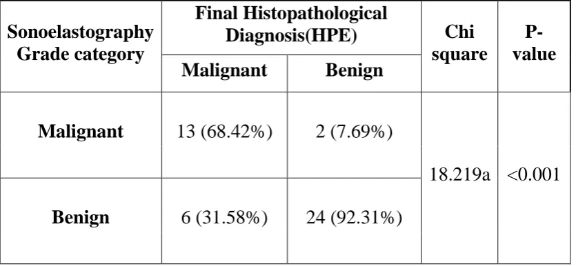
STATISTICAL ANALYSIS AND RESULTS
The Data was entered in a excel worksheet and double
checked.IBM SPSS version 22 software is used for statistical analysis.
Descriptive analysis:
Descriptive analysis was carried out using mean and standard
deviation for quantitative variables.
The frequency and proportion are used for categorical variables.
Data was also represented using appropriate diagrams like bar
diagram, pie diagram and box plots.
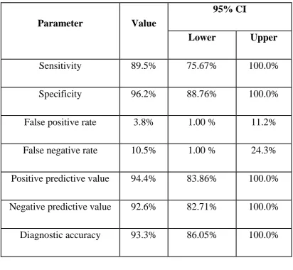
The sensitivity, specificity, positive and negative predictive values
and diagnostic accuracy of the Sonoelastography and Dynamic MR
mammogram against gold standard – the Histo Pathological Examination
(HPE) along with their 95% CI (Confidence Interval) are computed and
presented.
Reliability of the screening test is assessed by kappa statistics
along with its 95% CI and P Value.
Table 1: Descriptive analysis for Age in study population (N= 45)
Parameter Mean
±STD Median Min Max
95% C.I. for EXP(B)
Lower Upper
Age
39.71 ± 11.88
37.00 21.00 74.00 36.14 43.28
Table 2: Descriptive analysis of Age Group in study population (N=45)
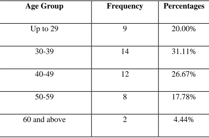
Age Group Frequency Percentages
Up to 29 9 20.00%
30-39 14 31.11%
40-49 12 26.67%
50-59 8 17.78%
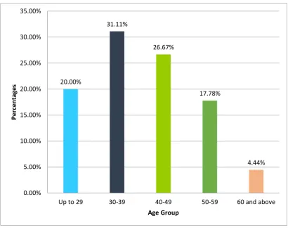
[image:81.595.110.475.452.693.2]Table 3: Descriptive analysis of Gender in study population (N=45)
Gender Frequency Percentage
Female 44 97.78%
Male 1 2.22%
Fig 1: Bar chart of Age Group distribution in study population (N=45) 20.00% 31.11% 26.67% 17.78% 4.44% 0.00% 5.00% 10.00% 15.00% 20.00% 25.00% 30.00% 35.00%
Up to 29 30-39 40-49 50-59 60 and above
Per
ce
n
tages
[image:82.595.103.514.375.699.2]Fig 2: Bar chart of Gender distribution in study population
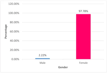
Table 4: Descriptive analysis of Right/Left in study population (N=45)
Right/Left Frequency Percentages
LEFT 20 44.44%
RIGHT 19 42.22%
BOTH 6 13.33%
2.22%
97.78%
0.00% 20.00% 40.00% 60.00% 80.00% 100.00% 120.00%
Male Female
P
e
rcen
ta
ge
[image:83.595.110.521.528.684.2]