The 3.3 Å structure of a plant geminivirus using
cryo-EM
Emma L. Hesketh
1, Keith Saunders
2, Chloe Fisher
1, Joran Potze
2, John Stanley
2,
George P. Lomonossoff
2& Neil A. Ranson
1Geminiviruses are major plant pathogens that threaten food security globally. They have a unique architecture built from two incomplete icosahedral particles, fused to form a geminate capsid. However, despite their importance to agricultural economies and fundamental bio-logical interest, the details of how this is realized in 3D remain unknown. Here we report the structure ofAgeratumyellow vein virus at 3.3 Å resolution, using single-particle cryo-electron microscopy, together with an atomic model that shows that the N-terminus of the single capsid protein (CP) adopts three different conformations essential for building the interface between geminate halves. Our map also contains density for ~7 bases of single-stranded DNA bound to each CP, and we show that the interactions between the genome and CPs are different at the interface than in the rest of the capsid. With additional mutagenesis data, this suggests a central role for DNA binding-induced conformational change in directing the assembly of geminate capsids.
DOI: 10.1038/s41467-018-04793-6 OPEN
1Astbury Centre for Structural Molecular Biology, School of Molecular & Cellular Biology, Faculty of Biological Sciences, University of Leeds, Leeds LS2 9JT,
UK.2Department of Biological Chemistry, John Innes Centre, Norwich Research Park, Colney, Norwich NR4 7UH, UK. These authors contributed equally: Emma L. Hesketh, Keith Saunders. Correspondence and requests for materials should be addressed to G.P.L. (email:[email protected]) or to N.A.R. (email:[email protected])
123456789
G
eminiviruses are thought to have been responsible for an unseasonal change in the appearance of the summer foliage in plants that was described in the poetry of the Empress Kōken in Japan in 752 AD1. Geminiviruses are thus probably the cause of thefirst available record of a viral disease in plants. In modern times, diseases associated with geminiviruses include maize streak disease and cassava mosaic disease in Africa; golden mosaic disease of beans in the Americas; tomato yellow leaf curl disease across much of the globe; and cotton leaf curl disease across India and Pakistan. The causative agents of many of these diseases areBegomoviruses, a genus in theGeminiviridae, which have circular, single-stranded2DNA genomes of total size ~2.7 to ~5.4 kb. The genetics of Begomoviruses are complex. Many have bipartite genomes, consisting of two separately encapsidated DNAs (DNA-A and B, each of ~2.7 kb). OtherBegomoviruses, such as Ageratum yellow vein virus (AYVV) studied here, have a single genomic DNA (DNA-A, ~2.7 kb), along with an array of satellite DNAs (α&β, each of ~1.3–1.4 kb). βsatellite (DNA-β), whose replication is totally dependent upon gene functions encoded within DNA-A, encodes a gene which is essential for AYVV infectivity in its natural host plant3. In contrast, αsatellite DNA (DNA-α) encodes aRep gene respon-sible for its own replication4. The basic requirement for a func-tional geminivirus capsid is thus that it must be able to encapsidate a single, 2.7 kb circular, single-stranded DNA mole-cule. The closest plant virus relatives of the geminiviruses are the nanoviruses, which have multipartite, single-stranded DNA genomes that are separately encapsidated and are ~1 kb in size5,6. Each such DNA molecule typically encodes a single gene product. Geminiviruses therefore appear to be able to package much larger amounts of DNA than nanoviruses, and to achieve this, they have evolved a unique capsid structure. Whereas nanoviruses have T=1 icosahedral capsids of ~18 nm in dia-meter (although no detailed structures are available), gemini-viruses are formed from two such isometric particles, or
‘hemicapsids’, fused to form a geminate particle (18 × 30 nm) which gives the virus family its name2,6. Until recently, no high-resolution structure for a geminivirus was available to describe how this architecture was realized. However, low-resolution cryo-EM studies on maize streak virus (MSV)7 and African cassava mosaic virus (ACMV)8, combined with homol-ogy modeling, suggested that the geminivirus coat protein (CP) adopts a structure similar to that of satellite tobacco necrosis virus (STNV), a single-stranded RNA virus7. Indeed, based on an ~25 Å resolution cryo-EM structure of MSV, a model for the geminate interface was proposed with an α-helix from each subunit (found in the STNV CP) extending across the geminate interface7. STNV also has a much smaller genome than a gemi-nivirus (~1250 vs ~2700 nucleotides), so geminate capsids again can package more genetic material than a simpleT=1 particle, despite encoding a single CP sequence. This is a similar leap in packaging capacity to that made by other viruses in the transition from T=1 to T=3 capsids described by Caspar & Klug in classical quasi-equivalence theory9. However, it seems unlikely that this arose from a simple expansion of the DNA genome beyond that which can be encapsidated within an isometric particle, as in such cases, aberrant structures with multiple, concentric CP shells have been observed10,11. A very recent structure of ACMV at 4.2 Å resolution12 has begun to reveal details of how the geminate structure is built, proposing that alternating conformations of an N-terminal segment could be a major determinant of the geminate interface. However, the resolution of this structure was insufficient to describe the interface between the two ‘hemicapsids’in atomic detail, and it contains no interpretable density for the genomic ssDNA or the interactions it makes with the capsid.
Our current knowledge of geminivirus structure thus leaves two questions of fundamental biological interest unanswered. Firstly, how does a single CP accommodate the different con-formations required to build a geminate particle? Secondly, no empty geminivirus particles have ever been reported, implying that DNA binding is intimately linked to capsid assembly; but how is DNA recognized and packaged? Here we present the cryo-EM structure of AYVV at 3.3 Å resolution. The structure shows that the capsid is built from three distinct conformations of a single CP, and that these conformational differences facilitate the formation of the interface between hemicapsids. We also show details of single-stranded DNA binding and propose a model for geminivirus capsid assembly.
Results
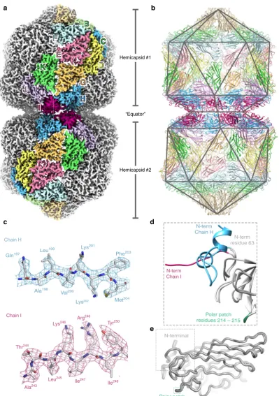
Structure of AYVV. To address these questions, we determined the structure of AYVV using single particle cryo-EM (Fig. 1). AYVV (and, indeed, the majority of the Begomoviruses) are phloem-limited and consequently are not mechanically trans-missible, so we usedAgrobacteriumtransformed with a plasmid containing a partial tandem copy of AYVV DNA-A13 (see Methods), to agroinfect14N. benthamianaplants (Supplementary Fig.1). Geminate particles were purified from symptomatic leaves (~2 mg/Kg of fresh leaf tissue) and a cryo-EM dataset collected (see Methods). Structure refinement with D5 symmetry, yielded a density map at 3.3 Å resolution (Fig. 1a and Supplementary Fig. 2), and an atomic model for the CP layer was built and refined (see Supplementary Table1), starting from a homology model of the STNV CP (see methods). The capsid is composed of 110 copies of the CP, and we built and refined a complete D5 asymmetric unit of 11 unique chains (chains A-K) (Fig.1b–e). As described previously7,8,12, each hemicapsid is formed from aT=
1 particle from which a penton of CP subunits is missing. The resulting facets on each hemicapsid associate to form the gemi-nate particle (Fig.1a). The body of the CP is similar to that of STNV and many other viruses, consisting of a jelly-roll fold with two twisted, 4-stranded β-sheets. This core domain structure is identical in all 11 chains in the asymmetric unit (Fig.1e).
N-terminal
Polar patch residues 214 – 215 Chain I
Tyr250
Ile249 Arg248
Ile247
Lys246
Leu245
Thr244
Ala243
Met204
Phe203
Lys202
Lys201
Val200 Leu199
Ala198 Gln197
Chain H
Polar patch residues 214 – 215
N-term Chain H
N-term residue 63
N-term Chain I
c
e d b a
Hemicapsid #1c
Hemi
A
A
B
F
E
K
C
D
G
H
I
J
Hemicapsid #2 “Equator”
[image:3.595.101.501.76.644.2]different roles in stabilizing the capsid. The helix-loop-helix motif from chain H is a major component of the equatorial interface (Fig.2c), making a series of H-bonding interactions to a patch of predominantly polar residues in the body of the CP subunit across the interface (residues 214 & 215 in chain I; see Figs.1d, e and 2c, and Supplementary Fig. 4), which is surrounded by van der Waals interactions. This polar patch is normally solvent-exposed on the surface of the virus in the subunits that form the hemicapsid (see Supplementary Fig. 4). The ordered segment from chain I extends across to stabilize the base of the helix-loop-helix motif in an adjacent H subunit, presumably stabilizing the ring of equatorial subunits (Fig.2d). This interaction is mediated by backbone hydrogen bonding; essentially, a short (~3 residue) domain-swapped, antiparallelβ-strand interaction is formed. The
geminate particle therefore critically relies on a previously dis-ordered segment of the N-terminus of the CP acquiring two different conformations to build the equatorial interface, which local resolution analysis suggests is marginally the most ordered part of the structure (Supplementary Fig.5).
The role of the N-terminal extension in chains H and I. Our structure makes a simple functional prediction: that interactions between chain H, and chain I are critical to particle assembly, and that disrupting these interactions might favor single rather than geminate particle formation. However, the geminate capsid is required to encapsidate AYVV’s genomic DNA, the 2.7 kb DNA-A; destabilization of the geminate interface could therefore lead to
100
* *
* c
d
e
a b
N215
R48A
M59D
M59 R48
M59 “Geminate” particles “Single” particles
90
80
P
ercentage of par
ticles (%)
70
60
50
40
30
20
10
0 AYVV DNA-A
wt CP +DNA-α
R48A +DNA-α
M59D +DNA-α wt CP + DNA-α
AYVV DNA-A
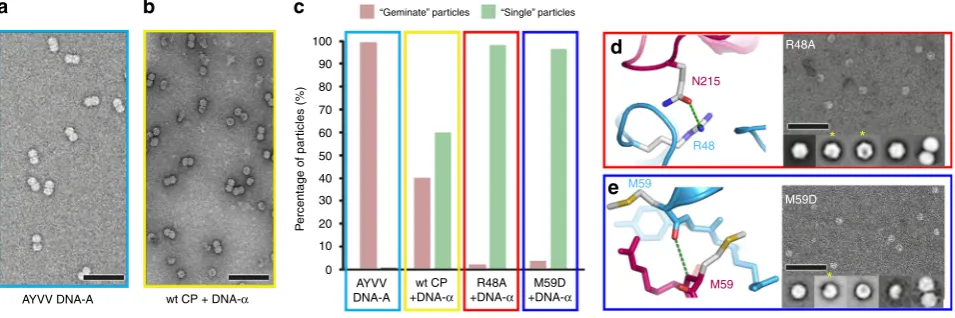
Fig. 3In vivo assembly assay for genome encapsidation. Representative negative stain micrographs of particles produced by infiltration with AYVV DNA-A (a) or co-infiltration with AYVV CP and DNA−α(b). Relative composition of“geminate”particles (pink in bar chart) compared with“single”particles (green in bar chart) from AYVV (c), and co-infiltrations of AYVV CP, R48A, and M59D, each with DNA−α(c). Representative negative stain micrographs and 2D class averages are shown for R48A variant (d) and M59D variant (e). The yellow asterisk highlights 2D class averages which appear to be“single” VLPs with a missing pentameric capsomer. Error bar is 100 nm
Chain H Residues 41–63
H H
H H I
I
I
H214
Y57 M59 N215
R48
P49
R61
M59
Chain I Residues 55–63
c b
a
d
[image:4.595.124.509.216.400.2] [image:4.595.60.538.525.684.2]degradation of the genomic DNA in vivo and abolish particle formation of all types.
We therefore exploited our knowledge of geminivirus genetics to establish an in vivo assay for encapsidation and assembly. AYVV infection of Ageratum conyzoides requires its 2.7 kb genomic DNA-A, together with its 1.3 kbβ-satellite DNA, to give the classic yellow vein symptoms on this host3. However, in experimental hosts such asN. benthamiana, leaf curl symptoms are evident with inoculations of DNA-A alone. In the natural host, this “A+β” infection complex is always associated with DNA-α (again, ~1.3 kb in length). DNA-α encodes a Rep gene that facilitates its own replication, but is encapsidated by the CP encoded by DNA-A, and it is this encapsidation that is essential for vector transmission and ultimately for maintenance of the DNA-α itself. We therefore took advantage of this by over expressing geminiviral CP, in the absence of a geminivirus infection, and at the same time introduced the self-replicating DNA-αinto the infiltration system. The result was the formation
of a mixture of “geminate” and “single” virus-like particles (VLPs). Importantly no VLPs are generated by the expression of geminivirus CP alone, strongly indicating that the provision of circular ssDNA is essential for capsid formation irrespective of whether “geminate” or “single” VLPs are generated. In the presence of wildtype CP and DNA-α, the ratio of geminate to single VLPs is ~40:60, compared to essentially 100% geminate particles for the wildtype virus (Fig. 3a–c). We then made two mutations within the CP to probe the effect of interface destabilization. Firstly, we made R48A, which we predicted would disrupt the H-bonding between to the ‘top’of the helix-loop-helix motif and chain I in the opposite hemicapsid (shown
in Fig. 2c). We also made M59D, which we predicted would disrupt the main chain H-bonding interaction between chain I and the base of the helix-loop-helix motif in chain H (shown in Fig. 2d). This region is stabilized by backbone hydrogen bonds between residue M59 in the two chains, as part of the shortβ -strand interaction described above, and we rationalized that the introduction of a negative charge in this region would disrupt this specific interaction. For both mutations, an almost complete switch from geminate to single particles was observed with DNA-α (Fig. 3c–e), confirming the importance of these regions for geminate capsid formation. Strikingly for both the R48A and M59D variants, some of the resulting‘single’VLPs appear to have a missing pentameric capsomer in negative stain EM image averages (Fig.3d, e).
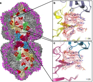
Interactions between the genomic DNA and CP subunits. A geminate capsid thus requires three distinct conformations, which although not classically‘quasi-equivalent’as they do not map to positions within an icosahedral lattice, fulfill a similar role in allowing a bigger capsid to be built. However, a fundamental question remains: how does a CP subunit‘know’what position it occupies within the overall architecture of the capsid? One solution would be for the conformation to be specified through interaction with the genome, as has been shown for single-stranded RNA viruses17,18,19. We therefore looked at the exten-sive density in our EM map that was unoccupied by the atomic model for the CP (Fig.4; Supplementary Movies1and2). Density is present beneath every CP in the capsid, consistent with an interaction with a ssDNA stem-loop. The map was of sufficient
CD(N)
HI(S)
b a
c
[image:5.595.136.462.387.672.2]quality to build 7 nucleotides into the density beneath each subunit, except for subunit H at the interface where there are 6. This density is as strong as that for the CP, suggesting that occupancy is high, but owing to the D5 symmetry averaging applied, we cannot definitively say that all sites in the capsid are occupied. However, assuming that they are, our structure resolves ~28% of the genomic DNA molecule at high resolution (Figs. 4 and 5). There is some suggestion from the density, which is slightly different for each of the 11 DNA chains in the asymmetric unit, as to whether each position is a purine or pyrimidine, and thus we have built a very tentative consensus sequence of YRRYYRY into the density (where Y represents a generic pyrimidine, and R a generic purine; using adenine for R, and cytosine for Y).
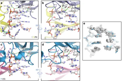
For each DNA chain in the body of the capsid (i.e. chains A-G and J-K), and for chain I, interactions are made between one CP by four residues (S114, Y116, R248, and Y250) to the top of the bound stem loop (nucleotides 3 and 4) (between the DNA and the blue subunit in Fig. 5a, b; Supplementary Movie 3). Additional contacts are made between arginine residues in a second CP subunit (R142, R144 & R174) (the yellow in Fig.5a, b; Supplementary Movie3) and the DNA backbone at the 3′end of the bound stem loop (nucleotides 5 & 7). In chain H, we again see a difference compared to the other chains. At the top of the stem loop, the interactions remain unchanged (between the DNA and the blue subunit in Fig. 5c, d; Supplementary Movie 4). More significantly, a new interaction occurs between the backbone of nucleotide 5 and R41, at the extreme N-terminus of the helix-loop-helix motif in chain H. This motif forms the equatorial
interface, and presumably the interaction with DNA stabilizes this structural element. Despite this, overall the conformation of the bound DNA is very similar at each position, including at the equatorial interface (Fig.5e).
To test the importance of DNA binding, we also made mutant R41A, to disrupt the interaction between R41 and the DNA backbone. When this was expressed alongside DNA-α, no particles were recovered from leaves, suggesting that DNA binding and/or assembly is severely compromised, as expected from our predicted role for R41.
Discussion
The structure of AYVV presented here helps to answer several long-standing questions in geminivirus biology. Early cryo-EM structures at low resolution (25–30 Å)7,8, together with the pio-neering work by Liljas and colleagues to determine the crystal structure of STNV20established a basic architecture for a gemi-nate capsid, and suggested that it would be built from viral CPs with an STNV-like, jelly-roll fold. McKenna and colleagues7 suggested an initial model, where the geminate interface was formed by conformational switching of the CPs at that interface, allowing the exchange of helices, tilted relative to the plane of the interface, between CPs. The STNV structure upon which this model was based does indeed have an N-terminal α-helix of varying length15,20. Recent work on ACMV12 at much higher resolution (~4.2 Å) moved this tentative model forward, by sug-gesting that alternate conformations of subunits occurred at the geminate interface. The structure at 3.3 Å resolution presented
R248
R174
a b
e
d c
R174
Y116
S114 R174
55°
CD(N)
HI(S)
55°
R41
Y250 S114
Y116 Y250
F203 nt4
nt3 nt5
nt2
nt6
nt1 nt7
nt4 nt3 nt5
nt2
nt6
nt1 F203
R144
R142
Y250
[image:6.595.91.508.382.660.2]here confirms this, and now provides molecular details of this conformational switching, the conformations adopted, and the interactions made. Two new conformations of portions of the N-terminus are observed in chains H and I at the geminate interface–one (chain H) in a helix-loop-helix motif, that forms an integral part of the geminate interface and the other (chain I) in an extended conformation that stabilizes chain H, via a short, domain-swapped β-strand interaction. These conformations are not compatible with the previous ‘helix-swapping’ model pro-posed for MSV; no discrete bridges of density are observed. Rather, the helix-loop-helix motif in our structure forms a rela-tively flat surface and thus a more intimate interface, with the centers of mass of the two hemicapsids moved closer together than has been proposed for MSV.
The structure presented here also demonstrates a role for DNA binding in specifying protein conformation. The recent 4.2 Å structure of ACMV resolves no ordered DNA12, but tentatively assigns amorphous density beneath the fivefold axis of each capsomer to DNA. We see very similar features in our map; however, this density disappears during map sharpening, which removes low resolution information from the reconstruction. This strongly suggests that, irrespective of whether this density corresponds to the N-terminal domains of the CP, genomic DNA, or a mixture of the two, no ordered structure exists at these positions in a geminivirus capsid. The structure presented here does, however, resolve large amounts of the genomic single-stranded DNA at high resolution, at a position beneath each CP subunit. This is not unprecedented for single-stranded DNA viruses, as structures of parvoviruses resolve longer segments (up to 11 bases of ssDNA in canine parvovirus)21. However, given that the parvovirus genome is rather larger (~5 kb), this repre-sents only ~13% of the genomic DNA, compared to ~28% in this 3.3 Å structure of AYVV. An examination of the unsharpened map, suggests that even a 2.7 kb genomic DNA would fill the entire available space within a particle, suggesting different
genomic components (~2.7 kb) and satellite geminivirus DNAs (~1.4 kb) are likely to be encapsidated separately.
The DNA loops resolved in the AYVV structure sit beneath each CP, but make interactions with a second, neighboring CP, immediately suggesting a role for the DNA in directing the assembly of the particle. This suggestion is then bolstered by the observations of DNA binding at the geminate interface, with a specific interaction between R41, at the extreme N-terminal end of the helix-turn-helix motif, and the DNA backbone of nucleo-tide 5. We therefore see a specific interaction, between a uniquely ordered protein segment at the interface and the DNA, which when disrupted by mutagenesis abolished capsid formation, suggesting a role for DNA in directing the assembly of the par-ticle. Despite this, overall the conformation of the 6 nucleotides found at every position in the bound DNA are strikingly similar, including at the equatorial interface (Fig.5e). This in turn suggest that perhaps the spatial arrangement of loops rather than a fundamentally different mode of binding may specify the gemi-nate capsid. However, our current data do not allow us to propose a consensus DNA sequence for binding with sufficient confidence to determine the number and/or position of binding sites within the genome.
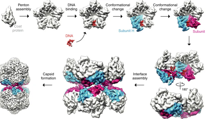
These data together with previous observations in other viral systems suggest a simple model for geminate particle formation (Fig.6). Given that each hemicapsid is missing a single penton, and that the interface between hemicapsids is pentameric, it seems plausible that the fundamental building block or ‘ cap-somere’of the geminate capsid is a penton of CP subunits. The penton as capsomere has been suggested or shown for a broad range of viruses with different genome types, including animal-and plant-infecting viruses of the order picornavirales22–25with ssRNA genomes, and indeed geminiviruses, where negative stain EM has shown that disassembly of ACMV occurs to iso-lated pentons26. Assembly of these ‘capsomeres’ appears to then be driven by DNA binding, explaining both the
Penton assembly
Coat protein
DNA binding
DNA
Subunit H Subunit I Conformational
change
Interface assembly Capsid
formation
180° Conformational
change
[image:7.595.98.500.447.680.2]observation that no empty capsids have ever been reported for any geminivirus, and that specific disruption of an interaction between DNA and CP (via R41A) abolishes capsid formation. Again, this fits well with observations from other systems. For example, a role for the ssDNA genome in canine parvovirus assembly has previously been postulated27, and more recently the assembly of beak and feather disease CP into higher order assemblies has been convincingly demonstrated to be stimu-lated by ssDNA binding28. The role of ssRNA is more solidly characterized, with structures of ribonucleoprotein complexes for viruses such as Pariacoto virus29and STNV15that suggest a role for nucleic acid in templating assembly. We speculate that a stem-loop that is different in either sequence or structure, promotes the conversion of a CP into the chain H conforma-tion. The unique stem-loop structure found within the origin of replication for all known geminiviruses13,30is a plausible can-didate for this role, but we have no experimental evidence to support this idea at present. Concurrently, either protein-protein and/or protein-protein-DNA interactions, promotes a neigh-boring subunit to adopt the chain I conformation. It is important to note, however, that this order is speculative. At this point however, the fundamental building blocks for a geminate capsid would be present, and an appropriate spatial distribution of stem loops would promote the assembly of these building blocks to form the capsid, with other nascent stem loops binding to non-equatorial positions in the high local concentration caused by encapsidation. Whilst speculative, this model provides a simple explanation to a long-standing puzzle as to how geminate particles are formed that is consistent with the high resolution structural observations of protein-protein and protein-DNA interactions described herein. A key goal of our future studies will be to validate this model by determining the sequence of the DNA bound to the AYVV CP, and thereby the number or position of these loops within the genomic DNA.
Methods
Infection of plants with AYVV DNA-A. The construction of pHNBin419 con-taining a partial tandem copy of AYVV DNA-A has been described13. Cultures of transformedAgrobacterium tumefaciensstrain GV 3850 harboring this plasmid were used to mechanically inoculate the stems of two-week-oldNicotiana ben-thamianausing the method previously described31. Inoculated plants were sub-sequently allowed to grow at 25 °C in an insect-proof glasshouse for 20–25 days with 16 hours supplementary lighting.
Expression of wild-type and specific mutants of AYVV CP. Oligonucleotides KS37 and KS38 (Supplementary Table2) were used with pHNBin419 DNA as template and Phusion polymerase (New England Biolabs) for PCR to obtain DNA encoding the AYVV CP that was subsequently cloned via the BP reaction into pDONR207 (Invitrogen). A sequenced-verified clone was transferred via the Gateway LR reaction into pEAQ-HT-DEST131to yield pEAQ-HT-D1-AYVVCP. The Agilent“QuikChange Primer Design”website,http://www.genomics.agilent. com/primerDesignProgram.jspwas used to design primer pairs to introduce mutations into the AYVV CP in pEAQ-HT-D1-AYVVCP at residues 41 (R to A: KS125P and KS126P), at residue 48, (R to A: KS127P and KS128P), and at residue 59 (M to D: KS129P and KS130P); Supplementary Table2. pEAQ-HT-D1-AYVVCP was used as the DNA template for each reaction as described in the GeneArt site-directed mutagenesis system (Invitrogen). The sequences of the wild-type and mutated clones were verified prior to transformingAgrobacterium
tumefaciensstrain LBA 4404 for transient expression in plants. The AYVV
satellite DNA-αpartial repeat clone, pBin AYVV 1/7, was pressure co-infiltrated with pEAQ-HT-D1-AYVVCP or its mutants as described previously32.
Isolation of virus and virus-like particles. Symptom-displaying leaves were harvested and ground at 4 °C in 3 volumes (1 g/3 mL) of extraction buffer (100 mM sodium phosphate buffer pH 6.0, 10 mM Na2SO3, 0.1% (v/v) 2-mercaptoethanol,
0.5% (v/v) Triton X-100, 0.1% (w/v) Driselase (Sigma-Aldrich)), supplemented with EDTA-free protease inhibitor tablets (Roche), 1 tablet/100 mL. After over-night incubation at 4 °C with constant stirring, the mixture was squeezed through Miracloth (Merck Chemicals) and clarified by centrifugation at 11,500×gfor 20 min. To the resulting supernatant, one quarter volume of chloroform was added and mixed for 10 min and the phases separated by centrifugation at 12,000×gfor
20 min. One quarter volume of a 1 M NaCl, 20% (w/v) PEG solution was added to the recovered aqueous phase and the resulting mixture was stirred overnight at 4 ° C. Precipitated virus particles were collected by centrifugation at 16,500×gfor 20 min. The pellet was resuspended in 100 mM sodium phosphate buffer, pH 7.0 and the solution clarified by centrifugation at 27,000×gfor 20 min. To the supernatant, Cs2SO4was added to afinal concentration of 36% (w/v) and the solution
cen-trifuged at 247,103×gfor 24 hours at 5 °C. Gradients were fractionated into 0.5 ml fractions and the fractions containing viral CP were identified by electrophoresis in 12% (w/v) NuPAGE MOPS-buffered gels (Life Technologies) followed by staining with Instant Blue dye (Expedeon Ltd). Viral CP identity was confirmed by sub-sequent MALDI-TOF analysis of the gel band containing the putative CP (John Innes Centre, platform facility). Selected fractions were dialyzed overnight at 4 °C against 100 mm sodium phosphate buffer pH7.0 and subsequently centrifuged at 210,000×gthrough a 20–60% (w/v) step sucrose gradient for 3 hours at 5 °C. Gradients were again fractionated, and the virus containing fractions identified by gel electrophoresis. After overnight dialysis against 100 mM sodium phosphate buffer, pH7.0, the samples were concentrated using Amicon Ultra-15 100,000 MWCO spin concentrators as described by the manufacturer.
Negative stain electron microscopy. Negative stain EM grids were produced by applying 3μl of particle preparations on to carbon- coated copper grids. Excess liquid was blotted away, the grids were washed in water twice and once in 2% (w/v) uranyl acetate, excess liquid was removed and grids were air-dried. The grids were imaged using either an FEI Tecnai F20 EMfitted with an FEI CMOS camera or FEI Tecnai 12 EMfitted with a Gatan US4000 (Astbury Biostructure Laboratory, University of Leeds). The number of particles in each analysis were as follows: WT virus 1,489 particles, WT CP and DNA-α4,164 particles, R48A mutant 16,015 particles, M59D mutant 12,639 particles.
Cryo-EM. Cryo-EM grids were prepared by applying AYVV to 400 mesh grids with a supporting carbon laceyfilm. The lacey carbon was coated with an ultra-thin carbon supportfilm < 3 nm thick (Agar Scientific, UK). Grids were glow-discharged for 30 seconds prior to applying the sample (easiGlow, Ted Pella). To increase the number of particles that adhere to the carbon, 3μl of the sample was incubated on the grid for 5 min. The majority of the liquid evaporated during the incubation, but at no point was the grid allowed to dry out; this was repeated three times. Thefinal 3μl was blotted immediately using a Leica EM GP plunge freeze (Leica Microsystems) device. Grids were frozen in liquid ethane cooled by liquid nitrogen, at 90% relative humidity, and a chamber temperature of 4 °C. AYVV data was collected on an FEI Titan Krios (Astbury Biostructure Laboratory, University of Leeds) EM at 300 kV, using a total electron dose of 110 e-/Å2and a magnifi-cation of 75,000 × . A total of 12,028 exposures were recorded using the EPU automated acquisition software on a FEI Falcon III direct electron detector, with a final object sampling of 1.065 Å/pixel. Each exposure movie had a total exposure of two seconds and contained 79 frames. AYVV has a strongly preferred orientation, with its 5-fold symmetry axis parallel to the ice layer. Particles with a view down thefive-fold axis are not visualized. To increase the range of views, we collected 1810 movies with the stage tilted to 10° and 322 movies with the stage tilted to 20°.
Image processing. A summary of image processing is shown in Supplementary Fig.2. Image processing was carried out using the RELION 2.0/2.1 pipeline33. Drift-corrected averages of each movie were created using MOTIONCOR234and the contrast transfer function of each determined using gCTF35. Any images showing signs of significant astigmatism were discarded. Approximately 5000 particles were manually picked and classified using reference-free 2D classifica-tion36. The resulting 2D class average views were used as templates for automated particle picking using gAutomatch37(see Supplementary Table1for particle numbers). Automated particle picking of particles on lacey carbon grids resulted in a large number of boxes picked on the edge of carbon (without AYVV particles). To classify these images away, 2D classification in RELION was used with CTF amplitude correction only performed from thefirst peak of each CTF onwards. Particles were further classified using several rounds of both reference-free 2D classification and 3D classification, with D5 symmetry imposed. A negative stain reconstruction was usedfiltered to ~60 Å resolution for the starting model. After each round, the best classes/class was taken to the next step of classification. Post-processing was employed to appropriately mask the model, estimate and correct for the B-factor of the maps38. Thefinal resolution was determined using the‘gold standard’Fourier shell correlation (FSC=0.143) criterion as 3.3 Å. Local resolu-tion was estimated using the local resoluresolu-tion feature in RELION33.
COOT. The model was refined using three rounds of REFMAC42with secondary structure restraints imposed using ProSMART42. After each refinement round, non-ideal rotamers, bond angles and Ramachandran outliers were improved using COOT. The resulting model of one subunit was symmetrized using Chimera and 11 subunits werefitted to represent one asymmetric unit, subunits A-K in one hemicapsid (Fig.1). Amino acids 40–62 were removed for all subunits other than H and amino acids 55-62 were added for subunit I. This asymmetric unit was refined using the‘real space refinement tool’in COOT followed by three rounds in Phe-nix43and four rounds in REFMAC42with non-crystallographic symmetry con-straints imposed. The quality of the asymmetric unit was assessed using MolProbity44(Supplementary Table1). Figures were generated using Chimera40, ChimeraX45and PyMOL46.
Data availability. Coordinates are deposited in the Protein Data Bank under accession code6F2S. Cryo-EM reconstructions of the AYVV are deposited in the EM Data Bank under accession codesEMD-4174. All reagents and relevant data are available from the authors upon request.
Received: 10 December 2017 Accepted: 24 April 2018
References
1. Saunders, K., Bedford, I. D., Yahara, T. & Stanley, J. Aetiology: the earliest recorded plant virus disease.Nature422, 831 (2003).
2. Harrison, B. D. et al. Plant-viruses with circular single-stranded-DNA.Nature
270, 760–762 (1977).
3. Saunders, K. et al. A unique virus complex causes Ageratum yellow vein disease.Proc. Natl Acad. Sci. USA97, 6890–6895 (2000).
4. Saunders, K. & Stanley, J. A nanovirus-like DNA component associated with yellow vein disease of Ageratum conyzoides: Evidence for interfamilial recombination between plant DNA viruses.Virology264, 142–152 (1999). 5. Grigoras, I. et al. Reconstitution of authentic nanovirus from multiple cloned
DNAs.J. Virol.83, 10778–10787 (2009).
6. Gronenborn, B. Nanoviruses: genome organisation and protein function.Vet. Microbiol.98, 103–109 (2004).
7. Zhang, W. et al. Structure of the maize streak virus geminate particle.Virology
279, 471–477 (2001).
8. Bottcher, B., Unseld, S., Ceulemans, H., Russell, R. B. & Jeske, H. Geminate structures of African cassava mosaic virus.J. Virol.78, 6758–6765 (2004). 9. Caspar, D. L. & Klug, A. Physical principles in the construction of regular
viruses.Cold Spring Harb. Symp. Quant. Biol.27, 1–24 (1962).
10. Perlmutter, J. D. & Hagan, M. F. Mechanisms of virus assembly.Annu. Rev. Phys. Chem.66, 217–239 (2015).
11. Prinsen, P., van der Schoot, P., Gelbart, W. M. & Knobler, C. M. Multishell structures of virus coat proteins.J. Phys. Chem. B114, 5522–5533 (2010). 12. Hipp, K., Grimm, C., Jeske, H. & Bottcher, B. Near-atomic resolution structure
of a plant geminivirus determined by electron cryomicroscopy.Structure25, 1303–1309 (2017).
13. Tan, P. H. N. et al. Genome organization of Ageratum yellow vein virus, a monopartite whitefly-transmitted geminivirus isolated from a common weed. J. Gen. Virol.76, 2915–2922 (1995).
14. Grimsley, N., Hohn, T., Davies, J. W. & Hohn, B. Agrobacterium-mediated delivery of infectious maize streak virus into maize plants.Nature325, 177–179 (1987).
15. Ford, R. J. et al. Sequence-specific, RNA-protein interactions overcome electrostatic barriers preventing assembly of satellite tobacco necrosis virus coat protein.J. Mol. Biol.425, 1050–1064 (2013).
16. Harrison, S. C., Olson, A. J., Schutt, C. E., Winkler, F. K. & Bricogne, G. Tomato bushy stunt virus at 2.9 A resolution.Nature276, 368–373 (1978). 17. Dent, K. C. et al. The asymmetric structure of an icosahedral virus bound to its
receptor suggests a mechanism for genome release.Structure21, 1225–1234 (2013).
18. Dai, X. et al. In situ structures of the genome and genome-delivery apparatus in a single-stranded RNA virus.Nature541, 112–116 (2017).
19. Stockley, P. G. et al. A simple, RNA-mediated allosteric switch controls the pathway to formation of a T=3 viral capsid.J. Mol. Biol.369, 541–552 (2007). 20. Liljas, L. et al. Structure of satellite tobacco necrosis virus at 3.0 A resolution.J.
Mol. Biol.159, 93–108 (1982).
21. Xie, Q. & Chapman, M. S. Canine parvovirus capsid structure, analyzed at 2.9 Å resolution.J. Mol. Biol.264, 497–520 (1996).
22. Tuthill, T. J. et al. Equine rhinitis A virus and its low pH empty particle: clues towards an aphthovirus entry mechanism?PLoS Pathog.5, 11–21 (2009). 23. Bakker, S. E. et al. Limits of structural plasticity in a picornavirus capsid
revealed by a massively expanded equine rhinitis A virus particle.J. Virol.88, 6093–6099 (2014).
24. Li, C. L., Wang, J. C. Y., Taylor, M. W. & Zlotnick, A. In vitro assembly of an empty picornavirus capsid follows a dodecahedral path.J. Virol.86, 13062–13069 (2012).
25. Hesketh, E. L. et al. Mechanisms of assembly and genome packaging in an RNA virus revealed by high-resolution cryo-EM.Nat. Commun.6, 10 (2015). 26. Kittelmann, K. & Jeske, H. Disassembly of African cassava mosaic virus.J.
Gen. Virol.89, 2029–2036 (2008).
27. Chapman, M. S. & Rossmann, M. G. Single-stranded DNA–protein interactions in canine parvovirus.Structure3, 151–162 (1995).
28. Sarker, S. et al. Structural insights into the assembly and regulation of distinct viral capsid complexes.Nat. Commun.7, 13014 (2016).
29. Tang, L. et al. The structure of Pariacoto virus reveals a dodecahedral cage of duplex RNA.Nat. Struct. Biol.8, 77–83 (2001).
30. Saunders, K., Lucy, A. & Stanley, J. DNA forms of the geminivirus African cassava mosaic virus consistent with a rolling circle mechanism of replication. Nucleic Acids Res.19, 2325–2330 (1991).
31. Klinkenberg, F. A., Ellwood, S. & Stanley, J. Fate of African cassava mosaic virus coat protein deletion mutants after agroinculation.J. Gen. Virol.70, 1837–1844 (1989).
32. Sainsbury, F., Thuenemann, E. C. & Lomonossof, G. P. pEAQ: versatile expression vectors for easy and quick transient expression of heterologous proteins in plants.Plant. Biotechnol. J.7, 682–693 (2009).
33. Fernandez-Leiro, R. & Scheres, S. H. W. A pipeline approach to single-particle processing in RELION.Acta Crystallogr. D73, 496–502 (2017).
34. Zheng, S. Q. et al. MotionCor2: anisotropic correction of beam-induced motion for improved cryo-electron microscopy.Nat. Methods14, 331–332 (2017).
35. Zhang, K. Gctf: real-time CTF determination and correction.J. Struct. Biol.
193, 1–12 (2016).
36. Scheres, S. H. RELION: implementation of a Bayesian approach to cryo-EM structure determination.J. Struct. Biol.180, 519–530 (2012).
37. Urnvicius, L. et al. The structure of the dynactin complex and its interaction with dynein.Science347, 1441–1446 (2015).
38. Scheres, S. H. & Chen, S. Prevention of overfitting in cryo-EM structure determination.Nat. Methods9, 853–854 (2012).
39. Jones, T. A. & Liljas, L. Structure of satellite tobacco necrosis virus after crystallographic refinement at 2.5- a resolution.J. Mol. Biol.177, 735–767 (1984).
40. Pettersen, E. F. et al. UCSF Chimera--a visualization system for exploratory research and analysis.J. Comput. Chem.25, 1605–1612 (2004).
41. Emsley, P., Lohkamp, B., Scott, W. G. & Cowtan, K. Features and development of Coot.Acta Crystallogr. D66, 486–501 (2010).
42. Brown, A. et al. Tools for macromolecular model building and refinement into electron cryo-microscopy reconstructions.Acta Crystallogr. D71, 136–153 (2015).
43. Adams, P. D. et al. PHENIX: a comprehensive Python-based system for macromolecular structure solution.Acta Crystallogr. D66, 213–221 (2010). 44. Chen, V. B. et al. MolProbity: all-atom structure validation for
macromolecular crystallography.Acta Crystallogr. D66, 12–21 (2010). 45. Goddard, T. D. et al. UCSF ChimeraX: meeting modern challenges in
visualization and analysis.Protein Sci.27, 14–25 (2017).
46. DeLano, W. L. The PyMOL Molecular Graphics System, Version 2.0 (Schrodinger, 2018).
Acknowledgements
This work was supported by the UK Biotechnological and Biological Sciences Research Council (Grants BB/L021250/1 (Leeds) & BB/L020955/1 (John Innes Centre). We thank the Astbury Biostructure Laboratory (ABSL), in particular Dr. Rebecca Thompson, for assisting with cryo-EM data collection. All Electron Microscopy was performed at ABSL which was funded by the University of Leeds and the Wellcome Trust (108466/Z/15/Z). Chloe Fisher was funded by a Dean’s Vacation Research Scholarship (University of Leeds). We also thank Drs. Matthew Iadanza and Shaun Rawson for their excellent script writing. At the John Innes Centre, this work was also supported by the BBSRC Institute Strategic Programme Grants“Understanding and Exploiting Plant and Microbial Sec-ondary Metabolism”(BB/J004596/1) and“Molecules from Nature”(BB/P012523/1) and by the John Innes Foundation.
Author contributions
N.A.R. and G.P.L. conceived the project. K.S. and J.P. constructed AYVV CP clones and purified the virus, J.S. provided infectious AYVV andAgrobacteriumcultures. E.L.H. performed the cryo-EM data collection and structure refinement. E.L.H., C.F., and N.A.R. built and analyzed the structure. All authors contributed to the writing of the manuscript.
Additional information
Competing interests:G.P.L. declares that he is a named inventor on granted patent WO 29087391 A1 that describes the system used for transient expression of the CP in this manuscript. The remaining authors declare no competing interests.
Reprints and permissioninformation is available online athttp://npg.nature.com/ reprintsandpermissions/
Publisher's note:Springer Nature remains neutral with regard to jurisdictional claims in published maps and institutional affiliations.
Open Access This article is licensed under a Creative Commons Attribution 4.0 International License, which permits use, sharing,
adaptation, distribution and reproduction in any medium or format, as long as you give appropriate credit to the original author(s) and the source, provide a link to the Creative Commons license, and indicate if changes were made. The images or other third party material in this article are included in the article’s Creative Commons license, unless indicated otherwise in a credit line to the material. If material is not included in the article’s Creative Commons license and your intended use is not permitted by statutory regulation or exceeds the permitted use, you will need to obtain permission directly from the copyright holder. To view a copy of this license, visithttp://creativecommons.org/ licenses/by/4.0/.