RESEARCH
Transcriptome-based analysis of putative
allergens of
Chorioptes texanus
Ran He
1, Xiao‑Bin Gu
1, Yue Xie
1, Xue‑Rong Peng
2, Christiana Angel
3and Guang‑You Yang
1*Abstract
Background: Mites of the genus Chorioptes are non‑burrowing and cause mange in a wide range of domestic and wild animals including cattle, horses, sheep, goats, panda, moose, camelids, mydaus and alpacas. Molecular biology and host‑parasite interactions of Chorioptes texanus are poorly understood, and only a few C. texanus genes and tran‑ script sequences are available in public databases including the allergen genes.
Methods: Chorioptes texanus RNA was isolated from mites, and the transcriptome of C. texanus was analyzed using bioinformatics tools. Chorioptes texanus unigenes were compared with the allergen protein sequences from the mite allergen database website to predict the potential allergens. Chorioptes texanus putative allergen unigenes were compared with hydrolase genes by building a C. texanus hydrolase gene library with the best match of the homolo‑ gous sequences. Three allergen genes were cloned and expressed, their recombinant proteins were purified and their allergenic activities were preliminarily investigated.
Results: Transcriptome sequencing (RNA‑Seq) of C. texanus was analyzed and results demonstrated that 33,138 unigenes were assembled with an average length of 751 bp. A total of 15,130 unigenes were annotated and 5598 unigenes were enriched in 262 KEGG signaling pathways. We obtained 209 putative allergen genes and 34 putative allergen‑hydrolase genes. Three recombinant allergen proteins were observed to induce different degrees of allergic reactions on rabbit skin.
Conclusions: The present transcriptome data provide a useful basis for understanding the host‑parasite interaction and molecular biology of the C. texanus mite. The allergenic activities of recombinant Euroglyphus maynei 1‑like (Eur m 1‑like) protein, Dermatophagoides ptreronyssinus 1‑like (Der p 1‑like) protein and Dermatophagoides ptreronyssinus 7‑like (Der p 7‑like) protein were preliminarily investigated by intradermal skin test. Meanwhile, differences in eosino‑ phil counts were observed in different injected sites of the skin. The identification of putative allergen genes and hydrolase genes offers opportunities for the development of new diagnostic, prevention and treatment methods. Keywords: Chorioptic mange, Chorioptes texanus, Transcriptome, RNA‑Seq, Allergen, Hydrolase
© The Author(s) 2019. This article is licensed under a Creative Commons Attribution 4.0 International License, which permits use, sharing, adaptation, distribution and reproduction in any medium or format, as long as you give appropriate credit to the original author(s) and the source, provide a link to the Creative Commons licence, and indicate if changes were made. The images or other third party material in this article are included in the article’s Creative Commons licence, unless indicated otherwise in a credit line to the material. If material is not included in the article’s Creative Commons licence and your intended use is not permitted by statutory regulation or exceeds the permitted use, you will need to obtain permission directly from the copyright holder. To view a copy of this licence, visit http://creat iveco mmons .org/licen ses/by/4.0/. The Creative Commons Public Domain Dedication waiver (http://creat iveco mmons .org/publi cdoma in/ zero/1.0/) applies to the data made available in this article, unless otherwise stated in a credit line to the data.
Open Access
*Correspondence: [email protected]
1 Department of Parasitology, College of Veterinary Medicine, Sichuan Agricultural University, Chengdu 611130, China
Full list of author information is available at the end of the article Background
Parasitic mites belonging to the genus Chorioptes
(Acariformes: Psoroptidae) are found worldwide, caus-ing mange (chorioptic mange: a skin disease) in a wide
range of domestic and wild animals [1, 2]. Chorioptes
legs, often infesting the base of the tail, perineum and
the udder, where it is normally not easily observed [8,
9]. Chorioptic mange causes economic loses in both
dairy and beef herds. The mites cause itching, rub-bing and scratching which results in leather damage. Itching also makes the animal restless and can result
in reduced production of beef and milk [10–12]. Based
on morphological, epidemiological and genetic
dif-ferentiation, Chorioptes bovis and Chorioptes texanus
are generally accepted as two distinct valid species
[1, 13, 14]. The egg, larva, protonymph, deutonymph
and adult constitute a single life-cycle which spans
approximately three weeks [7, 15]. Hosts may be
asymptomatic at an early stage of infestation or low mite density, but mange may become generalized later or at a high mite density. Chorioptic mange is a highly seasonal disease, and more common in colder peri-ods, particularly in winter and when cattle are stabled; however, it may recover when cattle return to pasture
after the winter season [16]. Characteristic clinical
signs include scratching and rubbing at the base of tail,
perineum and legs [17]. The diagnosis of chorioptic
mange is based on clinical signs and microscopic con-firmation by identification of mites in the scrapings of
infested skin from the host [18]. The control of
cho-rioptic mange depends mainly on treatment with anti-parasitic drugs including eprinomectin, moxidectin,
closantel, deltamethrin, ivermectin and selamectin [3,
10, 19]. Confirmatory laboratory diagnosis requires a
longer time, and an overuse of the anti-parasitic drugs has led to environmental damage and drug resistance; therefore, it is necessary to find new efficient tools for diagnosis, prevention and treatment of this dis-ease. Chorioptes texanus mites infest the host, eliciting
an inflammation reaction [13], leading to an allergic
skin response, hair loss, scratching and skin dam-age. Allergens are the main factors eliciting the hosts pro-inflammatory response, and most of the allergen genes are hydrolase genes, having crucial roles in mite
evasion and survival in the host [20, 21]. Moreover,
there are several allergen and hydrolase genes in mites which have been identified to be potential diagnostic
antigens and vaccine candidates [22–25]. Intriguingly,
analysis of C. texanus allergen and hydrolase genes
may provide a valuable database for exploring new potential diagnostic, prevention and treatment
meth-ods. However, our current understanding of C. texanus
molecular biology and host-parasite interactions is still incomplete. Additionally, available evidence regarding
allergen genes of C. texanus are mainly from case
stud-ies and epidemiological and clinical reports, only a few
C. texanus genes and transcript sequences are avail-able in public databases [6, 26–28]. Therefore, in the
present study, RNA-Seq was performed to analyze the
transcriptome of C. texanus and to obtain a valuable
sequence database of allergen and hydrolase genes. In
addition, three allergen genes of C. texanus were
sub-sequently cloned, expressed and their recombinant proteins were purified, and the allergenic activity was
preliminarily investigated via an intradermal skin test.
Methods
Mite collection
Chorioptes spp. mites were isolated from scabs in the distal extremities and the base of the tail of three natu-rally infested cows from a farm in Chengdu, Sichuan Province, China. The animals were maintained under the same feeding and environmental conditions. Scabs were incubated in glass plates at 36 °C and mites were collected on an hourly basis. Mites that emerged from
the scabs were collected and identified as C. texanus
based on their morphology, as previously described
[2, 26, 29] and were preserved immediately in liquid
nitrogen (LN) after harvesting.
RNA isolation and library construction
Total RNA was extracted from mite samples using Trizol reagent (Invitrogen, Carlsbad, USA) accord-ing to the manufacturer’s instructions. The purity and integrity of RNA was assessed by agarose gel electro-phoresis and quantified by measuring the absorbance at 260/280 nm using a Nanodrop spectrophotometer (Bio-rad, California, USA). Sequencing libraries were
obtained using the Illumina TruSeqTM RNA sample
preparation kits (Illumina, California, USA) follow-ing the manufacturer’s instructions and sequenced on an Illumina Hiseq 2000 platform (Illumina). Detailed
methods are described in our previous study [30]. PCR
products were purified using the AMPure XP system (Beckman Coulter, California, USA), and the quality of the sequencing libraries assessed on an Agilent Bio-analyzer 2100 (Agilent Technologies, California, USA).
RNA‑Seq data assembly and functional assignment
protein sequence database), PFAM (protein family), GO (Gene Ontology) and KOG (eukaryote-specific ver-sion of the Clusters of Orthologous Groups). Coding sequences (CDS) were predicted by comparison with sequences from other eukaryotes. The CDS and direc-tion of unigenes in databases were obtained based on the best alignment results. Unigenes which failed alignment to the databases were scanned with EST-Scan software
to predict the CDS and direction [32]. The Blast2GO
program and WEGO software were applied for further
analysis [33, 34]. Blast2GO was employed to obtain GO
annotations in the cellular component, molecular func-tion and biological processes. Then WEGO was used to perform GO functional classification of unigenes. Uni-gene sequences were aligned to the above protein library according to the priority order of Nr and Swissprot. If the results matched, then the open reading frame (ORF) information of the transcript was extracted from the alignment results, and the coding region sequence was translated into an amino acid sequence according to the standard codon table (in the order of 5′–3′); otherwise the ORF of the unigenes was predicted by EST-scan soft-ware, thereby obtaining the nucleic acid sequence and amino acid sequence encoded by the part of a gene.
Putative allergen genes and allergen‑hydrolase genes Chorioptes texanus unigenes were compared with the allergen protein sequences from the allergen database website (http://www.aller gome.org), and putative allergen
gene sequences were obtained. Additionally, C. texanus
putative mite allergen unigenes were compared with the
hydrolase gene sequences by building a C. texanus
hydro-lase gene library with the best match of the homologous sequences.
Cloning and expression of three allergen genes and purification of their recombinant protein
The putative allergen genes were obtained after con-ducting a BLAST search, and three allergen genes, i.e.
Dermatophagoides ptreronyssinus 1-like (Der p 1-like)
protein gene, Dermatophagoides ptreronyssinus 7-like
(Der p 7-like) protein gene and Euroglyphus maynei
1-like (Eur m 1-like) protein gene, were selected. The
cDNA was transcribed using a RevertAiTM First Strand
cDNA Synthesis Kit (Thermo, Massachusetts, USA) according to the manufacturer’s instructions and stored
at − 80 °C. Available gene sequences were used to design
primers for three genes as follows, Der p 1-like protein
gene was amplified from cDNA using a sense primer (5′
-CGC GGA TCC AAA AAA ATG AAA ATC ATA TCA
TCA ATC GCA ATT TTT TCG CT-3′) containing a
BamHI site (underlined) and antisense primer (5′-CCG
CTC GAG TCA AAC AGA TAA AAC AAC ATA TGG
ATA TTG TTC AAT GCC-3′) containing a XhoI site
(underlined); Der p 7-like protein gene was amplified
from cDNA using a sense primer (5′-CGC GGA TCC
GAT CCA ATT CAC TTT GAT AAA AT-3′) containing
a BamHI site (underlined) and antisense primer (5′-CCG
CTC GAG TAA TGT TGA TCG ATT GCT TTT TT-3′)
containing a XhoI site (underlined); Eur m 1-like protein
gene was amplified from cDNA using a sense primer (5′
-CGC GGA TCC CGT CCA TCA TCA ATT AAA AC-3′)
containing a BamHI site (underlined) and antisense
primer (5′-CCG CTC GAG TTA AAG AAT AAC AAC
ATA TGG ATA T-3′) containing a XhoI site (underlined).
The cDNA encoding the Der p 1-like protein gene and Der p 7-like protein gene were successfully sub-cloned
into the pET32a(+) expression vector (Novagen,
Darm-stadt, Germany), and the Eur m 1-like protein gene was
successfully sub-cloned into the pET28a(+) expression
vector and expressed in Escherichia coli BL21(DE3).
The expressed recombinant proteins were purified using Ni2+ affinity chromatography (Bio-Rad, California, USA)
according the manufacturer’s instructions.
Intradermal skin test and eosinophil count
An intradermal skin test was performed to investigate the allergenic activity of recombinant allergen proteins. Nine New Zealand White rabbits were injected subcutane-ously with 100, 200, 400 μg of purified recombinant aller-gen protein in 0.1 ml PBS. The histamine-injected group (4 mg/ml, 0.1 ml) was used as a positive control. The neg-ative controls were: (i) 0.1 ml PBS; (ii) physiological saline
(0.9%, 0.1 ml); and (iii) 400 μg of purified pET-32a (+)
empty expression vector in 0.1 ml PBS. Skin reactions were recorded every 30 min following an injection, and when the presence of a wheal and erythema was observed the skin reactions were defined as positive. After 2.5 h, skin samples were collected with an annular skin sam-pler (diameter of 7 mm), and fixed in 4% paraformalde-hyde in PBS (pH 7.4) for 24 h. A rotary microtome was used to serially cut the paraffin-embedded specimens at 5 μm thickness and sections were mounted on clean glass slides. H&E staining was performed to observe dif-ferential eosinophilic infiltration at different injected sites of the skin. For each slide, six microscopic fields (200×) were randomly selected and microphotographed. Image-Pro Plus 6.0 was used to count the number of eosinophils per field.
Validation of RNA‑Seq data by RT‑qPCR
RT-qPCR analysis was performed to validate the RNA-Seq results. Part of allergen genes were validated
allergens genes and genes from the allergen database website. A MX300P spectrofluorometric thermal cycler (Stratagene, California, USA) was used to conduct the RT-qPCR. The cycling conditions were as follows, an ini-tial denaturation at 95 °C for 2 min, followed by 40 cycles at 94 °C for 20 s and 58 °C for 20 s. qPCR was conducted in triplicate and the relative gene expression levels were calculated using the 2−ΔΔCq method [35].
Results
RNA‑Seq and assembly of C. texanus transcriptome data
RNA-Seq analysis of C. texanus yielded 48,497,838
raw reads from mites (GenBank project accession no. PRJNA495065). After quality assessment and filter-ing, 46,404,816 clean reads were obtained with a Q20 of 96.66% and a GG of 33.48%. Clean reads were used for assembly with Trinity software. A total of 51,868 tran-scripts with N50 values of 2406 and 33,138 unigenes with N50 values of 1340 were generated respectively (Addi-tional file 1: Figure S1).
Functional annotation and GO classification
Unigenes were annotated with multiple databases using different software and/or websites. After final assembly, 33,138 unigenes were compared to NR, NT, SwissProt
and KOG using NCBI blast 2.2.28+ with an E-value
cut-off of le-5 for NR, NT, SwissProt and E-value, cut-cut-off of le-3 for KOG. There were 7998 unigenes categorized into 25 molecular families when aligned through the KOG database, and unigenes distribution is shown in
Addi-tional file 2: Figure S2. Furthermore, unigenes were
ana-lyzed using the HMMER 3.0 package and hmmscan with the PFAM database, and were analyzed within Blast2GO with the GO database and annotated by KAAS, KEGG Automatic Annotation Server with the KEGG database
(Table 1). A total of 11,915 unigenes were mapped to GO
terms which were categorized into biological processes
(BP), molecular functions (MF) and cellular components (CC) (Additional file 3: Figure S3). The top three predom-inant terms for BP were cellular process, metabolic pro-cess and single-organism propro-cess; whereas the top three terms in MF were binding, catalytic activity and trans-porter activity; and for CC, the top three terms were cell, cell part and organelle.
KEGG pathway analysis of unigenes
KEGG pathway enrichment analysis revealed that a total of 5598 unigenes were enriched in 262 pathways. The KEGG pathways included five categories, cellular
pro-cessing (n= 17; 6.5%), environmental information
pro-cessing (n= 31; 11.8%), genetic information processing
(n= 22; 8.4%), metabolism (n= 123; 46.9%) and organis-mal systems (n= 69; 26.3%) (Additional file 4: Figure S4). Transport and catabolism (443 unigenes) was the most abundant subcategory in cellular processing; signal trans-duction (1393 unigenes) was the most abundant subcat-egory in environment information processing; translation (681 unigenes) was the most abundant subcategory in genetic information processing; carbohydrate metabo-lism (558 unigenes) was the most abundant subcategory in metabolism and organismal systems; endocrine sys-tem (947 unigenes) was the most abundant subcategory in organismal systems. The top 10 unigenes abundant pathways were ribosome (381 unigenes), carbon metabo-lism (182 unigenes), protein processing in endoplasmic reticulum (181 unigenes), oxidative phosphorylation (161 unigenes), PI3K-Akt signaling pathway (146 unigenes), biosynthesis of amino acids (145 unigenes), spliceosome (143 unigenes), MAPK signaling pathway (137 unigenes), purine metabolism (136 unigenes), and phagosome (130 unigenes). CDS were determined using the BLASTX pro-gram and EST-Scan propro-gram, unigenes length and uni-genes counts are shown in Additional file: 5 Figure S5 and Additional file 6: Figure S6, respectively.
Putative allergen genes and allergen‑hydrolase genes
According to results from the allergen blast, 1246
aller-gen homologous unialler-genes were obtained from the C.
texanus RNA-Seq data. The number of unigenes homol-ogous to allergens from the website was 1167. From these, 959 unigenes were non-mite putative allergens and 208 unigenes were putative allergens of seven
spe-cies of mite, i.e. Dermatophagoides farina,
Dermatopha-goides pteronyssinus, Euroglyphus maynei, Lepidoglyphus destructor, Psoroptes ovis, Sarcoptes scabiei, Tyrophagus putrescentiae and Acarus siro. A hydrolase gene sequence bank was constructed using the homologous
hydro-lase sequences from the C. texanus RNA-Seq data and
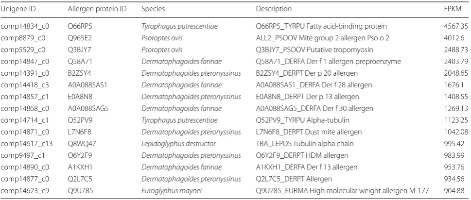
[image:4.595.57.290.581.725.2]34 of them were putative allergen genes of C. texanus,
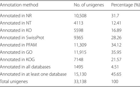
Table 1 Summary of assembled unigenes annotation
Annotation method No. of unigenes Percentage (%) Annotated in NR 10,508 31.7
including comp7640_c0, comp2015_c0, comp11218_c1, comp14904_c0, comp14553_c12, comp4959_c0, etc. The top 15 putative allergen genes were listed according to the fragments per kilobase of exon per million parts map-ping (FPKM) value (Table 2) [36].
Cloning and expression of three allergen genes and purification of their recombinant proteins
The cDNA encoding Der p 1-like protein, Der p 7-like protein and Eur m 1-like protein contained 984-bp, 660-bp and 915-660-bp ORF, respectively. The cDNA encoding these three allergen genes were successfully sub-cloned
into expression vectors and expressed in E. coli BL21
(DE3) cells with a molecular weight of approximately 56 kDa (Der p 1-like protein; including the His tag), 44 kDa (Der p 7-like protein; including the His tag) and 42 kDa (Eur m 1-like protein; including the His tag). Then recombinant proteins were purified using a Ni-chelating column and examined by SDS-PAGE.
Intradermal skin test and eosinophil count
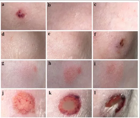
Recombinant Eur m 1-like protein injected at 100 μg, 200 μg and 400 μg doses produced a wheal, flare and erythema reaction, including a blistering and ulceration reaction. As for the recombinant Der p 1-like protein, 400 μg recombinant protein produced erythema reac-tion, whereas recombinant Der p 7-like protein at 100 μg, 200 μg and 400 μg doses produced a wheal and erythema reaction. No reaction was induced by 0.9% physiological saline, PBS or empty expression vector, whereas the hista-mine-positive control produced a wheal reaction (Fig. 1).
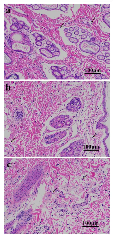
Histological assessment revealed an increased infiltration of eosinophils at the site of recombinant protein injection compared to the 0.9% physiological saline-injected site. As for the different recombinant protein-injected sites, the number of eosinophils in the Eur m 1-like protein site was greater compared to the Der p 1-like protein and Der p 7-like protein-injected sites, whereas eosinophil counts in Der p 1-like protein site showed similarity to that in the Der p 7-like protein-injected sites (Fig. 2).
RT‑qPCR validation
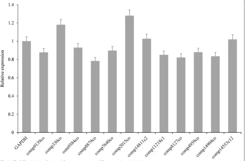
RT-qPCR was performed to validate allergen unigenes, which showed that our results were highly correlated with transcripts obtained by RNA-Seq, revealing that our RNA-seq results were reliable. Primers of aller-gen unialler-genes and the internal reference aller-gene GAPDH were designed for RT-qPCR. Expression of 12 aller-gens (comp7640_c0, comp2015_c0, comp14904_c0, comp14553_c12, comp4959_c0, comp8127_c0, comp9584_c0, comp330_c0, comp8879_c0, comp11218_ c1, comp9139_c0, and comp14811_c2) were normalized to GAPDH (Fig. 3).
Discussion
Unigenes of C. texanus were enriched in 262 signaling
[image:5.595.56.541.100.306.2]pathways as shown in KEGG pathway enrichment anal-ysis. The enriched pathways including immune related signaling pathways, MAPK signaling pathway, chemokine signaling pathway (90 unigenes), Fc gamma R-mediated phagocytosis (61 unigenes), Toll-like receptor ing pathway (51 unigenes), NOD-like receptor signal-ing pathway (46 unigenes), Jak-STAT signalsignal-ing pathway
Table 2 Putative allergen unigenes
(34 unigenes), RIG-I-like receptor signaling pathway (23 unigenes) and others. From these, the Jak-STAT signal-ing pathway, Toll-like receptor signalsignal-ing pathway, and immune deficiency signaling pathway are reported to be the main signaling pathways of the insect innate immune
response [37, 38]; however, the immune deficiency
sign-aling pathway was not identified in the KEGG pathway analysis result. Jak-STAT controls multiple biological processes in metazoan development and tissue homoeo-stasis, and it has been associated with several aspects of
the innate immune system [39]. The Jak-STAT
signal-ing pathway can affect the activation of neutrophils and macrophages, pro-inflammation response, and regulate B
cell and T cell differentiation [40, 41]. Toll-like receptor signaling has been studied by various approaches involv-ing genetic, biochemical, structural, cell biology and bio-informatics studies [42, 43]. Toll-like receptors (TLRs), are an important family of pattern recognition receptors (PRRs) and are responsible for the recognition of patho-gen-associated molecular patterns from infectious path-ogens. PRRs activate downstream signaling pathways which lead to the induction of innate immune responses by producing the inflammatory cytokines, type I
inter-feron (IFN), and other mediators [44]. These processes
not only trigger immediate host defensive responses such as inflammation, but also prime and orchestrate the
Fig. 1 Intradermal skin test. a Histamine (4 mg/ml, 0.1 ml). b 400 μg of purified pET‑32a (+) empty expression vector in 0.1 ml PBS. c 0.1 ml PBS.
[image:6.595.60.540.88.490.2]antigen-specific adaptive immune responses [44]. Toll-like receptor signaling appears to be divergent and plays an important role in many aspects of the innate immune responses in many pathogens [43, 45, 46].
Chorioptes texanus infested cattle suffer hair loss, scratching and skin damage. The pro-inflammatory cytokines commonly cause skin damage and these factors
come from the secretions/excretions of mites. Secre-tions from some parasites are important allergens which elicit a pro-inflammatory response in the host [47–49]. Although allergic reactions in host can lead to a protective immune response, allergic events can lead to immuno-logical hypersensitivity, tissue damage and may produce harmful effects. IgE antibody mediates a type I immu-nological hypersensitivity which is commonly caused by allergenicity of an allergen [50, 51]. The ability of an anti-gen to induce allergic sensitization is called alleranti-genicity, which is measured by the reactivity of allergen-induced IgE antibody, and indicates that immune system of the host has been elicited to an allergic state [52–54]. Twenty-one allergens have been widely studied (structural, chemi-cal and biologichemi-cal properties) in house dust mites [54, 55]. Allergens can activate the innate immune cells and induce immunologic responses by binding to C-type lectin recep-tors or Toll-like receprecep-tors [56]. In addition, group 1 and
2 allergens from Dermatophagoides pteronyssinus were
reported to boost the innate immune response and cleave IgE receptors, resulting in an increased allergic reaction in the host [57–59]. Several other allergens have been
pre-dicted in the transcriptome or genome of S. scabiei and
P. ovis, including triosephosphate isomerase, chitinase-like protein of S. scabiei and Pso 1, Pso 2, Pso 10, Pso 11 of P. ovis [23, 60–63]. These findings highlight that mite allergens may also play a crucial role in the pathogenesis of chorioptic mange. Proteases are mainly allergens of the house dust mite and hydrolysis of these proteases is reported to promote and aggravate the allergic reaction and inflammatory responses [20, 21, 64, 65]. Proteases can degrade fibrinogen in P. ovis, and provide flow of serous
exudate from the host during mite feeding [21].
Addition-ally, aspartic protease of S. scabiei can digest serum mol-ecules and skin of the host [66]. Similarly, hydrolases may also contribute to mite survival and invasion, and have a crucial role in the interaction of C. texanus mites and
their host. In the present study, comparisons between C.
texanus unigenes and the allergen website resulted in 209 putative mite allergen unigenes homologous to the aller-gen website. Also 34 putative alleraller-gen-hydrolase unialler-genes were obtained. Normally IgE ELISA and IgE dot blotting are performed to identify the allergenic activity of aller-gens. However, in the present study, IgE ELISA and IgE dot blotting were not feasible due to the lack of an effec-tive secondary anti-cow IgE antibody. Three allergen genes were selected from the 209 putative mite allergen unigenes, Der p 1-like protein gene, Der p 7-like protein gene and Eur m 1-like protein gene. Der p 1 and group
1 allergens from Dermatophagoides farina represent a
homologous pair of major allergens which possess both cross-reacting and species-specific epitopes [67], group 1
allergen (Der p 1) from Dermatophagoides pteronyssinus
[image:7.595.58.288.87.563.2]has been reported to boost the innate immune response and cleave IgE receptors which may have increased the allergic reaction in the host [57–59]. Recombinant fusion protein of Der p 1 activates basophils in mite-allergic
patients and triggers specific CD4+ T cell proliferation
[68]. Interestingly, although Eur m 1 showed 85% amino
acid identity with Der p 1 [69, 70], but 100 μg, 200 μg and 400 μg of recombinant Eur m 1-like protein produced the wheal, flare and erythema reaction, even blistering and ulceration, and only 400 μg recombinant Der p 1-like protein produced an erythema reaction. There is a large degree of T cell cross-reactivity between the whole puri-fied allergen from each species, according to the prolif-erative and cytokine response to the group 1 and group
7 allergens [71]. Der p 7 is a glycoprotein and performs
its function partially through glycan binding, it can acti-vate BMDCs through TLR4 and DC-SIGN, and establish a link between innate TLR4-C-type lectin receptors and
adaptive Th2 immunity [72]. Normally, the frequency of
IgE-binding to the allergen in sera from an allergic popu-lation is used to determine the relative importance of the individual house dust mite allergens, and the equivalent increased IL-5 response of PBMC to group 7 and group 1 allergen (different IgE-inducing activity) indicates that
allergens may be equally capable of contributing to an asthmatic response by inducing eosinophilia [73]. Der p 7 has a high IgE-binding activity but only reacts with 50% allergic sera, and this may explain the onset of a mild aller-gic skin reaction compared to the Eur m 1-like protein. Eosinophils participate in the adaptive immune response as antigen presenting cells and secrete Th cell chemokine, accounting about 1–3% in normal physiological condi-tions. Normally eosinophils are not present in skin, but many contributing factors, including hypersensitivity to arthropod bites and parasites, can cause eosinophilic infil-tration in the skin, resulting in skin disorders. In the pre-sent study, we observed that the number eosinophils in the Eur m 1-like protein-injected sites was higher than in the Der p 1-like protein and Der p 7-like protein-injected sites. These findings indicate that, these recombinant proteins can induce allergic reactions, further validating the reliability of our sequencing results and analysis. The Der p 1-like protein was a low-range allergen, the Der p 7-like protein and Eur m 1-like proteins were medium-range and high-medium-range allergens in C. texanus mite, respec-tively. Further focused functional studies on these genes
can improve our understating of C. texanus and host
[image:8.595.62.540.90.404.2]interactions, which may contribute to the discovery of novel interventions against this ecoparasite.
Conclusions
Comparisons between C. texanus unigenes and the
allergen database website resulted in 209 putative mite allergen unigenes. Also 34 putative hydrolase-allergen unigenes were obtained. The allergenic activity of recom-binant Eur m 1-like protein, Der p 1-like protein and Der p 7-like protein were preliminarily investigated by an
intradermal skin test. The transcriptome of C. texanus
provides a useful basis for understanding the host-par-asite interaction and molecular biology of mites. Iden-tification of putative allergen genes and hydrolase genes could offer opportunities for the development of new efficient diagnostic, prevention and treatment methods.
Supplementary information
Supplementary information accompanies this paper at https ://doi. org/10.1186/s1307 1‑019‑3843‑7.
Additional file 1: Figure S1. Length distribution of C. texanus unigenes and transcript.
Additional file 2: Figure S2. KOG classification of C. texanus unigenes.
Additional file 3: Figure S3. GO annotation of C. texanus unigenes.
Additional file 4: Figure S4. KEGG pathway analysis of C. texanus unigenes.
Additional file 5: Figure S5. Length distribution of CDS determined by Blastx program.
Additional file 6: Figure S6. Length distribution of CDS determined by EST‑Scan software.
Abbreviations
GO: Gene Ontology; KEGG: Kyoto Encyclopedia of Genes and Genomes; KO: KEGG Orthology; KOG: Eukaryotic Orthology Groups Database. CDS: coding sequences; RT‑qPCR: reverse‑transcription quantitative PCR.
Acknowledgements
We would like to thank Ruiqi Hua, Xiang Nong, Manli He, Haojie Zhang, Song Liu, and Ce Wang for their help and suggestions.
Authors’ contributions
RH and GYY conceived and designed this study. RH contributed sample col‑ lection, data analysis, experiment preformation and paper writing. CA contrib‑ uted to paper revision. XBG contributed to sample collection and bioinformat‑ ics analysis. XY, XRP, GYY contributed to sample collection. All authors read and approved the final manuscript.
Funding
This work was supported by a Grant from the Research Fund for the Chengdu Research of Giant Panda Breeding (Project No. CPF2014‑17). The funder had no role in study design, data collection and analysis, decision to publish, or preparation of the manuscript.
Availability of data and materials
The transcriptome raw sequence data has been submitted to the GenBank (Project Accession No. PRJNA495065). The other data supporting our findings and conclusions are available in the article and its additional files.
Ethics approval and consent to participate
The animal study was reviewed and approved by the Animal Care and Use Committee of Sichuan Agricultural University (SYXK2019‑187). All animal pro‑ cedures used in this study were carried out in accordance with the Guide for the Care and Use of Laboratory Animals (National Research Council, Bethesda, MD, USA) and recommendations of the ARRIVE guidelines (http://www.nc3rs .org.uk/arriv e‑guide lines ). All methods were carried out in accordance with relevant guidelines and regulations.
Consent for publication
Not applicable.
Competing interests
The authors declare that they have no competing interests.
Author details
1 Department of Parasitology, College of Veterinary Medicine, Sichuan Agricul‑ tural University, Chengdu 611130, China. 2 Department of Chemistry, College of Life and Basic Science, Sichuan Agricultural University, Chengdu 611130, China. 3 Department of Veterinary Parasitology, Faculty of Veterinary Sci‑ ences, Shaheed Benazir Bhutto University of Veterinary and Animal Sciences, Sindh 67210, Pakistan.
Received: 23 September 2019 Accepted: 9 December 2019
References
1. Yeruham I, Rosen S, Hadani A. Chorioptic mange (Acarina: Psoroptidae) in domestic and wild ruminants in Israel. Exp Appl Acarol. 1999;23:861–9. 2. Lusat J, Bornstein S, Wall R. Chorioptes mites: re‑evaluation of species
integrity. Med Vet Entomol. 2011;25:370–6.
3. Wang T, Xie Y, Zheng Y, Wang C, Li D, Koehler AV, et al. Parasites of the giant panda: a risk factor in the conservation of a species. Adv Parasit. 2018;99:1–33.
4. Fain A, Leclerc M. A case of mange in a giant panda caused by a new spe‑ cies of Chorioptes (Acarina: Psoroptidae). Acarologia. 1975;17:177. 5. Cremers H. Chorioptes bovis (Acarina: Psoroptidae) in some camelids from
Dutch zoos. Vet Quart. 1985;7:198–9.
6. Bochkov AV, Klimov PB, Hestvik G, Saveljev AP. Integrated Bayesian species delimitation and morphological diagnostics of chorioptic mange mites (Acariformes: Psoroptidae: Chorioptes). Parasitol Res. 2014;113:2603–27.
7. Sweatman GK. Life history, hon‑specificity, and revision of the genus Chorioptes, a parasitic mite of herbivores. Can J Zool. 1957;35:641–89. 8. Suh GH, Hur TS, Shin SM, Kwon J, Cho SH, Lee CY, et al. The first outbreak
of Chorioptes texanus (Acari: Psoroptidae) infestation in a cattle farm in Korea. Korean J Parasitol. 2008;46:273.
9. Vieira MIB, Bordin T, Agnol BD, Zanchin F, Motta ACD, et al. Re‑emergence of Chorioptes bovis (Acari: Psoroptidae) in cattle in the state of Rio Grande do Sul. Brazil. Rev Bras Parasitol. 2014;23:530–3.
10. Rehbein S, Winter R, Visser M, Maciel AE, Marley SE. Chorioptic mange in dairy cattle: treatment with eprinomectin pour‑on. Parasitol Res. 2005;98:21.
11. Nong X, Li SH, Wang JH, Xie Y, Chen FZ, He R, et al. Acaricidal activity of petroleum ether extracts from Eupatorium adenophorum against the ectoparasitic cattle mite, Chorioptes texanus. Parasitol Res. 2014;113:1201–7.
12. Villarroel A, Halliburton MK. Control of extensive chorioptic mange natural infection in lactating dairy cattle without milk withdrawal. Vet J. 2013;197:233–7.
13. Hestvik G, Zahler Rinder M, Gavier Widén D, Lindberg R, Mattsson R, Morrison D, et al. A previously unidentified Chorioptes species infest‑ ing outer ear canals of moose (Alces alces): characterization of the mite and the pathology of infestation. Acta Vet Scand. 2007;49:21. 14. Zahler M, Hendrikx WM, Essig A, Rinder H, Gothe R. Taxonomic recon‑
15. Wall RL, Shearer D. Veterinary ectoparasites: biology, pathology and control. 2nd ed. Hoboken, USA: John Wiley Sons; 2008.
16. Marcondes CB, Dantas‑Torres F. Diseases caused by Acari (ticks and mites). J Arthropod Bone Dis. 2017;537–48.
17. Cremers H. The incidence of Chorioptes bovis (Acarina: Psoroptidae) on the feet of horses, sheep, and goats in the Netherlands. Vet Quart. 1985;7:283–9.
18. Losson B, Mignon B, Bossaert K, Leclipteux T, Lonneux J. Field efficacy of injectable doramectin against Chorioptes bovis in naturally infected cattle. Vet Rec. 1998;142:18–9.
19. Rüfenacht S, Roosje PJ, Sager H, Doherr MG, Straub R, Goldinger Müller P, et al. Combined moxidectin and environmental therapy do not eliminate Chorioptes bovis infestation in heavily feathered horses. Vet Dermatol. 2011;22:17–23.
20. Arlian LG. Arthropod allergens and human health. Annu Rev Entomol. 2002;47:395–433.
21. Kenyon F, Knox D. The proteinases of Psoroptes ovis, the sheep scab mite‑their diversity and substrate specificity. Vet Parasitol. 2002;105:317–25.
22. Caraballo L, Coronado S. Parasite allergens. Mol Immunol. 2018;100:113–9.
23. He R, Shen N, Zhang H, Ren Y, He M, Xu J, et al. Molecular charac‑ teristics and serodiagnostic potential of chitinase‑like protein from Sarcoptes scabiei. Oncotarget. 2017;8:83995.
24. Xu J, Huang X, He M, Ren Y, Shen N, Li C, et al. Identification of a novel PYP‑1 gene in Sarcoptes scabiei and its potential as a serodiagnostic candidate by indirect‑ELISA. Parasitology. 2018;145:752–61. 25. Shen N, He R, Liang Y, Xu J, He M, Ren Y, et al. Expression and charac‑
terisation of a Sarcoptes scabiei protein tyrosine kinase as a potential antigen for scabies diagnosis. Sci Rep. 2017;7:9639.
26. Wang S, Gu X, Fu Y, Lai S, Wang S, Peng X, et al. Molecular taxonomic relationships of Psoroptes and Chorioptes mites from China based on COI and 18S rDNA gene sequences. Vet Parasitol. 2012;184:392–7. 27. Essig A, Rinder H, Gothe R, Zahler M. Genetic differentiation of
mites of the genus Chorioptes (Acari: Psoroptidae). Exp Appl Acarol. 1999;23:309–18.
28. Zheng J, Zhang X, Yang G, Gu X, Yu Z. Cloning and sequence analysis of Chorioptes texanus paramyosin gene. Chinese Vet Sci. 2008;38:38–41. 29. Zhang X, Yang G, Gu X, Jia X, Yang C, Zheng J. Morphological observa‑
tion of Chorioptes isolated from cattle in Sichuan Province. Chin Vet Sci. 2006;36:827–31.
30. He R, Gu X, Lai W, Peng X, Yang G. Transcriptome‑microRNA analy‑ sis of Sarcoptes scabiei and host immune response. PLoS ONE. 2017;12:e0177733.
31. Haas BJ, Papanicolaou A, Yassour M, Grabherr M, Blood PD, Bowden J, et al. De novo transcript sequence reconstruction from RNA‑seq using the Trinity platform for reference generation and analysis. Nat Protoc. 2013;8:1494–512.
32. Iseli C, Jongeneel CV, Bucher P. ESTScan: a program for detecting, eval‑ uating, and reconstructing potential coding regions in EST sequences. In: ISMB. 1999. p. 138–48.
33. Conesa A, Götz S, García‑Gómez JM, Terol J, Talón M, Robles M. Blast2GO: a universal tool for annotation, visualization and analysis in functional genomics research. Bioinformatics. 2005;21:3674–6. 34. Ye J, Fang L, Zheng H, Zhang Y, Chen J, Zhang Z, et al. WEGO: a web
tool for plotting GO annotations. Nucleic Acids Res. 2006;34:293–7. 35. Livak KJ, Schmittgen TD. Analysis of relative gene expression data
using real‑time quantitative PCR and the 2− ΔΔCT method. Methods. 2001;25:402–8.
36. Cole T, Williams BA, Geo P, Ali M, Gordon K, Baren MJ, et al. Transcript assembly and quantification by RNA‑Seq reveals unannotated transcripts and isoform switching during cell differentiation. Nat Biotechnol. 2010;28:511–5.
37. De Gregorio E, Spellman PT, Tzou P, Rubin GM, Lemaitre B. The Toll and Imd pathways are the major regulators of the immune response in Dros-ophila. EMBO J. 2002;21:2568–79.
38. Tanji T, Hu X, Weber AN, Ip YT. Toll and IMD pathways synergistically activate an innate immune response in Drosophila melanogaster. Mol Cell Biol. 2007;27:4578–88.
39. Dostert C, Jouanguy E, Irving P, Troxler L, Galiana‑Arnoux D, Hetru C, et al. The Jak‑STAT signaling pathway is required but not sufficient for the antiviral response of Drosophila. Nat Immunol. 2005;6:946–53. 40. Rawlings JS, Rosler KM, Harrison DA. The JAK/STAT signaling pathway. J
Cell Sci. 2004;117:1281–3.
41. Heinrich PC, Behrmann I, Serge H, Hermanns HM, Müller‑Newen G, Schaper F. Principles of interleukin (IL)‑6‑type cytokine signalling and its regulation. Biochem J. 2003;374:1–20.
42. Botos I, Segal DM, Davies DR. The structural biology of Toll‑like receptors. Structure. 2011;19:447–59.
43. Barton GM, Medzhitov R. Toll‑like receptor signaling pathways. Science. 2003;300:1524–5.
44. Janeway CA Jr, Medzhitov R. Innate immune recognition. Annu Rev Immunol. 2002;20:197–216.
45. Yamamoto M, Sato S, Hemmi H, Hoshino K, Kaisho T, Sanjo H, et al. Role of adaptor TRIF in the MyD88‑independent toll‑like receptor signaling pathway. Science. 2003;1:640–3.
46. Akira S, Takeda K. Toll‑like receptor signalling. Nat Rev Immunol. 2004;4:499.
47. Levy DA. Parasites and allergy. Clin Rev Aaaerg Immun. 2004;26:1–4. 48. Hewitson JP, Grainger JR, Maizels RM. Helminth immunoregulation: the
role of parasite secreted proteins in modulating host immunity. Mol Biochem Parasit. 2009;167:1–11.
49. Bennuru S, Semnani R, Meng Z, Ribeiro JMC, Veenstra TD, Nutman BT. Brugia malayi excreted/secreted proteins at the host/parasite interface: stage‑ and gender‑specific proteomic profiling. Plos Negl Trop Dis. 2009;3:e410.
50. Hales BJ, Martin AC, Pearce LJ, Laing IA, Hayden CM, Goldblatt J, et al. IgE and IgG anti‑house dust mite specificities in allergic disease. J Allergy Clin Immun. 2006;118:361–7.
51. Chapman MD, Pomés A, Breiteneder H, Ferreira F. Nomenclature and structural biology of allergens. J Allergy Clin Immun. 2007;119:414. 52. Ogunlade OA, Ige OM, Arinola OG, Onadeko BO. Allergen‑specific immu‑
noglobulin E (IgE) antibodies and skin test reactivity in patients with asthma in Nigeria. J Clin Immun Immunol Res. 2012;3:25–8.
53. Mizuma H, Tanaka A, Uchida Y, Fujiwara A, Manabe R, Furukawa H, et al. Influence of omalizumab on allergen‑specific IgE in patients with adult asthma. Int Arch Allergy Imm. 2016;168:165.
54. Thomas WR, Hales BJ, Smith WA. House dust mite allergens in asthma and allergy. Trends Mol Med. 2010;16:321–8.
55. Fischer K, Walton S. Parasitic mites of medical and veterinary importance—is there a common research agenda? Int J Parasitol. 2014;44:955–67.
56. Gao P. Erratum to “Sensitization to cockroach allergen: immune regula‑ tion and genetic determinants”. Clin Dev Immunol. 2012;63:1847. 57. Trompette A, Divanovic S, Visintin A, Blanchard C, Hegde RS, Madan R,
et al. Allergenicity resulting from functional mimicry of a Toll‑like receptor complex protein. Nature. 2008;457:585–8.
58. Gough L, Schulz O, Sewell HF, Shakib F. The cysteine protease activity of the major dust mite allergen Der p 1 selectively enhances the immuno‑ globulin E antibody response. J Exp Med. 1902;190:1897–902.
59. Posa D, Perna S, Resch Y, Lupinek C, Panetta V, Hofmaier S, et al. Evolution and predictive value of IgE responses toward a comprehensive panel of house dust mite allergens during the first 2 decades of life. J Allergy Clin Immun. 2017;139:541–9.
60. He R, Zhang H, Shen N, Guo C, Ren Y, Xie Y, et al. Molecular characteri‑ zation and allergenicity potential of triosephosphate isomerase from Sarcoptes scabiei. Vet Parasitol. 2018;257:40–7.
61. Nisbet A, MacKellar A, Wright H, Brennan G, Chua K, Cheong N, et al. Molecular characterization, expression and localization of tropomyosin and paramyosin immunodominant allergens from sheep scab mites (Psoroptes ovis). Parasitology. 2006;133:515–23.
62. Temeyer KB, Soileau LC, Pruett JH. Cloning and sequence analysis of a cDNA encoding Pso o II, a mite group II allergen of the sheep scab mite (Acari: Psoroptidae). J Med Entomol. 2002;39:384–91.
63. Nisbet A, MacKellar A, McLean K, Brennan G, Huntley J. Eukaryotic expres‑ sion of recombinant Pso o 1, an allergen from Psoroptes ovis, and its localization in the mite. Parasitology. 2007;134:83–9.
•fast, convenient online submission
•
thorough peer review by experienced researchers in your field
• rapid publication on acceptance
• support for research data, including large and complex data types
•
gold Open Access which fosters wider collaboration and increased citations maximum visibility for your research: over 100M website views per year
•
At BMC, research is always in progress.
Learn more biomedcentral.com/submissions
Ready to submit your research? Choose BMC and benefit from:
65. Aalberse RC. Structural biology of allergens. J Allergy Clin Immunol. 2000;106:228–38.
66. Mahmood W, Viberg LT, Fischer K, Walton SF, Holt DC. An aspartic pro‑ tease of the scabies mite Sarcoptes scabiei is involved in the digestion of host skin and blood macromolecules. PloS Negl Trop Dis. 2013;7:e2525. 67. Heymann PW, Chapman MD, Platts‑Mills TA. Antigen Der f I from the dust
mite Dermatophagoides farinae: structural comparison with Der p I from Dermatophagoides pteronyssinus and epitope specificity of murine IgG and human IgE antibodies. J Immunol. 1986;137:2841–7.
68. Laetitia B, Véronique BLF, Ingrid B, Henri C, Emmanuel N, Aurélie L, et al. Recombinant fusion proteins assembling Der p 1 and Der p 2 allergens from Dermatophagoides pteronyssinus. Int Arch Allergy Imm. 2010;153:141–51.
69. Kent NA, Hill MR, Keen JN, Holland PW, Hart BJ. Molecular characterisation of group I allergen Eur m I from house dust mite Euroglyphus maynei. Int Arch Allergy Imm. 1992;99:150–2.
70. Smith W, Mills K, Hazell L, Hart B, Thomas W. Molecular analysis of the group 1 and 2 allergens from the house dust mite, Euroglyphus maynei. Int Arch Allergy Immun. 1999;118:15–22.
71. Hales BJ, Shen HD, Thomas WR. Cross‑reactivity of T‑cell responses to Dermatophagoides pteronyssinus and D. farinae Studies with group 1 and 7 allergens. Clin Exp Allergy. 2010;30:927–33.
72. Tsai JJ, Wang HC, Chiu CL, Liao EC. The effect of Dermatophagoides ptero-nyssinus group 7 allergen (Der p 7) on dendritic cells and its role in T cell polarization. Immunobiology. 2016;221:1319–28.
73. Hales BJ, Shen HD, Thomas WR. Cytokine responses to Der p 1 and Der p 7: house dust mite allergens with different IgE‑binding activities. Clin Exp Allergy. 2010;30:934–43.
Publisher’s Note