0022-538X/81/010001-06$02.00/0 Vol. 37, No. 1
Comparative Study of Rabies
Virus Persistence
in Human
and
Hamster Cell
Lines
0. G. ANDZHAPARIDZE, N.N. BOGOMOLOVA, Y. S. BORISKIN,* M. S. BEKTEMIROVA, AND
I.D. DRYNOV
ResearchInstitute forViral Preparations,Moscow, USSR
Persistent infections by rabies virus in BHK-21/13S and HEp-2 cells were
studied comparatively. No evidence of interferon production, selectionof
virus-resistantcells,orintegration of the viralgenomecould be found. Persistingviruses
replicated efficiently at 34, 36, and 40°C. Both persistently infected cultures
released defective interfering virus particles.Acyclicalpatternof infection,which
wasnotcharacteristic of the persistently infectedHEp-2system,wasobserved in persistently infected BHK cultures. The virus from persistently infected BHK
cultures lost its virulence for mice, whereas the virus frompersistently infected
HEp-2 cultures retained mouse-killing capacity formorethan3years.
Persistent infectionby rabies virus has been studied extensively in cell cultures of hamster
origin (6, 8, 16); in general, the mechanism of rabies virus persistence is currently attributed
todefective interferingparticle (DIp)-mediated
cell sparing, as evidenced from several studies
with BHK-21 cells (6, 8, 17). However, a host
effect with respect to DIp generation (7) or
acquisition of virulence (3) has been observed
whencells distinctfromconventionalBHKcells were used. This communication is an examina-tion of the roleofseveral factorsthat may con-tribute to rabies viruspersistence intwo differ-ent cell cultures. The results indicate that the hostcell type maystronglyinfluencethe
estab-lishment and course of infection and the
viru-lence of the persistingvirus.
MATERIALS AND METHODS
Cells and viruses.Primarycultures ofJapanese
quail embryo(QE)cellswerepropagatedasdescribed
previously (2). BHK-21/13S and HEp-2 cells were
grown inEagle and 199media, respectively,
supple-mented with 10% bovine serum and antibiotics. A
Pasteur strain of rabies virus that was adapted to
growth inQEcells (2) wasused for the initiation of
persistent infection andas a control standard virus.
Bothpersistentlyinfected cell linesweremaintained
at360C.
Assayof viralinfectivity.The infectious titer of
rabies viruses was determined by the intracerebral
inoculation of mice orbya plaque assaywith CER
cells (kindly provided by J. J. Holland and T. J.
Wiktor) withaSephadex overlay (10).
Directfluorescent-antibody technique.The
di-rectfluorescent-antibodytechniquewasdescribed pre-viously (2).
Cellcloningexperiments. Carriercellswere
dis-persed,counted,andplatedatdilutionsthatprovided
veryfewcells per flask.Single cellsweremarked3 to
4days after seeding and observed until a colony had
formed. These coloniesweretransferredtoindividual
tubes, and thedeveloped subcultures were examined.
Immunogenicity of persistingviruses. The
im-munogenic activity of the viruseswasdetermined by
the standard National Institutes of Health potency
testinmice(11).
Concentration andpurification of rabies virus.
Rabies virus was pelleted from the culture fluid at
20,000 rpmfor2 hina 6x250mlrotorofanMSE SS
65 centrifuge, suspended in NTE buffer (13), and
clarified througha30%sucrosecushionat61,000xg
for 40 min. The visible band at the interface was
aspirated, diluted with NTE buffer, and pelleted at
45,000 rpm for2hin anSW50rotorofaBeckman L5
65centrifuge. Virus suspended in 0.5 to 0.8 ml was
layeredon a 5 to30% linearsucrosegradientin NTE
buffer made with the aid ofanLKBUltrograd gradient
mixer. CentrifugationwasperformedinanSW40rotor
at 34,000 rpm for60min. Gradients wererecovered
from the bottomthroughaflowcell ofaGilford
2400-2spectrophotometer, withacontinuousmonitoringof
theoptical density(OD)at260nm.
Polyacrylamide gelelectrophoresis.Samplesof
purified virusweredissociated andseparatedon7.5%
cylihdrical gelsinasodiumdodecyl sulfate-phosphate
systemasdescribedbyWeberetal.(15).
Electropho-resiswasconducted in 14-cmtubesat3mApertube
for 20 to 24 h. Gels were fixed and stained in a
methanol-glacialacetic acid-Coomassiebrilliant blue
R-250mixture and after destainingwere scannedin
theGilfordspectrophotometerat550nm.The
molec-ular weight standardswere bovinealbumin,
ovalbu-min, and myoglobin (Schwarz/Mann, Orangeburg,
N.Y.; 68,000, 45,000,and18,600,respectively).
Preparation of rabies virus ['H1RNA and
[tHl-cDNA. For thepreparation of[3H]RNA, RNA was
extractedfrompurifiedrabiesvirus(a totalof5 x10'0
50% lethaldoses[LD50])that had beenpropagatedin
QE cellsby treatmentwith sodium dodecyl
sulfate-phenol-chloroform (9).Aportionof this RNA
prepa-ration was used forlabelingbythe direct introduction
on November 10, 2019 by guest
http://jvi.asm.org/
2 ANDZHAPARIDZE ET AL.
of tritium into the polynucleotide in the course of RNA treatment with 3H atoms (12). [3H]RNA
ob-tained in thismanner wasdialyzed against25volumes
ofO.lxSSC buffer(lx SSC is0.15MNaClplus0.015
M sodium citrate) containing 0.5% sodium dodecyl
sulfate inanMMC Amiconsystemthrougha10PM
membrane and stored under ethanol at-25°C. The
specificactivity of thepreparationwas0.9x106cpm/
Ag-[3H]complementary DNA ([3H]cDNA) of rabies
vi-rusRNAwasobtainedessentiallyasdescribed
previ-ously (1) with50U of Escherichia coli DNA
polym-erase (Sigma Chemical Co.,St. Louis, Mo.) and
3H-labeled deoxytriphosphates ([3H]dGTP and
[3H]-dCTP[AmershamCorp., Arlington Heights,Ill.],
spe-cificactivities,10.3and25Ci/mmol,respectively;and
[3H]dATP [Isotop, Leningrad, USSR],specific
activ-ity, 7.2 Ci/mmol). The composition ofthe reaction
mixture, the isolation ofcDNA, and thepreparation
of thehybridwerethesame aspreviously described
(1).Thespecific activity of cDNAwas6.7 x107cpm/
ug as determined from the specific activities ofthe
input3H-labeleddeoxytriphosphates. After synthesis,
cDNA sedimented inan alkalinesucrosegradientat
apositioncorresponding to 8to 9S and annealedto
excessvirion RNAto an extentof about 90%.
Hybridization of carrier cell DNA to rabies
virus [3H]RNA orcDNA. The DNA was isolated
from carrier cultures at a level of 104 passages for
persistently infected HEp-2 cultures(HEp-2p.j.)andat
alevel of117passagesforpersistently infected BHK
cultures(BHKp.i.)byachloroform-isoamyl alcohol (24:
1)technique,including thestepsof RNase andpronase
digestions between subsequent deproteinizations, as
described by Cooper and Temin (4). The DNA
ob-tained in thismannerhadaratio ofabsorbanceat 260
nm toabsorbance at280nmof1.8 to1.9,whichwas
comparabletothat ofcalfthymus DNA(Serva,
Hei-delberg, Germany). The composition of the annealing
reactionmixtures and theprotocol for the
hybridiza-tionwereidenticaltothose describedpreviously(1).
Transfectionexperiments with DNA from
car-riercells. Confluentmonolayersof 2x10⁷BHKcells
were pretreated for 5 min with 300
iLg
ofDEAE-dextranperml in 0.15 MNaCl. Afterthe aspiration of
DEAE-dextran,cellswereexposedtoDNAdissolved
in 0.15 M NaCl. The DNA exposure levels ranged
from0.5 to 7 mg perflask. Aftera15-min adsorption
at roomtemperature,growth mediumwasaddedand
cells were incubated at 36°C. Transfected cultures
weresplittwiceweekly,with periodic assays of cultural
fluid forinfectiousvirus and of cells for viral antigen
byimmunofluorescence.
RESULTS
Establishment and time course of per-sistent infection. Confluent monolayer
cul-turesofHEp-2 or BHK cells were infected with
rabies virus at a multiplicity of 1 LD50per cell.
Aftera1.5-hadsorption period, growth medium
was added and the cultures were incubated at
36°C.Nosigns ofcelldestruction were observed in
HEp-2p.i.
at 7 to 10 days postinfection. In contrast, inBHKp.i.,
manycells diedbypostin-fection day 4, and the addition of fresh BHK
cellswasrequired during eight subsequent
pas-sages to preventcompletecelldestruction.After
the persistent infection had stabilized,
BHKp.i.
werepassed every5 to 7days,withnosigns of
visible cytopathology.
Differentpatterns of infection werefound in the two persistently infected cultures (Fig. 1). For
BHKp.i.,
cyclical fluctuations of antigen-pos-itive cellsranging from 100toless than1%wereobserved, although the antigen never
disap-peared completely. The persisting virus
gradu-ally lost its virulence for mice; soon after 50 passages of
BHKp.i.,
mouse-lethalviruswas nolonger detectable.
In contrast to
BHKp.i.,
a significantly moreconstantproductionof infectious virus and viral
antigenwasfound in
HEp-2p.i.
(Fig. 1).Properties ofpersistingviruses. Starting
from theearly stages ofinfection,thepersisting viruses fromboth cultures
characteristically
dis-playedincubationperiodsthatwereabout twice
aslongin miceasthose withstandardvirus(12
to 14 versus 6 to 8days).The virus from
HEp-2p.;.
retained its virulence for mice formorethan 3years, whereas the virusfromBHKp.i.
lostitspathogenicity after 50 passages ofthe culture. Thisloss of
mouse-killing
abilityremained after three intracerebral passages of the virus insuck-ling-mousebrains.
Persisting viruses could induce plaques on
CER cellmonolayers.Thespecificityofplaques
wasverifiedinaplaque reduction neutralization
testwith antiserumtooriginalviruspropagated
in QE cells.
BHKp.i.
virus plaques were 0.1 to0.15 cm in
diameter,
whereasHEp-2p.i.
virusformedtiny pinpointplaques.
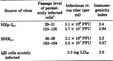
The immunogenic ability of unconcentrated
persisting viruses was poor. However,
concen-FIG. 1. Timecourseofpersistent infections by
ra-bies virus in
HEp-2p.,.
(A) andBHKP,.. (B). Symbols:0,infectivity;0,percentage ofantigen-positivecells.
(Abscissa) Number of cell transfers; (left ordinate)
percentageoffluorescingcells;(right ordinate) virus titerinlogLD50permilliliter.
J. VIROL.
on November 10, 2019 by guest
http://jvi.asm.org/
[image:2.496.264.455.470.606.2]trated viruses displayed their immunogenicity
evenatlatestagesof persistence(Table 1).
To determine the effectoftemperatureonthe multiplication of persisting viruses, BHK cells wereinfectedwith
HEp-2p.i.
virus frompassage134,
BHKp.i.
virus frompassage163,orstandard virus and incubatedat34, 36,and400C.Samplesofculture fluidwere taken on 3, 4, and 5days
postinfection and titratedin miceorbyplaquing. As shown in Table 2, the virus from
HEp-2p.j.
replicated almost equally well atdifferent
tem-peratures. The virus from
BHKp.i.
replicatedsomewhat better at 340C than it did at 400C. However, there were nosignificant differences in the numbers of antigen-positive cells when BHK cultures infected with
BHKp.i.
virus andincubated at various temperatures were com-pared (datanotshown). The original virus
dis-played pronouncedtemperaturesensitivity (Ta-ble2).
Incontrasttothe originalvirusgrown inQE
cells, persisting viruses from
HEp-2p.i.
andBHKp.i.
couldreadilyestablishacarrierstateinBHK and Vero cell cultures, with a similarly fluctuating pattern of infection in BHK cells
(datanotshown).
Detection of rabies virus DIpin
persist-ently infected cultures. Several lines of evi-dence suggested the presenceof interfering ac-tivity in ourpersistently infected cultures: (i)a consistently observed autointerference
phenom-enonin initial virus dilutions when titratedby
plaquing; (ii) alonger incubation period of per-sisting viruses in mice whencompared with that
ofstandardvirus; and (iii) acyclical patternof
infection in
BHKp.i.
thatwasreportedly dueto [image:3.496.48.241.475.579.2]DIp (8). Totestdirectly for thepresenceof DIp,
TABLE 1. Immunogenicproperties ofrabies viruses
releasedbypersistently infectedculturesa
Passagelevel Infectiousvi-
Immuno-Source ofvirus of persist- rustiter(per genicity
entlyinfected mll) index
cellsb
HEp-2.i. 29-31 3.1x10"PFU 2.4
123-126 3.7x10⁷PFU 0.94
BHKpi. 36-39 2.1x10⁸PFU 2.2
163-164 2.5x10⁷PFU 0.57
QEcellsacutely 5.5logLD5o 2.5
infected
aPersistingviruseswereconcentrated109-foldandinjected intraperitoneallyinto mice. For each dilutionof thevirus,
groupsof 14animalswereused. Theimmunogenicityindex is
expressedasthe ratio of thereciprocalof thehighestdilution
of the virusprotecting50%ofthe mice tothat of thestandard
reference vaccine supplied byTarasevichState Institute of
BiologicalStandardizationandControl,Moscow.
'Asimultaneouscomparisonbetweenviusesof different
cell passages waspossible due to the storage ofthe early
passagecarrier cellculturesinliquidnitrogen.
TABLE 2. Replicationofpersistingviruses in BHK
cells atdifferenttemperatures
Timepjt- Titer atfollowingtemp Source of virus inoculation _ _ C_ _
(h) 34 36 40
HEp-2p.L 72 3.0 2.5 3.0
96 4.25 3.5 2.75
120 3.75 3.0 4.0
BHKp.5. 72 4.5 4.0 3.0
96 NTb NT NT
120 NT NT NT
QEcells acutely 72 4.5 3.5 2.0
infected 96 6.0 3.5 2.0
120 6.0 3.75 2.0
a
All
of the titers are expressed in logLD50
permilliliter,
except forBHKp.L,
wherethe titers are expressed in log PFU permilliliter.b
NT,
Not tested.persistingviruses were
sedimented
from 800- to1,500-mlvolumes and inoculatedinto mice in a mixturewith 10-fold dilutions of standard virus.
Nodifferenceswereobservedbetween the titers
of the virus mixture and the standard virusonly.
However, after a
preliminary
one-step amplifi-cation on BHK cells (6), the titer ofamixturewas0.75 to 1.5log
LDio
perml lowerthan thatof similarly amplified standard virus. When BHKcellswereinfectedwithundiluted and
100-fold-diluted culture fluidfrom
HEp-2.i.,
the final virustiterwas 1log
LD5o
permlhigher
in thelattercase.Releasedvirusesharvested from
per-sistently
infected cellsatdifferentpassagelevelswereconcentrated and
analyzed
insucrosegra-dients. Viruses from
HEp-2p.i.
orBHKp.i.
formed ODbands that couldnotbedetectedinconcen-trated
materials
frommock-infectedorstandardvirus-infected
QE
cells(Fig. 2).
BHKp.i.
virususually
formed one broadband,
whereas twobandscould oftenbeobservedin
HEp-2p.i.
virus gradients(Fig.
20).
Theinfectivity
peak
sedi-mented ahead of the OD band(Fig.
2B),
sug-gesting
that OD structuresweresmallerin size thanrabiesvirusvirions.Thepolypeptide
com-position of
purified
ODstructureswassimilartothat of standard virus
propagated
inQE
cells (Fig. 3).Themolecularweights
ofthe polypep-tides were78,000,
62,000,
43,000,
and26,000,
whichare in agreementwithreported
data onrabies
virus proteins (14). Thus,
we concluded thatthese ODstructures weretruerabies virusdefective
particles.
Purified defectiveparticles
were not infectious when
injected
intracere-brally into mice and did not protect animals against
challenge
with virulent rabies virus. However, when theseparticles
weremixed with theinfectiousvirus andamplified
onBHKcells,
a
reproducible
reductioninthevirustiter,
on November 10, 2019 by guest
http://jvi.asm.org/
4 ANDZHAPARIDZE ET AL.
'x
IsoK
°o . I 1/:.6
6
A
4
2
3
2
I
[image:4.496.57.458.54.570.2]ib I5
FIG. 2. Sucrosegradient profiles ofrabiesviruses
releasedfrom acutelyinfected QEcells(A)and
HEp-2p.,.
(B). (C) ODprofilesoftwoparallel gradients (I,HEp-2p..;II, BHKp.i)recordedonthesame sheetof
paper. ( ) OD at 260nm; (....) infectivity.
(Ab-scissa) Fraction number; (left ordinate) OD at260
nm;(right ordinate) virus titerinlogLDroper
milli-liter.Centrifugationwasfrom lefttoright.
ing from 0.5 to 1.25 log LD50 per ml, was ob-served.
Examination of cell clones isolated from
persistentlyinfectedcultures. Todetermine
whether the resistance of persistently infected
culturestohomologous viruschallenge was as-sociated with the selection of rabies virus-resist-antcells,wecloned
BHKp.i.
andHEp-2p.i.
atthe levels of 100 and about 70% ofantigen-positivecells, respectively. A total of 32 clones were
0.5
B
C
FIG. 3. Polypeptide composition of QE
cell-prop-agated standard rabies virus (A) and DIp from
BHKp.L.
(B) andHEp-2p.1.
(C). (Ordinate) ODat 550 nm.obtained from
HEp-2p.i.,
17of which were anti-gen positive and virusproducing. Theantigen-free clones were susceptible to rabies virus to
thesame extent as werecontrol uninfected
HEp-2 cells. Of26
BHKp.i. clones,
24 were antigenJ. VIROL.
on November 10, 2019 by guest
http://jvi.asm.org/
negative and rabies virus susceptible. This result is intriguing because 100% of the cells in pre-cloned
BHKp.i.
contained viral antigen. More-over, two antigen-positive clones fromBHKp.i.
were spontaneously destroyed after four pas-sages, whereasall antigen-negative cloneswere splitatleast another five times without notice-ablechanges.
Attemptstodetect interferon production in
BHKp.i.
andHEp-2p.i.
BHKp.i.,
HEp-2p.i.,
and control uninfected cellswere infected with the Indiana serotype of vesicular stomatitis virus andaninterferon-susceptible measles virus var-iant. At 72 h postinfection, challenge viruses weretitrated by cytopathogenicityonBHK (ve-sicular stomatitis virus) or HEp-2 (measles vi-rus) cells. Challenge viruses consistently multi-plied equally well both inpersistently infected cultures and in uninfected control cultures, sug-gesting the lack of detectable interferon produc-tioninpersistently infected cultures.Search for virus-specific sequences in
DNA of the carrier cell cultures. At a Cot ranging from 137 to 6,567 mol.s/liter and a carrier cell DNA concentration of5mg/ml, the percentageofhybridization wasintherangeof
from 5to7% with eithera[3H]cDNAora
[3H]-RNAprobe. Control calfthymusDNA did not
differsignificantly from carrier cell DNAin its annealingrate.However, the cDNA probe
pres-entinthe reaction mixture wasstillcapable of specific annealing to excessvirion RNA atthe end of thehybridization period (59to62%ata
Cot
of3,283 mol.s/liter). Repeatedtransfectionattemptswith carriercellDNA toinduce
infec-tious virus or viral antigen in BHK cells were also unsuccessful.
DISCUSSION
Persistent infectionsby rabies virus have been established in rabbit (5) and reptilian (16) cell cultures, but have been studied extensively in
BHK-21 cells (6, 8, 16). However, an apparent hostcelleffect has been observedonrhabdovirus DIp generation in human cells (7) and on the virulence of rabies virus in neuroblastoma cells (3). We have studied thehost cell effecton (i) the course ofpersistent rabies virus infection, (ii) properties ofpersisting virus, and (iii) the
contribution of several factors currently re-garded as the mechanisms of viralpersistence into the establishment and maintenance of a carrierstate.To thisend, wehaveuseda well-characterized
BHKp.i.
system andHEp-2p.i.
ofhumanorigin.Incontrast to
BHKp.i.,
inwhicha cyclicalpatternof infection has beenobserved, the course of infection inHEp-2p.i.
was moreconstant. The persisting virus from
BHKp.i.
atthe level of50cellpassageslost itsvirulence for
mice; this is in accord with the finding of
Fer-nandes et al. (5). In contrast, the virus from
HEp-2p.;.
retained itsvirulence formiceformore than 3 years despite the production of DIp. Recently, Wunner and Clark have presentedevidence that the virulenceof rabies virus is not
mediatedbyDIp (18).
An examination of persistently infected cul-tureswith respect to a mechanismofvirus
per-sistencerevealedthatneither interferonnor
in-tegration ofvirus and cell genomes seemed to
playarole.Temperature sensitivity ofpersisting viruses was poor and never reached the level of that of the original virus. A selection of
virus-resistant cells is alsounlikelydue to the observed
susceptibility
of virus-negative clonestoreinfec-tion. Both carrier cell cultures were found to
produce DIp, which is apparently inherent to
rabies virus persistence (6, 8, 17). The viruses
readily establishedpersistent infections in Vero
and BHK
cells,
incontrast totheQE cell-prop-agated non-DIp-containing virus, whichcausedregular cytopathic effectinthesamecells.This
finding
suggeststhat inBHKp.i.
the generationandprotective effectof DIp are prerequisites for
the initiationandmaintenanceof persistent
in-fection.In
HEp-2p.i.,
however, the role ofDIp inviruspersistence is questionable.In contrast to
BHKp.i.,
persistent rabies virus infection could bereadily andconsistently institutedinHEp-2cells,
without anysigns of initial cytopathic ef-fect. Wealso did notfind anyevidence for therapid generation of DIp within the firstpassage
of rabies virus-infected HEp-2 cells (data not
shown). These observations leadus to suggest
that, in
HEp-2p.i.,
DIp areproduced
as aby-product of rabies virus
replication
which is not necessaryforHEp-2cellsparing.Thus, host cellsmay exhibit pronounced
ef-fects on the course of persistent rabies virus
infectionand thevirulence ofthevirus,whereas
inherent viralproperties, e.g.,thegenerationof
DIp, could neverthelessbepreserved in various cellsystems.
ACKNOWLEDGMENTS
WeareindebtedtoJ. J.Holland,B.Janis, and T. J. Wiktor foranintroduction into rabies virusplaqueassaysandtoA. V.Shishkov for in vitrolabelingof RNA. The expert technical assistanceofI.Snyatkova,T.Luckjanova,and I.Kasatochkina isgratefullyacknowledged.
L1TERATURE CITED
1.Andzhaparidze, 0.G.,I.D.Drynov,N. N.
Bogomo-lova,N. V. Chelyapov, and Y.S. Boriskin. 1979. Tick-borne encephalitis virus-specified sequences in persistently infected cell culture revealed by DNA-DNAhybridization.Experientia35:601-602. 2. Bektemirova,M.S.,D.F. Osidze,E. R. Pille,L. V.
Nadaichik,K.S.Matevosyan,F. G.Nagieva,N. N. Bogomolova, Y.S. Boriskin, and N. N. Yanova. 1979. Properties of rabies virus (MNIIVP-74) strain
on November 10, 2019 by guest
http://jvi.asm.org/
6 ANDZHAPARIDZE ET AL.
adaptedtoJapanesequail embryo cell culture. Arch. Virol. 61:61-68.
3. Clark, H. F. 1978. Rabies virus increase in virulence when propagated inneuroblastoma cell culture. Science 199: 1072-1075.
4. Cooper, G.M.,and H. M. Temin. 1974. Infectious Rous sarcoma andreticuloendotheliosis virus DNAs. J. Virol. 14:1132-1141.
5. Fernandes, M. V., T. J.Wiktor, and H. Koprowski. 1964. Endosymbioticrelationship between animal vi-ruses and hostcells. J. Exp. Med. 120:1099-1116. 6. Holland, J. J., and L. P. Villarreal.1975.Purification
of defectiveinterfering T particles of vesicular stoma-titis and rabies virusesgenerated in vivo in brains of newborn mice.Virology 67:438-449.
7. Holland, J. J., L. P.Villarreal,and M. Breindl.1976. Factorsinvolved inthe generation andreplication of rhabdovirus defective T particles. J. Virol. 17:805-815. 8. Kawai,A., S.Matsumoto, and K. Tanabe. 1975. Char-acterization of rabies viruses recovered from persist-ently infected BHK cells.Virology 67:520-533. 9.Perry, R. P., J. La Torre, D. E. Kelly, and J. R.
Greenberg.1972.On thelabilityofpoly (A)sequences
during extraction of messenger RNAfrom
polyribo-somes.Biochim.Biophys.Acta262:220-226. 10.Schneider,L. 1973.Cellmonolayer plaquetest, p.
339-342.In M.M. Kaplan and H.Koprowski (ed.), Labo-ratory techniques in rabies. World HealthOrganization, Geneva.
11.Seligmann, E. B. 1973. The NIHtestfor potency, p. 279-286. In M. M. Kaplan and H. Koprowski (ed.), Labo-ratory techniques in rabies. WorldHealth Organization, Geneva.
12.Shishkov, A. V., E. S. Filatov, E. F. Simonov, M. S. Unukovich, V. I.Goldansky, and A. N. Nesmey-anov. 1976.Production oftritium-labelled biologically activecompounds. Proc. Natl. Acad. Sci. USSR 228: 1237-1239 (In Russian).
13.Sokol, F., E.Kuwert, T. J.Wiktor, K. Hummeler,
and H. Koprowski. 1968. Purification of rabies virus grown intissue culture. J. Virol.2:836-849.
14.Sokol, F.,D.Stancek,and H.Koprowski.1971. Struc-tural proteins of rabies virus. J. Virol. 7:241-249. 15.Weber, K.,J. R.Pringle,and M.Osborn.1973.
Mea-surement of molecularweights by electrophoresis on SDS-acrylamide gel. Methods Enzymol. 26C:3-27. 16.Wiktor,T.J., andH. F. Clark. 1972.Chronic rabies
virus infection ofcell cultures. Infect.Immun. 6:988-995.
17. Wiktor,T.J.,B.Dietzschold,R. N.Leamnson,and H.Koprowski.1977.Induction andbiological proper-tiesof defective interfering particles of rabies virus. J. Virol. 21:626-635.
18. Wunner,W.H., and H. F. Clark.1978.Study of virulent and avirulent rabies viruses and their defective RNA-containing particles, p.599-6. In B. W. Mahy and R. D. Barry (ed.), Negative strand viruses and the host cell. AcademicPress, Inc., New York.
J. VIROL.