City, University of London Institutional Repository
Citation:
Myint, Joy (2013). A study of case finding for chronic open angle glaucoma by UK community optometrists. (Unpublished Doctoral thesis, City University London)This is the unspecified version of the paper.
This version of the publication may differ from the final published
version.
Permanent repository link:
http://openaccess.city.ac.uk/2735/Link to published version:
Copyright and reuse: City Research Online aims to make research
outputs of City, University of London available to a wider audience.
Copyright and Moral Rights remain with the author(s) and/or copyright
holders. URLs from City Research Online may be freely distributed and
linked to.
City Research Online: http://openaccess.city.ac.uk/ [email protected]
A Study of Case Finding
for Chronic Open Angle
Glaucoma by UK
Community Optometrists
Joy Myint
Submitted for the degree of
Doctor of Philosophy
City University
Division of Optometry and Visual
Science
July 2013
Contents
Contents List of Tables List of Figures Acknowledgements Declaration
Abstract Abbreviations
Chapter 1 INTRODUCTION
1.1 Definition of glaucoma
1.2 Classification of the glaucomas 1.2.1 Terminology
1.2.2 Angle Closure Glaucoma (ACG) 1.2.3 Normal Tension Glaucoma 1.3 Ocular Hypertension
1.4 Epidemiology of glaucoma
1.4.1 Ethnic variations in OAG prevalence 1.5 Chronic Open Angle Glaucoma
1.5.1 Aqueous production and drainage 1.6 Mechanism of damage in OAG
1.7 Structural changes in glaucoma and their assessment 1.8 Changes in visual function in glaucoma and their detection 1.9 Burden of glaucoma in the UK
1.9.1 Visual impairment and registration as a result of glaucoma
1.9.2 Burden of glaucoma in secondary care 1.10 Risk Factors for OAG
1.10.5 Systemic Disease 1.10.6 Family History 1.10.7 Other factors 1.11 Disease Progression
1.12 Case Finding Strategies for OAG 1.13 The profession of optometry in the UK 1.14 Optometry Education and Training 1.15 Aims of this thesis
Chapter 2 A SURVEY OF GLAUCOMA DETECTION AND REFERRAL IN COMMUNITY PRACTICE
2.1 Introduction
2.2 Case-Finding Strategies for OAG
2.2.1 Tests used by optometrists for the diagnosis of OAG
2.3 Referral for OAG
2.4 Impact of the NICE glaucoma guideline on glaucoma case-finding
2.5 Optometry in the Netherlands 2.6 Aims and Objectives
2.7 Methods
2.7.1 Survey of Case-finding Practice Reported by UK Optometrists
2.7.2 Survey Validation (UK)
2.7.3 Impact of NICE Guidance on referral practice 2.7.4 Survey of glaucoma case-finding in the Netherlands 2.8 Results of the UK Survey of Optometrists Case-finding Practice
2.8.1 Respondent Demographics
2.8.2 Mode of practice and practice organisation
2.8.3 Case-finding strategies and screening equipment used 2.8.3.1 Sub Analysis
2.8.4 Referral practice
2.8.5 Validation of self-reporting on referral practice 2.8.6 Perceived barriers to case-finding
2.9 Results of the Survey of Case-finding Practice Reported by Optometrists in the Netherlands
2.9.1 Respondent demographics, mode of practice and practice organization
2.9.2 Screening for COAG 2.10 Discussion
2.10.1 How representative is the survey sample? 2.10.2 Equipment and Case-Finding Strategies
2.10.2.1 Visual field testing 2.10.2.2 IOP measurement
2.10.2.3 Optic nerve head assessment
2.10.2.4 Specialised equipment for the detection of COAG
2.10.2.5 Referral practice for glaucoma suspects 2.10.2.6 Barriers to case finding for COAG
2.10.2.6.1 Time and financial barriers 2.10.2.6.2 Equipment
2.10.2.6.3 Patient education 2.10.2.6.4 Practice management 2.10.2.6.5 Clinical information 2.10.2.6.6 Training
2.10.2.6.7 Communication
2.10.3 COAG case-finding practice in the Netherlands 2.11 Limitations of this study
2.12 Conclusions
Chapter 3 DEVELOPMENT OF A COMPETENCY FRAMEWORK FOR OPTOMETRISTS WITH A SPECIAL INTEREST IN GLAUCOMA
3.1 Introduction
3.1.1 Competence
3.1.2 Competency Based Training 3.1.3 Competency Framework
3.1.4 Delphi Method 3.1.5 Glaucoma Training 3.1.6 Aim of Chapter 3 3.2 Methods
3.3 Results
3.3.1 Delphi Round 1 3.3.2 Delphi Round 2 3.3.3 Workshop
3.3.4 Stakeholder Consultation 3.4 Discussion
3.4.1 Possible limitations of the Study 3.5 Conclusion
Chapter 4 EDUCATION AND TRAINING
4.1 Introduction
4.2 The need for glaucoma training and accreditation 4.3 Post-registration glaucoma training in the UK 4.4 The role of optic disc analysis in glaucoma detection
4.5 Online and computer-based training for, and assessment of, optic disc analysis
4.5.1 The GONE project 4.5.2 The Discus program 4.6 Aim of Chapter 4
4.7 Methods
4.7.1 Subjects
4.7.2 Evaluation of effectiveness of glaucoma training 4.7.2.1 Disc Analysis
4.7.2.2 Clinical Decision Making 4.7.2.3 Discus Program
4.8 Results
4.8.1 Knowledge of important features of the optic disc in glaucoma detection
4.8.2 Clinical Decision Making
4.8.3 The Discus program for disc evaluation
4.8.4 Comparisons between the MSc and Control cohorts 4.8.5 ROC curves
4.8.6 Distribution of mean scores 4.9 Discussion
4.9.1 Knowledge of important features of the optic disc in glaucoma detection
4.9.2 Clinical decision making (CDM)
4.9.3 The Discus program for disc evaluation 4.10 Limitations of the study
4.11 Conclusion
Chapter 5 SUMMARY AND DIRECTIONS FOR FUTURE WORK
5.1 Plans for future work
References List of Appendices
Appendix 1 Community Optometrist Survey Appendix 2 Dutch Survey
Appendix 3 Delphi Survey - Round 1 Appendix 4 Delphi Survey - Round 2 Appendix 5 Educational Intervention
Appendix 6 Competency Framework for Glaucoma List of Publications and Presentations
Conference Presentations: Published Abstracts Other Publications and Presentations
List of Tables
PAGE
Table 1.1
Table 1.2
Estimates of the prevalence of Open Angle Glaucoma in White
adult populations from well-designed population-based studies.
Estimated prevalence of open angle glaucoma (OAG) in the
over-40s in different regions as reported by Quigley and
Broman (Quigley and Broman, 2006).
22 23 Table 2.1 Table 2.2 Table 2.3 Table 2.4 Table 2.5 Table 2.6 Table 2.7 Table 2.8
Distribution of optometrists by country according to the 2007/8
Annual Report, AOP membership database and among survey
respondents.
Distribution of practices by the nature of their location across
the four countries of the UK.
Reported screening tests for the investigation of COAG
(free-text question).
Reported combinations of screening tests for the investigation
of COAG.
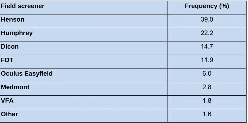
Relative frequency of field screener use by community
optometrists.
Relative frequency of the different methods of disc
examination.
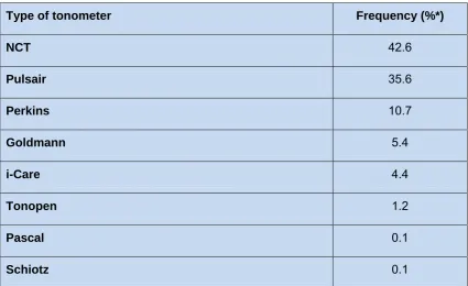
Relative frequency of the different types of tonometer used for
the measurement of IOP.
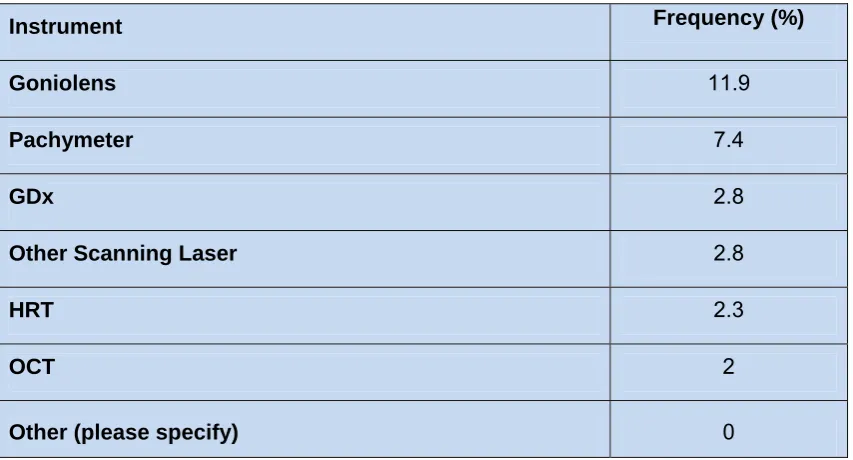
Relative frequency of the availability of specialist equipment in
community optometric practice.
Table 2.9 Table 2.10 Table 2.11 Table 2.12 Table 2.13 Table 2.14 Table 2.15 Table 2.16 Table 2.17 Table 2.18 Table 2.19
Relative frequency of pre-screening by mode of practice.
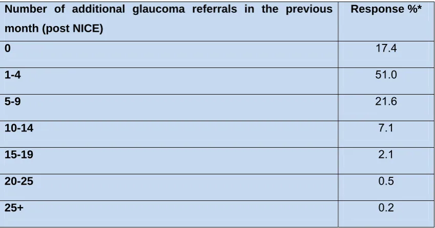
Estimate of the number of additional glaucoma referrals made
in a month following publication of the NICE guidelines.
Information included in a referral letter for suspect glaucoma.
Self-reported test combinations for the ‘standard’ screening
triad included in a referral letter for suspect glaucoma.
Test combinations for the ‘standard’ screening triad included in
actual referral letters for suspect glaucoma.
Criterion validity: Correspondence between the survey
responses (self-reports) and actual referral letters obtained
from consultant ophthalmologists.
Main barriers reported by survey respondents.
Barriers by region.
Number of eye examinations performed by respondents from
the Netherlands and the UK in a typical week.
Relative frequency of the different types of tonometer by
optometrists in the Netherlands compared to the UK.
Relative frequency of the availability of specialist equipment in
community optometric practice in the Netherlands and UK.
58 60 61 61 62 63 64 65 67 68 68
Table 3.1 NICE recommendation for diagnosis of OHT and suspected
COAG.
90
Table 3.3 Round 2 ratings for competencies required by optometrists
involved in the diagnosis of glaucoma.
97 Table 3.4 Table 4.1 Table 4.2 Table 4.3 Table 4.4 Table 4.5 Table 4.6 Table 4.7 Table 4.8
Round 2 ratings for competencies required by optometrists
involved in the monitoring and treatment of glaucoma.
The format of the simple table used to record participants’
choices of the five most relevant disc features to observe when
assessing a patient’s disc.
Number of optic disc features correctly identified by the MSc
cohort (n = 53) pre- and post- the educational intervention.
Number of optic disc features correctly identified by the Control
cohort (n = 20) at baseline (“Pre-intervention”) and after a
3-month interval (“Post-intervention”).
Performance in the four clinical decision making scenarios for
the MSc Cohort (n = 53) pre- and post-intervention.
Performance in the four clinical decision making scenarios for
the Control Cohort (n = 20) at baseline (“Pre-intervention”) and
after a 3-month interval (“Post-intervention”).
Performance in the Discus program for the MSc Cohort (n =
53) pre- and post-intervention.
Performance in the Discus program for the Control Cohort (n =
20) at baseline (“Pre-intervention”) and after a 3-month interval
(“Post-intervention”).
The average latency for decision making for the MSc Cohort (n
= 53) pre- and post- intervention.
Table 4.9 The average latency for decision making for the Control cohort
(n = 20) pre- and post-intervention.
List of Figures
PAGE
Figure 1.1 Simplified Classification of the Glaucomas. 20
Figure 1.2 A glaucomatous optic disc showing focal loss of inferior
neuro-retinal rim tissue
25
Figure 1.3 The flow of Aqueous Humour. 26
Figure 1.4
Figure 2.1
Figure 2.2
Figure 2.3
Typical Field Loss associated with Open Angle Glaucoma.
Distribution of respondents based on year of GOC
registration.
Number of eye examinations performed by respondents in a
typical week.
Delegated screening tests in practices using pre-screening.
29
52
54
59
Figure 3.1 Miller’s Pyramid of Clinical Competence. 83
Figure 3.2
Figure 4.1
Figure 4.2
The new College of Optometrists’ Higher Qualification
structure, which illustrates how optometrists can progress
from the entry level Professional Certificate in Glaucoma,
through the Professional Higher Certificate to the Professional
Diploma in Glaucoma.
Screenshot of the sample disc available on the GONE
website http://www.gone-project.com
Screenshot of the analysis of the results of the sample disc
available on the GONE website http://www.gone-project.com
105
112
Figure 4.3
(a-c)
Figure 4.4
Figure 4.5
Figure 4.6
Figure 4.7
Figure 4.8
Figure 4.9
Three screenshots of typical Discus images. Figure 4.3 (b)
shows a close-up of the rating scale and of the “Next” button
which the participant clicks to move on to the next image.
These are the first three images of a typical Discus program
in which the 126 disc images are presented in random order.
Reference ROC Curve.
Composite ROC curves for the MSc cohort pre- and
post-intervention.
Composite ROC curves for the Control cohort pre- and
post-intervention.
Box and whisker plots of mean scores for each of 100 images
for the pre-intervention Control cohort and pre-intervention
MSc cohort.
Box and whisker plots of mean scores for each of 100
images for the pre- and post-intervention Control cohort.
Box and whisker plots of mean scores for each of 100 images
for the pre-intervention MSc cohort and estimated mean
scores for the Discus Expert Panel (DH_Estimate).
115
117
137
138
139
140
Acknowledgments
I will be forever indebted to my supervisors Professor John Lawrenson and Professor David Edgar without whom this project, and this thesis, would not have been possible. They believed in me enough to give me this opportunity and have shown me unfaltering patience, encouragement, guidance, reassurance and kindness. Their support and endless efforts through related and unrelated difficulties as well as their vast academic knowledge and expertise, have been invaluable to me. I am extremely proud to call them supervisors, mentors and friends and owe them my deepest gratitude. An enormous credit for this thesis must be afforded to them both.
Thanks also to my advisors Mr. Ian Murdoch and Dr. Aachal Kotecha, for generously and selflessly giving encouragement, inspiration, advice and time. To my examiners Professor Andrew McNaught and Dr Paul Spry, I am grateful for a very pleasant experience, and for allowing me the opportunity to learn from their wisdom.
It has been an honour to have once again studied and worked in the Division of Optometry and Visual Science, City University, London, and to collaborate with esteemed colleagues at Moorfields Eye Hospital.
Heartfelt thanks also should go to Pfizer Ophthalmology, Ms. Anita Lightstone, the Association of Optometrists, Professor David Henson, Dr. Helen Baker, Professor David Crabb, Mr Henri Obstfeld, Professor Chris Hull (“The Chair”), to Dutch colleagues and of course to the Delphi panel and my many “respondents”.
To my friends and colleagues who were there when I needed enthusiasm, wisdom and moral support. There are too many to thank but I will mention (in alphabetical order) Andrew, Caroline, Colin, D and D, David, Duncan, Jane M, Jane P, Janet (is it Friday yet?), Kez, Liam, Matilda, Miriam, Paul, Peter and B & C and T & T. To Bill, Clare, Kalpana, Karen and especially Lynne to whom I will always be eternally grateful, you deserve a special mention, your patience, thoughtfulness and care has been seemingly untiring and limitless. Thank you also to the various medical and surgical experts I have visited in recent times.
To my family for their endless faith and belief in me and for their love, nurture, care and understanding which has aided me immensely. Even in my darkest hours, they were there for me. For my late (much missed) father, my mother, Steven, Fiona, Tim, Nick,
Declaration
Abstract
A Study of Case Finding for Chronic Open Angle Glaucoma (COAG) by UK Community Optometrists
In 2009 approximately 480,000 people were affected by COAG in England. Furthermore, glaucoma sufferers and suspects are responsible for over one million glaucoma-related outpatient visits annually. Community optometrists make over 95% of suspect COAG referrals, identifying suspects through opportunistic case-finding. Optometrists’ case-finding is largely based on a triad of tests: optic nerve head assessment, tonometry, and visual fields. There has been little research into optometrists’ COAG case-finding strategies.
Chapter 2 reports on a national survey regarding COAG case-finding methodologies and referral criteria. Survey response validity was confirmed by comparing these with a national sample of referral letters. UK optometrists are well-equipped to detect COAG. Optometrist’s skills and scope of practice in the detection of glaucoma have evolved since the last national survey in the late 1980’s. The level of funding and nature of the GOS contract in England limits development of effective services for glaucoma detection. For comparison, the survey was also performed in the Netherlands. Dutch optometrists own fewer automated field screeners but more goniolenses and pachymeters, and are more likely to use binocular indirect ophthalmoscopy than UK optometrists.
Chapter 3 describes the development of a competency framework for optometrists with a specialist interest in glaucoma utilising Delphi methodology. The Delphi technique is a robust method for gaining autonomous expert opinion. This approach has led to the development of an accepted national competency framework for optometrists with a special interest in glaucoma.
Chapter 4 evaluated the impact of a postgraduate educational intervention on aspects of glaucoma detection. The intervention increased awareness of disc changes in glaucoma, but was less effective for clinical decision-making and for improving performance in the Discus program for disc assessment. The traditional didactic teaching style is unsuited for training optometrists in the clinical competencies required for glaucoma detection and management.
Abbreviations
ABDO Association of British Dispensing Opticians
A/C Anterior Chamber
ACG Angle Closure Glaucoma AOP Association of Optometrists
AUROC Area under the receiver operating characteristic curve BSc Bachelor of Science
BEH Bristol Eye Hospital CCT Central corneal thickness CDM Clinical Decision Making CDR Cup-disc ratio
CET Continuing Education and Training CHANGES Community and Hospital Allied Network
Glaucoma Evaluation Scheme
CIGTS Collaborative Initial Glaucoma Treatment Study CNTGS Collaborative Normal-Tension Glaucoma Study COAG Chronic open angle glaucoma
CoO College of Optometrists
CPD Continuing Professional Development
DH David Henson
EMGT Early Manifest Glaucoma Treatment
F+ False Positive
F- False Negative
FDT Frequency Doubling Technology
FH Family History
FODO Federation of Ophthalmic and Dispensing Opticians GAT Goldman applanation tonometry
GIST Glaucoma Inheritance Study in Tasmania GOC General Optical Council
GON Glaucomatous Optic Neuropathy
GONE Glaucomatous Optic Neuropathy Evaluation Gonio Goniolens
HES Hospital Eye Service HFA Humphrey Field Analyser HRT Heidelberg Retina Tomograph HTA Health Technology Assessment IGA International Glaucoma Association IOP Intraocular pressure
ISNT Inferior, Superior, Nasal, Temporal (Jonas’ Rule) LOCSU Local Optical Committee Support Unit
MD Mean Deviation
MRA Moorfields Regression Analysis MREH Manchester Royal Eye Hospital MYOC Myocilin
NCT Non-contact tonometry NHS National Health Service
NICE National Institute of Health and Clinical Excellence NIHR National Institute of Health Research
N. Ireland Northern Ireland
NPC National Prescribing Centre NRR Neural Retinal Rim
NTG Normal tension glaucoma
NTGS Normal Tension Glaucoma Study OAG Open-angle glaucoma
OCT Optical Coherence Tomography OHT Ocular hypertension
OHTS Ocular Hypertension Treatment Study OLGA Optometric Led Glaucoma Assessment OMP Ophthalmic Medical Practitioner
ONH Optic Nerve Head
OVN Optometristen Vereniging Nederland PCT Primary Care Trust
POAG Primary open-angle glaucoma PPA Peripapillary Atrophy
PPV Positive Predictive Value
PQE Professional Qualifying Examination PSD Pattern Standard Deviation
RNFL Retinal Nerve Fibre Layer
ROC Receiver Operating Characteristic SAP Standardised automated perimetry
SD Standard Deviation
SfR Scheme for Registration SP Standardised patient
SWAP Short wavelength automated perimetry
T+ True Positive
T- True Negative
UK United Kingdom
VF Visual Fields
Chapter 1: Introduction
1.1 Definition of glaucoma
The word “glaucoma” is derived from the Greek word “glaukos” which means blue-green glow (Tsatos & Broadway, 2007). Glaucoma is actually not a single disease entity but a group of diseases. There are many definitions of glaucoma, but one frequently used is that published by the European Glaucoma Society: “Glaucoma is a group of diseases that result in a progressive optic neuropathy that causes characteristic changes in the optic nerve head and retinal nerve fibre layer” (European Glaucoma Society, 2003). The biological basis or pathogenesis of the disease is not fully understood (Weinreb and Khaw, 2004), though it is undoubtedly multifactorial in nature (Anderson, 1989; Drance, 1997; Bonomi et al, 2001; Foster et al, 2002).
The association between raised intraocular pressure (IOP) and glaucoma has been known since the 19th century, but since the late 1980s and early 1990s (Sponsel, 1989; Quigley, 1993) IOP has been omitted from the definitions of open angle glaucoma, instead being regarded as an important risk factor for the condition.
1.2 Classification of the glaucomas
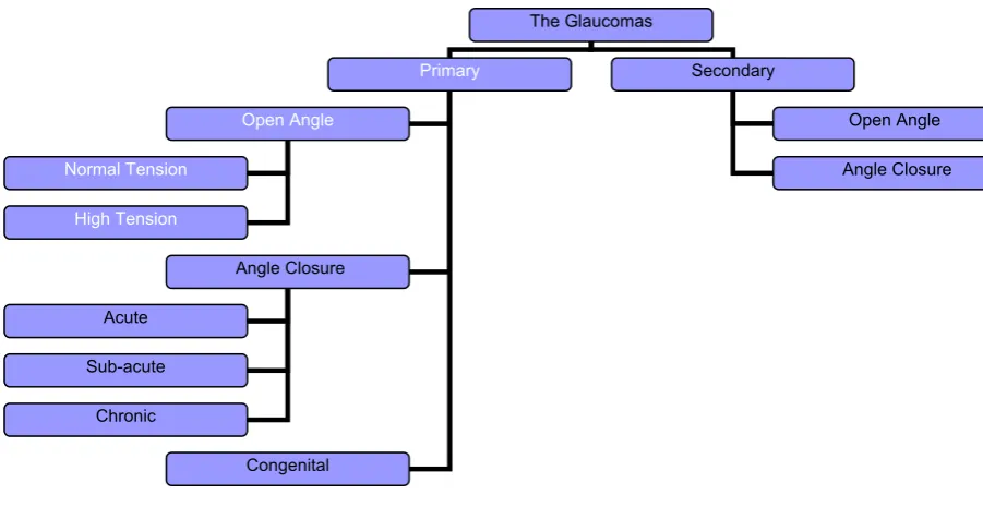
The glaucomas can be classified in several ways, for example according to the mechanism of damage, or by the aetiology of IOP elevation (Allingham et al., 2005). The classification chosen for this thesis is based on the cause of IOP elevation (Spry & Harper, 2010), and a simplified version of this classification is shown in Figure 1.1.
Figure 1.1: Simplified Classification of the Glaucomas.
1.2.1 Terminology
An ongoing issue in glaucoma is accommodating the different terms that can be used to describe the same condition. During the course of the research studies which are reported in this PhD thesis, the National Institute for Health and Clinical Excellence (NICE) Clinical Guideline 85 was published (NICE, 2009) and this has led to the increased use of the term Chronic Open Angle Glaucoma (COAG). NICE defined COAG as: Glaucoma without evident secondary cause, which follows a chronic time
course and occurs in the presence of an open anterior chamber angle
The author adopted the term COAG following the publication of the Guideline, and this term has been used in three publications to emerge from this thesis. Two earlier publications based on the research presented in the thesis use the term Primary open angle glaucoma (POAG) to refer to what is now called COAG. Furthermore, the term POAG was used in many of the publications referenced in this thesis and is still used in many current publications. There is clearly potential for confusion in the use of terminology here and, in an effort to address this issue, the author has adopted the following strategy in the thesis:
The Glaucomas
Primary Secondary
Open Angle
Angle Closure
Congenital
Normal Tension
High Tension
Acute
Sub-acute
Chronic
Open Angle
Where there is no scope for confusion (i.e. where either the author or the publications referred to in the text have used the term COAG) the term COAG is used.
Where the possibility for confusion exists (i.e. where either the author or the publications referred to in the text have used the term POAG in place of COAG) the term OAG has been used.
1.2.2 Angle closure glaucoma (ACG)
A common feature of the group of conditions that comprise ACG is closure of the angle of the anterior chamber, a closure which can result from a number of possible causes. Angle closure leads to elevated IOP which causes glaucomatous optic neuropathy. There are a number of risk factors for ACG, including increasing age, hypermetropia, ethnicity and female gender (Spry & Harper, 2010). Unlike COAG, ACG is sometimes accompanied by symptoms. The prevalence of ACG in European populations was estimated to be 0.25% in 2010 (Quigley & Broman, 2006). The focus of the current thesis is on case-finding for COAG, but community optometrists have an important role to play in the detection and appropriate management of acute, chronic and intermittent ACG (College of Optometrists Clinical Management Guidelines, 2009a).
1.2.3 Normal Tension Glaucoma
Normal Tension Glaucoma (NTG) is defined by NICE (2009) as: “A type of chronic open-angle glaucoma where intraocular pressure has rarely been recorded above 21 mm of Hg (a figure frequently taken as the ‘statistical’ upper limit of the normal range)”.
1.3 Ocular Hypertension
optometrists have had a key role to play in the detection and appropriate referral of OHT.
1.4 Epidemiology of glaucoma
There have been many major population-based studies related to glaucoma (Friedmann et al., 2004a). Among the most notable of these are the Baltimore Eye Survey (US), the Beaver Dam Eye Study (US), the Blue Mountains Eye Study (Australia), the Roscommon study (Irish Republic), the Melbourne project (Australia) and the Rotterdam Eye Study (The Netherlands). They have identified the prevalence of OAG in adults, with some studies including those aged over 40 years of age (e.g. Baltimore and Melbourne) up to one study including patients over 55 years of age (Rotterdam). The prevalence figures vary, reflecting different inclusion criteria in terms of age and different definitions of glaucoma, but a broad consensus emerges from these well-designed and well-executed studies: the prevalence of OAG varies from around 1.1% to 2.4% in adult White populations (Table 1.1) (Coffey et al., 1993; Dielemans et al., 1994; Mitchell et al., 1996; Sack et al., 1996; Kroese et al., 2002; Owen et al., 2006).
Table 1.1: Estimates of the prevalence of Open Angle Glaucoma in White adult
populations from well-designed population-based studies.
Study Prevalence
Baltimore (1990) 1.1% (40 years of age and over)
Beaver Dam (1992) 2.1% (43 years of age and over)
Blue Mountains (1996) 2.4% (49 years of age and over)
Roscommon (Ireland) (1992) 1.9% (50 years of age and over)
Melbourne (1997) 1.7% (40 years of age and over)
Rotterdam (1996) 1.1% (55 years of age and over)
1.4.1 Ethnic variations in OAG prevalence
estimates for 2010 are presented in Table 1.2 as percentages of the population over 40 years of age in each region predicted to have OAG. Africans are most likely to develop OAG (prevalence 4.16%) compared to all other ethnicities and compared to the world prevalence of 1.96%, which is virtually identical to the prevalence in Europe.
Table 1.2: Estimated prevalence of open angle glaucoma (OAG) in the over-40s in
different regions as reported by Quigley and Broman (Quigley & Broman, 2006).
World Region % with OAG
Africa 4.16%
Japan 3.31%
Latin America 3.16%
Europe 1.97%
India 1.75%
China 1.40%
Middle East 1.31%
South East Asia 1.18%
World 1.96%
and/or standard refractive correction, and has a visual acuity of less than 6/18 to light perception, or a visual field less than 10 degrees from the point of fixation, but who uses, or is potentially able to use, vision for the planning and/or execution of a task for which vision is essential.” Blindness is defined by the WHO, as included in ICD-10, as a visual acuity <3/60 in the better eye or visual field constricted to <=10 degrees in the better eye.
The World Health Organisation (Thylelfors et al., 1995) indicated from blindness survey data that glaucoma accounted for blindness in 5.2 million people, or 15% of total global blindness (Thylefors et al., 1994). Three million of these blind people were blind as a result of OAG. The numbers of those classified as blind as a result of glaucoma has increased dramatically since then, with a prediction from Quigley and Broman (2006) that bilateral blindness would be present in 4.5 million people suffering from OAG in 2010, rising to 5.9 million people by 2020.
1.5 Chronic open angle glaucoma
Figure 1.2: A glaucomatous optic disc showing focal loss of inferior neuro-retinal rim
tissue (image credit: Broadway et al., Surv Ophthalmol 43 [Suppl 1] :S223–S243,
1999).
1.5.1 Aqueous production and drainage
routes and the episcleral venous pressure. It should be noted that outflow resistance increases with advancing age in a normal eye in the absence of glaucoma (Tamm, 2009). Figure 1.3 illustrates the dynamics of aqueous production and drainage.
Figure 1.3: The flow of Aqueous Humour (Image Credit: National Eye Institute, National
Institutes of Health).
1.6 Mechanism of damage in OAG
1.7 Structural changes in glaucoma and their assessment
OAG is typified by damage to retinal ganglion cells and their axons which lead to characteristic visual field loss. It is the characteristic pattern of damage to the optic nerve head, related to the distribution and arrangement of the retinal nerve fibres, “that differentiates glaucoma from other causes of visual morbidity” (Foster et al., 2002). The retinal nerve fibres that originate from the retina temporal to the fovea do not cross the fovea as they approach the optic nerve head because to do so would impair sharp image formation at the fovea. Instead these nerve fibres arch above and below the fovea, entering the optic disc at its upper and lower poles. Nor do these nerve fibres cross the midline (Horizontal Raphé), and these anatomical configurations give rise to the characteristic arcuate shape of the nerve fibre bundles on the retina. The initial damage to ganglion cells and their axons in glaucoma is primarily noted at the inferior and superior poles of the disc, where these arcuate fibres from the temporal retina enter the optic nerve head, and many practitioners will routinely record vertical cup/disc ratio and comment on the neuro-retinal rim in order to detect any glaucomatous changes (Kotecha, 2009).
Regression Analysis (MRA) database and the Glaucoma Probability Score (GPS) as aids for detection of glaucomatous ONH change (Andersson et al., 2011). Scanning laser polarimetry (SLP) can objectively measure the retinal nerve fibre layer (RNFL) thickness surrounding the optic disc by taking advantage of the fact that the RNFL is a bi-refringent tissue. The SLP technique uses low intensity polarised laser light to measure the retardation or change in polarisation when light illuminates the bi-refringent RNFL. A challenge in this technique is to separate retardation resulting from the nerve fibre layer from retardation caused by the cornea and lens, with algorithms being employed to compensate for the retardation introduced by the cornea in particular (Lemij & Reus, 2008).
1.8 Changes in visual function in glaucoma and their detection
Although glaucoma can affect many aspects of visual function, including contrast sensitivity, colour vision, motion sensitivity, and eventually visual acuity (Sinclair, 2012), it is the effects of the disease on the visual field that is the aspect of visual function most frequently tested in both primary care optometry and secondary care. Over the years many tests have been developed for the assessment of the visual field but static automated perimetry (using both supra-threshold and threshold techniques) is now the established method. The field testing equipment routinely used by community optometrists in glaucoma case-finding has been investigated in the current research and the results are presented in Chapter 2 of this thesis. In hospital outpatient departments in the UK it is Standard Automated Perimetry (SAP) using the Humphrey Field Analyser (HFA) which predominates. Figure 1.4 shows an example of typical arcuate pattern of field loss in OAG as plotted on the HFA, with the shape of the visual field defects reflecting the damage to the arcuate nerve fibre bundles.
community optometric practice as an alternative to conventional methods of perimetry. Automated perimetry is a non-selective test, in the sense that it tests all three subtypes of retinal ganglion cell (magnocellular, koniocellular and parvocellular). FDT makes use of the frequency doubling illusion, first described by Kelly (1981), in which a low spatial frequency grating is counter-phase modulated at a high temporal frequency. The illusion is said to be mediated principally by the magnocellular pathway. The gratings are presented at 16 locations in the visual field and the patient indicates if they can detect the grating against the uniform grey background. The test is fast, but recent research (Jampel et al., 2011) suggests that it there is no clear advantage to be obtained by using FDT compared with standard automated perimetry.
Figure 1.4: Typical Field Loss associated with Open Angle Glaucoma (Image Credit:
1.9 Burden of glaucoma in the UK
1.9.1 Visual impairment and registration as a result of glaucoma
Analysis of blind (severely sight impaired) and partial sight (sight impaired) registrations in England and Wales between April 1990 and March 1991 revealed that 11.7% of blindness was caused by glaucoma in all age groups (Evans, 1995). A similar analysis covering the period between April 1999 and March 2000 found that glaucoma accounted for 10.9% of all blindness certifications and 10.2% of all partial sight registrations (Bunce & Wormald, 2006). In a more recent study, glaucoma accounted for 8.4% of all blindness certification and 7.4 % of all partial sight registration for the period April 2007-8 (Bunce et al., 2010). These studies indicate that, despite improvements in treatment modalities, glaucoma is a disease which continues to account for a significant proportion of those registered as sight impaired and severely sight impaired in the UK. There is evidence that these registration data may be an underestimate of the extent of the problem, as some of those eligible for registration may not wish to be registered. Also the criteria applied for registration have an element of subjectivity in their interpretation (Burr et al., 2007).
A study investigating visual impairment in a small sample of the North London elderly population established that 3% had open angle glaucoma while 7% had suspect glaucoma (Reidy et al., 1998). A study which quantified visual impairment in a sample of 75 year olds identified that 7.9% were visually impaired as a result of glaucoma (Evans et al., 2004).
1.9.2 Burden of glaucoma in secondary care
approximately 480,000 people affected by OAG in England. Furthermore, glaucoma sufferers and suspects are responsible for over one million glaucoma-related outpatient visits to the Hospital Eye Service (HES) each year (NICE, 2009).
It is noteworthy that several population studies in the UK and Australia have indicated that only 50% approximately of cases of OAG are diagnosed (Crick, 1994; Mitchell et al., 1996; Wensor et al., 1998). This would equate to approximately 250,000 people in the UK with undetected glaucoma.
1.10 Risk Factors for OAG
During the past decade there has been a notable increase in our understanding of risk factors for OAG. A summary of aspects of this research that are particularly relevant to community optometrists is presented in this section.
1.10.1 Intraocular Pressure
Elevated intraocular pressure (IOP) is an extremely important risk factor for OAG (Bengtsson, 1980; Sommer et al., 1991; Leske et al., 1995), and the only proven treatable risk factor (Pohjanpelto & Plava, 1974; Anderson 1989; Vogel, 1990; Sommer et al., 1991; Quigley, 1993; Kass & Gordon, 2000; Kroese & Burton, 2003; Weinreb & Khaw, 2004). There is considerable evidence to demonstrate the importance of IOP in the development and progression of glaucoma. For example, as IOP increases so the risk of developing OAG also increases, and as IOP increases in those with OAG there is a greater risk of progression of visual field defects (Leske et al., 1999; Kass & Gordon, 2000; Heijl et al., 2002). Also, patients who are diagnosed with advanced glaucoma are found to have higher IOPs at the time of diagnosis than those with less advanced glaucoma (Sommer et al., 1991; Grødum et al., 2002)
glaucoma (NTG) although Spry and Harper (2010) point out that this distinction between ‘high’ pressure and ‘normal’ pressure types of COAG is arbitrary, with both types belonging to the spectrum of disease that is COAG.
IOP is the only modifiable risk factor for OAG, and for many years lowering IOP by surgery or medication has been the method for managing OAG. Evidence for the benefits of IOP-lowering treatment in NTG has emerged from the Collaborative Normal-Tension Glaucoma Study Group (NTGS). They reported that lowering IOP by 30% from its baseline level can be effective in reducing the rate at which patients lose their visual field (or have progression of disc changes) in normal tension glaucoma (CNTGS 1998a; CNTGS 1998b; Anderson, 2003). The Early Manifest Glaucoma Treatment Study (EMGTS) also reported significantly lowered IOP from baseline (by an average of 25%) but used a patient sample which included patients with baseline pressures of up to 29mmHg. Results from this study demonstrated that lowering IOP significantly succeeded in delaying progression of OAG in patients with NTG and in those with higher pressures (Heijl et al., 2002).
1.10.2 Age
There have been a number of studies that have clearly demonstrated a strong association between age and OAG, with evidence showing that both incidence and prevalence of the disease increase with age (Tielsch et al., 1991b, Klein et al., 1992; Klein et al., 1993; Dielemans et al., 1994; Leske et al., 1994; Friedman et al., 2004a; Coleman & Miglior, 2008). The strength of this association varies considerably across populations, however Rudnicka and Owen (2007) note that on average the risk of OAG in those people over 70 years of age is 3 to 4 times greater than those in their 40s.
1.10.3 Myopia
A more recent meta-analysis confirmed that myopes had twice the risk of developing OAG (Marcus et al., 2011).
Myopic eyes tend to be large eyes and tend to have large optic discs. A number of studies have identified optic disc diameter as a risk factor for glaucoma (Healey et al., 1997; Quigley et al., 1999; Healey & Mitchell, 2000). Myopia is often associated with an elongation of the eye, and it is possible that this may lead to changes in the lamina cribrosa. It has been noted that the changes in the lamina cribrosa observed in eyes with myopia are similar to the changes seen in glaucoma (Quigley et al., 1983).
1.10.4 Ethnicity
There are striking ethnic variations in the prevalence of OAG (See also Section 1.4 Epidemiology of glaucoma). These were highlighted in the Baltimore Eye Survey, conducted in an inner city mixed Black and White population (Tielsch et al., 1991b). The glaucoma prevalence in the Black population aged 40 years and above was 4.2% compared with 1.1% in the equivalent White population. A Bayesian meta-analysis, which examined 46 published studies that investigated age, gender and race in relation to OAG, demonstrated that the prevalence of OAG in different racial groups varied with age (Rudnicka et al., 2006). In 40 to 49 year olds, the prevalence of OAG in Black populations was approximately 7 times higher than that in White populations, whereas by age 80 to 89 years the prevalence was only approximately 2.5 times higher in Black populations. In the 40 to 69 age group, the prevalence in Asian populations was similar to the prevalence in White populations but in the older age groups it was higher in White populations.
1.10.5 Systemic Disease
(2011) reviewed 18 epidemiological trials looking for any association between glaucoma and diabetes. Of these, 7 found an association and 11 failed to find an association. They explained these discrepancies by the use in these studies of different definitions of glaucoma, different ways of classifying diabetes, sample sizes that in some studies were too small, and variations in the statistical methods used. However, they concluded from laboratory-based research that there was good evidence for an association between the two diseases.
Associations between systemic hypertension and vascular regulatory disorders (e.g. cold extremities, migraine and Raynaud’s phenomenon) have been found in some studies, although the research evidence is often contradictory (Pache & Flammer, 2006). If these conditions are associated with OAG then the link is likely to be through the vascular mechanism for development of the disease (Section 1.6). According to the vascular theory, a combination of low blood pressure and elevated IOP can lead to a reduction of perfusion pressure at the optic nerve head, leading to damage to the retinal ganglion cells. Paradoxically, elevated blood pressure has also been associated with increased risk of developing OAG because it too can reduce the perfusion pressure at the optic nerve head (Memarzadeh et al., 2010). The interaction between blood pressure and IOP is clearly complex. Nicolela (2008) reviewed the evidence that could link vasospasm (or vascular regulatory disorders), which manifests as migraine and Raynaud’s phenomenon etc., and glaucoma. He concluded that there was increasing evidence, both clinical and epidemiological, of an association between vascular regulatory disorders and glaucoma, at least in certain subgroups of the population.
1.10.6 Family History
glaucoma. Further evidence was provided by The Barbados Family Study which found that 10% of living relatives of those diagnosed with OAG also had the disease. They estimated that a further 13% probably had OAG (Nemesure et al., 2001).
McNaught et al (2000) investigated 5 long-established pedigrees comprising over 400 glaucoma sufferers in Tasmania, Australia (GIST study). 13% of this sample had already been diagnosed as having OAG or as being OAG suspects, and a further 16% were identified during the GIST study. This was the first study to examine such a large sample belonging to glaucoma families in such detail and it was striking that so many new, admittedly mostly suspect, OAGs were detected. Interestingly, 27% of those with a family history of glaucoma were unaware of it.
Optineurin (OPTN) and myocilin (MYOC) are among the genes that can independently cause glaucoma, (Boland & Quigley 2007; Weinreb & Khaw 2004). However, glaucoma is a most complex disease and in many cases it is likely that multiple genes are acting to cause the condition and that interaction between these genes may account for the inter-individual variations that occur in glaucoma (Carbonaro & Hammond, 2007).
1.10.7 Other factors
The relationship between OAG and corneal thickness is particularly important when investigating intraocular pressure, as a thinner than average cornea will lead to underestimation of the IOP as measured with an applanation tonometry, while a thicker than average cornea will lead to an overestimation of IOP (Ehlers et al., 1975). Recent studies have shown no association between glaucoma and central corneal thickness (CCT) (Terai et al., 2011; Wanga et al., 2011; Brandt et al., 2012). However, the European Glaucoma Society reports that the measurement of CCT is a requirement when managing ocular hypertension (OHT) (European Glaucoma Society, 2003). The importance of CCT measurement in the diagnosis and monitoring of OHT is highlighted in the NICE Clinical Guideline (NICE, 2009). Furthermore, the Ocular Hypertension Treatment Study (OHTS) (Gordon et al., 2002) identified CCT as being the best predictor for conversion of their OHT subjects to open angle glaucoma.
size that thwarted many earlier studies. They identified a 1.23 times greater risk for OAG in males than in females in Whites, with similar increased risks in Black and Asian populations. Czudowska et al, (2010) subsequently also found increased risk for OAG in males. Other suggested risk factors include socio-economic status (Leske & Rosenthal, 1979; Fraser et al., 2001; Ng et al., 2010), and alcohol abuse (Katz & Sommer, 1988). A UK-based study found that approximately two-thirds of glaucoma patients (66.6%) had no academic qualification, which is higher than national statistics figures would predict (Sharma et al., 2010). Smoking has been suggested as a risk factor for glaucoma but studies have yet to find a definite association (Katz and Sommer, 1988; Rudnicka et al, 2006).
1.11 Disease Progression
If left untreated all glaucomas can lead to permanent visual impairment, which in some cases will be severe. OAG is usually slowly progressing as aforementioned, with initially characteristic arcuate paracentral scotomata, and is usually asymptomatic due to overlapping central fields of the right and left eye, but the advanced stages of the disease are more likely to be symptomatic, especially when the field loss approaches or involves fixation, when it will be coupled with an associated loss in acuity. A study examining the rate of OAG progression from cross-sectional, population-based data found that progression rates are not affected by age; and rates were not different between different ethnic groups (Broman et al., 2008).
1.12 Case-finding Strategies for OAG
In the UK, the current practice of chronic open angle glaucoma (OAG) detection depends largely on community optometrists, who are responsible for over 95% of suspect OAG referrals to secondary care (Bowling et al., 2005). Although 5.3 million NHS sight tests were conducted on patients over 60 in England and Wales in the year ending March 2011, significant numbers of the population in this age group who are ‘at risk’ of OAG do not consult optometrists or do not consult them on a sufficiently regular basis. Moreover, higher rates of late presentation are associated with living in areas of high social deprivation where optometrists’ premises are poorly represented (Day et al., 2010).
Given this background, it could be argued that there is a case for initiating a national screening programme for the detection of OAG. This question was addressed by the Health Technology Assessment (HTA) programme, which is part of the National Institute for Health Research (NIHR). Its primary remit is to research the effectiveness of healthcare within the NHS.
In the absence of a formal screening programme, optometrists identify glaucoma suspects through opportunistic case-finding. Optometrists’ case-finding approach to OAG is largely based on the results of three diagnostic tests: assessment of the optic nerve head, tonometry, and assessment of the central visual field. The College of Optometrists (CoO) has developed guidelines for Examining the Patient at Risk from Primary Open Angle Glaucoma (College of Optometrists, 2009b) and these, together with a more detailed discussion of the triad of tests commonly used in community practice are included in Chapter 2 of this thesis.
1.13 The profession of optometry in the UK
Compared to medicine, optometry is a relatively new profession. With the creation of the NHS in 1948, the anticipation was that eyecare would be provided in a hospital setting. However, this proved unrealistic because of the huge numbers involved - over 80% of eye examinations in the UK were provided by community-based ophthalmic opticians (http://www.optical.org/goc/filemanager/root/site_assets/publications/celebra ting_ 50_years.pdf). There was a need to provide regulation of eyecare services but it was not until 1958 that this came about with the passing of The Opticians Act and the formation of the General Optical Council (GOC) (Taylor, 1986). The GOC is the statutory regulatory body for optometrists and dispensing opticians, one of several health and social care regulatory bodies which also includes the General Medical Council (GMC). The GOC has, as its primary purpose, the protection of the public but it also maintains the registers of all optometrists and dispensing opticians, oversees all training and provides disciplinary powers, not just regarding clinical practice but professional behaviour.
The original legislation was subsequently consolidated and amended to the current 1989 act (Taylor, 1991) (http://www.legislation.gov.uk/ukpga/1989/44/pdfs/ ukpga_19890044_en.pdf). Further minor amendments have been included since then, most notably in 2005 when mandatory Continuing Education and Training (CET) was introduced for all optometrists. This initiative is partly funded by the NHS, with individual grants for registrants.
disease or abnormality in the eye or elsewhere as the regulations require”, which would include detection of glaucoma.
The GOC annual report for the period 2007/8 states that there were 11,094 optometrists on the register for the UK (GOC, 2008a). Subsequent annual reports showed that this figure increased by 3.7% to 11,559 for the period 2008/9 and again to 12,414 for the period 2009/10 (GOC, 2009; GOC, 2010). Though there are opportunities for optometrists to work in secondary care, the majority of optometrists work in community-based primary care settings (Burr et al., 2007). Although patients may often consult their general medical practitioners (GMP) regarding eye problems, GMPs are rarely able to access the necessary specialist equipment, or do not usually have the essential training and skills to adequately detect certain eye diseases, notably glaucoma (Smeeth, 1998)
1.14 Optometry Education and Training
UK optometrists have to obtain a Bachelor’s degree qualification at one of the 9 universities (six in England, one in Wales, one in Scotland and one in Northern Ireland) which offer BSc (or equivalent) degrees in Optometry. Students follow syllabi to ultimately satisfy the core competencies set out by the General Optical Council (GOC Optometry Core Curriculum, Core Competencies and Learning Outcomes).
Before they are able to practice, students must first obtain at least a second division second class (2:2) degree in Optometry and then can commence their pre-registration period where they work under supervision, and also participate in the College of Optometrists Scheme for Registration (SfR) where they need to complete a number of worked-based assessments and a final OSCE examination to satisfy competencies set out by the GOC (General Optical Council Stage 2 Core Competencies for Optometry, 2005). Post-registration, optometrists can elect to work in High Street practice, either for an independent or multiple; the hospital eye service; laser eye clinics, academia or a combination. Optometrists can often elect to be employed, self-employed, a locum or again a combination.
qualifications, accreditation or higher degrees are taken through an individual’s personal choice. In terms of glaucoma there may be local accreditation processes for enhanced schemes, or a more formal certificate or diploma. As part of their modular MSc in Clinical Optometry, City University London has a glaucoma-specific module, aspects of which are evaluated as part of this research (see Chapter 4 of the thesis). The College of Optometrists has a number of specialist higher qualifications which during the course of this PhD research comprised two separate glaucoma certificates which jointly led to a diploma. The current higher qualifications are being phased out from 2012 and the CoO has introduced a new pathway to gain higher qualifications. The new higher qualifications framework has a modular approach to achieving a new set of professional higher qualifications
(http://www.college-optometrists.org/en/professionaldevelopment/hq/new-college-accredited-courses/index.cfm).
1.15 Aims of this thesis
This research has four primary aims which are discussed in detail in the next four chapters.
1. To carry out a national survey of optometrists’ self-reported practice for glaucoma case-finding.
2. To evaluate strategies used by optometrists for the detection of glaucoma. 3. To identify the training needs of optometrists involved in the detection and
management of glaucoma.
4. To study the impact of an educational intervention on clinical decision making in glaucoma.
Chapter 3 focuses on the third primary aim and describes the development of a competency framework for optometrists with a specialist interest in glaucoma utilising Delphi methodology.
Chapter 4 addresses the final primary aim and evaluates the impact on clinical decision making of a current, established postgraduate educational course in glaucoma.
Chapter 2: A Survey of Glaucoma Detection and Referral in Community
Practice
2.1 Introduction
This chapter describes the results of a national survey regarding COAG case-finding methodologies/referral criteria used by community optometrists in the UK. The survey was delivered entirely online and was conducted in mid-2008, prior to the introduction of the National Institute for Health and Clinical Excellence (NICE) Glaucoma Clinical Guideline CG85 (NICE, 2009). The survey included sections on strategies for glaucoma detection, screening equipment used, barriers to case-finding and processes for referral, including the content of referral letters to an ophthalmologist.
Because questionnaires can only act as a proxy measure for actual clinical practice (Theodossiades et al., 2012), the validity of self-reporting by optometrists was assessed by comparing the survey responses in relation to referral with a national sample of referral letters obtained from consultant ophthalmologists across the UK. The chapter also reports on the findings of a version of the glaucoma survey translated into Dutch carried out in the Netherlands in early 2009.
2.2 Case-Finding Strategies for COAG
COAG is an insidious blinding disease that leads to a slowly progressive loss of visual field. Sufferers are often unaware of their field defect until it encroaches into their central vision. Since glaucomatous optic nerve damage is irreversible, early detection would provide access to effective pressure-lowering therapeutic interventions. However, population screening for glaucoma presents a considerable challenge; COAG is asymptomatic, has a low prevalence and there is no consensus definition for diagnosis. Consequently, there insufficient evidence for the effectiveness of a COAG screening programme that targets the general population (Hatt et al., 2006). In all parts of the developed world, the detection of COAG continues to rely heavily on opportunistic case-finding (Lawrenson, 2013).
available to everyone over 60 years and those over 40 with a family history of glaucoma through the General Ophthalmic Services (GOS). In Scotland, NHS-funded Sight Tests are available to all. The choice of equipment and the actual glaucoma case-finding protocol used is at the discretion of the individual optometrist, which can lead to significant variation in practice (Ang et al., 2009; Shah et al., 2009a).
Guidance for all UK optometrists has been published by their professional body (College of Optometrists, 2005), regarding the ‘examination of patients at risk from glaucoma’ (College of Optometrists, Code of Ethics and Guidelines for Professional Conduct, Section D3 Examining patients at risk from glaucoma). This guidance states that: “It is for the practitioner to satisfy him/herself that procedures are included or excluded according to the patient’s clinical need but in addition to the guideline on the eye examination, good practice for these patients should normally include:
Assessment of the optic nerve head;
Tonometry. Where pressures are high or borderline, arrangements should be made for the test to be repeated, noting the time of day of each test; the examination may also include:
Central visual field assessment using perimetry with threshold control. Where necessary, practitioners should consider repeating visual fields assessment to obtain a meaningful result.”
The College of Optometrists guidelines also state that “Non-contact applanation
tonometry is acceptable for screening but good practice would suggest that equivocal
results be followed up with contact applanation tonometry.” And additionally that both for tonometry and perimetry, these tests should be repeated to obtain a significant result.
2.2.1. Tests used by optometrists for the diagnosis of COAG
last 10 years has seen considerable changes within the optical sector, including: the scope of optometric practice, developments in the training and accreditation of optometrists and the adoption of new technology.
There is a strong body of opinion that combining structural and functional tests improves the ability to diagnose glaucoma (Malik et al., 2012). The presence of structural damage is conventionally assessed by a subjective assessment of the optic nerve head. Although direct ophthalmoscopy provides a magnified view of the optic disc, monocular viewing does not allow an appreciation of the three-dimensional morphology of the optic nerve head. Indirect slit-lamp ophthalmoscopy overcomes this problem, although usually requires pupil dilation to ensure a consistent stereoscopic view.
Conventional standardised automated perimetry (SAP) is the most widely used test of visual function for glaucoma diagnosis and monitoring. For screening, suprathreshold testing is typically employed, using stimuli of greater intensity than the estimated threshold at each test location. Although this test strategy does not always quantify the depth of any visual field defect, its principal advantage for routine case-finding is that the test duration is considerably shorter than full threshold testing.
The measurement of IOP is an integral part of glaucoma diagnosis and there is good-quality evidence to support ocular hypertension being a significant risk factor for the development of glaucoma (Kass et al., 2002). IOP can be determined by both contact and non-contact methods. The slit-lamp mounted Goldmann applanation tonometer is considered to be the reference standard for the determination of IOP (Burr et al., 2007). A hand-held version (Perkins applanation tonometer) is also widely used. Non-contact tonometers (NCT) have been available since the 1970’s (Grolman, 1972). These devices use a jet of air to applanate the cornea. Topical anaesthesia is not required and the technique is simple to use allowing the measurement of IOP in community optometric practice to be delegated to optical assistants.
2.3 Referral for COAG
If, when examining a patient, an optometrist suspects that glaucoma may be present, the optometrist has a duty of care to refer the patient to the appropriate practitioner for diagnosis and/or treatment.
The College of Optometrists Code of Ethics and Guidelines for Professional Conduct (2005) state that:
“During the course of professional practice, the optometrist has a duty to refer
the patient for appropriate ongoing clinical care and/or management whenever
s/he observes a sign or symptom of a condition that cannot be managed within
his/her competence and scope of practice, whether the observation is made
during the eye examination or at any other time in the course of practice.”
Optometrists conventionally would refer patients they suspect of having COAG to the hospital eye service (HES) via their General Practitioner (GP). The responsibility then essentially lies with the GP to decide if onward referral is necessary. GPs can choose to forward on the referral by the optometrist, or may alternatively choose to write their own referral including the information supplied by the optometrist (Scully et al., 2009).
The challenge for case detection in a primary care setting is that COAG has a low prevalence. Consequently, even when a combination of screening tests is used to maximise sensitivity and specificity the positive predictive value (PPV) of referrals is likely to be low. Reported PPVs are generally in the region of 30-40% (Harrison et al., 1988; Bell & O’Brien, 1997; Theodossiades & Murdoch 1999; Bowling et al., 2005). Since inappropriate referrals place high demands on the HES and may also result in longer waiting times and considerable financial costs (Vernon, 1998; Henson et al., 2003), there have been several attempts to reduce the number of false positive referrals through a process of community refinement of glaucoma referrals using accredited community optometrists (Henson et al., 2003; Parkins & Edgar, 2011).
2.4. Impact of the NICE glaucoma guideline on glaucoma case-finding
the guideline did not encompass case-finding and screening (Sparrow, 2012), the publication of the guideline had an immediate and unintentional impact on case-finding practice and patterns of referral. Immediately following publication in April 2009, the Association of Optometrists (AOP), Association of British Dispensing Opticians (ABDO) and the Federation of Dispensing Opticians and Optometrists (FODO) issued advice to its members to refer all patients with an IOP >21mmHg irrespective of the tonometer used and even if the discs and fields were normal (AOP: April 2009 and reiterated in June and October 2009).
When the NICE guidance was issued, colleagues at City University were in the final stages of developing a web-based questionnaire to collect data on the patterns of referrals made by optometrists to medical practitioners. The timing of this survey provided an opportunity to assess the effects of the NICE guidance on referral numbers. An additional question was included at the start of the questionnaire which asked each optometrist for the number of extra referrals, based on the NICE glaucoma guidelines only, made in the previous working month. These data provided the first national and profession-wide snapshot of the immediate impact of the NICE guidance on the number of glaucoma referrals.
2.5 Optometry in the Netherlands
Optometry is a well established profession in the UK, with perhaps the most significant milestone being statutory regulation with the creation of the Opticians Act and the General Optical Council in 1958, though opticians had been practicing unregulated prior to this point.
a three year advanced course. There is no equivalent of the pre-registration year and no compulsory requirement for continuing education and training (CET).
There are only about 700 registered optometrists in the Netherlands (Stevens et al,. 2002) as opposed to the 11,000+ in the UK. The profession is regulated by the Ministerie Van Volksgezondheid, which monitors entry into the profession, registration, use of titles and scope of practice. Sale of optical appliances is not regulated.
2.6. Aims of Chapter 2
1. To conduct a national web-based survey to determine:
diagnostic tests used by optometrists for glaucoma case-finding referral behaviour in relation to the detection of glaucoma perceived barriers to case-finding
2. To determine the impact of the publication of the NICE glaucoma guideline on referral behaviour
3. To estimate the validity of self-reporting as a measure of optometrist case-finding practice for glaucoma and the appropriate referral of suspects
4. To report on the findings of a version of the glaucoma survey translated into Dutch and carried out in the Netherlands.
2.7 Methods
2.7.1 Survey of Case-finding Practice Reported by UK Optometrists
A survey to investigate UK community optometrists’ current practice in the detection of COAG was developed. The survey was entirely web-based and hosted by a US provider of online surveys (Survey Monkey; http://www.surveymonkey.com; Oregon, USA).
survey was open for 16 weeks between April and July 2008. See Appendix 1 for a copy of the final questionnaire.
All optometrists on the Association of Optometrists (AOP) electronic database were invited to participate. The AOP represents the professional interests of UK optometrists. Seven thousand four hundred and thirty emails were sent to AOP members, but this total included practicing and retired optometrists, and non-community practitioners (e.g. hospital-based optometrists). The GOC annual report for 2007/8 stated that there were 11094 optometrists on the register for the UK, considerably greater than the AOP membership. There were also some duplicate email addresses. The email invited members to participate in the survey online via a hyperlink to the website. Two reminders were sent and news features promoting the survey were included in AOP membership publications.
The survey was anonymous and no incentives to participate or feedback were offered. It consisted of 27 forced choice or free-text questions covering different aspects of glaucoma case-finding practice. All questions required an answer, and once a section was completed respondents could not return to alter an answer.
The final survey consisted of five sections totalling 27 questions.
The first question asked respondents “Are you currently practising as a community optometrist?” This question was designed to screen out non-practising optometrists and those not working in community practice. Respondents providing a negative response to this question did not enter the survey and were presented with an acknowledgement page.
The first section consisted of 8 questions relating to mode of practice. The initial questions asked the principal mode of practice (question 2) and the proportion (%) of working time spent working in the principal practice (question 3). Subsequent questions asked for information regarding how many days a week they spent in their principal practice (question 4), and the location of the practice (questions 5-7).
The second section consisted of two free-text boxes to investigate strategies for glaucoma detection. The first asked for details regarding how the optometrist would investigate for suspect OAG, including the elements of the eye examination they regarded as most important. The second asked the optometrist to comment on any potential barriers that they felt would compromise effective detection of primary open angle glaucoma in community optometric practice and how they felt these barriers constrained implementation of practice.
The third section had nine questions relating to equipment used for glaucoma detection, and additionally practice organisation. This consisted of questions regarding pre-screening, screening equipment available in practice, any involvement in local glaucoma schemes and whether the individual had completed any further postgraduate training specifically related to glaucoma.
The fourth section asked how many referrals the optometrist made and how many specifically were related to glaucoma. It also enquired to whom referrals were made and what information was included in the referral.
The final section collected personal demographic information relating to gender and year of registration on the GOC register. A message thanking the optometrist for their participation was then displayed.
The questionnaire was designed such that once a page of questions had been completed and the respondent had advanced to the next page they were unable to return to the previous page to amend the answers. All questions were mandatory.
2.7.2 Survey Validation (UK)
Three methods were used to validate the survey responses:
1. Internal validation: the use of forced choice questions following a free-text question regarding referral information (Questions 24 and 25). Respondents could not return to the free-text question once they had advanced to the next (validation) question.