A STUDY OF THE EXTENT, BRANCHING
PATTERN AND APPLIED ASPECTS OF
ABDOMINAL AORTA
A Dissertation submitted toThe Tamil Nadu Dr. M. G. R. Medical University,
Chennai.
In partial fulfillment of the requirements for the degree of
M.D. DEGREE EXAMINATION BRANCH – XXIII (ANATOMY)
GOVERNMENT STANLEY MEDICAL COLLEGE AND HOSPITAL
CHENNAI – 600 001.
THE TAMILNADU DR. M.G.R. MEDICAL UNIVERSITY
CHENNAI
CERTIFICATE
This is to certify that the dissertation work on “A STUDY OF THE EXTENT,
BRANCHING PATTERN AND APPLIED ASPECTS OF ABDOMINAL AORTA” is a bonafide research work done by Dr. J. SENTHIL KUMAR, post graduate (2015-2018) in the Department of Anatomy, Govt. Stanley Medical College and Hospital, Chennai under my direct guidance and supervision, in partial fulfillment of the regulations laid down by the TAMILNADU DR. M.G.R. MEDICAL UNIVERSITY Chennai for the award of M.D. Anatomy (Branch XXIII) degree examination to be held in APRIL 2018.
Prof. Dr. S. Ponnambala Namasivayam, Dr. T. Vasantha Kumar, M.S.,
M.D., D.A., D.N.B., Professor and Head of Department
The Dean Department of Anatomy
Stanley Medical College Stanley Medical College,
DECLARATION
I hereby declare that this dissertation entitled in “A STUDY OF THE EXTENT, BRANCHING PATTERN AND APPLIED ASPECTS OF ABDOMINAL AORTA” was written by me in the Department of Anatomy, Government Stanley Medical College and Hospital, Chennai under the guidance and supervision of Prof. Dr. T. VASANATHA KUMAR, M.S., Professor and Head of the Department of Anatomy, Government Stanley Medical College and Chennai – 600 001.
This dissertation is submitted to The Tamilnadu Dr. M.G.R. Medical
University, Chennai in partial fulfilment of the university regulations for the award of Degree of M.D., Anatomy (Branch XXIII) Examination to be held in
April 2018.
Date:
ACKNOWLEDGEMENT
I wish to express my sincere thanks and gratitude to, Prof. Dr. S.Ponnambala Namasivayam, M.D., D.A., D.N.B., Dean, Stanley Medical College and Hospital, Chennai – 1 for having permitted me to utilise the facilities in this college for the conduct of the study.
My heartfelt thankfulness, gratitude and gratefulness to Dr. T. Vasantha Kumar, M.S., Professor and Head of the Department of Anatomy, Government Stanley Medical College, Chennai for his invaluable guidance, motivation and persistent support, encouragement and for providing all necessary arrangement to make the study a reality.
I sincerely thank Dr. S. Chitra, M.S., (Retd. Professor & HOD, Department of Anatomy), for her invaluable guidance, motivation and encouragement.
My sincere thanks to Dr. S.Balasubramanian, B.Sc., M.D., D.C.H., Professor and Head of the Department of Forensic Medicine, Govt. Stanley Medical College, Chennai-1 and his faculty for their kind cooperation and timely help.
I am grateful to Dr. C. Amarnath, M.D., (RD)., Professor and Head of the Department of Radiology, Govt. Stanley Medical College, Chennai-1 and his faculty for their help in radiological study.
Dr. J.K. Raja, M.D., Dr. S. Elizabeth Priyadarisini M.D., Dr. M.Anuradha M.D., Dr.Adline Misba, M.D., Dr. E. Anitha, M.D., Dr.B. Ramkumar, M.D., Dr. M.R.Manimegalai, M.D., Dr. F.Stelina, M.D., Assistant Professors for their valuable suggestions and constant encouragement throughout the study.
I also specialy thank earnestly my colleague Dr. V.Shanthi, M.D., and my seniors Dr.C.Sasikala, M.D., Dr.S.Manonmani, MD., and my juniors Dr.P.J.Seeja,
Dr.S.Sivakumari, Dr. P.Maharathi, Dr. Mohammed Sammiullah for their help rendered to me during the study.
I am also thankful to lab technicians Smt.K.Rajalakshmi, Smt. E.Jayanthi, and departmental staffs, Thiru.C.Birammaiah, Thiru.A.Kadar Basha, Thiru.M.Jagadeesan, Mr. Srinivasan and Tmt. Susila for helping me in carrying out the study.
It gives me great pleasure in preparing this dissertation and I take this opportunity to thank everyone who made this possible.
CONTENTS
S.NO. TITLE PAGE NO.
1 INTRODUCTION 1
2 ANATOMICAL CONSIDERATIONS 3
3 AIM OF THE STUDY 14
4 REVIEW OF LITERATURE 16
5 MATERIALS AND METHODS 33
6 OBSERVATION 42
7 DISCUSSION 65
8 CONCLUSION 85
INTRODUCTION
The abdominal aorta is the continuation of thoracic aorta and is the chief artery supplying oxygenated blood to the abdominal wall and abdominal organs (Prakash et al 2011). This arterial trunk extends from the lower border of the twelfth thoracic verterbra at the aortic opening of the diaphragm to the level of the fourth lumbar verterbra and to the left of the midline(Datta A.K 8th edit).
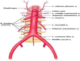
The branches of abdominal aorta can be classified into anterior or ventral, lateral and dorsal branches. The three ventral branches are coeliac trunk which supplies the foregut derivatives, superior mesenteric artery which supplies the midgut derivatives and the inferior mesenteric artery which supplies the hindgut derivatives. The paired lateral branches includes the inferior phrenic artery, middle suprarenal artery, renal and gonadal artery. The dorsal branches include four pairs of lumbar arteries and median sacral artery(Ranganathan.T.S 6th edit).
Abdominal aorta and its branches show wide variations which have been documented in the literature since decades (Mane UddhavWamanrao2016). These variations have gained practical importance during surgical and interventional procedures. According to Pennington.N 2004, variation in the morphology and branching pattern are of interest as it plays an important role in pathological conditions like atherosclerosis, aneurysms etc.
The knowledge of the anatomical variations regarding the abdominal aorta is essential in surgical procedures like laparoscopic surgeries, liver and kidney transplantation and oncological resections in the abdominal region and interventional procedures like arterial chemo-embolisation for treatment of liver carcinoma and surgical treatment of aortic aneurysms (Prakash et al 2013)
ANATOMICAL CONSIDERATIONS
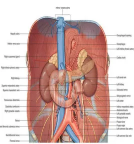
The abdominal aorta is the main arterial trunk supplying abdominal wall and organs. It is the continuation of thoracic aorta and extends from the lower border of twelfth thoracic vertebra at the level of aortic opening of the diaphragm to the level of the fourth lumbar vertebra and to the left of the midline (Ranganathan.T.S 6th edit). It descends on the posterior abdominal
wall infront of the vertebral bodies and their intervertebral discs. Infront, it is covered by peritoneum and it is placed to the left of the inferior vena cava. It terminates at the lower border of the fourth lumbar vertebra by dividing into right and left common iliac arteries (Gray’s Anatomy 41st edit, Borley N 2005)
fig 1.
[image:10.612.108.439.429.682.2]RELATIONS:
ANTERIORLY:
From above downwards (Datta.A.K 8thedit)
1. Origin of coeliac artery with coeliac plexus of nerves surrounding it. This part of aorta is covered anteriorly with peritoneum which separates it from the lesser sac and lesser omentum.
2. Body of pancreas:This portion of aorta behind the pancreas gives origin to the superior mesenteric artery and it is crossed transversely by the splenic vein above and the left renal vein below.
3. Third part of duodenum: This part gives origin to the inferior mesenteric artery.
4. Below the third part of duodenum, the aorta is crossed by the left gonadal vein and is related to the coils of small intestine(fig 2)
POSTERIORLY:
1. Bodies of upper four lumbar vertebrae, intervening intervertebral discs and anterior longitudinal ligament.
RIGHT SIDE:
In the lower part, the aorta is related to the inferior vena cava on the right side. In the upper part, they are separated by the right crus of the diaphragm and the right coeliac ganglion.
LEFT SIDE:
On the left side of the aorta, above downwards,
1. Left crus of diaphragm and left coeliac ganglion. 2. Duodeno-jejunal flexure.
[image:12.612.103.370.385.673.2]3. Fourth part of duodenum and 4. Inferior mesenteric artery.
BRANCHES:
The branches of the abdominal aorta can be classified as
Ventral
Lateral
Dorsal
And terminal branches.
The ventral and lateral branches are distributed to the viscera. The dorsal branches supplying the body wall, vertebral column, canal and its content (Inecikli M et al 2010).
VENTRAL BRANCHES:
1) Coeliac artery
2) Superior mesentric artery 3) Inferior mesentric artery
1. COELIAC ARTERY:
(Ranganathan.T.S 6th edit, Borley N 2005). It may also give off one or both
inferior phrenic arteries (Pamidi N 2008).
The left gastric artery is the smallest coeliac branch ascends along the lesser curvature between the layers of the lesser omentum (Pennington N 2005). The left gastric artery supplies the cardiac part of the stomach and it also gives of oesophageal branch which supplies the oesophagus. An accessory left gastric artery may arise from the left branch of the hepatic artery reaching the lesser curvature through the lesser omentum (Mburu K 2010).
The hepatic artery is intermediate in size between the left gastric and splenic artery (Gray’s Anatomy edit 41stedit). It passes forward and right
below the epiploic foramen to the upper aspect of the superior aspect of the duodenum. The hepatic artery divides into common hepatic artery which arise from the coeliac trunk to the origin of the gastroduodenal artery and hepatic artery proper from that point to its bifurcation. It gives off right gastric artery which ends by anastamosing with the left gastric artery, gastroduodenal trunk descends behind the first part of the duodenum and divides into right gastro-epiploic artery and superior pancreatico-duodenal artery (Last R.J edit ,Ranganathan T.S 6thedit )
bile duct and reaches the porta-hepatis where it divides into right and left branches.These branches enters the respective lobes of the liver. The right branch gives of cystic artery to supply the gall bladder.
1) SUPERIOR MESENTERIC ARTERY:
It is the ventral branch of abdominal aorta arising 1.25cm below the origin of coeliac artery and supplies the derivatives of the midgut arising at the level of L1-2 intervertebral disc (DahiphaleVarshaPrabhakar 2014). It emerges out between the body of the pancreas and its uncinate process, crosses in front of the uncinate process and third part of duodenum to get into the root of the mesentry. The branches of the superior mesenteric artery are
1. Inferior pancreatico-duodenal artery: It runs upwards to anastamose with the superior pancreatico-duodenal artery to form anterior and posterior pancreatico-duodenal arcades.
2. Jejunal and ileal branches: About 10-15 in number arises from the left side of the artery and pass between the layers of the mesentry. They form a series of arcades before reaching the jejunum and ileum.
Anterior and posterior caecal branches to the caecum. c) Appendicular artery. d) Colic branch to supply lower one third of the ascending colon)
4. Right colic branch divides into ascending and descending branch. The descending branch anastamoses with the ascending branch of the ileocolic artery to form the marginal artery. The right colic artery supplies the upper 2/3rd of the ascending colon and the hepatic flexure of the colon.
5. Middle colic artery passes between the layers of the transverse mesocolon divides into right and left branch. The right branch anastamoses with the ascending branch of the right colic artery. The left branch anastamoses with the left colic branch of the inferior mesenteric artery. The middle colic artery supplies the right two third of the transverse colon (Ranganathan T.S 6thedit)
2) INFERIOR MESENTERIC ARTERY:
The inferior mesenteric artery is the ventral branch of the abdominal aorta supplying the derivatives of the hindgut. It arises at the level of L3 vertebra behind the third part of the duodenum. The derivatives of the hindgut are the left one third of the transverse colon, descending colon, sigmoid colon, rectum and upper part of the anal canal upto the white line of Hilton.
Anteriorly it is covered with parietal peritoneum, posteriorly it is related to left psoas major muscle and termination of left common iliac artery.
The branches of the inferior mesenteric artery are
1. Superior left colic artery: It divides into ascending and descending branch. The ascending branch anastamoses with the left branch of middle colic artery. The descending branch anastamoses with the ascending branch of the left colic artery to continue the marginal artery of the colon. 2. Sigmoid artery (Inferior left colic artery): The sigmoid artery supply the
lower descending colon and the sigmoid colon, anastamosing above with the left colic artery and below with the superior rectal artery. Anastamoses with the branches of the left colic artery and the sigmoid arteries form a continous ‘marginal artery’ of colon near the colon (Sinnatamby CS edit).
DORSAL BRANCHES:
1. LUMBAR ARTERIES:
The lumbar arteries are usually four in number, present on each side in series with the posterior intercostals arteries (Gray’s Anatomy 41st edit). They
2. MEDIAN SACRAL ARTERY:
The median sacral artery is the terminal continuation of the abdominal aorta arise from the dorsal aspect, runs downwards on the middle of the sacrum to end in the coccygeal body infront of the tip of the coccyx.
LATERAL BRANCHES:
1) Inferior phrenic artery 2) Middle suprarenal artery 3) Renal artery
4) Gonadal artery
1) INFERIOR PHRENIC ARTERIES:
2) MIDDLE SUPRARENAL ARTERIES:
They are three or four small vessels arising from the sides of the aorta, passes laterally over the crura of the diaphragm to supply the suprarenal glands. They anastamose with the suprarenal branches of the inferior phrenic and renal arteries (Gray’s Anatomy 41st edit ).
3) RENAL ARTERIES:
The renal arteries arise at the level of lower part of L1 lumbar vertebra and supplies the kidneys. The right renal artery is normally longer than the left renal artery due to the position of the aorta and the inferior vena cava. The renal artery divides into four or five branches before it reaches the hilum, the anterior branches lie between the renal vein and ureter, the vein being in front, the ureter behind and the posterior branches are usually situated behind the ureter. The blood supply to the kidney is variable as there may be one or more renal arteries supplying each kidney (Saldarriaga et al 2008)
4) GONADAL ARTERIES:
TERMINAL BRANCHES:
They are the right and left common iliac arteries at the level of fourth lumbar vertebra. They end in front of the sacroiliac joint, one on each side and each artery bifurcate into external and internal iliac arteries.
AIM AND OBJECTIVES
AIM:The aim of the study is to study the morphological changes of the abdominal aorta, its branching pattern and its variation in both dissection and radiological study.
OBJECTIVES:
DISSECTION METHOD:
I) To study the course of abdominal aorta in its full extent.
II) To find out the number of ventral and lateral branches of abdominal aorta.
III) To find out the vertebral level of origin of
i) Coeliac artery
ii) Superior mesenteric artery
iii)Inferior mesenteric artery
iv)right and left Renal artery
v) Right and left gonadal artery
vi)Bifurcation of abdominal aorta.
IV) To measure the length of abdominal aorta from the aortic opening of diaphragm upto the aortic bifurcation.
i) at the level of coeliac artery
ii) at the mid-aortic level
iii) aortic bifurcation.
VI) To measure the inter-arterial distance between
i) Coeliac artery to Superior mesenteric artery.
ii) Superior mesenteric artery to Inferior mesenteric artery.
iii) Inferior mesenteric artery to Aortic bifurcation.
To measure the angle of bifurcation of aorta.
To find out the variations in branching pattern of abdominal aorta.
RADIOLOGICAL METHOD:
REVIEW OF LITERATURE
1. The study of morphology and importance of abdominal aorta dates long back to Thane(1892), Schwalbe and Pfitaner(1893) and Monguidie(1893). 2. The various studies of abdominal aorta was done by different authors: Corsy
and Aubert(1913), Lipshutz(1917), Ssoson-Jarosche-Witsch(1926),
Adachi(1928), Tsukamoto(1929), Heidseilk(1928), Taniguchi(1931),
George(1935), Anson and M’cVay(1936), Cauldwell and Anson(1943),
Fellar wood and Burne (1960).
I) VERTEBRAL LEVEL OF ORIGIN OF BRANCHES OF ABDOMINAL AORTA:
3. Early studies of origin of visceral branches of Abdominal Aorta was done by
George Ruggles in 1935. This study was done in 100 cadavers and the vertebral origin of the main branches of abdominal aorta and its bifurcation level was reported.The results were
Coeliac artery arises at the first lumbar vertebra(L1)
Superior mesenteric artery originates opposite to lower border of first lumbar vertebra.
Inferior mesenteric artery arises opposite to body of third lumbar vertebra.
4. The visceral branches of the abdominal aorta were studied by. Bluch and Michon, Achachi, Heidsieck(1936) and Earl W.Cauldwell et al (1943). This study gives insight regarding the point of origin of the visceral arteries with relation to the vertebral bodies, distance between arterial origin and the aortic bifurcation.
5. The vertebral level of branches of abdominal aorta was studied by Anson and M’cVay (1936). The mean vertebral level of, origin of unpaired branches of abdominal aorta was reported in 100 cadavers. Their results were
Mean vertebral level of coeliac axis at the first lumbar vertebra.
Mean vertebral level of superior mesenteric artery at the lower border of first lumbar vertebra.
Mean vertebral level of inferior mesenteric artery at the body of third lumbar vertebra.
6. The topographic relationship of visceral branches of abdominal aorta was done by Cauldwell and Anson in 1943. The vertebral level of origin of the visceral branches of abdominal aorta was reported in 300 cadavers. The results were:
Coeliac axis arises at the first lumbar vertebra
Inferior mesenteric artery originates at the body of third lumbar vertebra.
Aortic bifurcation occurs at the lower border of fourth lumbar vertebra.
7. Fellar wood and Burn 1960 studied the mean vertebral level of origin of abdominal aorta in 100 cadavers and the results were
Coeliac axis- at the first lumbar vertebra(L1).
Superior mesenteric artery at the lower border of first lumbar vertebra(LL1).
Inferior mesenteric artery at the body of third lumbar vertebra(BL3).
The right and left renal arteries at the second lumbar vertebra(L2).
Aortic bifurcation at the lower border of fourth lumbar vertebra(LL4).
8. S.R.Satchidhanandam in 1987 studied the origin of ventral branches of abdominal aorta. The results were
The mean vertebral level of origin of coeliac axis varies from the intervertebral disc between the twelfth thoracic vertebra and first lumbar vertebra(T12-L1).
The mean vertebral level of inferior mesenteric artery was at the body of the third lumbar vertebra(BL3).
Aortic bifurcation at the body of fourth lumbar vertebra(BL4). 9. Neil Pennington et al in 2005 studied the mean vertebral level of anterior
visceral branches of abdominal aorta.
Coeliac axis below the mid-point of twelfth thoracic vertebra.
Superior mesenteric artery at the upper third of body of first lumbar vertebra(UL1).
Inferior mesenteric artery at the lower third of body of third lumbar vertebra(LL3).
Right and left renal arteries at the lower third of body of first lumbar vertebra(LL1) and the right renal artery was proximal than the left renal artery in 60%.
Aortic bifurcation at the lower third of body of fourth lumbar vertebra(LL4).
10.Songur et al (2005) ‘Morphometric variations of abdominal aorta and its branches’ in 95 autopsied aorta specimens observed the mean vertebral level of the visceral branches of abdominal aorta.
Superior mesenteric artery at the inter-vertebral disc between the first and second lumbar vertebra.
Renal arteries at the second lumbar vertebra(L2).
Aortic bifurcation at the lower border of fourth lumbar vertebra(L4).
11.The position of aortic bifurcation with respect to lumbar vertebra was studied by Lakchayapakorn in 2008. He reported the aortic bifurcation was most commonly seen at the level of fourth lumbar vertebra(63%) and it was commonly seen between the third and the fifth lumbar vertebra. 12.The vertebral level of origin of branches of abdominal aorta was done by
Prakash et al in 2011.
Coeliac artery at the level of twelfth thoracic vertebra(T12).
Superior mesenteric artery at the level of first lumbar vertebra(L1).
Inferior mesenteric arterty at the level of third lumbar vertebra(L3).
Renal arteries at the level of first lumbar vertebra(L1).
Aortic bifurcation at the level of fourth lumbar vertebra(L4). 13.The results of the various studies were summarized below.
S.No Authors CA SMA IMA LRA RRA AB
1 George Ruggles (1935) L1 LL1 BL3 - - L4 2 Anson and
McVay(1936)
L1 LL1 BL3 - - LL4
Cauldwell(1943)
5 Fellarwood (1960) L1 LL1 BL3 L2 L2 LL4
6 Satchidhanandam(1987) L1 BL1 BL3 - - BL4 7 Neil Pennington (2005) BT12 UL1 LL3 LL1 LL1 LL4 8 Songur et al (2010)
T12-L1
L1-L2
L3 L2 L2 LL4
9 Prakash et al (2011) T12 L1 L3 L1 L1 L4
(T12- Twelfth thoracic vertebra, BT12-body of twelfth thoracic vertebra, L1-first lumbar vertebra, UL1-upper first lumbar vertebra, LL1-lower border of first lumbar vertebra, L1-L2-intervertebral disc between first and second lumbar vertebra, L3-third lumbar vertebra, BL3-body of third lumbar vertebra, L4-fourth lumbar vertebra, LL4-lower border of fourth lumbar vertebra)
14. According to various textbooks, Last RJ edit, G.J.Romanes15th edit, Grays Anatomy 41st edit, the abdominal aorta enters the abdominal
of second lumbar vertebra. The gonadal artery arise just below the renal artery at the level of second lumbar vertebra.
II) LENGTH OF ABDOMINAL AORTA:
15. According to Wood Jones(1953) the length is 5inches and Wood Burne (1961)is 13cm.The mean length of abdominal aorta was 13.2cm with standard deviation of 1.43 (range of 11-17cm) was reported by Mane Uddhav Wamanrao, Kulkarni Yashwant Ramakrishna (2016) in their study ‘Anatomical study of Abdominal Aorta and its branches for multiple variations’.
16.A.K.Datta manual of Human Anatomy 8th quoted the length of
abdominal aorta as 13cm.
III) INTER-ARTERIAL DISTANCE BETWEEN INDIVIDUAL VESSELS:
17. The inter-arterial distance between the individual vessels was measured by various authors. According to George Ruggles (1935), the inter-arterial distance between
Coeliac trunk to aortic bifurcation-13.3cm
Superior mesenteric to inferior mesenteric artery-7.8cm
Inferior mesenteric to aortic bifurcation-4.6cm.
18.According to Anson and McVay (1936), the inter-arterial distance between
Inferior mesenteric artery to aortic bifurcationis 4.2cm.
19.The inter-arterial distance and their topographic relationship of the visceral branches were studied by Anson and Cauldwell(1943). The inter-arterial distance between
Coeliac trunk to aortic bifurcation is 12.9cm.
Coeliac trunk to superior mesenteric artery is 1cm(1-1.9cm).
Superior mesenteric artery to inferior mesenteric artery is 6.95cm (3.5 -10.8cm).
Inferior mesenteric artery to aortic bifurcation is 4.5cm.
20.Morriesin 1953 reported the inter-arterial distance between the coeliac axis to superior mesenteric artery ranges from 1cm to 2.2cm and from the inferior mesenteric artery to aortic bifurcation is 3.7cm.
21.The inter-arterial distance was also studied by Fellar wood burne (1960)
who reported the inter-arterial distance between
Coeliac trunk to aortic bifurcation -12.7cm
Coeliac artery to superior mesenteric artery -1.6cm
Inferior mesenteric artery to aortic bifurcation -4.2cm
They also reported the mean length of abdominal aorta as 13cm. 22. S.R.Satchidhanandam (1987) measured the inter-arterial distance
between the visceral branches of abdominal aorta. The results were
Superior mesenteric artery to inferior mesenteric artery is 5.2-7.2cm
Inferior mesenteric artery to aortic bifurcation is 3.9cm (3-5cm).
23.Songur et al (2010) in their results
Coeliac axis to superior mesenteric artery is 1.4±0.26cm.
Superior mesenteric artery to Inferior mesenteric artery is 5.7 to 8cm.
Inferior mesenteric artery to aortic bifurcation is 3.5cm.
24.The inter-arterial distance of aortic branches were sparsely reported in various textbooks. According to G.J.Romanes 15th edit, the distance between the coeliac axis to the superior mesenteric artery is 0.5cm and from the inferior mesenteric artery to aortic bifurcation is 3.75cm. According to R.J.Last, the inter-arterial distance between the coeliac axis to superior mesenteric artery is 1.25cm. According to Grays Anatomy 41st edit, the inter-arterial distance between the coeliac axis to superior
mesenteric artery is 1cm and from inferior mesenteric artery to aortic bifurcation is 3.4cm Wood Jones (1960) stated that the inter-arterial distance between the coeliac axis to superior mesenteric artery is 0.6cm and between the inferior mesenteric artery to aortic bifurcation is 3.75cm. 25.The inter-arterial distance of branches of abdominal aorta from textbooks
INTER-ARTERIAL DISTANCE
S.No Authors CA-SMA
SMA-IMA
IMA-AB
AB-CA
1 George Ruggles (1935) - 7.8cm 4.6cm 13.3cm
2 Anson and McVay (1936) 1-2cm - 4.2cm -
3 Anson and Cauldwell(1943)
1cm 6.95cm 4.5cm 12.9cm
4 Morries (1953) 1-2.2cm - 3.7cm -
5 Fellarwood-Burne(1960) 1.6cm - 4.2cm 12.7cm 6 S.R.Satchidhanandam.(198
7)
1.2cm 5.2-7.2cm
3-5cm -
7 Songur et al (2010) 1.4±0.26 cm
5.7-8cm 3.5cm -
(CA-coeliac axis, SMA-superior mesenteric artery, IMA-inferior mesenteric artery, AB-aortic bifurcation.)
IV) EXTERNAL DIAMETER OF THE ABDOMINAL AORTA:
26. Datas for the width of the abdominal aorta was done only in few studies.
27. Ultrasonographic study of the width of abdominal aorta was done by various authors. Hasan et al(1994) on his study in Saudi population in 100 samples stated that the aortic diameter,
At the level of coeliac axis is 1.99±0.36cm.
At the level of mid-aorta is 1.91±0.45cm.
Just aortic bifurcation is 1.52±0.59cm.
28. Kadachi et al (2000) reported the ultrasonographic study of abdominal aortic diameter in Japense population as 1.72±0.2cm.
29. J.I.Spark et al(2001) in their epidemiological study of abdominal aortic aneurysm reported that the Caucasian population had larger aortic diameter than Asian population.
30. Neil Pennington (2005) measured the width of the abdominal aorta at three levels.
At the level of coeliac axis is 2.49±0.48cm.
At the level of superior mesenteric artery is 2.44±0.42cm.
At the level of inferior mesenteric artery is 2.11±0.55cm.
The average mean of all the three measurement is 1.8cm±0.2cm.
V) ANGLE OF BIFURCATION:
individuals have an smaller bifurcation angle and asymmetry than the younger individuals.
32.Shakeri A.B et al stated that the angle of aortic bifurcation is an independent risk factor for aorto-illiac occlusive disease. The study was done in 33 normal individual and 26 patients with occlusive disease of the aorta. The angle varied from 19 to 83 degrees with an mean angle of 34.6± 7.3 in normal individual and 58.2±11.2 in aorto-illiac atherosclerotic patients. The author concludes that eventhough the angle of bifurcation was an independent risk factor for atherosclerosis, age and bifurcation asymmetry were not significant.
33. Mane Uddhav Wamanrao 2016 stated that the angle of bifurcation was 47.2 with no statistical difference between males and females. Angle of bifurcation was also done by Arvind Deswal. In their study, measurements were done in 25 specimens and the range of aortic bifurcation was 50.16±8.64° with no statistical variation between both sexes.
VI) VARIATION IN BRANCHING PATTERN
34. Gray’s Anatomy (41ST edit) stated the common origin of coeliac axis
and superior mesenteric artery as common coeliaco-mesentric trunk. It was also reported by S.R.Satchidhanandam(1987).
Population’ stated that the renal arteries presented great variability regarding their origin andtheir ramification level. This study was conducted in 185 renal block specimens and reported the origin, branching pattern and variations in the emergence pattern of renal artery. The incidence of variations of abnormal origin of renal artery as reported by this study is 24.90%. This study concludes that renal artery represents a broad spectrum of variability in their morphological expressions, branching pattern and such aspects should be considered when considering surgeries and in interpreting diagnostic images. Other authors like Palmieri et al(23.5%), Eisendrath(4.5%), Talvic et al (30.76%)
reported their incidence of variations in their respective studies.
37. Salve VM, Ratanprabha C (2011) reported the following variations in their study.
The coeliac trunk gave off three branches. The first branch was left inferior phrenic artery which arose directly from coeliac trunk. The second branch trifurcated into two left gastric arteries and one accessory hepatic artery for the left lobe of the liver. The third branch gave off splenic artery and common hepatic artery which divide into two branch before entering the right lobe of the liver.
Presence of bilateral aberrant renal arteries for the lower pole of both kidneys.
The right testicular artery was found to arise from right aberrant renal artery.
38. Suresh T, Sangeeta M 2013 ‘Variation in the branching pattern of coeliac trunk’ reported three accessory hepatic arteries arising from left gastric artery, and two from distal to origin of hepatic artery. The cystic artery was seen arising from accessory hepatic artery.
trunk which divides into right and left gastric arteries. The splenic artery arose from the superior mesenteric artery.
40. Tiwari 2014 on their study ‘Symmetrical variations in the branching pattern of abdominal aorta’ reported that the accessory renal arteries were present on both sides and were related anteriorly to the main trunk of renal arteries and enters the kidney through the hilum anterior to the main artery. Another variation reported in this study was both inferior phrenic arteries arose from the coeliac trunk instead of abdominal aorta and were slightly more torturous than usual.
41. Bali Sharma et al 2015 described the incidence of variations of the coeliac trunk in CT angiography in 80 patients. This study reported that 22.5% of the coeliac trunk were found to have variations pertaining to the morphology of the coeliac trunk.
42. Chaitra D et al (2016) on their study ‘Variation in branching pattern of coeliac trunk-a case report’ stated that instead of three main branches, the coeliac trunk gave five branches that is left gastric artery, common hepatic artery, splenic artery, left inferior phrenic artery and middle colic artery.
cases (20% of aortic origin, 7.5% of renal origin and 2.5% of superior mesenteric origin) on the right side and 15/40 (37.5%) cases (27.5% of aortic origin and 10% of renal origin) on the left side. This study concludes the awareness of variations of renal artery during investigational procedures and in surgery.
44. Mane Uddhav Wamanrao, Kulkarni Yashwant Ramakrishna 2016
MATERIALS AND METHODS
This study was done in 50 unclaimed human adult cadavers and 10 angiography images irrespective of sex. The study was conducted in the Department of Anatomy, Stanley Medical College, Chennai. The specimens were obtained from the adult cadavers present in the Department of Anatomy and from the forensic department, Stanley Medical College, Chennai and CT images were obtained from the Department of Radiology, Stanley Medical College, Chennai. The cadaveric specimens were numbered in a serial order from 1-50 and the measurement were recorded and tabulated. Only aorta with normal morphology was studied and diseases pertaining to aorta such as aortic aneurysm and aortic dissection were excluded from the study.
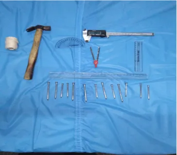
MATERIALS:
Fig 3: Instruments used in the study.
METHODS:
I) DISSECTION METHOD
II) RADIOLOGICAL METHOD
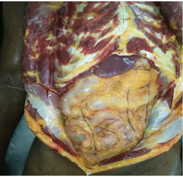
I) DISSECTION METHOD:
The guidelines of the Cunningham’s manual of practical Anatomy 15th edition were followed. The abdominal aorta is exposed by reflecting the
and the diaphragmatic curare and the arcuate ligament were exposed to see the aortic hiatus (fig-4).
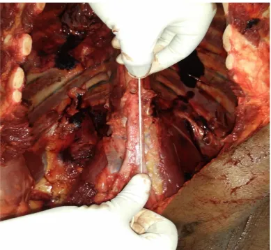
1. The course of the abdominal aorta were noted.
[image:41.612.94.467.205.567.2]2. Number of branches of abdominal aorta were recorded.
Fig 4: Exposing the abdominal aorta after midline incision.
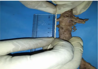
identified, then a two-inch nail was hammered into the vertebral coloumn through the centre of the origin of each branch. The nail were inserted in an horizontal plane through each vertebral origin. After the abdominal aorta was stripped off, the vertebral origin of each artery and the inter-arterial distance between each branches were measured and recorded by vernier caliper, thread and scale(Mane Uddhav Wamanrao 2016)
Fig 5: Exposing the abdominal aorta with its branches.
5. The external diameter of the aorta were measured at three levels- i) Aorta at the aortic hiatus.
ii) Mid-aortic diameter.
iii)At the level of aortic bifurcation.
6. The external diameter of the aorta was measured in an horizontal direction in its outer wall using vernier caliper (fig-7,8). The mean of all the three diameter were calculated and recorded.
7. The inter-arterial distance between the origin of coeliac artery to the superior mesenteric artery, from superior mesenteric artery to the inferior mesenteric artery and from the inferior mesenteric artery to the aortic bifurcation were measured using thread, scale and recorded (fig-9). 8. The angle of the aortic bifurcation is also measured using protractor and
recorded in all specimens. The axis of the abdominal aorta is noted and the angle is measured from the long axis of commom iliac artery to other.
Fig 7: Measuring the width of abdominal aorta at the aortic bifurcation level.
[image:45.612.111.501.370.648.2]Fig 9 Measuring the inter-arterial distance between SMA and IMA
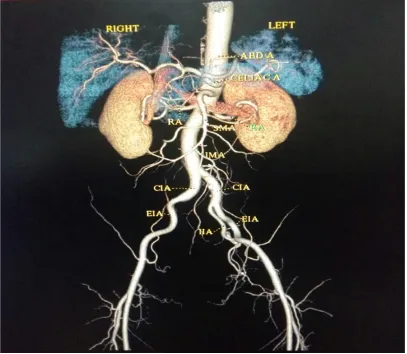
[image:46.612.96.504.418.670.2]I) RADIOLOGICAL METHOD:
[image:47.612.99.506.269.623.2]10 CT angiography images with the proper consent obtained from the patient were taken up for the study. Variations of the abdominal aorta with its branches were studied. Any pathological conditions like aortic aneurysm or aortic atherosclerosis were not taken for the study.The angiography images were obtained by spiral CT scanner to evaluate the arterial anatomy.
OBSERVATION
I) COURSE OF THE ABDOMINAL AORTA:
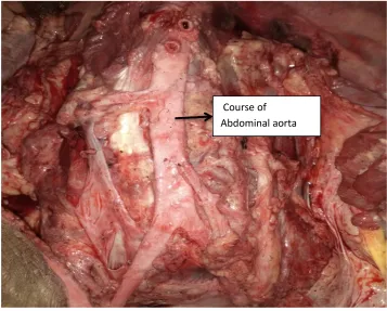
[image:48.612.145.502.405.692.2]In all the fifty specimens studied, the abdominal aorta takes a straight course with a slight deviation to the left from its point of origin in the aortic hiatus to its bifurcation. The aorta descends on the vertebral column and its inter-vertebral disc and it bifurcates into two common iliac arteries at the level of fourth lumbar vertebra. The course of abdominal aorta is straight or deviated was noted and photographed(fig11).
Fig 12 -Course of Abdominal aorta
II) NUMBER OF VENTRAL AND LATERAL BRANCHES:
In this study, ventral branches including coeliac artery, superior mesentric artery, inferior mesentric artery and the lateral branches including the paired renal and gonadal arteries were taken into this study. In all the specimens, the abdominal aorta gives off three ventral branches namely the coeliac artery, superior mesenteric artery and inferior mesenteric artery; lateral branches namely right and left renal and gonadal artery. The number of branches of abdominal aorta is noted and tabulated.
Ventral branches Lateral branches
CA SMA IMA RRA LRA RGA LGA
50 50 50 50 50
+ 1
50 50
TABLE -1 Number of ventral, lateral visceral and parietal branches of abdominal aorta
III. VERTEBRAL LEVEL OF ORIGIN OF BRANCHES OF ABDOMINAL AORTA(Table 2):
A) VENTRAL BRANCHES:
i) COELIAC AXIS:
In 18 out of 50 specimen, the coeliac artery arise at the level of T12-L1 intervertebral disc.
In 14 out of 50 specimens, the coeliac artery arise at the level of lower border of T12 thoracic vertebra.
In 13 out of 50 specimens, the coeliac artery arise at the level of upper border of L1 lumbar vertebra.
In 5 out of 50 specimens, the coeliac artery arise at the level of body of L1 lumbar vertebra(fig 12)
ii) SUPERIOR MESENTERIC ARTERY:
In 18 out of 50 specimens, the superior mesenteric artery arise at the level of upper border of L1 lumbar vertebra.
In 24 out of 50 specimens, the superior mesenteric artery arise at the level of body of L1 lumbar vertebra.
In 8 out of 50 specimens, the superior mesenteric artery arise at the level of lower border of L1 lumbar vertebra.
In 25 out of 50 specimens, the inferior mesenteric artery arises at the level of lower border of L3 lumbar vertebra.
In 16 out of 50 specimens, the inferior mesenteric artery arises at the level of body of L3 lumbar vertebra.
In 9 out of 50 specimens, the inferior mesenteric artery arises at the level of L3-L4 intervertebral disc.
B) LATERAL BRANCHES:
i) RENAL ARTERIES:
The renal artery arises from upper border of L2 lumbar vertebra in 30 specimen on the right side and 31 specimen on the left side.
The renal artery arises from the lower border of L1 lumbar vertebra in 18 specimens on the right side and 17 specimens on the left side.
The renal artery arises from the body of the L1 lumbar in 2 specimens on both sides.
ii) GONADAL ARTERIES:
In 48 out of 50 specimens, gonadal arteries arise from the abdominal aorta at the level of lower border of L2 lumbar vertebra on the right side and in 49 specimens on the left side.
In one specimen, the gonadal arteries on both sides arise from the abdominal aorta at the upper border of L2 lumbar vertebra.
On the left side, the gonadal artery arise from the renal artery (specimen no.15).
C) AORTIC BIFURCATION:
In 37 out of 50 specimens, aorta bifurcates at the level of body of L4 lumbar vertebrae.
In 8 out of 50 specimens, aorta bifurcates at the level of upper border of L4 lumbar vertebrae.
In 4 out of 50 specimens, aorta bifurcates at the level of lower border of L4 lumbar vertebrae.
Fig no 13- Vertebral level of origin of branches of Abdominal aorta using nail Vertebral level of aortic
bifurcation
Vertebral level of coelic artery Vertebral level of SMA
Specimen CA SMA IMA RRA LRA RGA LGA AB
1 T12-L1 UL1 LL3 UL2 UL2 LL2 LL2 UL4
2 LT-12 UL1 BL3 LL1 LL1 LL2 LL2 UL4
3 LT-12 UL1 BL3 UL2 UL2 LL2 LL2 BL4
4 LT-12 BL1 LL3 UL2 UL2 LL2 LL2 BL4
5 UL-1 BL1 LL3 UL2 UL2 UL2 UL2 BL4
6 T12-L1 UL1 LL3 LL1 LL1 LL2 LL2 L4-L5
7 T12-L1 BL1 LL3 UL2 UL2 LL2 LL2 BL4
8 UL1 UL1 BL3 UL2 UL2 LL2 LL2 BL4
9 LT-12 BL1 BL3 LL1 LL1 LL2 LL2 BL4
10 T12-L1 BL1 BL3 UL2 UL2 LL2 LL2 BL4
11 T12-L1 UL1 LL3 UL2 UL2 LL2 LL2 UL4
12 T12-L1 UL1 LL3 UL2 UL2 LL2 LL2 BL4
13 UL-1 BL1 BL3 LL1 LL1 LL2 LL2 BL4
14 UL-1 BL1 L3-L4 UL2 UL2 LL2 LL2 BL4
15 LT-12 BL1 BL3 UL2 UL2 LL2 LL2 BL4
16 T12-L1 UL1 LL3 BL1 BL1 LL1 LL2 LL4
17 T12-L1 BL1 L3-L4 UL2 UL2 LL2 LL2 UL4
18 UL-1 LL1 LL3 UL2 UL2 LL2 LL2 BL4
19 LT-12 LL1 BL3 LL1 LL1 LL2 LL2 BL4
20 LT-12 BL1 BL3 UL2 UL2 LL2 LL2 BL4
21 T12-L1 BL1 LL3 UL2 UL2 LL2 LL2 BL4
22 UL-1 BL1 L3-L4 LL1 LL1 LL2 LL2 BL4
23 LT-12 LL1 BL3 LL1 LL1 LL2 LL2 UL4
24 LT-12 LL1 BL3 LL1 LL1 LL2 LL2 LL4
25 T12-L1 UL1 L3-L4 LL1 LL1 LL2 LL2 BL4
26 T12-L1 BL1 LL3 UL2 UL2 LL2 LL2 BL4
27 BL-1 LL1 L3-L4 LL1 UL2 LL2 LL2 BL4
28 UL-1 LL1 LL3 UL2 UL2 LL2 LL2 BL4
29 LT-12 BL1 LL3 UL2 UL2 LL2 LL2 UL4
30 T12-L1 UL1 BL3 LL1 LL1 LL2 LL2 BL4
31 T12-L1 BL1 LL3 UL2 UL2 LL2 LL2 BL4
32 UL-1 BL1 L3-L4 LL1 LL1 LL2 LL2 BL4
33 LT-12 LL1 LL3 UL2 UL2 LL2 LL2 BL4
34 BL-1 BL1 BL3 UL2 UL2 LL2 LL2 LL4
35 LT-12 UL1 L3-L4 UL2 UL2 LL2 LL2 BL4
37 UL-1 BL1 LL3 UL2 UL2 LL2 LL2 BL4
38 UL-1 BL1 LL3 BL1 BL1 LL2 LL2 BL4
39 BL-1 LL1 L3-L4 UL2 UL2 LL2 LL2 BL4
40 LT-12 UL1 BL3 UL2 UL2 LL2 LL2 UL4
41 T12-L1 UL1 LL3 UL2 UL2 LL2 LL2 BL4
42 UL-1 BL1 BL3 LL1 LL1 LL2 LL2 BL4
43 BL-1 UL1 LL3 UL2 UL2 LL2 LL2 LL4
44 LT-12 UL1 LL3 LL1 LL1 LL2 LL2 UL4
45 T12-L1 BL1 LL3 UL2 UL2 LL2 LL2 BL4
46 T12-L1 BL1 LL3 LL1 LL1 LL2 LL2 BL4
47 UL-1 UL1 BL3 UL2 UL2 LL2 LL2 BL4
48 BL-1 UL1 L3-L4 UL2 UL2 LL2 LL2 BL4
49 T12-L1 BL1 LL3 LL1 LL1 LL2 LL2 BL4
50 UL-1 BL1 LL3 LL1 LL1 LL2 LL2 BL4
TABLE -2 VERTEBRAL ORIGIN OF VARIOUS BRANCHES OF ABDOMINAL AORTA
1V) LENGTH OF ABDOMONAL AORTA:
Specimen Total length of abdominal aorta(in
cm)
Specimen Total length of abdominal aorta(in cm)
1 10.5 26 13.2
2 10.8 27 13.4
3 12.7 28 13.3
4 12.9 29 13.7
5 12.8 30 11.2
6 13.7 31 11.9
7 13.5 32 12.2
8 10.7 33 12
9 11.8 34 13
10 12.2 35 12.8
11 12.3 36 13.2
12 12.5 37 12.3
13 12.8 38 12.3
14 13 39 12.3
15 13.2 40 11.5
16 13.7 41 11.7
17 13.6 42 12.2
18 11 43 13.2
19 12.2 44 12.2
20 11.8 45 13.2
21 12.3 46 11
22 12.7 47 10.7
23 12.9 48 13.2
24 12.3 49 12.8
25 13.1 50 12.7
The mean length of abdominal aorta is 12.45cm and range from 10.5 to 13.7 cm. The maximum frequency ranges from 12 to 12.9cm in 22 specimens(fig 13)
V) EXTERNAL DIAMETER OF AORTA:
Specimen Aortic opening at the level
of coeliac artery ( in cm) diameter (in cm) Mid-aortic Diameter just above bifurcation (in cm)
1 1.6 1.5 1.2
2 1.6 1.5 1.2
3 1.7 1.3 1.1
4 1.8 1.4 1.1
5 1.7 1.5 1.2
6 1.6 1.5 1.2
7 1.6 1.5 1.2
8 1.9 1.4 1.1
9 1.8 1.3 1.1
10 1.7 1.4 1.1
11 1.6 1.5 1.2
12 1.6 1.5 1.2
13 1.7 1.4 1.1
14 1.6 1.5 1.2
15 1.8 1.4 1.1
16 1.6 1.5 1.2
17 1.6 1.5 1.2
18 1.7 1.5 1.2
19 1.6 1.4 1.4
20 1.9 1.5 1.2
21 1.6 1.5 1.2
22 1.9 1.4 1.1
23 1.7 1.5 1.2
24 1.7 1.5 1.2
25 1.6 1.5 1.2
26 1.7 1.5 1.2
27 1.6 1.5 1.2
28 1.7 1.4 1.1
29 1.8 1.4 1.1
30 1.6 1.5 1.2
31 1.6 1.5 1.2
32 1.7 1.5 1.2
33 1.6 1.4 1.1
34 1.7 1.4 1.1
35 1.8 1.6 1.2
36 1.6 1.5 1.2
37 1.6 1.5 1.2
38 1.7 1.4 1.1
39 1.8 1.5 1.2
40 1.6 1.4 1.1
41 1.6 1.5 1.2
43 1.6 1.5 1.2
44 2.2 1.8 1.3
45 1.6 1.5 1.2
46 1.6 1.5 1.2
47 1.7 1.6 1
48 2.1 1.7 1.3
49 2.2 1.7 1.2
50 1.6 1.5 1.2
TABLE 4 EXTERNAL DIAMETER OF AORTA AT VARIOUS LEVELS.
The mean external diameter of the aorta at the level of aortic hiatus is 1.7cm and maximum frequency ranges from 1.4cm to 1.6cm in 24 specimens(fig 14).
The mean external diameter of mid-aortic diameter is 1.5cm and the maximum frequency occurs in 1.5cm in 31 specimens(fig 15).
Fig no15- Measurement of diameter of Abdominal aorta at the level of coeliac artery
[image:60.612.96.486.411.676.2]Fig 16- Diameter just above bifurcation
VI) INTER-ARTERIAL DISTANCE:
Specimen CT-SMA ( in cm) SMA-IMA(in cm) IMA-AB (in cm)
1 1.3 5.6 2.4
2 1.0 6.0 3.2
3 1.3 6.2 2.8
4 1.3 6.2 3.2
5 1.3 6.0 3.0
6 1.3 4.5 2.6
7 1.2 6.6 3.2
8 1.3 6.2 3.2
9 1.3 5.4 3.3
10 1.3 6.6 3.3
11 1.3 6.7 2.8
12 1.0 6.0 3.5
14 1.3 6.6 3.3
15 1.6 6.7 4.3
16 1.3 6.2 2.8
17 1.6 6.0 3.8
18 1.3 6.7 2.6
19 1.3 6.6 3.5
20 0.8 5.6 3.0
21 1.4 6.6 3.7
22 1.3 6.6 4.3
23 1.3 6.2 3.8
24 1.3 6.7 3.0
25 1.4 6.6 3.3
26 1.3 5.6 2.4
27 1.3 6.6 3.2
28 1.3 6.0 3.0
29 1.0 6.6 3.3
30 1.3 6.7 2.8
31 1.4 6.2 3.7
32 1.3 6.7 3.3
33 1.3 6.6 3.2
34 1.3 5.4 4.0
35 1.0 6.8 2.8
36 1.3 6.0 3.2
37 1.3 6.2 3.2
38 1.2 6.7 2.6
39 1.3 6.4 3.7
40 1.4 7.2 3.0
41 1.6 6.6 3.3
42 1.3 6.2 4.0
43 1.3 6.6 2.8
44 1.3 6.7 3.5
45 1.2 6.0 3.3
46 1.3 6.8 3.5
47 1.3 5.6 2.6
48 1.3 6.2 3.0
49 0.8 6.6 3.8
50 1.3 7.0 3.3
[image:62.612.90.492.65.647.2] The mean distance between the coeliac and superior mesenteric artery is 1.3cm and the maximum frequency ranges from 1.2cm to 1.3cm in 37 specimens(fig 17).
The mean distance between the superior mesenteric and the inferior mesenteric artery is 6.3cm and the maximum frequency ranges from 6cm to 6.6cm in 41 specimens(fig 18).
Fig no.18- Measurement of distance between coeliac and superior mesenteric artery.
Fig no.19- Distance between superior and inferior mesenteric artery. Coeliac artery
Superior mesenteric artery
Superior mesenteric artery
[image:64.612.96.490.378.674.2]Fig no.20 Distance between inferior mesenteric artery and aortic bifurcation.
III) ANGLE OF BIFURCATION: Specimen No Angle of
bifurcation (in degrees)
Specimen No Angle of
bifurcation (in degrees)
1 46.1 26 46.0
2 33.2 27 50.6
3 46.2 28 50.4
4 34.4 29 51.0
5 35.6 30 46.0
6 37.4 31 51.0
7 46.5 32 46.1
8 46.4 33 50.0
9 38.2 34 46.1
Inferior mesenteric artery
10 46.6 35 52.0
11 39.4 36 46.1
12 40.2 37 53.0
13 41.2 38 54.6
14 46.8 39 55.4
15 46.4 40 56.2
16 48.2 41 46.4
17 46.4 42 46.5
18 46.1 43 57.2
19 49.1 44 56.2
20 49.1 45 46.1
21 46.1 46 46.4
22 46.0 47 58.4
23 46.0 48 46.3
24 46.2 49 59.2
[image:66.612.102.471.62.320.2]25 49.4 50 46.4
TABLE NO 6: Angle of bifurcation
The mean angle of bifurcation of the abdominal aorta is 46.36 degrees measured at the body of L4 vertebra(fig 20)
[image:66.612.96.466.435.681.2]VIII) VARIATIONS:
The morphological variations pertaining to the abnormal origin and accessory vessels were noted in one specimen in the present study. The variations were
An accessory renal artery were found to arise from the abdominal aorta just below the origin of left renal artery at the lower level of L1 lumbar vertebra in one specimen. The accessory renal artery was found to arise from the lateral aspect of the abdominal aorta. The accessory renal artery entered the kidney through hilum in the lower pole and divides into smaller branches. There were no anastamosis between the main branch and the accessory vessels and there is no change in external architecture of the kidneys.
The gonadal artery on the left side was found to arise from the left renal artery at the level of lower border of L1 lumbar vertebra(fig 21). It then passes downwards anteriorly to the psoas major, ureter and genito-femoral nerve (Datta A.K edit 8th). The left gonadal artery then enters
RADIOLOGICAL STUDY:
[image:69.612.95.481.207.513.2]All 10 Computerized Tomographic images of abdominal aorta showed normal course and branching pattern. No variation were found pertaining to the course and branching pattern of the abdominal aorta.
DISCUSSION
The knowledge of morphology of abdominal aorta and its variations is a pre-requisite for radiologists and surgeons for diagnostic, interventional and surgical procedures. The variations in the course and branches of abdominal aorta should not be ignored during abdominal investigational and operative procedures because many of the complications can be avoided with the sound knowledge of anatomical variations (Mane Uddhav Wamanrao 2016).
In this study, morphology of abdominal aorta and its branches in 50 specimens were studied and compared with the previous studies.
I) COURSE OF ABDOMINAL AORTA IN THE PRESENT STUDY:
should be taken into consideration especially during surgical procedures (Mane UddhavWamanrao 2016).
II) NUMBER OF VENTRAL AND LATERAL BRANCHES:
An accessory renal artery arising from the abdominal aorta was seen on the left side.
The ventral branches of abdominal aorta (coeliac artery, superior mesenteric and inferior mesenteric artery) do not show any variations in the number. The accessory renal artery was found to arise just below the renal artery at the level of lower border of L2 vertebra on the left side. The accessory renal artery originates from abdominal aorta just below the main renal artery. The caliber of accessory renal artery is less than that of main renal artery. The accessory renal artery is related anterior to the renal vein as it gains entry into the hilum of the left kidney.
III) VERTEBRAL LEVEL OF ORIGIN OF BRANCHES OF ABDOMINAL AORTA:
i) COELIAC AXIS:
Out of 50 specimens, the coeliac artery arises from the intervertebral disc at the level of T12-L1 in 18 specimens, at the lower border of T12 thoracic vertebra in 14 specimens, from the upper border of L1 lumbar vertebra in 13 specimens, from the body of L1 lumbar vertebra in 5 specimens.
Vertebral level No of specimens
Intervertebral disc (T12-L1) 18
Lower border of T12 14
Upper border of L1 13
Body of L1 5
Pie chart no 1- showing vertebral origin of coeliac axis.
ii) SUPERIOR MESENTERIC ARTERY: In majority of specimens, the superior mesenteric artery arises from body of L1 lumbar vertebra in 24 out of 50 specimens. It also arises from upper and lower border of L1 lumbar vertebra in 26 specimens.
Vertebral origin No.of specimens
UL1 18
BL1 24
[image:74.612.126.485.63.307.2]LL1 8
Table No 8- showing vertebral origin of superior mesenteric artery.
T12-L1 36%
LT12 28% UL1
26%
BL1 10%
Pie chart 2: showing vertebral origin of superior mesenteric artery.
iii) INFERIOR MESENTERIC ARTERY: In majority of specimens, inferior mesenteric artery arises from lower border of L3 lumbar vertebra (25 out of 50 soecimens). It also arises from body of L3 and intervertebral disc between L3-L4 vertebra in 25 out of 50 specimens.
Vertebral origin No.of specimens
BL3 25
LL3 16
[image:75.612.93.456.62.368.2]L3-L4 9
Table no 9: Showing vertebral origin of inferior mesenteric artery.
UL1 36%
BL1 48%
LL1 16%
Pie chart 3- showing vertebral origin of inferior mesenteric artery.
iv) VERTEBRAL ORIGIN OF LATETRAL VISCERAL BRANCHES:
The majority of renal arteries on both sides arises from upper border of L2 lumbar vertebra (in 30 specimens on the right side and 31 specimens on the left side) and rest of the specimens arises from lower border and body of L1 lumbar vertebra.
The majority of gonadal artery arise from the lower border of L2 lumbar vertebra on both sides.
BL3 50%
LL3 32%
L3-L4 18%
v) AORTIC BIFURCATION: In 37 specimens , aorta bifurcates at the level of L4 lumbar vertebra. In 8 specimens, aorta bifurcates at the upper border of L4 vertebra and in 4 specimens, aorta bifurcates in lower border of L4 lumbar vertebra. In one specimen, aorta bifurcates in the intervertebral disc between L4-L5 vertebra.
Vertebral origin No.of specimens
BL4 37
UL4 8
LL4 4
[image:77.612.152.463.363.651.2]L4-L5 1
Table no10- showing vertebral level of bifurcation of aorta.
Pie chart 4-showing vertebral level of aortic bifurcation.
BL4 74% UL4
16% LL4 8%
L4-L5 2%
ART ERY
VERTEBRAL LEVEL OF BRANCHES OF ABDOMINAL AORTA
T12
T12-L1
UL1 BL1 LL1 L1-L2 UL2 LL2 UL3 BL3 LL3 L3-L4
UL4 BL4 LL
4
CA 28% 36% 26% 10% - - - -
SMA - - 36% 48% 16% - - - --
IMA - - - 32% 50% 18% - - -
RRA - - - 4% 36% - 60% - - - -
LRA - - - 4% 34% - 62% - - - -
RGA - - - - -- - 3% 97% - - - -
LGA - - - - -- - 2% 98% - - - -
AB - - - 16% 74% 8%
Table no 11- Percentage of vertebral origin of branches of abdominal aorta
S.no Authors CA SMA IMA RRA LRA RGA LGA AB
1. Satchidhanandam(1987) L1 BL1 BL3 - - - - BL4 2. Neil pennington(2005) BT12 UL1 LL3 LL1 LL1 - - LL4
3. Songur et al(2010) T12-L1
L1-L2
L3 L2 L2 - - LL4
4. Prakash et al (2011) T12 L1 L3 L1 L1 - - LL4
5. Present study T12-L1
[image:79.612.56.529.65.284.2]BL1 LL3 UL2 UL2 LL2 LL2 BL4
Table No.12- Comparison with other studies.
iv)LENGTH OF ABDOMINAL AORTA:
The length of abdominal aorta was measured from the aortic hiatus which marks the approximate length of the upper most part of abdominal aorta. The mean length of abdominal aorta in our present study is 12.45cm. According to
A.K.Datta 8thedit. the length of abdominal aorta is 13cm, Wood Jones(1953)
5inches, Wood Burne (1961) 13cm and Keith Moore (2010) is 13cm.
Length of AA(range in cm) Number of specimens
10.5cm to 12cm 12
12cm to 12.9cm 22
13cm to 13.7cm 16
Pie chart 5 showing range of length of abdominal aorta.
10.5cm-12cm 24%
12cm-12.9cm 44% 13cm-13.7cm
32%
The length of the abdominal aorta in our present study is compared with the previous studies.
Authors Length of Abdominal
aorta
Wood Burne (1961) 13cm
Mane Uddhav Wamanrao (2016) 13.2cm
Ruggles George 13.2cm
Present study 12.45cm
v) EXTERNAL DIAMETER OF AORTA AT VARIOUS LEVELS.
In the present study, the external diameter (width) of the abdominal aorta was done at three levels. The mean diameter at various levels are,
i) The mean diameter of Aortic opening at the origin of coeliac trunk is 1.7cm and the maximum frequency ranges from 1.4cm to 1.6cm in 24 specimens.
ii) The mean diameter at mid-aortic level is 1.52cm and the maximum frequency occurs in 1.5cm in 32 specimens.
EXTERNAL DIAMETER AT THE AORTIC OPENING External diameter (ranges in cm) Number of specimens
1.4cm-1.6cm 24
1.7cm-2cm 23
2.1cm-2.2cm 3
Pie chart 6 : showing the external diameter at the level of coeliac trunk.
1.4-1.6cm 48% 1.7-2cm
46%
2.1-2.2cm 6%
MID AORTIC DIAMETER
External diameter (ranges in cm) Number of specimens
1.3cm-1.4cm 16
1.5cm 32
1.6cm 2
Pie chart 7 : showing external diameter at mid-aortic level.
1.3-1.4cm 32%
1.5cm 64%
1.6cm 4%
EXTERNAL DIAMETER JUST ABOVE BIFURCATION External diameter (ranges in
cm) Number of specimens
1cm -1.1cm 16
1.2 33
1.3cm -1.4cm 1
Pie chart 8 : showing external diameter at aortic bifurcation.
Very few studies mentioned about the external diameter at various levels in adult cadavers. Neil Pennington reported the diameter at the level of coeliac axis2.49cm±0.48cm, at the level of superior mesenteric artery -2.44cm±0.42cm, and at the level of inferior mesenteric artery-2.11±0.55cm. In
1-1.1cm 32%
1.2cm 66%
1.3-1.4cm 2%
this study, the mean external diameter at the level of aortic opening is 1.7cm, at the mid-aortic level is 1.52cm and at the level of aortic bifurcation is 1.2cm. The result were comparable with Adachi et al who reported the mean aortic diameter is 1.72cm. Mane Uddhav Wamanrao reported the mean diameter at the aortic opening is 2.19cm, at the mid-aortic diameter is 1.67cm and at the aortic bifurcation is 1.67cm.
The diameter of the abdominal aorta progressively decreases from proximal to distal in its length. This decrease in diameter is due to supply to visceral organs. (Mane Uddhav Wamanrao).The diameter of abdominal aorta is smaller in Asian population than in the Caucasian population as reported by Spark J.I and it signifies the prevalence of abdominal aneurysm is higher in Caucasian population.
VI) INTER-ARTERIAL DISTANCE:
The inter-arerial distance was stated by various authors and in different textbooks. In this study, the mean distance between the coeliac artery and the superior mesenteric artery is 1.3cm with the maximum frequency of 1.2 to 1.3cm. G.J.Romanes 16th edit stated that the average distance between the two
vessel in 1cm, Last edit 1.25cm, Gray’s Anatomy 40th edit 1cm. The
Inferior mesenteric artery and the aortic bifurcation is 3.2cm. This is comparable to Songur et al -3.5cm.
S.no Authors CA-SMA
(in cm)
SMA-IMA
(in cm)
IMA-AB
(in cm)
1 Cauldwell and Anson 1-1.9cm 6.9cm 4.2
2 Woodburne (1966) 1.6cm - -
3 Songur et al (2010) 1.4cm±0.26cm 5.7to8cm 3.5cm
4 Present study 1.3cm 6.3cm 3.2cm
VII ) ANGLE OF BIFURCATION:
VIII) VARIATIONS:
The present study report two variation in the branching pattern.
1) Accessory or supernumerary renal artery is arising on the left side of the abdominal aorta. The supernumerary artery is smaller in size when compared to the main renal artery and it divides into smaller branches at the lower pole of the kidney. The accessory (supernumerary) renal artery can be encountered in 23% of the population (Banowsky LHW). Singh G et al also stated that the accessory renal artery were more common on the left side either at the upper or the lower pole of the kidney. Apurba Patra 2016 stated that 20% of the origin of accessory renal artery originated from the abdominal aorta. The knowedge of these anatomical variations should be kept in mind before interventional procedures like renal angiograms and during surgical procedures like renal transplantation (Raikos A 2010).
and fourth are middle group supplying the kidneys, sixth to ninth are caudal arteries supplying the gonads through gonadal arteries. The persistence of cranial and the middle group result in high-origin of gonadal artery arising from the suprarenal and the renal arteries respectively. The knowledge of the variations should be taken into account in avoiding complications during surgery and diagnostic techniques.
Fig no 25: Showing Left Accessory renal artery arising from abdominal aorta
CONCLUSION
The abdominal aorta was studied in fifty specimens obtained from the Department of Anatomy and from the Department of Forensic Medicine. The course, number of ventral and lateral branches, vertebral level of origin of branches, external diameter, length, inter-arterial distance, angle of bifurcation and variations were observed and compared with the findings of the previous studies. The following conclusions were derived from these parameters.
1. The course of the abdominal aorta was found to be normal from its exit from the thorax to the bifurcation in all 50 specimens.
2. The number of branches of abdominal aorta were found to be normal in 49 specimens. In one specimen, accessory renal artery was found to arise above the main renal artery on the left side from the aorta.
3. The vertebral origin of coeliac artery were at the T12-L1 level, superior mesenteric artery at the body of L1 vertebra, inferior mesenteric artery at lower border of L3 vertebra, bilateral renal artery at lower border of L1 vertebra, gonadal artery at L2 vertebra on both sides and aortic bifurcation at the body of L4 lumbar vertebra.
4. The mean length of abdominal aorta in the present study is 12.45cm.
5. The mean external diameter at the coeliac level is 1.7cm, at the mid aortic level is 1.5cm and just above bifurcation is 1.2cm.
artery is 6.3cm and from the inferior mesenteric artery to the aortic bifurcation is 3.2cm.
7. The angle of bifurcation was at the level of body of L4 lumbar vertebra and the mean angle is 45.36 degrees.
8. The two variations noted in the present study includes super numerary renal artery arising from abdominal aorta just below the origin of renal artery on the left side and abnormal origin of gonadal artery from renal artery on the left side.
BIBLIOGRAPHY
1. Adachi B., Das Arteriensystem der Japaner, Band II, Verlag der Kaliserlich- Japanischenuniversitatzu Kyoto, Maruzen publishing Co., 1928, 28,38,54.
2. Adachi, B. (2000) Anatamie der Japaner.I.Das.Arterian system der Japaner. II.Aorta. Thoracalis. Arcus.Plantarie Profundus Maruzen. Kyoto quoted by cauld well and Anson in Americian Jr. of Anat.
3. Akhilandeswari.B, PriyaRanganath ‘Variations in the source of origin of inferior phrenic artery: a cadaveric study’ 2013, Journal of the Anatomical society of India 6(2) pg 6-9.
4. Anson BJ McVay CB The topographical positions and the mutual relations of the visceral branches of the abdominal aorta: a study of 100 consecutive cadavers. Anat Rec. 1936:67:7-15.
6. ArvindDeswal, Binod Kumar Tamang, AnjuBala ‘Study of Aortic- Common Iliac Bifurcation and Its Clinical Significance’ J. ClinDiagn Res 2014 Jul 8(7).
7. Balli Sharma, Dhiraj Saxena, Sangita Chauhan, S.K. Agarwal ‘CT angiography based Study of Variations in Coeliac Trunk and its surgical implications’ IOSR Journal of Dental and Medical Sciences .Volume 14, Issue 12, 2015.
8. Banowsky LHW, Surgical anatomy. In: Novick AC, Streem SB, Pontes JE (eds), Stewart’s operative urology, Williams & Wilkins, Baltimore, 1989.
9. Bargeron CB, Hutchins GM, Moore GW, Deters OJ, Mark FF, Friedman MH (1986) Distribution of the geometric parameters of human aortic bifurcations. Arteriosclerosis, 6: 109–113.
10. Borley N. R. Chapter 68 Posterior abdominal wall and retroperitoneum. In: Standring S., Collins P., Wigley C., editors. Gray’s Anatomy. 39th Edition. London: Churchill Livingstone, Elsevier Ltd; 2005- 06. p. 1116-1120.