Open Access
Research article
An inactivated nuclease-like domain in RecC with novel function:
implications for evolution
Daniel John Rigden*
Address: School of Biological Sciences, University of Liverpool, Crown St., Liverpool L69 7ZB, UK
Email: Daniel John Rigden* - [email protected] * Corresponding author
Abstract
Background: The PD-(D/E)xK superfamily, containing a wide variety of other exo- and endonucleases, is a notable example of general function conservation in the face of extreme sequence and structural variation. Almost all members employ a small number of shared conserved residues to bind catalytically essential metal ions and thereby effect DNA cleavage. The crystal structure of the RecBCD prokaryotic DNA repair machinery shows that RecB contains such a nuclease domain at its C-terminus. The RecC C-terminal region was reported as having a novel fold.
Results: The RecC C-terminal region can be divided into an alpha/beta domain and a smaller alpha-helical bundle domain. Here we show that the alpha/beta domain is homologous to the RecB nuclease domain but lacks the features necessary for catalysis. Instead, the domain has a novel function within the nuclease superfamily – providing a hoop through which single-stranded DNA passes. Comparison with other structures of nuclease domains bound to DNA reveals strikingly different modes of ligand binding. The alpha-helical bundle domain contributes the pin which splits the DNA duplex.
Conclusion: The demonstrated homology of RecB and RecC shows how evolution acted to produce the present RecBCD complex through aggregation of new domains as well as functional divergence and structural redeployment of existing domains. Distantly homologous nuclease(-like) domains bind DNA in highly diverse manners.
Background
The largest evolutionary superfamilies of proteins cover such a large range of sequence space that the relationships shared by members may not be apparent by standard means of sequence comparison, and hence are often only recognized after structural determinations. Such has fre-quently been the case for the PD-(D/E)xK superfamily of nucleases. Within the superfamily, structures were first obtained for four restriction enzymes, of such diverse sequences that they were initially assumed not to share
homology (reviewed in [1]). Since then structures have confirmed distant and often unexpected homologies of those four with many other restriction enzymes, as well as exo- and endo-nucleases involved in such diverse cellular processes as DNA repair [2], transposition [3], Holliday junction resolution [4] and recombination [5].
The unifying catalytic site characteristic of the superfamily is the presence of one or more catalytically essential diva-lent cations [6,7]. The conserved acidic residues of the
PD-Published: 28 June 2005
BMC Structural Biology 2005, 5:9 doi:10.1186/1472-6807-5-9
Received: 15 April 2005 Accepted: 28 June 2005
This article is available from: http://www.biomedcentral.com/1472-6807/5/9
© 2005 Rigden; licensee BioMed Central Ltd.
these catalytic site variants some interesting exceptions have been noticed. Thus, while clearly containing a PD-(D/E)xK superfamily-like domain structure [10], the tRNA splicing endoribonuclease EndA, has evolved an unre-lated catalytic site on the opposite side of the fold to the conventional site [11]. A catalytically inactive version of the fold has also been seen in the N-terminal domain of
S. cerevisiae RPB5, an RNA polymerase subunit, where evi-dence suggests that it functions in protein-protein interac-tions [12].
Although extremely diverse in structure and sequence, modern sequence comparison methods have played their part in elucidating the full range of PD-(D/E)xK super-family members [9,13-15]. Nevertheless, structure deter-minations and structure-informed bioinformatics [16] will continue to be crucial in this most diverse of super-families. Some five years ago it was predicted that the nuclease activity associated with the C-terminus of RecB [17] resulted from the presence of a domain homologous to that of λ-exonuclease, despite RecB not possessing a PD-(D/E)xK motif [13,14]. This prediction has been recently confirmed with the crystal structure determina-tion of the structure of the RecBCD heterotrimer [18]. This remarkable complex (see [18] and references therein) which functions to process double-stranded breaks in DNA, contains two distinct helicase activities, contributed by RecB and RecD. Also present is a catalytically inactive subunit, RecC. Among its proposed roles is recognition of the Chi DNA sequence [18]. Remarkably, twin helicase(-like) motor domains (canonically named 1A and 2A) are present in all three subunits, although those in RecC are inactivated and only those in RecB and RecC contain α -helical insert domains in each motor domain (named 1B and 2B, respectively). As mentioned, the helicase domains of RecB are followed by a PD-(D/E)xK superfamily nucle-ase domain 3. In contrast, the C-terminal 'domain 3' of RecC was reported as being of novel fold [18].
Here we show that the C-terminal region ('domain 3') of RecC can actually be dissected into two domains, the first of which is clearly related to PD-(D/E)xK superfamily nuclease domains (hereafter called simply nuclease
terminus, as well as by altering function of, and reposi-tioning of, existing domains.
Results and discussion
An unsuspected nuclease-like domain in RecC
Domain 3 of RecC has been described as being of novel fold [18]. Structural examination suggested that it could, in fact, be divided into two domains, an α/β domain and a C-terminal all α-helical domain. Although the division was made by eye initially, analysis with Protein Domain Parser [19] produced a result that differed by just two res-idues. When the α/β domain (comprising residues 828– 1033) was submitted to DALI [20], the most closely related structure in the database was reported as phospho-serine phosphatase but in second place was λ-exonuclease (PDB code 1avq; [5]). A root mean squared (rms) devia-tion between the third RecC domain and λ-exonuclease of 4.2Å for 121 Cα atoms was obtained (yielding a DALI Z score of 4.1). λ-exonuclease is the nearest structural neigh-bour to the nuclease domain of RecB [18]. For that pair, 131 Cα atoms can be superimposed with an rms devia-tion of 3.5 Å (Z score of 6.2). From these data and visual inspection (later additionally supported by PSI-BLAST results – see below), it is clear that the third RecC domain is a relative of the nuclease domain common to RecB and
λ-exonuclease (Figures 1 and 2). Notably, the further divi-sion of the C-terminal RecC 'domain 3' into two domains was essential for this relationship to become apparent. In contrast, the fourth, α-helical bundle domain of RecC has no close neighbours in the present database.
site, like those shown in Figure 1, is not present in RecC (Figure 3). Indeed, the overall sequence identity between the RecB and RecC sequence segments shown in Figure 3 is just 2–11 %. Thus, just as domains 1 and 2 of RecC are inactivated helicase domains [18], so its domain 3 is an inactivated nuclease.
Interestingly, comparison of the nuclease domains of RecB, RecC and λ-exonuclease shows that the Rec subu-nits clearly share a more recent ancestor than the common ancestor of all three structures. As Figures 1 and 2 show, a single helix present in λ-exonuclease is replaced in both RecB and RecC by a three-helix α-helical bundle. This bundle is not present in the more distant relatives of λ -exonuclease highlighted by the CE server [21] such as archaeal Holliday junction resolvase, tRNA endonuclease and the PvuII restriction enzyme. Curiously, the degree of structural superposition that can be achieved between the RecB and RecC nuclease domains and λ-exonuclease sug-gests no closer relationship between the former pair. For example, 71 Cα atoms of RecB nuclease domain may be superimposed on their equivalents in RecC to produce an rms deviation of 2.19 Å. In comparison, 82 Cα atoms of RecB superimpose on equivalents of λ-exonuclease with a lower rms deviation of 1.71Å. However, the superimpos-able three-helix α-helical bundle shared only by RecB and RecC (Figures 1 and 2) show that they are more closely evolutionarily related to each other than to other homol-ogous structures. The closer structural superposition of RecB and λ-exonuclease seems likely to arise from their shared nuclease activity, while RecC has evolved a differ-ent function.
Novel function of the nuclease-like domain in RecC
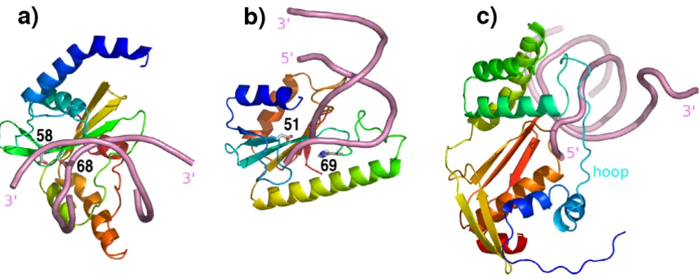
As mentioned, nuclease domains as represented in the present PDB are extremely diverse in sequence but share conserved residues that bind essential metal ions and are almost invariably catalytically active. The recognition of the third domain of RecC as an inactivated nuclease domain highlights a wholly unexpected new function for a non-catalytic but clearly nuclease-like domain. As shown in Fig 2e, the nuclease-like domain of RecC pro-vides a hoop through which a single strand of the newly separated DNA duplex is passed. The hoop is the entrance to the 5' channel leading to RecD in the RecBCD complex [18]. The pin responsible for separating the two DNA strands consists of a loop extending out of the α-helical bundle domain 4 of RecC.
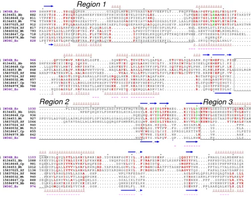
[image:3.612.57.555.86.233.2]Structural comparisons show that a series of three struc-tural adaptations have been required in RecC in order to achieve this novel ssDNA-hoop function. These involve three regions of sequence marked on Figures 2 and 3. Region 1 comprises a long linker sequence between the extended structure that starts the domain and the three helix α-helical bundle subdomain. This linker region is very poor in regular secondary structure and adopts dra-matically different conformations in the two domains. Significant sequence identity between RecB and RecC seems absent in the region. In RecB this linker lies along the surface of the remainder of the domain. In dramatic contrast, region 1 in RecC has few contacts with the rest of the domain (although it contacts other parts of RecC – see below) and forms most of the rim of the hoop through which ssDNA is passed. Region 2 is the connection Stereo structural superposition of the nuclease(-like) domains of RecB and RecC produced with LSQMAN [29]
Figure 1
between the two strands forming an antiparallel β-sheet. In E. coli RecC the connection is a minimal β-turn and connections in other RecC sequences are also very short (Figure 3). In contrast, Region 2 in RecB is usually much larger, tracing out, in the E. coli RecB structure, an 11-resi-due α-helix and a substantial stretch lacking regular sec-ondary structure. Structure comparison shows the reason
for the short connectors in RecC (Figure 2); larger connec-tors occupy the same space as the fourth domain of RecC. Thus, a larger connector would be incompatible with a RecC-style pin domain. Region 3, providing the connector between a β-strand and an α-helix, is again larger in RecB than in RecC and again contains an α-helix in RecB. Here the reason for the shorter connector in RecC is even more Comparisons of structurally aligned nuclease(-like) domains in λ-exonuclease, RecB and RecC
Figure 2
fundamental; were it to have the longer connector of RecB, the aperture whereby ssDNA passes through the RecC nuclease-like domain would be sterically obstructed.
DNA interactions with nuclease and nuclease-like domains
Unfortunately, no structure of λ-exonuclease in complex with DNA is yet available. However, other enzymes
[image:5.612.56.555.85.477.2]shar-ing the same fold, includshar-ing many type II restriction enzymes, have been crystallized in complex with DNA. Therefore, DNA-bound structures were sought for the enzymes identified as closest structural neighbours for λ -exonuclease by the CE server [21]. This analysis pin-pointed the restriction enzyme PvuII (PDB code 1pvi; [22]) and the vsr exonuclease (PDB code 1odg; [23]) Structure-based sequence alignment of the nuclease(-like) domains of RecB and RecC
Figure 3
involved in repair of bacterial G:T mismatches. Further analysis (not shown) showed that the mode of binding of DNA to Pvu II was, in fact, typical of many restriction enzymes, irrespective of dimeric vs tetrameric quaternary state and of differing modes of dimerization.
[image:6.612.56.556.83.283.2]Remarkably, as shown in Figure 4, the axes of duplex DNA binding to PvuII and to vsr exonuclease are almost orthog-onal, a difference that seems to have escaped notice. The catalytic sites of both enzymes, although differing in detail, are similarly placed at one edge of the β-sheet, defining the 'front' of catalytic nucleases. Most unexpect-edly, the inactivated nuclease-like domain of RecC which also, in the context of RecBCD, binds duplex DNA, prior to strand splitting by the fourth domain, does so in a com-pletely novel manner. First, the axis of the bound duplex DNA is approximately orthogonal to both PvuII and vsr exonuclease modes. Secondly, the binding involves the 'back' of the domain; only a single strand of the DNA arrives at the 'front' side after passing through the aperture (Figure 4). These results make clear that few assumptions can be made regarding modes of DNA binding by nucle-ase(-like) domains in the absence of experimental data such as structures in complex with DNA.
Homology of RecB and RecC
The observation of inactivated helicase-like domains in RecC was not considered reason enough to propose the
existence of homology between RecB and RecC extending over their whole length [18]. Indeed, both sequence and structural comparisons at first suggest that RecB more closely resembles other helicases than it does RecC. For example, in the results of PSI-BLAST [24] starting with E. coli RecB, PcrA, another helicase that contains large heli-cal-insert domains in each helicase domain [25], appears as a significant hit (e = 6 × 10⁻⁹) in the results of the first
iteration. In contrast, using an e-value cut-off of 0.0001 four iterations are required before RecC sequences, including that of E. coli RecC, appear among the signifi-cant hits. While the BLAST alignments centred on the hel-icase(-like) domains the C-terminal nuclease(-like) domains were sometimes matched, although PSI-BLAST runs of the nuclease domain of RecB failed to hit the nuclease-like domain of RecC, and vice versa. Similarly, structural comparisons show that both helicase domains and both α-helical insert domains of RecB are more simi-lar to their counterparts in PcrA than to the corresponding RecC domains (not shown). Nevertheless, the clear homology of the RecB and RecC nuclease(-like) domains, evident in their common three α-helical bundle (see above) strongly suggests that RecB and RecC share a more recent common ancestor than they have in common with other extant helicases. How then to explain the apparently closer relationship of RecB with PcrA than with RecC? As was proposed for the nuclease(-like) domains (see above) it seems like the dramatic functional differences between Comparison of modes of DNA binding to superimposed nuclease(-like) domains
Figure 4
corresponding RecB and RecC domains are responsible. As discussed above, the RecC nuclease-like domain is sig-nificantly shorter than the RecB nuclease domain in two key regions, each associated with its new role as provider of an ssDNA hoop. Thus, it seems plausible that the
[image:7.612.53.566.81.531.2]main-tenance of helicase activity by the helicase domains of PcrA and RecB is responsible for their apparently closer relationship, the structural changes accompanying evolu-tion of the helicase-like domains in RecC for new roles Domain comparison of PcrA, RecB and RecC
Figure 5
This picture of aggregation of novel functionality through domain addition is complemented by alterations in func-tion of homologous domains. Thus, as described, the nuclease-like domain of RecC continues to bind duplex DNA, but using a different surface of the domain, as well as providing the entrance to the 5' ssDNA channel leading to RecD. This modification is paralleled in the helicase-like domains by a change from catalytic helicase activity to Chi sequence recognition ([18] and references therein). The α-helical inserts into the helicase(-like) domains also have different functions in RecB and RecC [18], including, in the case of the RecC domain 1B binding to the RecC nuclease-like domain and the rim of its ssDNA aperture (Figure 5). Although homologous, the structural compar-ison of complete RecB and RecC subunits shows large dif-ferences in relative domain orientations and positions, most dramatically with regard to the position of the nucle-ase(-like) domains relative to the helicnucle-ase(-like) domain cores (Figure 5).
There is an interesting parallel to be drawn between RecBCD and AddAB (also known as RexAB), a different DNA repair system found in Gram positive bacteria where RecBCD is lacking (see [26] for a review). AddA and AddB also appear homologous and each possesses helicase and nuclease motifs. Within AddAB, it is AddB that recognises the Chi sequence and therefore is the counterpart of RecC in RecBCD. Most interestingly, however, both the nucle-ase domains of AddA and AddB appear to be active [27]. The AddAB system may therefore resemble an evolution-arily intermediate stage, through which the RecBCD machine passed before inactivation of the RecC nuclease domain and recruitment of RecD.
In summary, the improved domain dissection of RecC presented here and its ramifications enhance our under-standing of the evolutionary processes responsible for the remarkable DNA processing machinery that is the RecBCD complex [18]. It is now even more apparent that relatively straightforward addition of modular functional-ity has been accompanied by quite dramatic functional evolution of homologous domains.
which was also used for general sequence manipulation. Protein sequence alignment was carried out using MUS-CLE [35] and T-COFFEE [36]. Formatting of sequence alignments was done with ESPRIPT [37] using default options for colouring of sequence conservation.
Acknowledgements
I am grateful for the helpful remarks of one of the anonymous referees regarding AddAB.
References
1. Aggarwal AK: Structure and function of restriction endonucleases. Curr Opin Struct Biol 1995, 5:11-19.
2. Tsutakawa SE, Muto T, Kawate T, Jingami H, Kunishima N, Ariyoshi M, Kohda D, Nakagawa M, Morikawa K: Crystallographic and functional studies of very short patch repair endonuclease.
Mol Cell 1999, 3:621-628.
3. Hickman AB, Li Y, Mathew SV, May EW, Craig NL, Dyda F: Unex-pected structural diversity in DNA recombination: the restriction endonuclease connection. Mol Cell 2000,
5:1025-1034.
4. Bond CS, Kvaratskhelia M, Richard D, White MF, Hunter WN:
Structure of Hjc, a Holliday junction resolvase, from Sulfolo-bus solfataricus. Proc Natl Acad Sci U S A 2001, 98:5509-5514. 5. Kovall R, Matthews BW: Toroidal structure of
lambda-exonu-clease. Science 1997, 277:1824-1827.
6. Kovall RA, Matthews BW: Type II restriction endonucleases: structural, functional and evolutionary relationships. Curr Opin Chem Biol 1999, 3:578-583.
7. Pingoud A, Jeltsch A: Structure and function of type II restric-tion endonucleases. Nucleic Acids Res 2001, 29:3705-3727. 8. Skirgaila R, Grazulis S, Bozic D, Huber R, Siksnys V:
Structure-based redesign of the catalytic/metal binding site of Cfr10I restriction endonuclease reveals importance of spatial rather than sequence conservation of active centre residues.
J Mol Biol 1998, 279:473-481.
9. Feder M, Bujnicki JM: Identification of a new family of putative PD-(D/E)XK nucleases with unusual phylogenomic distribu-tion and a new type of the active site. BMC Genomics 2005, 6:21. 10. Bujnicki JM, Rychlewski L: Unusual evolutionary history of tRNA splicing the endonuclease EndA: relationship to the LAGLI-DADG and PD-(D/E)XK deoxyribonucleases. Protein Sci 2001,
10:656-660.
11. Li H, Trotta CR, Abelson J: Crystal structure and evolution of a transfer RNA splicing enzyme. Science 1998, 280:279-284. 12. Todone F, Weinzierl RO, Brick P, Onesti S: Crystal structure of
RPB5, a universal eukaryotic RNA polymerase subunit and transcription factor interaction target. Proc Natl Acad Sci U S A 2000, 97:6306-6310.
13. Daiyasu H, Komori K, Sakae S, Ishino Y, Toh H: Hjc resolvase is a distantly related member of the type II restriction endonu-clease family. Nucleic Acids Res 2000, 28:4540-4543.
Publish with BioMed Central and every scientist can read your work free of charge "BioMed Central will be the most significant development for disseminating the results of biomedical researc h in our lifetime."
Sir Paul Nurse, Cancer Research UK
Your research papers will be:
available free of charge to the entire biomedical community
peer reviewed and published immediately upon acceptance
cited in PubMed and archived on PubMed Central
yours — you keep the copyright
Submit your manuscript here:
http://www.biomedcentral.com/info/publishing_adv.asp
BioMedcentral 15. Bujnicki JM, Rychlewski L: Grouping together highly diverged
PD-(D/E)XK nucleases and identification of novel super-family members using structure-guided alignment of sequence profiles. J Mol Microbiol Biotechnol 2001, 3:69-72. 16. Bujnicki JM: Crystallographic and bioinformatic studies on
restriction endonucleases: inference of evolutionary rela-tionships in the "midnight zone" of homology. Curr Protein Pept Sci 2003, 4:327-337.
17. Yu M, Souaya J, Julin DA: The 30-kDa C-terminal domain of the RecB protein is critical for the nuclease activity, but not the helicase activity, of the RecBCD enzyme from Escherichia coli. Proc Natl Acad Sci USA 1998, 95:981-986.
18. Singleton MR, Dillingham MS, Gaudier M, Kowalczykowski SC, Wigley DB: Crystal structure of RecBCD enzyme reveals a machine for processing DNA breaks. Nature 2004, 432:187-193. 19. Alexandrov N, Shindyalov I: PDP: protein domain parser.
Bioin-formatics 2003, 19:429-430.
20. Holm L, Sander C: Dali: a network tool for protein structure comparison. Trends Biochem Sci 1995, 20:478-480.
21. Shindyalov IN, Bourne PE: Protein structure alignment by incre-mental combinatorial extension (CE) of the optimal path.
Protein Eng 1998, 11:739-747.
22. Cheng X, Balendiran K, Schildkraut I, Anderson JE: Structure of PvuII endonuclease with cognate DNA. EMBO J 1994,
13:3927-3935.
23. Bunting KA, Roe SM, Headley A, Brown T, Savva R, Pearl LH: Crystal structure of the Escherichia coli dcm very-short-patch DNA repair endonuclease bound to its reaction product-site in a DNA superhelix. Nucleic Acids Res 2003, 31:1633-1639.
24. Altschul SF, Madden TL, Schäffer AA, Zhang J, Zhang Z, Miller W, Lip-man DJ: Gapped BLAST and PSI-BLAST: a new generation of protein database search programs. Nucleic Acids Res 1997,
25:3389-3402.
25. Subramanya HS, Bird LE, Brannigan JA, Wigley DB: Crystal struc-ture of a DExx box DNA helicase. Nature 1996, 384:379-383. 26. Chedin F, Kowalczykowski SC: A novel family of regulated
heli-cases/nucleases from Gram-positive bacteria: insights into the initiation of DNA recombination. Mol Microbiol 2002,
43:823-834.
27. Quiberoni A, Biswas I, El Karoui M, Rezaiki L, Tailliez P, Gruss A: In vivo evidence for two active nuclease motifs in the double-strand break repair enzyme RexAB of Lactococcus lactis. J Bacteriol 2001, 183:4071-4078.
28. Berman HM, Westbrook J, Feng Z, Gilliland G, Bhat TN, Weissig H, Shindyalov IN, Bourne PE: The Protein Data Bank. Nucleic Acids Res 2000, 28:235-242.
29. Kleywegt GJ: Use of non-crystallographic symmetry in protein structure refinement. Acta Cryst 1996, D52:842-857.
30. Murzin AG, Brenner SE, Hubbard T, Chothia C: SCOP: a structural classification of proteins database for the investigation of sequences and structures. J Mol Biol 1995, 247:536-540 [http:w.sciencedirect.com/science?_ob=IssueURL&_method=view MIN Page&_tockey=%23TOC%236899%239999%23997529995%239999 %23MIN%23Volume_247,_Issue_4&_acct=C000044499&_version=1 &_urlVersion=0&_userid=822084&md5=a0c105250d976d9d488358 b33d8562fd].
31. Jones TA, Zou JY, Cowan SW, Kjeldgaard M: Improved methods for building protein models in electron density maps and the location of errors in these models. Acta Cryst 1991,
A47:110-119.
32. PyMOL Home Page [http://pymol.sourceforge.net]
33. Tatusov RL, Natale DA, Garkavtsev IV, Tatusova TA, Shankavaram UT, Rao BS, Kiryutin B, Galperin MY, Fedorova ND, Koonin EV: The COG database: new developments in phylogenetic classifica-tion of proteins from complete genomes. Nucleic Acids Res 2001, 29:22-28.
34. Clamp M, Cuff J, Searle SM, Barton GJ: The Jalview Java alignment editor. Bioinformatics 2004, 20:426-427.
35. Edgar RC: MUSCLE: multiple sequence alignment with high accuracy and high throughput. Nucleic Acids Res 2004,
32:1792-1797.
36. Notredame C, Higgins DG, Heringa J: T-Coffee: A novel method for fast and accurate multiple sequence alignment. J Mol Biol 2004, 302:205-217.
37. Gouet P, Courcelle E, Stuart DI, Metoz F: ESPript: analysis of mul-tiple sequence alignments in PostScript. Bioinformatics 1999,
15:305-308.
38. Velankar SS, Soultanas P, Dillingham MS, Subramanya HS, Wigley DB:
![Figure 1Stereo structural superposition of the nuclease(-like) domains of RecB and RecC produced with LSQMAN [29]Stereo structural superposition of the nuclease(-like) domains of RecB and RecC produced with LSQMAN [29]](https://thumb-us.123doks.com/thumbv2/123dok_us/8061353.226025/3.612.57.555.86.233/structural-superposition-nuclease-produced-structural-superposition-nuclease-produced.webp)