Biology Theses Department of Biology
5-1-2019
DETECTION OF NECROPTOSIS AND
PYROPTOSIS-ASSOCIATED MOLECULES
DURING EXPERIMENTAL MAIDS-RELATED
CYTOMEGALOVIRUS RETINITIS
LAUREN-ASHLEY DUNCAN Georgia State University
Follow this and additional works at:https://scholarworks.gsu.edu/biology_theses
This Thesis is brought to you for free and open access by the Department of Biology at ScholarWorks @ Georgia State University. It has been accepted for inclusion in Biology Theses by an authorized administrator of ScholarWorks @ Georgia State University. For more information, please contact
Recommended Citation
DURING EXPERIMENTAL MAIDS-RELATED CYTOMEGALOVIRUS RETINITIS
by
LAUREN-ASHLEY DUNCAN
Under the Direction of Richard D. Dix, PhD
ABSTRACT
Sight threatening human cytomegalovirus (HCMV) retinitis still remains a cause for concern
among AIDS patients who do not respond to or do not have access to current antiretroviral
(ART) therapy [1]. However, little is currently known about the degenerative mechanisms
behind HCMV retinal destruction during retroviral immunosuppression. The well-established
murine AIDS (MAIDS) related murine cytomegalovirus (MCMV) retinitis model closely mimics
disease progression seen in AIDS patients [4]. Previous work using this model has shown that
while the cell death pathway apoptosis is involved in disease progression, it is not fully
responsible [1]. It has been found that the mRNA of molecules associated with two other cell
death pathways, necroptosis and pyroptosis, are also correlated with the pathophysiology of
MAIDS-related MCMV retinitis [1]. We propose that retinal degeneration seen during CMV
retinitis involves necroptosis/pyroptosis programmed cell death correlating with the increased
mRNA/protein expression of associated molecules observed in this study.
INDEX WORDS: Necroptosis-associated molecules, Pyroptosis associated molecules,
DURING EXPERIMENTAL MAIDS-RELATED CYTOMEGALOVIRUS RETINITIS
by
LAUREN-ASHLEY DUNCAN
A Thesis Submitted in Partial Fulfillment of the Requirements for the Degree of
Master of Science
in the College of Arts and Sciences
Georgia State University
Copyright by Lauren-Ashley Duncan
DURING EXPERIMENTAL MAIDS-RELATED CYTOMEGALOVIRUS RETINITIS
by
LAUREN-ASHLEY DUNCAN
Committee Chair: Richard Dix
Committee: Julia Hilliard
John Houghton
Electronic Version Approved:
Office of Graduate Studies
College of Arts and Sciences
Georgia State University
DEDICATION
Every step leading me up to this point has been driven by a goal to somehow help make the
world a better place for my beloved family, human and non-human members alike. The faith
placed in me by my family has simultaneously been a heavy burden and a duty I proudly follow;
ACKNOWLEDGEMENTS
It is with my sincerest appreciation that I thank my advisor, Dr. Richard Dix, for giving me a
chance. I have learned far more than I imagined I would and it is thanks in no small part to his
guidance. Additionally, I thank my committee members Dr. Julia Hilliard and Dr. John
Houghton for their guidance. Furthermore, I could not have come so far without the help of
members of our lab, past and future. I especially have to thank Jessica Carter who has been a wonderful mentor and friend. Finally, I can’t forget all the remarkable professors and classes I
TABLE OF CONTENTS
ACKNOWLEDGEMENTS ... V
LIST OF FIGURES ... VIII
LIST OF ABBREVIATIONS ... XI
1 INTRODUCTION ... 1
2 MATERIALS AND METHODS ... 16
2.1 Cell Lines ... 16
2.2 Viruses ... 16
2.3 Animals ... 17
2.4 Real-Time RT-PCR ... 19
2.5 Immunohistochemistry (IHC) Fluorescent Staining ... 19
2.6 Western Blot ... 21
2.7 Statistical Analysis... 22
3 MLKL EXPRESSION IN WHOLE EYES DURING MCMV RETINITIS ... 23
3.1 MLKL mRNA Expression in MCMV Infected Eyes ... 27
3.2 MLKL Protein Expression in MCMV Infected Eyes ... 31
4 LOCALIZED EXPRESSION OF NECROPTOSIS-ASSOCIATED MOLECULES IN THE RETINA FOLLOWING MCMV INFECTION OF MAIDS-10 MICE ... 35
4.2 RIPK3 Expression Following Ocular MCMV Infection at Days 6 and 10
Post-Infection... 41
4.3 MLKL Expression Following Ocular MCMV Infection at Days 6 and 10
Post-Infection... 45
5 LOCALIZED EXPRESSION OF PYROPTOSIS-ASSOCIATED MOLECULES IN
THE RETINA FOLLOWING MCMV INFECTION OF MAIDS-10 MICE ... 49
5.1 Caspase 1 Expression Following Ocular MCMV Infection at Days 6 and 10
Post-Infection... 52
5.2 Caspase 11 Expression Following Ocular MCMV Infection at Days 6 and 10
Post-Infection... 57
5.3 GSDMD Expression Following Ocular MCMV Infection at Days 6 and 10
Post-Infection... 62
5.4 Interleukin 1β Expression Following Ocular MCMV Infection at Days 6 and 10
Post-Infection ... 66
5.5 Interleukin 18 Expression Following Ocular MCMV Infection at Days 6 and 10
Post-Infection ... 70
6 DISCUSSION AND CONCLUSIONS ... 73
LIST OF FIGURES
Figure 1. Taxonomy of human cytomagalovius (HCMV) in relation to murine cytomegalovirus
(MCMV). ... 2
Figure 2. Cytomegalovirus Structure. ... 3
Figure 3.Comparison of HCMV and MCMV genome. ... 3
Figure 4. Viral Replication of HCMV/MCMV. ... 4
Figure 5. The structure of the retina. ... 6
Figure 6. Retinal folding and full retinitis. ... 7
Figure 7. Visual Representation of the site of delivery in subretinal injection. ... 8
Figure 8. Comparison of MAIDS 4 and MAIDS 10 MCMV infected mouse eyes... 10
Figure 9. Necroptosis cell death signaling pathway. ... 11
Figure 10. Pyroptosis cell death signaling pathway... 11
Figure 11. Types of cell death outcomes. ... 13
Figure 12. Central Dogma of molecular biology. ... 24
Figure 13.Reverse Transcriptase Polymerase Chain Reaction (RT-PCR). ... 25
Figure 14. RIPK1 and RIPK3 mRNA expression in MAIDS-10 subretinally infected eyes compared with media control eyes. ... 25
Figure 15. Schematic representation of an antibody. ... 26
Figure 16. MLKL mRNA expression in MAIDS-10 subretinally infected eyes compared with media control eyes. ... 27
Figure 17. MLKL protein is constitutively expressed in MAIDS-10 mice following subretinal injection of either MCMV or media control. ... 31
Figure 19. RIPK1 expression in the retina of MAIDS-10 mice Day 6 post-infection with MCMV
or media control. ... 39
Figure 20. RIPK1 expression in the retina of MAIDS-10 mice Day 10 post-infection with
MCMV or media control. ... 40
Figure 21. RIPK3 expression in the retina of MAIDS-10 mice Day 6 post-infection with MCMV
or media control. ... 43
Figure 22. RIPK3 expression in the retina of MAIDS-10 mice Day 10 post-infection with
MCMV or media control. ... 44
Figure 23. MLKL expression in the retina of MAIDS-10 mice Day 6 post-infection with MCMV
or media control. ... 47
Figure 24. MLKL expression in the retina of MAIDS-10 mice Day 10 post-infection with
MCMV or media control. ... 48
Figure 25. Caspase 1 mRNA expression in MAIDS-10 subretinally infected eyes compared with
media control eyes. ... 51
Figure 26. Cleaved caspase 11 protein in MAIDS-10 mice following subretinal injection of either
MCMV or media control. ... 51
Figure 27. Caspase 1 expression in the retina of MAIDS-10 mice Day 6 post-infection with
MCMV or media control. ... 55
Figure 28. Caspase 1 expression in the retina of MAIDS-10 mice Day 10 post-infection with
MCMV or media control. ... 56
Figure 29. Caspase 11 expression in the retina of MAIDS-10 mice Day 6 post-infection with
Figure 30. Caspase 11 expression in the retina of MAIDS-10 mice Day 10 post-infection with
MCMV or media control. ... 61
Figure 31. GSDMD expression in the retina of MAIDS-10 mice Day 6 post-infection with
MCMV or media control. ... 64
Figure 32. GSDMD expression in the retina of MAIDS-10 mice Day 10 post-infection with
MCMV or media control. ... 65 Figure 33. Interleukin 1β expression in the retina of MAIDS-10 mice Day 6 post-infection with
MCMV or media control. ... 68 Figure 34. Interleukin 1β expression in the retina of MAIDS-10 mice Day 10 post-infection with
MCMV or media control. ... 69
Figure 35. Interleukin 18 expression in the retina of MAIDS-10 mice Day 6 post-infection with
MCMV or media control. ... 71
Figure 36. Interleukin 18 expression in the retina of MAIDS-10 mice Day 10 post-infection with
LIST OF ABBREVIATIONS
RIPK1 Receptor-interacting sering/threonine-protein kinase 1 RIPK3 Receptor-interacting sering/threonine-protein kinase MLKL Mixed lineage kinase domain like psuedokinase GSDMD Gasdermin D
CMV Cytomegalovirus
MCMV Murine cytomegalovirus
AIDS Acquired immunodeficiency syndrome
MAIDS Murine acquired immunodeficiency syndrome TNF Tumor necrosis factor
TRAIL TNF-related apoptosis inducing ligand
TUNEL Terminal deoxynucleotidyl transferase dUTP nick end labeling
cFLIP Cellular FLICE (FADD-like IL-1β converting enzyme)-inhibitory protein DAMPs Damage-associated molecular patterns
TLRs Toll-like receptors
FADD Fas-associated protein with death domain
TRADD Tumor necrosis factor receptor type 1-associated DEATH domain protein TRIF TIR-domain-containing adapter-inducing interferon-β
CYLD Ubiquitin carboxyl-terminal hydrolase CYLD (cyclindromatosis) RHIM RIP homotypic interaction motif
1 INTRODUCTION
Human cytomegalovirus (HCMV) is a large DNA betaherpesvirus that establishes
lifelong myeloid lineage latency in individuals after initial infection of immunocompetent
individuals [1][2]. These individuals usually remain asymptomatic carriers and by the age of 40,
approximately 80% of the population is infected in this way [4][19][82]. In some individuals
however, immune suppression can increase the risk of HCMV reactivation and subsequent
inflammatory/degenerative pathologies respective to the type of immunosuppression [1][3]. Of
concern to our lab is the HCMV induced retinal destruction (retinitis) that can occur during
AIDS retroviral mediated immunosuppression [1].
HCMV exists under the Herpesviridae family of viruses along with Herpes
simplex 1 and 2, Varicella-zoster and Epstein-Barr among others (Fig. 1)[20][21]. Herpesviridae
viruses share a similar basic structure with a large double stranded (ds) genome contained by an
icosahedral nucleocapsid, tegument proteins and then finally an envelope with associated
glycoproteins needed for host cell binding and invasion (Fig. 2)[22]. Some of these glycoproteins
needed for successful viral entry (gB, gH, gL) are also highly conserved between Herpesvirdae
viruses [57]. Additionally, other closely related species-specific CMV fall within the same
Betaherpesvirinae subfamily (Fig. 1)(Fig. 3) and due to the stable nature of dsDNA viruses, there
is a little possibility of viral change [23][24]. Thus, other CMV viruses can be of use in modeling
human pathologies in non-human systems.
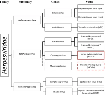
Figure 1. Taxonomy of human cytomagalovius (HCMV) in relation to murine cytomegalovirus (MCMV).
Human herpesvirus (solid lines) in Family Herpesviridae as they relate to HCMV and MCMV (dotted lines). Information from [20][21].
Family Subfamily Genus Virus
He
rpes
virida
e
Alphaherpesvirinae SimplexvirusHerpes simplex virus type 1
Herpes simplex virus type 2
Varicellovirus Varicella-zoster virus (VZV)
Betaherpesvirinae
Cytomegalovirus Human cytomegalovirus
(HCMV)
Muromegalovirus Murine cytomagolvirus
(MCMV) Roseolovirus
Human Herpesvirus 6 (HHV6)
Human Herpesvirus 7 (HHV7)
Gamaherpesvirinea
Lymphocryptovirus Epstein-Barr virus (EBV)
Figure 2. Cytomegalovirus Structure.
Representation of the elements in an HCMV/MCMV virion. From [22].
Figure 3.Comparison of HCMV and MCMV genome.
Boxes: inverted repeats. TRL: long terminal repeats. UL: long unique sequence. IRL: long
internal repeats. IRS: short internal repeats. TRS: short terminal repeats. Modified from
[23]. MCMV IRS not shown [24].
Transmission of CMV infections is similar across affected species and can occur with
direct contact of infected bodily fluids (saliva/urine), sexual activity, blood transfusions/organ
transplants, breast feeding and in-utero transfer [58]. After viral transmission, glycoproteins
initiate viral binding and entry in host cells [57]. Lytic replication, as reviewed in Figure 4, can
then take place during initial infection. In immunocompetent individuals, the immune response is
adequate to stop active infection and the production of virion progeny [59]. Subsequently the
virus enters a period of latency in the genome of myeloid progenitor cells as provirus where it HCMV
continues to be able to replicate its genome during host cell replication [59]. During
inflammatory events and the resulting differentiation of the infected progenitor cells into
macrophages (Mϴ) and dendritic cells (DC), the viral genome is activated by the same
regulatory signals acting on Mϴ/DC which leads to the production of certain viral replication
promoters (such as immediate-early) and proteins that down-regulate antigen presenting cell
(APC) activity [59][60]. In healthy individuals, robust APC activity is enough to overwhelm
viral attempts of re-activation. In non-healthy individuals who have immunosuppression, the
virus can take advantage of the reduced immune activity and overwhelm host cells defenses
leading to re-activation of viral lytic activity [59].
Figure 4. Viral Replication of HCMV/MCMV.
Viral coat proteins bind to cell membrane, virus invades, uncoating, entry into the
nucleus, nuclear genetic replication machinery takeover, viral genome replication, capsid assembly, vesicle bound, exocytosis and recoating. From [25].
In humans, AIDS related HCMV retinitis results in retinal tissue destruction that
ultimately leads to blindness. Before the advent of ART (anti-retroviral therapy), up to 30% of
AIDS patients lost their vision this way; unfortunately this is still a significant risk for those who
retinitis can occur outside of AIDS in other immunosuppressed populations such as in donor
organ/bone marrow recipients (15.2% out of non-AIDS HCMV retinitis cases) [1], those
suffering from rheumatological conditions (19.1%), individuals of advanced age (33.1%) and
even healthy individuals with localized immunosuppression (~5%) [3][61]. Regardless of the
mode of immunosuppression, clinical presentation of HCMV retinitis appears to be similar. Not
all cases are symptomatic bus usually there are initial visual disturbances in the form of floaters
and some loss of vision to the periphery or even more central areas of sight. Some reduction in
overall visual acuity may also be reported. During examination, aside from retinal necrosis, other
secondary pathologies may present such as macular oedema, optic nerve inflammation
(papillitis) and retinal detachment [61][62]. As the retinitis progresses, the field of visual loss
progressively increases until blindness results [1][62]. Even minimal inflammation and
destruction of the retina proves problematic due to the relative permanency of damage and the
consequent quality of life issues that may cause. For those individuals who are not under an
effective treatment, the effects of HCMV retinitis are far more concerning.
The retina itself is located at the back of the eye, encompassing nearly the whole interior
of the globe and creating a wide field of vision. It consists of several cellular and connecting
layers as described in Figure 5 with the ganglion layer situated facing into the eye and the
photoreceptors at the back. As light enters the eye, it passes to the back of the retina activating
the rods and cones of the photoreceptor cells which then signal the bipolar cells in another layer
(via synaptic activity). From there the signal is passed to the ganglion cells where they act as the
neuronal interface between retinal visual signaling and the image processing of the brain [29]. Of
non-retina areas may be of interest. The RPE (non-retinal pigment epithelium) located behind the
[image:19.612.149.463.140.380.2]photoreceptors is responsible for nourishing the retinal cells and degrading damaged ones [63].
Figure 5. The structure of the retina.
Nuclear layers: Ganglion cell layer, inner nuclear layer (bipolar cells) and the outer nuclear layer (nucleus containing portion of the photoreceptor cells). These are the layers that are counterstained with DAPI. From [29].
Behind it, is the choroid which is the vasculature structure providing blood supply and bringing
with it myeloid cells and their progenitors – potentially primed for inflammation and/or infected
with virus [63]. During advanced HCMV retinitis, full-thickness retinal necrosis can be seen
upon examination as large patchy white areas with interspersed hemorrhaging (Fig. 6D as
compared to Fig. 6C). There may also be widespread secondary inflammation in other parts of
Figure 6. Retinal folding and full retinitis.
A) Ocular histopathology of MAIDS related MCMV retinitis at 8 days post infection (dpi) showing both retinal folding and full retinal necrosis. We use retinal folding as a characteristic of 6 dpi however, there can still be some retinal folding by 10 dpi as
retinitis is patchy. B) Non-diseased/healthy mouse retina C) Non-diseased/healthy human retina D) Similar ‘patchy’ disease pathology is seen in human patients with AIDS related HCMV retinitis. From Dr. Dix, Dr. Cousins, Bascom Palmer Eye Institute, Miami, FL and Dix Lab.
A
B
Understanding disease pathogenesis of HCMV induced retinitis during opportunistic
pathogenicity may allow for better understanding of the cellular mechanisms at play in cell death
and tissue destruction. In order to explore these concepts, a murine model was used to replicate
AIDS-related HCMV retinitis in-lab. As previously discussed, HCMV is closely related to other
species-specific CMV viruses including murine CMV (MCMV) (Fig. 1)(Fig. 3) [22][23][57]. To
induce retroviral immunosuppression similar to AIDS, a murine leukemia cocktail is used
(MAIDS) [1][4]. Mice are infected with MAIDS at 4 weeks of age and allowed to progress to
systemic immunosuppression. Then 10 weeks later mice are directly injected subretinally with
MCMV (Fig. 7) to prompt MCMV retinitis in this model. Lastly, in order to evaluate the
progress of retinal destruction over time, groups of mice are euthanized at different time points
Figure 7. Visual Representation of the site of delivery in subretinal injection.
2 µl injection containing MCMV in MAIDS mice in the subretinal space. Mice under full anesthesia during the procedure. Modified from [26].
(3, 6 and 10 days post infection - dpi) and eyes are harvested. MAIDS-related MCMV retinitis
eyes are phenotypically characteristic and distinguishable between the different time points. This
is especially clear when observing 6 dpi and 10 dpi. Retinal folding is associated with 6 dpi with
full necrosis expected at 10dpi; although because retinitis is patchy in how it spreads, there can
Fig. 6B). In PCR work, mRNA was usually found to be more highly expressed at day 6 and
dramatically decreased by day 10 [1], likely due to the drop in retinitis activity at the endpoint of
full retinal necrosis. Day 3 mRNA and protein data was usually not highly stimulated.
Ultimately, these time points were picked based on prior data in order to better define the steps
of disease progression. In that same thought, MAIDS-10 (10 weeks MAIDS duration) mice are
used because previous work showed that MCMV could not induce any retinitis without full
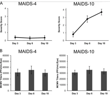
immunosuppression being attained by MAIDS over the course of at least 10 weeks [1][12]. Viral
titer remains the same when compared to mice unsusceptible to MCMV retinitis (MAIDS-4
mice) but only MAIDS-10 mice see progressively worsening retinitis at 6 dpi and 10 dpi (Fig. 8)
[1]. All data shown going forward was collected from MAIDS-10 mice unless otherwise
specified.
The mechanistic process leading to the dramatic destruction of the retina in both HCMV
and MCMV retinitis is not yet fully elucidated. Findings using the MAIDS-related MCMV
model in C57BL/6 mice seem to indicate that CMV infection and replication is not solely
responsible for retinal tissue destruction [1] and that while cell death signaling can be variable it
is a large purveyor of retinal degeneration [7][1]. In MAIDS-10 mice, it was seen that severe
retinal destruction occurred in tandem with significant up-regulation of apoptotic molecules -
tumor necrosis factor alpha (TNF-α), TNF receptor 1 and 2, cleaved caspase 8, cleaved caspase
3, TNF-related apoptosis-inducing ligand (TRAIL), TRAIL receptor (TRAIL-R), Fas, and Fas
ligand [1]. However, TUNEL assay revealed only partial contribution by apoptosis to cell death
(only ~8% and ~ 4% at 6 and 10 days post infection respectively); overall, apoptosis is
associated with limited cell death activity compared to other cell death pathways in many retinal
Figure 8. Comparison of MAIDS 4 and MAIDS 10 MCMV infected mouse eyes.
A) Severity of retinitis: MAIDS 4 eyes showed low levels or retinitis in comparison to the more fully developed MCMV disease pathophysiology in MAIDS 10 mouse eyes. B) There was no significant difference in MCMV titer levels between MAIDS 4 and
MAIDS 10. These experiments were performed by Dr. Hsin Chien under the direction of Dr. Richard D. Dix, Georgia State University [1].
regulating specific coordinated cell death activity – turning off one pathway results in turning on
another [2]. Identification of mRNA from other cell death pathway molecules seems to be
indicative of both necroptosis (Fig. 9) and pyroptosis (Fig. 10) activity [1]. Given that the
involvement of both has been documented in other retinal degenerative diseases [2][5][6], it
could be postulated that they also play a role in MCMV retinitis alongside apoptosis.
Necroptosis is an alternative programmed pathway induced when apoptosis is inhibited
by caspase blocking activity [8]. As a result, necroptosis shares the same receptor ligand binding
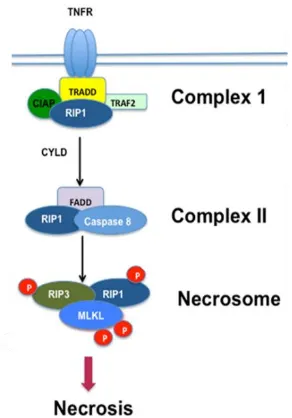
Figure 9. Necroptosis cell death signaling pathway.
Schematic of the programmed cell death pathway with associated molecules. Modified from [11].
Figure 10. Pyroptosis cell death signaling pathway.
Active caspase 1 and caspase 11 cleave GSDMD to initiate cellular death and inflammation by pyroptosis. From [27].
TNFR, Fas, TRAIL or TLRs induces the intracellular formation of a death complex with FADD,
[image:24.612.179.430.344.581.2]serine/threonine-protein kinase 1 (RIPK1). Cellular stimuli that upregulate the deubiquitinating
enzyme CYLD or the cellular inhibitor of apoptosis protein 1 (cIAP1) cause RIPK1 to dissociate
from the first complex in order to form a second death complex with uncleaved caspase 8 and
FADD [10][2]. From this point on, the two pathways diverge dependent on caspase 8 activity.
Necroptosis signaling is prompted when Fas-associated protein with death domain (FADD)
cleavage of pro-caspase 8 is inhibited via the competitive formation of uncleaved caspase 8 /
cellular FLICE inhibitory protein (cFLIPSorR) heterodimers [8]. After which, RIPK1 recruits
RIPK3 into the necrosome complex through their RIP homotypic interaction motif (RHIM) domains where autotransphosphorylation of both Ser227 also occurs via each other’s kinase
domain [10][2][64]. Lastly, the now active RIPK3 then recruits and phosphorylates mixed
lineage kinase domain-like (MLKL). Active MLKL forms an oligomer complex with other
active MLKL before it migrates to the plasma membrane and in a process still not yet fully
understood, ruptures the membrane via pore formation [11][64]. This results in a necrotic cell
death in which the release of damage-associated molecular patterns (DAMPs) trigger a highly
pro-inflammatory response [9]. A brief schematic of this signaling pathway is shown in Figure 9.
This is a well regulated form of necrosis controlled by a caspase independent signaling pathway – as opposed to passive necrosis, which occurs due to cell damage and a subsequent failure to
maintain homeostasis with the result being cell swelling and eventual membrane disruption
before death (Fig. 11)[5][28].
Pyroptosis on the other hand is similar to both apoptosis and necroptosis but exhibits its
own specific pathway. Similarly to apoptosis, it is a caspase dependent mode of cell death that
generally occurs in response to pathogen infection. Furthermore, it triggers an inflammatory
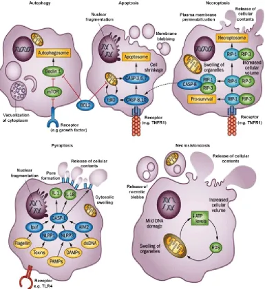
Figure 11. Types of cell death outcomes.
Schematic representation on cell destruction in relation to type of cell death. Programmed cell death: Apoptosis, Autophagy, Necroptosis and Pyroptosis. Non-programmed cell death: Necrosis/Oncosis. Of interest to this study Necroptosis and Pyroptosis. From [28].
associated molecular patterns (PAMPs) are exhibited intracellularly due to cellular infection [2].
Pattern recognition receptors (PRRs) such as nod-like receptors (NLR) and absent in melanoma
2 (AIM2) protein detect these PAMPs and form oligomer complexes that become caspase
activating inflammasomes upon the recruitment of apoptosis-associated speck-like adaptor
inflammasomes at the adaptor protein, ASC, and cleaved into active caspase 1. Final pyroptosis
activity is then initiated by caspase 1 cleavage activity of both gasdermin D (GSDMD) and the
inflammatory cytokines pro-interleukin (IL)-1β and pro-IL-18. Activated GSDMD proteins
oligomerize at the cell membrane and form pores which serve as an avenue of cytokine release
and ultimately cause cell swelling and lysis [27][2]. Thus pyroptosis is also an inflammatory
form of cell death but unlike necroptosis, instigates a more specific immune response by means
of extracellular cytokine signaling. A brief overview of both the canonical and non-canonical
pathway can be seen in Figure 10. As can be seen in the non-canonical pathway, caspase 11 can
also cleave and activate GSDMD but cannot stimulate cytokine release [27][2]. However,
pyroptotic cell death can still occur due to GSDMD disruption of the plasma membrane [2].
Both necroptosis and pyroptosis pathways are potent forms of cell death and a
pro-inflammatory state [2][6][9][56]. This might help explain the devastating levels of retinal
destruction and the speed with which it spreads. In contrast, apoptosis cell death does not signal
for cell death in surrounding cells nor does it seem capable of inducing inflammatory pathologies
known to co-occur with CMV retinitis [1][2][8][10]. In past studies, this lab has already shown
that necroptosis associated RIPK1and RIPK3 mRNA are expressed in MAIDS-related MCMV
retinitis eyes. RIPK1 mRNA is significantly upregulated with the greatest increase at 6 days post
MCMV infection (dpi); RIPK3 by comparison exhibits minor but clear upregulation as well [1].
Additionally, the mRNA of several pyroptosis associated molecules were previously found to be
upregulated in MAIDS-related MCMV retinitis as well [1]. Caspase 1, caspase 11, GSDMD, IL-1β and IL-18 all displayed significant upregulation by 6 dpi and dropped again in mRNA
expression by 10 dpi [1][performed by Jessica Carter under the direction of Dr. Richard D. Dix,
programmed cell death via necroptosis and pyroptosis play roles in MCMV induced retinal cell
death.
Therefore, we will expand on the central hypothesis that necroptosis-associated
molecules RIP1, RIP3, and/or MLKL and pyroptosis-associated molecules caspase 1, caspase
11, GSDMD, IL-1β and/or IL-18 are stimulated intraocularly during the pathogenesis of
MAIDS-related MCMV retinitis. Specific Aim 1 will investigate whether necroptosis cell death
is involved in the retinal destruction observed in the experimental model. Although mRNA for
necroptosis-associated molecules, RIPK1 and RIPK3, were found to be stimulated during
MAIDS-related MCMV retinitis, the full extent of necroptosis in MCMV retinitis has yet to be
elucidated. Therefore, the hypothesis of specific aim 1 postulates MLKL will be upregulated in
MCMV infected eyes and that all tested necroptosis molecules RIPK1, RIPK3 and MLKL will
be stimulated in retinal layers displaying MCMV retinitis in MAIDS-10 mice. This will be tested
by assessment of MLKL mRNA via real-time RT-PCR quantitative analysis and RIPK1, RIPK3,
MLKL protein via IHC staining and western blot in the murine model eyes. All data will be
evaluated by comparison with non-infected controls. Specific Aim 2 will then follow the first in
investigating the potential role of pyroptosis cell death in retinal tissue destruction in the
experimental model. Multiple pyroptosis-associated molecules were found to have increased
mRNA expression in MAIDS-related MCMV retinitis eyes but pyroptosis activity in the form of
protein expression is still ongoing. Hypothesis of specific aim 2 postulates that pyroptosis
molecules caspase 1, caspase 11, GSDMD, IL-1β and IL-18 will be stimulated in retinal layers
displaying MCMV retinitis in MAIDS-10 mice. This will be tested by protein analysis of caspase
1, caspase 11, GSDMD, IL-1β and IL-18 via IHC staining in the murine model eyes. All data
2 MATERIALS AND METHODS
2.1 Cell Lines
All cell lines used were for the cultivation of viral stocks to be used for murine in vivo
infection and disease initiation.
For MAIDS induction as a murine retroviral model, SC-1 fibroblast (ATCC #CRL-1404)
and LP-BM5 – MuLV infected SC-1 - (AIDS Research and Reference Reagent Program,
Germantown, MD) were utilized. SC-1 cells with and without murine leukemia viral (MuLV) infection were grown in Dulbecco’s modified eagle media (DMEM, Corning Life Sciences) with
10% fetal bovine serum (FBS)(Atlas Biologicals), 24 mM L-glutamine, 1%
penicillin/streptomycin, 0.1 mg/mL gentomicin.
MCMV stocks for sub-retinal injections were grown and titered in mouse embryonic
fibroblast (MEF) (ATCC #SCRC-1002) cells. Cells were maintained in DMEM with 15% FBS,
4 mM L-glutamine, 1% penicillin/streptomycin, and 0.1 mg/mL gentomicin.
2.2 Viruses
LP-BM5 (MuLV infected SC-1) and SC-1 cell lines were thawed and grown in SC-1
media until confluent. Established cultures of the two cell lines were mixed in a 1:1 ratio and
separated in 16 T-150 flasks with 12 mL of SC-1 growth media. After 6 days, the cell
monolayers in the flasks were gently scraped and transferred along with media into 150 mL
conical tubes and stored at -80°C until use. For injection, MuLV retroviral cocktail was thawed
and centrifuged to dispose of cellular debris.
MCMV stock, for use in subretinal injections, was propogated in BALB/c
(Harlan/Envigo) mouse salivary glands [12]. 20 mice were injected intraperitoneally (IP) with
infection (dpi), the mice were euthanized and the salivary glands harvested. Salivary glands were
homogenized in 1.5 mL DMEM with 10% fetal bovine serum and centrifuged before aliquoting
the supernatant into 0.5ml and storing in liquid nitrogen. Each viral stock was titered via plaque
assay in advance of sub-retinal injections.
2.3 Animals
Adult female wild-type C57BL/6 (Jackson Laboratory) mice of 2 weeks of age and adult
female wild-type BALB/c mice (Harlan/Envigo) of 8-12 weeks of age were acquired and acclimatized in Georgia State University’s Animal Facilities to the following husbandry
conditions: 5 mice to a habitat, alternative 12-h light-dark cycles, an enrichment article and free
access to food and water. Experimental procedures followed guidelines from the National
Institutes of Health (NIH) and the Association for Research in Vision and Ophthalmology
Resolution (ARVO) on the Use of Animals in Research. Experimental protocols received prior
approval by The Institutional Animal Care and Use Committee (IACUC) at Georgia State
University.
Induction of MAIDS: MAIDS was induced in groups of 4 week old wild-type C57BL/6
mice via intraperitoneal (IP) injection of 1 mL of MuLV retroviral cocktail (LP-BM5/SC-1
mixture at ~103 PFU). Infection was allowed to progress for 10 weeks (MAIDS-10 mice) post
injection before the next experimental step (mice aged 14 weeks); previous work indicated such
late stage MAIDS mice are more susceptible to retinitis than earlier stages (MAIDS 4 mice)
[1][12].
Subretinal MCMV Injections: Systemic MCMV does not induce retinitis despite viral
presence in the choroid and RPE [13]; however, it was found that directly injecting the viral load
AIDS related HCMV retinitis [12][14]. Therefore, a subretinal route of infection is a suitable
murine model for representation of the human disease.
For subretinal MCMV infection, the previously described groups of MAIDS-10 mice
underwent a series of steps to prepare them for subretinal injections. Both eyes for each mouse
were dilated over the course of 3 rounds of atropine and tropicamide ophthalmic drops.
Following the final round of eye drops, anesthesia procedures were initiated and mice were given
their first injection of 0.1 mL xylazine (1.72 mg/mL) intramuscularly (IM) in a hind leg. After
each mouse within a group received the first injection, a secondary injection of 0.1 mL
acepromazine (0.28 mg/mL) was given in the alternative leg. Induction into full anesthesia was
achieved with a final IP injection of 0.1–0.2 mL ketamine (8.58 mg/mL). Upon complete
non-response to stimuli, mice were given a final round of phenylephrine ophthalmic drops before
being subretinally (supraciliary) injected with 2µL of MCMV (prepared as previously discussed)
in the left eye and 2µL of DMEM media in the right eye (contralateral injections). The media
injected eye served as a control for the MCMV injected eye within one individual mouse due to
the findings that MCMV does not pass from the infected to the non-infected eye in the durations
under consideration here [15]. Groups of mice were euthanized at 3, 6 and 10 days post MCMV
infection via a two step process: isoflurane induced anesthesia followed by cervical dislocation.
Subsequently, the eyes were harvested and stored depending on how they would be later
analyzed for necroptosis and pyroptosis associated molecules; RNAlater (whole eye mRNA
analysis via real-time RT-PCR), frozen in liquid nitrogen (whole eye protein analysis via
Western Blot), or stored in 10% buffered formalin solution at 4°C (eye section protein staining
2.4 Real-Time RT-PCR
Whole MCMV-infected eyes and contralateral media injected eyes (control) at 3, 6, and
10 days post infection were stored in RNAlater solution (Ambion) at −80°C . For analysis, each
eye was thawed and homogenized in 1 ml of TRIzol reagent (Invitrogen Life Technologies).
RNA was extracted from each homogenized sample following the PureLink® RNA Mini Kit
total RNA purification system (Invitrogen Life Technologies) protocol. RNA concentration for
each sample was quantified via SmartSpec 3000 spectrometer (Bio-Rad Laboratories) and
equalized between samples. Samples were then stored at -80°C until needed.
Samples were thawed prior to undergoing the steps involved in real-time reverse
transcriptase polymerase chain reaction (RT-PCR). Extracted RNA was used to synthesize
complementary DNC (cDNA) using the SuperScriptTM III first-strand synthesis system
(Invitrogen) and protocol. Mouse specific MLKL and GAPDH primers obtained from QIAgen
(Valencia, CA) and SYBR green PCR master mix (Applied Biosystems) was used to help
determine levels of MLKL mRNA transcripts in each sample during thermocycling using a ABI
Prism 7500 real-time PCR instrument with sequence detection software (Applied Biosystems).
System parameters for cycling was set up for 10 min at 95°C, followed by 40 cycles consisting
of 15 s at 94°C, 31 s at 55°C, and 35 s at 70°C. MLKL mRNA transcript cycles to threshold
(CT) were determined for MCMV infected and contralateral control eyes compared to
constituently expressed GAPDH.
2.5 Immunohistochemistry (IHC) Fluorescent Staining
Eyes were collected at 6 and 10 days post MCMV infection and immediately fixed with
sections and every 6th section embedded in paraffin on slides by the Pathology Department of the
Emory Eye Center.
Prior to immunohistochemical staining, slides were soaked in Xylene substitute (Sigma-
Aldrich, St. Louis, MO) for 3 5 minute intervals to remove paraffin. Then sections were
rehydrated in decreasing 70% ethanol/PBS concentrations (95%, 75%, 50%) for 15 seconds
each, washed in PBS for 30 seconds, treated with 10 mM sodium citrate retrieval solution for 10
minutes, washed with PBS for another 5 minutes, and finally blocked with 5% normal goat
serum containing 0.2% Triton X-100 (Ambion/ThermoFisher) for 30 min at room temperature.
Another wash in PBS for 3 intervals of 5 minutes prepares the sections for primary antibody
application. Sections were incubated with primary antibodies specific for necroptosis and
pyroptosis specific molecules overnight at 4°C. Necroptosis: rabbit anti-mouse RIPK1 (1:200)
(Antibodies Online, Atlanta, GA), rabbit anti-mouse RIPK3 (1:200) (Antibodies Online, Atlanta,
GA), rabbit anti-mouse MLKL (1:200) (Abcam, Cambridge, MA). Pyroptosis: rabbit anti-mouse
caspase-1 (1:200) (Abcam, Cambridge, MA), rabbit anti-mouse caspase 11 (1:200) (Abcam,
Cambridge, MA), rabbit mouse GSDMD (1:200) (Abcam, Cambridge, MA), rabbit
anti-mouse IL-1β (1:200) (Abcam, Cambridge, MA), rabbit anti-anti-mouse IL-18 (1:200) (Abcam,
Cambridge, MA). A matched isotype rabbit IgG was used as a negative (1:200) (Abcam,
Cambridge, MA) for all primary antibodies. After incubation, three 5-min washes with PBS were
done prior to secondary staining. Retinal sections subsequently were incubated at room
temperature in the dark for 1 h with the secondary antibody conjugated goat anti-rabbit Cy3 (red)
(1:100) (Jackson ImmunoResearch) for fluorescent visualization of each molecule in the retina.
After secondary antibody incubation, sections were washed in PBS for three 5 minute and then
Laboratories) to counterstain nuclear material. Sections were inspected and photographed by
fluorescence microscopy (Nikon Eclipse). ImageJ was used to background correct images.
Presence of each of the previously mentioned necroptosis and pyroptosis-associated molecules
was qualitatively assessed by comparison of control (media injected) and MCMV infected eyes.
2.6 Western Blot
Western blot was used to evaluate levels of necroptosis-associated molecules present in
MCMV infected whole eyes compared to contralateral media injected (control). Eyes were
collected at 3, 6 and 10 days post infection (dpi), eyes harvested and stored in liquid nitrogen
until needed. Eyes were used individually, representing one group each per blot.
In preparation for western blot, eyes were thawed before protein extraction. 2X extraction
buffer was prepared using a protease inhibitor (complete Mini, EDTA-free) (Roche) in
phosphate-buffered saline (PBS) (Sigma, St. Louis, MO). Eyes were first homogenized in 0.5mL
cold extraction buffer and then spun at 13,000 RPM for 1 minute. 130µl of the supernatant was
transferred and added to equal amounts of cold DMEM for later viral quantification. Protein concentration of each sample was quantified following the “Microtitier Plate Protocols” of the
protein assay kit used (Bio-Rad –Bradford). The spun down pellet of the remaining sample
portion was resuspended and an equal volume of 2x extraction buffer was added before storing at
-20⁰C overnight. Samples were pulse sonicated following protein extraction steps and then centrifuged at 5,000xg for 5 minutes at 4⁰C. Protein concentration of each sample was equalized
to one another via PBS dilution based on the protein quantification results. Finally, 5x sample buffer was added to each sample in a 1:4 ratio before boiling at 99⁰C for 15 minutes (PCR
A Western blot protocol was performed to evaluate MLKL protein levels. Samples were
subjected to SDS polyacrylamide gel electrophoresis (SDS-PAGE) and transferred to a
nitrocellulose membrane using a Power Blotter Transfer Stack (ThermoFisher Scientific,
Waltham, MA, USA). Samples were blocked in 5% non-fat skim milk and probed for primary
antibodies specific for rabbit anti-mouse MLKL antibody (1:500, Abcam, Cambridge, MA) and
rabbit anti-mouse GAPDH (1:1000, Sigma-Aldrich, St. Louis, MO). Goat-anti-rabbit IgG used
(heavy plus light chains [H+L]) (1:2000, ThermoFisher) conjugated with horseradish peroxidase
was used as a secondary antibody. An enhanced chemiluminescent (ECL) horseradish peroxidase
(HRP) substrate (SuperSignalTM West Pico Plus - ThermoFisher) was then applied to the
membrane for 5 minutes before exposure to HyBlot film (Denville, Holliston, MA).
2.7 Statistical Analysis
Statistical analysis of protein and mRNA levels between MCMV infected and media
injected control eyes was performed where necessary with P values ≤0.05 considered significant.
Quantitative data from real-time RT-PCR of MLKL mRNA values from each MCMV infected
eye compared to the contralateral media injected eye from the same mouse was determined by
the 2-ΔΔCT method. Statistical analysis was done using student t-test yielding P values for each
group (* p<0.05, ** p<0.01). A standard error of deviation (values from at least 3 mice per
group) was also calculated for each group and labeled with an error bar. Graphed results
demonstrate mean fold change of mRNA levels in MCMV infected mice compared to that of the
control eyes.
Protein levels of MLKL were determined via western blot. Band density was quantified
using ImageJ and graphed to visualize protein level differences in MCMV infected eyes
3 MLKL EXPRESSION IN WHOLE EYES DURING MCMV RETINITIS
The final and critical determinant step of necroptosis signaling is the activation and
oligomerization of MLKL [11][64][65]. As discussed earlier, real-time RT-PCR work was
previously done for RIPK1 and RIPK3 to evaluate the potential involvement of these upstream
necroptosis-associated molecules in MAIDS-related MCMV retinitis [1]. However MLKL was
not investigated at that time because little was known then about MLKL and its role in the
necroptosis signal transduction pathway [65]. Current literature has brought to light MLKL’s
significant involvement in necroptosis as well as suggests other potential functions. Here the aim
was to determine if MLKL was stimulated in MCMV-infected eyes of MAIDS mice and thus
potentially confirm or refute the possibility of necroptotic cell death in MAIDS-related MCMV
retinitis.
Real-time RT-PCR is widely used to determine mRNA levels of intra- and extracellular
molecules in tissues and is generally done before protein assays. As an initial step in the
production of cellular products (illustrated in Fig. 12), transcription is indicative of cellular
driven regulatory activity aimed at promoting particular gene expression [16]. Quantification of
mRNA thus allows for an estimation of downstream protein production activity. Through the
process of mRNA amplification by RT-PCR (Fig. 13), even minute amounts of a given mRNA
transcript can be accurately detected in real-time [16][18]. On the other hand, western blots and
IHC provide important insights into final protein expression of a gene but are not as accurate or
precise due to the potential for antibody and antigen variability [17][32]. They are also not
necessarily able to gauge the total effect an experimental state has on gene expression [17] which
may be integral for identifying the likelihood regulatory interplay by other molecules of interest
and prevents the wasting of valuable time on subsequent protein work should mRNA be absent.
Hence why preliminary probing for necroptosis-associated RIPK1 and RIPK3 mRNA levels
were previously done by Chien et al. (2012) (Fig. 14). With the postliminary identification of
MLKL as a necroptosis participant by scientific literature, its mRNA expression in
MAIDS-related MCMV infected eyes was therefore investigated.
[image:37.612.93.332.265.421.2]
Figure 12. Central Dogma of molecular biology.
Flow of genetic information from DNA to RNA to Protein.
Replication
Transcription
Translation
DNA
mRNA Replication
Protein
*Nucleus*
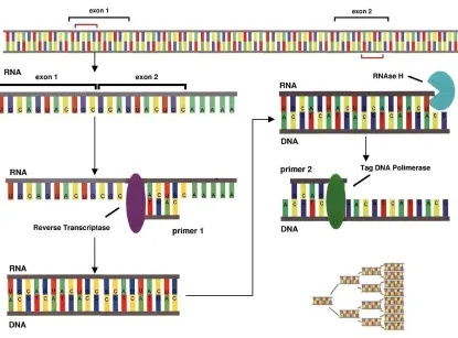
Figure 13.Reverse Transcriptase Polymerase Chain Reaction (RT-PCR).
Schematic representation of the reverse transcriptase and polymerase chain reaction used to amplify mRNA (which is quantified via real-time methods for this study). Modified from [18].
Figure 14. RIPK1 and RIPK3 mRNA expression in MAIDS-10 subretinally infected eyes compared with media control eyes.
[image:38.612.78.510.495.648.2]quantified by real-time RT-PCR and statistically analyzed by student T test with P values ≤0.05 being significant **p≤0.001, *p≤0.05 (n=5). (A) RIPK1 mRNA shows a
significant upregulation at 6 dpi and downregulation by 10 dpi. (B) RIPK3 mRNA shows downregulation from 6 dpi to 10 dpi. These experiments were performed by Dr. Hsin Chien under the direction of Dr. Richard D. Dix, Georgia State University [1].
In the case of mRNA expression, subsequent protein assays are necessary to assess true
protein levels as mRNA does not always correlate with protein expression [37][68]. This is due
in part to the adaptable nature of gene expression regulation; both mRNA and protein can be
modified in a number of ways that may ultimately result in alternative products or premature
degradation [66][67][104]. Therefore to confirm any mRNA based predictions of protein production, it is necessary to quantify the tissue’s protein product. In comparison to mRNA
quantification via real-time RT-PCR and the localization of protein via IHC, westerns can
quantify relative total protein through the use of specific antibody binding and signaling (Fig. 15
antibody schematic) [68]. Thus, it is a well-established method for providing an insight into
overall cellular protein production/processing activity throughout the tissue of interest.
Suggested as the key player in the necroptosis signaling pathway [65], overall MLKL activity in
the eye was of interest. MLKL relative total protein in whole eyes as compared to mRNA
expression also in whole eyes was done by western blot.
Figure 15. Schematic representation of an antibody.
‘antigen/primary Fc’ by its Fab and is conjugated by a fluorescent tag on its Fc. From [30].
3.1 MLKL mRNA Expression in MCMV Infected Eyes
Figure 16. MLKL mRNA expression in MAIDS-10 subretinally infected eyes compared with media control eyes.
Days 3, 6 and 10 post MCMV infection are shown. Values represent mRNA fold change of MCMV infected eyes compared to media injected control eyes. Expression of mRNA quantified by real-time RT-PCR and statistically analyzed by student T test with P values ≤0.05 being significant **p≤0.001, *p≤0.05. n=3-5
As was previously mentioned, apoptosis was found to not be the sole progenitor of cell
death in MCMV retinitis [1]. In evaluation of other possible cell death pathways, RIPK1
upregulation could be indicative of either apoptosis or necroptosis cell death signaling and the
minor upregulation of RIPK3 alone was not enough to significantly confirm necroptosis (Fig. 14)
[1]. MLKL involvement was thus investigated in order to clarify the suggestive but ambiguous
nature of this previous RIPK1 and RIPK3 data. During programmed necroptotic cell death, MLKL acts as the ‘executioner’ in the final steps - initiating and ultimately causing cell death
[2][64][65][79]. This final process involves the activation of MLKL by phosphorylation which
membrane; resultant selective pore formation and eventual cell lysis is characteristic of
necrosis-like death [43][11][64][65]. Therefore, in support of RIPK1/RIPK3 mRNA findings,
upregulation of MLKL mRNA was expected to occur during MCMV retinitis.
To test this, eyes from MAIDS model mice were taken at days 3, 6 and 10 days after
MCMV and contralateral mock (media) infection in accordance with earlier selected time points.
Real-time RT-PCR was run on homogenized whole eyes with a commercial primer
complementary for MLKL mRNA sequence; this was repeated three times with groups of three
to five animals each time. Results from MCMV infected eyes were normalized using results
from mock infected eyes and graphed as mRNA fold change; overall findings were significant
and repeatable (Fig. 16). MLKL mRNA was increased 3 dpi by ~36 fold in MCMV infected eyes
when compared to the media injected eyes, and was highly significant with little variability
between samples. The increase in mRNA expression (~38 fold change) by day 6 was also found
to be significant. The mRNA expression seen for day 10, while not significant due to sample
variability, displays a noticeable decrease. This indicates a general trend of high day 6 expression
that seems to greatly abrogate by day 10 and has been seen both in previous and subsequent data
[1]. In comparison of mRNA data for the three necroptosis-associated molecules investigated
(Fig. 14) (Fig. 16), it is pertinent to evaluate their combined results both in terms of regulation
and sequential signaling along the necroptosis pathway.
Discussed beforehand, we know that RIPK1 is the first required molecule to initiate
classical necroptosis signaling [2][11]. However it is also involved in several other pathways and
thus highly regulated with specific responses likely being conserved between individuals
[10][2][43][69][103]. Perhaps for that reason, mRNA values remain tightly clustered and
result from a reduced need for strict RIPK3 regulation due to its limited roles and the
unlikelihood of non-specific signaling [2]. Furthermore, it’s signaling activity is largely transient
[79] and indirectly controlled by other activating/deactivating molecules already under
tight/specific regulation such as caspase 8, RIPK1 and MLKL [43][65][72][75][80]. A
proteasome system has been suggested as a method for direct RIPK3 regulation; variable
poly-ubiquitination of different lysine residues by several non-specific ubiquitin ligases and the
potential for proteasome degradation could result in differential outcomes that vary between cells
[10][77][81]. Even at basal levels, necroptosis signal transduction can still occur as a few RIPK3
can transiently activate many more MLKL [2][10][79]. Even then, total absence of RIPK3 is
relatively well tolerated as can be seen with RIPK3 knockout mice [76]. Overall RIPK3 may be
an incredibly useful alternative for inducing necessary signaling in a compensatory but
non-essential capacity; if so, this could indicate there is less selective pressure on RIPK3 expression
between cells and individuals. Increased regulatory control is likely seen again though with the
final effector of necroptotic cell death, MLKL [11][64]. It is crucial to necroptosis [65][78] and
knocking it down/blocking it inhibits necrosis characterized death [65][77]. Current research also
seems to indicate the possibility of alternative MLKL activation and functions [33][70][71].
Taken together it could be assumed that MLKL is integral to cellular function but potentially
harmful if disregulation were to occur. Variation in expression however is still observed to
increase with each successive time point with loss of significance by day 10. Unfortunately a lot
is still unknown about MLKL [65], so there could be several reasons for the trend in its mRNA.
It could simply be that the rate of necroptosis progression differs between individuals, perhaps in
Despite any differences in mRNA expression between individuals, there is still a very
clear overall trend between day 6 and 10. Initial mRNA upregulation differ between RIPK1,
RIPK3 and MLKL but likely for some of the reasons discussed above. RIPK1 is expressed in
healthy cells at higher amounts due to its multi-functionality [2][11][69][103]; during early
disease there may not be a large change in expression between infected and non-infected eyes
due to the abundance of readily available RIPK1. When infection and/or inflammation reach
critical levels though, cells likely respond in kind in order to clear the infection such as seen at
day 6 [2][43]. This spike in mRNA expression may be due to the combined effect of necroptosis
and other RIPK1 mediated pathways sent into transcriptional overdrive in order to increase
activity of certain pathways and to replenish the rapid turnover of RIPK1 [72]. In regards to
RIPK3 initial upregulation, there is not yet a confirmed consensus on the possible expression of
constitutive RIPK3 [77] or the lack thereof [70]. What is known is that low levels of RIPK3 are
still able to efficiently signal for necroptosis [72] [2][10] and it plays a part in the cross-talk
between cell death pathways, allowing for the cell to respond to different scenarios via
alternative immune/cell death signaling [2][76]. Necroptosis is also well known as an anti-viral
response [65]; transcriptional upregulation of RIPK3 mRNA at day 3 might be indicative of
cellular prep for necroptosis upon apoptosis inhibition [1][2]. Expression decreases by day 6 but
is still noticeably higher than control. MLKL on the other hand is constitutively expressed at
basal levels [71] but is needed in greater amounts in order to oligomerize, form pores and
successfully orchestrate cell death [2][11][64]. Again, likely in order to prep for potential
necroptosis activity, MLKL mRNA expression is also increased at day 3 and even further at day
6. Overall, the increased mRNA expression of all three might suggest significant stimulation of
from day 6 to 10 (Fig. 14 and 16). The interim between these two time points happens to be
when the greatest changes to the retina occurs during MCMV retinitis. The decrease in
transcription of these cell death pathway molecules correlates with the decrease in active retinitis
once it has progressed to full retinal necrosis [1].
The assessment of MLKL expression via real-time RT-PCR quantitative analysis found
that mRNA was upregulated in MAIDS-related MCMV infected eyes. The comparison with
previous RIPK1/RIPK3 findings further links these necroptosis-associated molecules and known
disease progression. Due to the integral nature of MLKL, its increased mRNA expression makes
for the most definitive case of possible necroptosis so far. The translation of mRNA to actual
protein levels must first be determined before the presence of MLKL can be fully confirmed.
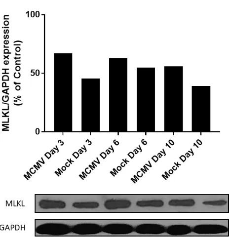
[image:44.612.82.315.411.653.2]3.2 MLKL Protein Expression in MCMV Infected Eyes
Western Blot. Top: bar graph of quantified total protein levels at days 3, 6 and 10 post MCMV infection (dpi) compared to contralateral mock non-infected eyes (media
injected) with total % of ubiquitous housekeeping molecule GAPDH as a control for each sample. Bottom: blotted protein; band density measured in ImageJ to quantify. Statistical analysis via student T-test.
In necroptosis cell death signaling, MLKL is synonymous with the pathway more so than
RIPK1 and RIPK3 [78][79]. It is also linked to RIPK3 activity in which one RIPK3 molecule
can activate several MLKL via phosphorylation [2][11]. To function as the cell executioner,
several active MLKL must form an oligomer [2][64]. MLKL mRNA must then correlate to an
increased concentration of MLKL protein in order to support necroptosis cell death [64]. To
further evaluate relevance of mRNA upregulation, total MLKL protein concentration was
quantified via western blot to confirm the presence of this death determinant
necroptosis-associated molecule in MAIDS-related MCMV retinitis eyes [11][64][65].
Whole eyes again were taken at the same time points, homogenized and prepped for
western blot against a phospho S345 MLKL antibody. Each sample tested consisted of a pool of
5-10 animals. Results for MCMV eyes were compared directly to mock infected eyes by band
density and graphed as a % of the control, GAPDH (Fig. 17). MLKL protein is constitutively
expressed in mock injected eyes at all time points, and although not significantly different, there
appears to be a trend of increased MLKL protein production in MCMV infected eyes when
compared to the media injected controls. It is possible that MCMV infected eyes may not show
substantially increased levels of MLKL protein due to alternative activation of MLKL or protein
localization to specific areas of tissue. It is of interest to note that the MLKL antibody used is
specific to phosphorylated MLKL at the 345 serine residue which is thought to signify its active
state. This phosphorylation site is independent of two other phosphorylation sites along the
Th349 phosphorylation for activation [65][74], Ser347 has been found to be necessary for
oligomerization [74][79]. Even with Ser347 phosphorylation, the ability to form oligomers is
further blocked until a critical threshold of MILKL is reached [71]. Structurally, MLKL contains
a c-terminal psuedokinase domain with an activation loop, a hinge and an attached N-terminal 4HB ‘killing’ domain [75][79]. When it is phosphorylated at its active loop, a conformational
change moves the C-terminal and N-terminal apart [75][92]. The 4HB domain can then associate
with the membrane through its two charged residue faces [70][75]. That being said, small
inhibitory molecules can bind at the activation loop concurrently to phosphorylation activation
[75][79]. These small molecules, such as ATP, can prevent conformational change and
subsequent oligomerization/cell death [71][75]. MLKL can still translocate to the membrane but
that alone is not sufficient to induce cell death [92]. Therefore, P345 MLKL might not always
equate to active/functioning MLKL. Validation of other MLKL antibodies would be useful in
elucidating the exact mechanism of MLKL involvement in MCMV infected eyes.
Constitutive expression of MLKL and other unknown effectors above threshold levels is
maintained by constitutive IFN signaling [70][71]. This mechanism of cellular homeostasis is
responsible for endosome activity and extracellular vesicle formation by MLKL [70]. Normally
autocrine IFN signaling works through the cGAS – Sting DNA sensor pathway to respond to
DNA damage, however, it can also respond to viral DNA and induce necroptosis augmentation
[71]. This occurs commonly in autoimmune and viral mediated cases and primes cells for
necroptosis [71]. In the case of MLKL activation, IFN signaling can either inhibit its activity
through small molecule binding [79] or utilize it for enhanced extracellular vesicle formation
[71]. For the latter, this enhanced activity helps maintain homeostasis in a stressed cell and/or
activated MLKL in an effort to hold off necroptotic cell death [70][71]. Depending on cell type
and the reason for necroptosis priming in the cell, some never succumb and instead are able to
reestablish homeostasis [70]. The immunomodulation and suppression that results from MAIDS
may be priming cells for heightened responses to normal cell housekeeping activity. The mock
infected eyes could very well be displaying active MLKL without undergoing cell death in the
retina.
While presence of MLKL can signify programmed necroptosis, total amount of protein
should not be taken to equate to levels of necroptosis activity in a 1:1 ratio. Western blots for
RIPK1 and RIPK3 (not shown) were indicative of increased total protein in MCMV infected
whole eyes. RIPK1 showed only cleaved protein until day 10, at which time both a cleaved and
full protein appeared. Cleavage is usually a sign of some sort of caspase 8 mediated activity such
as apoptosis or NF-kB mediated cell survival/inflammation [43][69][72]. That considered
together with the eventual shift to non-cleaved RIPK1 could be suggestive of either simultaneous
apoptosis necroptosis or early apoptosis secondary necroptosis; regardless the ultimate outcome
is necroptotic cell death [2][73][97]. If apoptosis was the main or sole cell death pathway at play,
we would expect to see cleaved RIPK3 and MLKL [43][73][81]. RIPK3 also had two bands at
different molecular weights. A band with a higher than normal RIPK3 weight was shown very
prominently at day 3 and increased in size by day 10. The normal RIPK3 band was barely visible
at day 3 but did increase in density by day 10. While it is difficult to accurately predict what the
heavier RIPK3 protein is, there is at least some evidence for the existence of two RIPK3
isoforms [76]. Alternatively, post translational modifications are not uncommon during RIPK3
signaling and regulation; phosphorylation, ubiquitination or oligomerization may have resulted
infected eyes indicate the possible stimulation of the necroptosis pathway, protein level alone
fails to tell us if these proteins are functionally responsible for the observed MCMV retinitis
pathology, nor does it actually localize them to the retina.
4 LOCALIZED EXPRESSION OF NECROPTOSIS-ASSOCIATED MOLECULES IN
THE RETINA FOLLOWING MCMV INFECTION OF MAIDS-10 MICE
Immunohistochemical staining (IHC) allows for the localization of target molecules to
specific areas of tissue [40]. Using fluorescently labeled antibodies (Fig. 18) differential
patterning of molecules can be visualized across tissue and during different time points in disease
pathogenesis [41]. The amount and location of a molecule of interest can thus be qualitatively
assessed. This makes IHC an incredibly useful technique that is commonly used in disease
pathology studies [41][52]. Therefore to confirm that mRNA and total protein findings associate
with the retinitis disease model during MAIDS-related MCMV infection, localized staining of
[image:48.612.215.399.470.651.2]necroptosis-associated molecules at the retina was necessary.
Figure 18. Indirect Immunofluorescence.
tissue) > anti-antigen primary antibody (hosted in rabbit) > anti-rabbit secondary antibody labeled with Cy3 (hosted in goat). Modified from [36].
MCMV and contralateral-mock infected whole eyes were paraffin embedded eyes and
sectioned then stained using specific antibodies for RIPK1, RIPK3 and MLKL respectively (Fig.
18). Day 6 and day 10 post-infected eye sections were examined with day 6 displaying retinal
folding and day 10 displaying full retinal necrosis. These time points were selected in accordance
with the observed largest change in disease state indicated by mRNA results (Fig. 14) (Fig. 16)
and disease pathology in this model (Fig. 6). Results from MCMV eyes were normalized to
mock infected (media injected) eyes and to an isotype negative control. It was expected that
RIPK1, RIPK3 and MLKL expression would be upregulated in the retina during MAIDS-related
MCMV infection.
While IHC is integral for confirmation of localization to the site of interest, it is not
without its drawbacks. Due to antibody difficulty in binding effectively to antigens in tissue
[17][32] there occasionally might be a loss of signal and the representation that certain molecules
of interest are under-expressed. Additionally, issues can arise when diseased tissue is normalized
to control tissue. Mock infected sections do not exhibit retinitis pathology however, they may be
subject to inflammatory processes mediated by local tissue injury from the injection itself and/or
can be subject to such problems as retinal detachment which is associated with necroptotic cell
death [7][8]. Furthermore, in in-bred lab strains of mice, there could be possible
inflammatory/immune related over-reactions triggered by this event or already in process due to
the normal environmental stressors the eye undergoes [35]. This is due to the immunomodulatory
nature of inbreeding-depression and other genetic effects [31]. Aside from any additional
immune pressures mediated by this model, there is also a case to be made for normal levels of
molecules outside of active cell death signaling [2][33][45][65][69]. In part, healthy mock
infected tissue could exhibit observable levels of RIPK1, RIPK3 and MLKL expression that has
to be accounted for by normalizing and thus potentially reducing observable true expression in
diseased tissue. IHC provides final confirmation of pathological association but is done with
support from mRNA and total protein data in order to form a more complete picture of
necroptosis associated cell death. Overall, it was indeed found that MCMV infected retinas
expressed more RIPK1, RIPK3 and MLKL; especially at day 6 post-MCMV infection.
Necroptosis signaling appears to be present during disease pathogenesis and could be inferred to
be actively inducing cell death during the retinal destruction apparent in this model.
4.1 RIPK1 Expression Following Ocular MCMV Infection at Days 6 and 10
Post-Infection
Presence of RIPK1 protein in the retina was qualitatively evaluated via IHC to determine
what differences, if any, there were in RIPK1 localized activity across MCMV infected and
non-infected retina at different time points. As RIPK1 is also associated with apoptosis cell death
signaling [2][42], its expression alone doesn’t determine what cellular death machinery is at
work. However, RIPK1 is the branching off point of many cellular signaling pathways:
apoptosis, necroptosis, cell survival and cytokine release through NFkβ [43][69]. Meaning
cellular activity in response to different stimuli can have differential effects on RIPK1 that then
determine its downstream signaling [2][8][10]. RIPK1 is an important molecule in understanding
what other players may be at work or inhibited during certain cellular processes. It can also serve
as a good indicator of overall initiation of programmed death signaling when tissue destruction is