This is a repository copy of
Peripheral blood leucocyte functional responses to acute
eccentric exercise in humans are influenced by systemic stress, but not by
exercise-induced muscle damage
.
White Rose Research Online URL for this paper:
http://eprints.whiterose.ac.uk/548/
Article:
Saxton, J. M., Claxton, D., Winter, E. et al. (1 more author) (2003) Peripheral blood
leucocyte functional responses to acute eccentric exercise in humans are influenced by
systemic stress, but not by exercise-induced muscle damage. Clinical Science, 104
(6977). pp. 69-77. ISSN 1470-8736
[email protected]
https://eprints.whiterose.ac.uk/
Reuse
Unless indicated otherwise, fulltext items are protected by copyright with all rights reserved. The copyright
exception in section 29 of the Copyright, Designs and Patents Act 1988 allows the making of a single copy
solely for the purpose of non-commercial research or private study within the limits of fair dealing. The
publisher or other rights-holder may allow further reproduction and re-use of this version - refer to the White
Rose Research Online record for this item. Where records identify the publisher as the copyright holder,
users can verify any specific terms of use on the publisher’s website.
Takedown
If you consider content in White Rose Research Online to be in breach of UK law, please notify us by
Peripheral blood leucocyte functional responses
to acute eccentric exercise in humans are
influenced by systemic stress, but not by
exercise-induced muscle damage
John M. SAXTON
*
, David CLAXTON
*
, Edward WINTER
*
and A. Graham POCKLEY†
*
The Centre for Sport and Exercise Science, Sheffield Hallam University, Collegiate Crescent Campus, Sheffield S10 2BP, U.K.,
and †Division of Clinical Sciences (North), University of Sheffield, Northern General Hospital, Herries Road, Sheffield S5 7AU,
U.K.
A
B
S
T
R
A
C
T
The effects of comparable lower-limb eccentric exercise that induces high (bench-stepping ; STEP) and low (repeated eccentric muscle action ; ECC) systemic stress on neutrophil and monocyte phagocytic and respiratory burst activity, and activation antigen (CD11b, CD66b, CD64) expression, were compared in recreationally active subjects (20–37 years old). Leucocyte responses were determined before and 4, 24, 48 and 72 h after exercise using whole-blood flow cytometry. Serum creatine kinase (CK) activity and perceived muscle soreness [delayed-onset muscle soreness (DOMS)] were assessed at the same time points up to 96 h ; as a control, measurements were taken during 5 days of rest. DOMS in quadriceps and contralateral triceps surae peaked 24–72 h after STEP (P!0.05) and 48–72 h after ECC (P!0.05), whereas serum CK activity (mean³S.E.M.) was only higher than baseline after ECC (15 123³3488 at 96 h compared with 115³29 units[l−1 pre-exercise ; P!0.01). The total leucocyte count increased from (5.4³0.4)¬109[l−1 and (5.7³0.5)¬109[l−1 at baseline to (7.6³0.5)¬109[l−1 and (7.0³0.5)¬109[l−1at 4 h after STEP and ECC respectively ; this was largely attributable to changes in the neutrophil count (P!0.05). The proportion of neutrophils undergoing phagocytosis and respiratory burst was unchanged 4 h after ECC and STEP, which, given the increase in neutrophil count after exercise, would suggest an overall improvement in systemic neutrophil microbicidal potential. The intensity of neutrophil (P¯0.01) and monocyte (P!0.05) phagocytosis and neutrophil respiratory burst responses (P!0.05) was only increased 24 h after STEP, whereas no changes in these measures were observed after ECC. Activation antigen expression was unchanged in all groups. These findings suggest that systemic stress evoked during an acute bout of eccentric exercise has a greater influence on subsequent leucocyte functional responses than the degree of muscle damage induced.
INTRODUCTION
Interest in the immunomodulatory effects of exercise has been prompted by evidence that chronic strenuous exercise training can have detrimental effects on immune
Key words :adhesion molecules, eccentric, exercise, leucocyte function.
Abbreviations : CK, creatine kinase ; DOMS, delayed-onset muscle soreness ; ECC, bout of eccentric exercise involving the quadriceps and triceps surae ; IL-1 (etc.), interleukin-1 (etc.) ; STEP, bout of bench-stepping exercise.
Correspondence:Dr John Saxton (e-mail j.m.saxton!shu.ac.uk).
70 J. M. Saxton and others
effects of exercise-induced immunomodulatory changes on the ability of the immune system to respond to infectious disease.
Exercise that involves a substantial proportion of eccentric muscular activity, in which the active muscles are forcibly lengthened, increases the degree of ultra-structural injury within myofibres [3,4], and this can evoke a significant local inflammatory response [5]. This is characterized by leucocyte infiltration into exercise-damaged tissue [6,7], an increased local production of inflammatory mediators such as cytokines, prosta-glandins and leukotrienes [8–10], and a systemic acute-phase response [11]. The inflammatory response to mechanical trauma could account for the progressive release of muscle-specific enzymes into the serum and feelings of soreness that are often observed in untrained subjects in the days after eccentric exercise [5,12].
Neutrophils and monocytes act as the first line of defence against potentially infectious agents, and they also have an important role in the post-exercise inflam-matory response to muscle fibre injury [13–15]. The infiltration of neutrophils and monocytes into exercise-damaged muscle as part of the repair}regeneration process is likely to be facilitated by an increased ex-pression of surface adhesion molecules. Adhesion molecules play an important role in leucocyte migration from the circulation to the site of inflammation. Eccentric exercise evoking muscle damage in untrained subjects has been reported to increase the expression of CD11b and CD64 on peripheral blood neutrophils and monocytes [16]. Since there was evidence of a diminished activation response for individuals who were less susceptible to eccentric exercise-induced injury [16], the implication was that leucocyte activation was associated with the degree of exercise-induced muscle damage. Changes in adhesion molecule expression and the involvement of neutrophils and monocytes in post-exercise inflam-matory responses might alter the microbicidal potential of these immune cells in peripheral blood.
The functional responsiveness of neutrophils and monocytes in relation to changes in activation antigen expression following eccentric exercise has not been studied previously. Furthermore, no studies have com-pared the responses between eccentric exercise models that evoke different degrees of systemic stress. The hypothesis of the present study was that changes in activation antigen expression (and immune cell function) are linked to the degree of exercise-induced muscle damage evoked by exercise, irrespective of the level of systemic stress encountered. To test this hypothesis, we compared the responses to serial repetitions of eccentric muscular work performed on an isokinetic dynamometer with those induced following a 40 min bout of intense bench-stepping exercise. Serial repetitions of eccentric muscle actions induce a substantial increase in serum creatine kinase (CK) activity and significant muscle
soreness in the muscle groups involved [12], despite evoking a relatively low level of systemic stress. Bench-stepping, on the other hand, can be used to subject the same muscle groups to eccentric damage, but is a form of exercise that evokes a considerably higher level of systemic (cardiovascular and metabolic) stress.
MATERIALS AND METHODS
Participants
Participants were recreationally active, healthy volunteers (age 20–37 years) who regularly undertook two or three exercise sessions per week (involving weight-bearing activity). Subjects were instructed not to perform any exercise training from 48 h before the study until the end of the blood sampling period. Eight participants performed a bout of eccentric exercise involving the quadriceps and triceps surae (ECC), with responses to ECC compared with control measurements taken during 5 days of rest. The order of control and experimental conditions was counterbalanced. A second group of eight age-matched recreationally active partici-pants performed a bout of bench-stepping exercise (STEP), which induces soreness in the same muscle groups [17].
Approval for this study was given by the South Sheffield Research Ethics Committee, and subjects gave their written informed consent according to University guidelines.
Exercise models
ECC
eccentric muscle actions using a similar protocol for the contralateral triceps surae.
STEP
The STEP model was based on that used previously by Newham et al. [17] to induce exercise-induced muscle soreness, and comprised 40 min of intense bench-step-ping exercise at a rate of 20 steps[min−"using a constant lead leg (chosen randomly). The bench height was set at 0.45 m and the step cadence was controlled precisely by a metronome (verified by observers). This exercise model subjects the triceps surae of the lead leg and the quadriceps of the trail leg to eccentric muscle actions, and evokes evidence of ultrastructural muscle fibre injury in these muscle groups [17]. Heart rate was measured continuously using a heart rate monitor (Sport Tester PE-3000 ; Polar Electro) and was recorded during the final 1 min of exercise.
Blood sampling
Blood samples for leucocyte analysis were drawn from an antecubital vein immediately before and 4, 24, 48 and 72 h after both bouts of exercise, and at precisely the same time points in the non-exercise control condition. With the exception of the 4 h sample, all blood was drawn between 08.00 and 08.30 hours. An extra blood sample was taken at 96 h for serum CK analysis, as the increase in serum CK activity after the ECC and STEP protocols often peaks 4–5 days after exercise [17]. Blood was collected into Vacutainer tubes (Becton Dickinson Ltd, Oxford, U.K.) that contained no anticoagulant for serum CK analysis, K
$EDTA for leucocyte counts and
activation antigen expression, and lithium heparin for leucocyte functional assays by flow cytometry. Serum samples were stored at®80°C until analysis.
Criterion measures
Muscle soreness and serum CK activity
Perceived muscle soreness [delayed-onset muscle sore-ness (DOMS)] was assessed using a muscle soresore-ness questionnaire that comprised a 10-point soreness scale for six regions of the anterior}posterior thigh and calf. The scale ranged from 1 (normal) to 10 (very, very sore), and the mean ratings for the muscle groups subjected to eccentric exercise (quadriceps and contralateral triceps surae) provided an overall assessment of soreness encountered in these regions. Serum CK activity was assayed using the method of Szasz et al. [18] with an enzymic kit (no. 47-10 ; Sigma, Poole, Dorset, U.K.) ; the interassay coefficient of variation was 5.6 %.
Leucocyte analysis
Differential leucocyte counts and other haematological parameters were measured within 6 h of blood sampling using a Coulter STKS automated haematological analyser
(Beckman Coulter U.K. Ltd, High Wycombe, U.K.) which was calibrated on a daily basis.
Leucocyte microbicidal function and activation antigen
expression
Neutrophil and monocyte phagocytosis and respiratory burst responses were measured using commercial kits (Phagotest2 and Phagoburst2; Orpegen, Heidelberg, Germany). The expression of CD11b, CD64 and CD66b was assessed using a modified three-colour whole-blood flow cytometry technique [19], using FITC-, Cychrome-and phycoerythrin-conjugated monoclonal antibodies. CD11b (Mac-1 ; complement receptor 3) is theαsubunit of the CD11–CD18 heterodimeric complex and plays a key role in localization at inflammatory sites ; its ex-pression is up-regulated rapidly upon leucocyte acti-vation [20]. CD64 (FcγR1) is expressed on monocytes and activated granulocytes ; it mediates antibody-de-pendent cytotoxicity and triggers phagocytosis, super-oxide production and degranulation [21–23]. CD66b is a member of the carcinoembryonic-like glycoprotein fam-ily and is expressed on peripheral blood granulocytes, but not lymphocytes or monocytes [24]. Negative control tubes were stained using appropriate FITC-, Cychrome-and phycoerythrin-conjugated isotype-matched non-reactive negative controls (Becton Dickinson, Serotec Ltd). All samples were stained within 2 h of collection to avoidex vivoup-regulation of CD11b expression [19].
Flow cytometric analysis
Flow cytometric analysis was performed on a Becton Dickinson FACSortTMflow cytometer (BD Biosciences, Oxford, U.K.). For the functional assays, both the percentage of positively stained cells (cells exhibiting phagocytic}respiratory burst response) and the median channel fluorescence intensity (which correlates with the number of ingested bacteria per cell, or the intensity of the respiratory burst response) were used as the criterion measures of functional status (interassay coefficients of variation were !2 % and !3 % respectively). For analysis of activation antigen expression, the expression of CD11b, CD64 and CD66b by neutrophils (10%cells) and of CD11b and CD64 by monocytes (5¬10$
cells) was determined. The percentage of cells express-ing each antigen and the median channel fluorescence intensity of expression (which correlates with antigen density) were used as the criterion measures of activation antigen expression (interassay coefficients of variation were!2 % and!4 % respectively).
Data analysis
72 J. M. Saxton and others
activity were analysed using repeated-measures ANOVA. Non-parametric muscle soreness data were analysed using the Wilcoxon signed-ranks test (paired data) and the Mann–WhitneyUtest (unpaired data). All values are expressed as means³S.E.M., and differences were considered to be of statistical significance at P!0.05. Statistical analyses were performed using SPSS
for Windows (SPSS UK Ltd, Woking, U.K.).
RESULTS
Heart rate response to STEP protocol
The STEP protocol was very demanding on the car-diovascular system, even for our recreationally active subjects. Heart rate was measured in the final 1 min of the STEP protocol and was used as an index of cardiovascular stress. The mean³S.E.M. heart rate in the final 1 min of exercise was 176³10 beats[min−", corresponding to a heart rate of 91³3 % of the predicted maximum (based on 220®age for prediction of maximum heart rate).
Muscle soreness and serum CK activity
The DOMS score peaked 48 h (P!0.05 ; Wilcoxon test) and 48–72 h (P!0.05 ; Wilcoxon test) after ECC in the quadriceps and contralateral triceps surae respectively (Table 1). DOMS in the same muscle groups was also observed after STEP (P!0.05), peaking 24–72 h after exercise (Table 1). The degree of soreness experienced inTable 1 Muscle soreness ratings after STEP and ECC
Data are presented as means, with ranges in parentheses. Statistical significance :
*P!0.05 compared with baseline ;†P!0.05,††P!0.01 between STEP and ECC conditions at that time point.
DOMS score
Pre-exercise 24 h 48 h 72 h
Quadriceps
STEP 1 (1–1) 3 (1–6)* 3 (2–5)* 2 (1–3) ECC 1 (1–1) 5 (2–7)* 6 (2–9)*† 5 (2–10)*††
Triceps surae
STEP 1 (1–1) 4 (1–6)* 4 (1–7)* 4 (1–7)*
[image:5.612.327.543.119.311.2] [image:5.612.98.316.490.608.2]ECC 1 (1–1) 5 (1–7)* 6 (2–9)* 6 (2–10)*
Table 2 Serum CK activity after STEP, after ECC and in the control condition (CON)
Data are presented as means³S.E.M. Statistical significance :*P!0.05,**P!0.01 compared with CON and STEP conditions.
Condition
CK level (units[l−1)
Pre-exercise 24 h 48 h 72 h 96 h
STEP 146³59 161³66 106³43 83³34 76³31
ECC 115³29 2776³1597 8368³3080* 14 673³3979** 15 123³3488**
CON 117³41 90³32 82³30 109³38 117³40
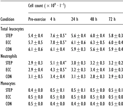
Table 3 Peripheral blood leucocyte counts after STEP, after ECC and in the control condition (CON)
Data are presented as means³S.E.M. Statistical significance : *P!0.05 compared with baseline.
Condition
Cell count (¬109[l−1)
Pre-exercise 4 h 24 h 48 h 72 h
Total leucocytes
STEP 5.4³0.4 7.6³0.5* 5.6³0.4 6.0³0.4 5.8³0.3 ECC 5.7³0.5 7.0³0.5* 6.1³0.6 6.3³0.5 6.0³0.4 CON 6.1³0.6 6.1³0.4 5.9³0.3 5.6³0.4 5.9³0.4 Neutrophils
STEP 2.9³0.3 5.1³0.4* 3.0³0.3 3.2³0.3 3.2³0.2 ECC 2.9³0.4 4.3³0.5* 3.2³0.3 3.4³0.4 3.0³0.3 CON 3.1³0.5 3.4³0.4 3.1³0.3 2.8³0.3 2.9³0.3 Monocytes
STEP 0.4³0.0 0.5³0.1 0.5³0.1 0.5³0.0 0.5³0.1 ECC 0.5³0.0 0.5³0.0 0.5³0.0 0.5³0.0 0.5³0.0 CON 0.5³0.0 0.4³0.0 0.4³0.0 0.4³0.0 0.5³0.0
the quadriceps muscle group was more marked after ECC (P!0.01). Serum CK activity increased progress-ively from a mean³S.E.M. baseline value of 115³29 units[l−" to 15 123³3488 units[l−" (P!0.01 ; ANOVA) 96 h after ECC, whereas no change in serum CK activity from baseline values was observed after STEP or in the control condition (Table 2).
Leucocyte counts
The total leucocyte count was increased at 4 h after both ECC and STEP (P!0.05), but had returned to pre-exercise levels after 24 h (Table 3). In both cases, the increase in total leucocyte count was largely attributable to changes in the number of circulating neutrophils (P!0.05). Neither exercise protocol had any effect on the peripheral blood monocyte count (Table 3), and there was no change in leucocyte count at any time point in the control condition.
Leucocyte phagocytosis and respiratory
burst responses
[image:5.612.99.547.654.738.2]Figure 1 Neutrophil and monocyte phagocytosis after STEP, after ECC and in the control condition (CON)
Data are presented as individual responses (thin lines) and as mean³S.E.M. responses (thick lines) for the group ;*P!0.05 compared with baseline.
Figure 2 Neutrophil and monocyte respiratory burst after STEP, after ECC and in the control condition (CON)
[image:6.612.68.506.426.697.2]74 J. M. Saxton and others
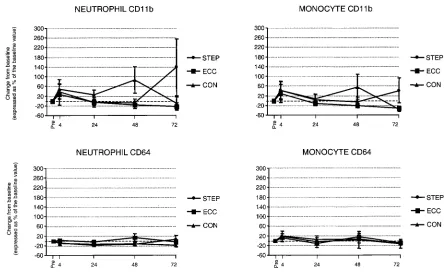
Figure 3 Neutrophil and monocyte activation antigen expression after STEP, after ECC and in the control condition (CON)
Data are presented as mean³S.E.M. responses.
at any time point after either exercise bout, and 85–95 % of neutrophils were functionally responsive throughout the time course of the study. For monocytes, the proportion of cells that exhibited phagocytic (40–50 %) and respiratory burst (30–40 %) responses was lower than that observed for neutrophils, and this proportion remained unchanged following both exercise bouts and in the control condition.
At the cellular level, there was an increase in the intensity of neutrophil phagocytosis at 24 h after STEP (P¯0.01) ; this was not observed after ECC or in the control condition (Figure 1). This change was mirrored by a concomitant increase in the neutrophil respiratory burst response (P!0.05) at the same time point (Figure 2). A potentiation of monocyte phagocytosis was also observed at the 24 h time point (P!0.05) after STEP, but not after ECC or in the control condition (Figure 1). The intensity of the monocyte respiratory burst response was unchanged after both exercise bouts and in the control condition (Figure 2).
Leucocyte activation antigen expression
The proportion of peripheral blood neutrophils and monocytes expressing each activation antigen was un-changed after both bouts of exercise and in the control condition. Neutrophils constitutively express CD66b, and the majority of peripheral blood neutrophils were also expressing CD11b. However, the proportion ofneutrophils expressing CD64 was lower, generally in the range 30–40 %. Monocytes do not express CD66b, but, as observed for neutrophils, a high proportion of the cells (generally"80 %) were expressing CD11b at all time points. CD64 was also expressed in the majority of peripheral blood monocytes. The intensity of all ac-tivation antigens on peripheral blood neutrophils and monocytes was unchanged after both bouts of exercise and in the control condition (Figure 3).
DISCUSSION
The present study compared phagocytic and respiratory burst responses and activation antigen expression in peripheral blood neutrophils and monocytes before and after acute bouts of exercise associated with low and high systemic stress. Both exercise protocols subjected the quadriceps and contralateral triceps surae to eccentric exercise ; however, the level of metabolic and cardio-vascular stress evoked by the STEP protocol was con-siderably greater than that evoked by the ECC protocol. The magnitude of changes in the indirect correlates of exercise-induced muscle damage suggests that a greater degree of temporary, repairable ultrastructural injury to muscle fibres was induced by ECC.
muscle-specific enzymes into the serum and significant post-exercise muscle soreness [11,17], as well as a rapid acute-phase response in untrained individuals [11]. In the present study, STEP evoked only moderate post-exercise muscle soreness without any increase in serum CK activity in our recreationally active participants. This attenuated response could reflect the training status of our participants, as trained skeletal muscle is less sus-ceptible to eccentric-induced injury [26].
The total leucocyte count was increased at 4 h after ECC and STEP, but had returned to pre-exercise levels after 24 h. In both instances, the increase in total leucocyte count was largely attributable to changes in the numbers of circulating neutrophils. Given that there was no change in the proportion of neutrophils (85–95 %) that exhibited phagocytic or respiratory burst responses at any time point after either exercise bout, the increase in the number of circulating neutrophils indicates an overall improvement in systemic neutrophil microbicidal po-tential at this time point.
The increase in the peripheral blood neutrophil count 4 h after STEP was probably attributable to the secondary neutrophil leucocytosis associated with non-specific ex-ercise stress that commonly occurs within 1–3 h of the cessation of intense}prolonged physical exertion. This response is probably due to the effects of elevated plasma cortisol levels on bone marrow [27,28]. It is notable that although a 2-fold increase in neutrophil count was detected 4 h after STEP in the present study, this response was lower than that observed previously after a similar exercise stimulus in untrained subjects [11]. The dif-ference could be explained by a blunted cortisol response to the STEP protocol in our recreationally active partici-pants, or a lower degree of muscle fibre injury, resulting in an attenuated inflammatory response after STEP. The increase in peripheral blood neutrophil count following ECC, on the other hand, could have been indicative of an inflammatory response to muscle fibre injury. This is indicated by the magnitude of the change in the indirect correlates of exercise-induced muscle damage.
At the cellular level, there was an increase in the intensity of the neutrophil phagocytosis and respiratory burst responses 24 h after STEP that was not observed after ECC. A potentiation of monocyte phagocytosis was also observed at the 24 h time point after STEP, but not after ECC. The increase in the intensity of leucocyte functional responses 24 h after STEP could have been induced through the effects of certain cytokines. Studies have demonstrated that strenuous exercise induces the release of interleukin-1 (IL-1) and other cytokines, including IL-6 and tumour necrosis factorα[10,29], that prime the various pathways that contribute to the activation of NADPH oxidase and can amplify immune responses in a concentration-dependent manner [30]. Although exercise-induced increases in circulating levels of IL-6 have usually returned to baseline within 24 h
[9,10], at the tissue level, the release of IL-1βfollows a protracted time course after exercise that has a large eccentric component and is associated with high systemic stress [6]. Furthermore, circulating IL-6 levels were recently shown to peak 24 h after eccentric resistance training exercise with large muscle groups that could have induced considerable systemic stress but minimal muscle damage [31]. Unfortunately, this could not be confirmed, because serum CK and the degree of soreness encountered by the subjects, who were described as ‘active ’, were not reported. Neuroendocrine factors such as growth hormone, catecholamines and glucocorticoids and a range of neuropeptides also play a role in modulating immune function in response to exercise [32,33]. However, there is a lack of published data on longer-term changes (12–24 h) in these factors following acute exercise, at the time when potentiation of neutro-phil and monocyte microbicidal function was observed in our study.
In contrast with previous studies that have reported an increase in neutrophil and monocyte activation antigen expression in untrained subjects following eccentric exercise with low and high systemic stress [16,34], we observed no changes in activation status after either ECC or STEP. Previous work has reported no change in neutrophil CD11b expression following moderate, sus-tained cycle ergometry exercise [35] and sub-maximal running exercise [36]. However, there is evidence that increases in neutrophil and monocyte CD11b and CD64 expression and in the concentration of circulating in-tercellular adhesion molecule-1 (ICAM-1) are influenced by the extent of muscle fibre injury after eccentric exercise [16,34,37]. This seems feasible, as increased expression of cell surface adhesion molecules could mediate the migration and infiltration of neutrophils and monocytes into exercise-damaged tissue, in which they contribute to the repair}regeneration process. Expression of CD11b is increased rapidly and dramatically (300– 400 %) upon exposure to chemotactic stimuli by the transport of preformed granular stores of the protein to the surface of the cell [38]. Although most integrins require a conformational change in order to acquire full adhesive function [39], there is evidence that newly mobilized CD11b is capable of functioning in adhesive events if the cells are subsequently exposed to an additional stimulus level [40]. Interestingly, a positive correlation between the number of tissue macrophages and CD11b expression in the days after eccentric cycling exercise has been reported [41].
76 J. M. Saxton and others
exercise-induced damage. Furthermore, our study used an established whole-blood flow cytometry technique [42] that is potentially more representative of in vivo responses, as cell isolation procedures can affect leucocyte function and the expression of surface antigens [43,44].
In summary, our results show that a potentiation of peripheral blood neutrophil and monocyte phagocytic and respiratory burst responses can occur without any changes in activation antigen expression following acute exercise that has a large eccentric component and that evokes a high level of systemic stress. Exercise-induced muscle damage appears to have no effect on the micro-bicidal potential of circulating leucocytes, nor on the expression of activation antigens at the cellular level. However, the increased number of functional circulating neutrophils at 4 h after both the ECC and STEP protocols indicates an overall increase in peripheral blood micro-bicidal potential after eccentric exercise, independent of the level of systemic stress. These findings suggest that the level of systemic stress evoked during an acute bout of eccentric exercise has a greater influence on subsequent leucocyte functional responses than the degree of muscle damage induced.
ACKNOWLEDGMENTS
We are grateful for the technical support of Penny Stephens and Brenka Swales.
REFERENCES
1 Keast, D., Cameron, K. and Morton, A. R. (1988) Exercise and the immune response. Sports Med.5, 248–267
2 Nieman, D. C. (1994) Exercise, infection, and immunity. Int. J. Sports Med.15(Suppl. 3), S131–S141
3 Friden, J., Sjostrom, M. and Ekblom, B. (1983) Myofibrillar damage following intense eccentric exercise in man. Int. J. Sports Med.4, 170–176
4 Newham, D. J., McPhail, G., Mills, K. R. and Edwards, R. H. (1983) Ultrastructural changes after concentric and eccentric contractions of human muscle. J. Neurol. Sci. 61, 109–122
5 Smith, L. L. (1991) Acute inflammation : the underlying mechanism in delayed onset muscle soreness ? Med. Sci. Sports Exercise23, 542–551
6 Fielding, R. A., Manfredi, T. J., Ding, W., Fiatarone, M. A., Evans, W. J. and Cannon, J. G. (1993) Acute phase response in exercise. III. Neutrophil and IL-1 beta accumulation in skeletal muscle. Am. J. Physiol.265, 166–172
7 Round, J. M., Jones, D. A. and Cambridge, G. (1987) Cellular infiltrates in human skeletal muscle : exercise induced damage as a model for inflammatory muscle disease ? J. Neurol. Sci.82, 1–11
8 Konig, D., Berg, A., Weinstock, C., Keul, J. and Northoff, H. (1997) Essential fatty acids, immune function, and exercise. Exercise Immunol. Rev.3, 1–31 9 Northoff, H., Enkel, S. and Weinstock, C. (1995)
Exercise, injury and immune function. Exercise Immunol. Rev.1, 1–25
10 Rhind, S. G., Shek, P. N. and Shephard, R. J. (1995) The impact of exercise on cytokines and receptor expression. Exercise Immunol. Rev.1, 97–148
11 Gleeson, M., Almey, J., Brooks, S., Cave, R., Lewis, A. and Griffiths, H. (1995) Haematological and acute-phase responses associated with delayed-onset muscle soreness in humans. Eur. J. Appl. Physiol.71, 137–142
12 Clarkson, P. M., Nosaka, K. and Braun, B. (1992) Muscle function after exercise-induced muscle damage and rapid adaptation. Med. Sci. Sports Exercise24, 512–520 13 Nieman, D. C., Nehlsen-Cannarella, S. L., Fagoaga, O. R.
et al. (1998) Effects of mode and carbohydrate on the granulocyte and monocyte response to intensive, prolonged exercise. J. Appl. Physiol.84, 1252–1259 14 Ortega, E. (1994) Physiology and biochemistry : influence
of exercise on phagocytosis. Int. J. Sports Med.15, S172–S178
15 Smith, J. A. and Pyne, D. B. (1997) Exercise, training, and neutrophil function. Exercise Immunol. Rev.3, 96–116 16 Pizza, F. X., Davis, B. H., Henrickson, S. D. et al. (1996)
Adaptation to eccentric exercise : effect on CD64 and CD11b}CD18 expression. J. Appl. Physiol.80, 47–55 17 Newham, D. J., Jones, D. A. and Edwards, R. H. (1983)
Large delayed plasma creatine kinase changes after stepping exercise. Muscle Nerve6, 380–385 18 Szasz, G., Gruber, W. and Bernt, E. (1976) Creatine
kinase in serum : 1. Determination of optimum reaction conditions. Clin. Chem.22, 650–656
19 Saxton, J. M. and Pockley, A. G. (1998) Effect of ex vivo storage on human peripheral blood neutrophil expression of CD11b and the stabilizing effects of Cyto-Chex. J. Immunol. Methods214, 11–17
20 Kishimoto, T. K., Jutila, M. A., Berg, E. L. and Butcher, E. C. (1989) Neutrophil Mac-1 and MEL-14 adhesion proteins inversely regulated by chemotactic factors. Science245, 1238–1241
21 Akerley, III, W. L., Guyre, P. M. and Davis, B. H. (1991) Neutrophil activation through high-affinity Fc gamma receptor using a monomeric antibody with unique properties. Blood77, 607–615
22 Shen, L., Guyre, P. M. and Fanger, M. W. (1987) Polymorphonuclear leukocyte function triggered through the high affinity Fc receptor for monomeric IgG. J. Immunol.139, 534–538
23 Unkeless, J. C., Boros, P. and Fein, M. (1992) Structure, signalling and function of FcγR. In Inflammation: Basic Principles and Clinical Correlates (Gallin, J. I., Goldstein, I. M. and Snyderman, R., eds), pp. 921–924, Raven, New York
24 Schlossman, S., Boumsell, I., Gilks, W. et al. (eds) (1994) Leukocyte Typing V : White Cell Differentiation Antigens, 5th International Conference on Leukocyte Differentiation Antigens, pp. 132–133, Oxford University Press, Oxford
25 Dill, D. B. and Costill, D. L. (1974) Calculation of percentage changes in volumes of blood, plasma, and red cells in dehydration. J. Appl. Physiol.37, 247–248 26 Clarkson, P. M. and Tremblay, I. (1988) Exercise-induced
muscle damage, repair, and adaptation in humans. J. Appl. Physiol.65, 1–6
27 Nieman, D. C., Miller, A. R., Henson, D. A. et al. (1994) Effect of high- versus moderate-intensity exercise on lymphocyte subpopulations and proliferative response. Int. J. Sports Med.15, 199–206
28 Pedersen, B. K., Bruunsgaard, H., Klokker, M. et al. (1997) Exercise-induced immunomodulation – possible roles of neuroendocrine and metabolic factors. Int. J. Sports Med.18(Suppl. 1), S2–S7
29 Camus, G., Deby-Dupont, G., Deby, C., Juchmes-Ferir, A., Pincemail, J. and Lamy, M. (1993) Inflammatory response to strenuous muscular exercise in man. Med. Inflamm.2, 335–342
30 Smith, J. A. (1994) Neutrophils, host defense, and inflammation : a double-edged sword. J. Leukocyte Biol. 56, 672–686
31 Smith, L. L., Anwar, A., Fragen, M., Rananto, C., Johnson, R. and Holbert, D. (2000) Cytokines and cell adhesion molecules associated with high-intensity eccentric exercise. Eur. J. Appl. Physiol.82, 61–67 32 Jonsdottir, I. H. (2000) Exercise immunology:
33 Jonsdottir, I. H. (2000) Neuropeptides and their interaction with exercise and immune function. Immunol. Cell Biol.78, 562–570
34 Pizza, F. X., Mitchell, J. B., Davis, B. H., Starling, R. D., Holtz, R. W. and Bigelow, N. (1995) Exercise-induced muscle damage: effect on circulating leukocyte and lymphocyte subsets. Med. Sci. Sports Exercise27, 363–370
35 Kurokawa, Y., Shinkai, S., Torii, J., Hino, S. and Shek, P. N. (1995) Exercise-induced changes in the expression of surface adhesion molecules on circulating granulocytes and lymphocytes subpopulations. Eur. J. Appl. Physiol. 71, 245–252
36 Jordan, J., Beneke, R., Hutler, M., Veith, A., Haller, H. and Luft, F. C. (1997) Moderate exercise leads to decreased expression of beta1 and beta2 integrins on leucocytes. Eur. J. Appl. Physiol.76, 192–194 37 Akimoto, T., Furudate, M., Saitoh, M. et al. (2002)
Increased plasma concentrations of intercellular adhesion molecule-1 after strenuous exercise associated with muscle damage. Eur. J. Appl. Physiol.86, 185–190 38 Elliott, M. J. and Finn, A. H. (1993) Interaction between
neutrophils and endothelium. Ann. Thorac. Surg.56, 1503–1508
Received 12 April 2002/27 September 2002; accepted 11 November 2002 39 Hynes, R. O. (1992) Integrins: versatility, modulation,
and signaling in cell adhesion. Cell69, 11–25 40 Hughes, B. J., Hollers, J. C., Crockett-Torabi, E. and
Smith, C. W. (1992) Recruitment of CD11b}CD18 to the neutrophil surface and adherence-dependent cell locomotion. J. Clin. Invest.90, 1687–1696 41 Malm, C., Nyberg, P., Engstrom, M. et al. (2000)
Immunological changes in human skeletal muscle and blood after eccentric exercise and multiple biopsies. J. Physiol. (Cambridge, U.K.)529, 243–262
42 Webster, G. A. and Pockley, A. G. (1993) Effect of red cell lysis protocols on the expression of rat peripheral blood lymphocyte subset and activation antigens. J. Immunol. Methods163, 115–121
43 Watson, F., Robinson, J. J. and Edwards, S. W. (1992) Neutrophil function in whole blood and after purification : changes in receptor expression, oxidase activity and responsiveness to cytokines. Biosci. Rep.12, 123–133