1
DISSERTATION ON
POST OPERATIVE EVALUATION OF EXCISION OF
ELONGATED STYLOID PROCESS
Dissertation submitted to
THE TAMILNADU DR.M.G.R. MEDICAL UNIVERSITY
In the partial fulfillment of the regulations
for the award of the degree of
M.S. IN OTORHINOLARYNGOLOGY
BRANCH – IV
GOVERNMENT STANLEY MEDICAL COLLEGE
CHENNAI -600 001.
THE TAMILNADU DR.M.G.R. MEDICAL UNIVERSITY
CHENNAI -600 032.
DECLARATION
I Dr. MALARVIZHI .R solemnly declare that the dissertation,
titled “POST OPERATIVE EVALUATION OF EXCISION OF
ELONGATED STYLOID PROCESS” a clinical study submitted by me
is a result of the original work carried out by myself under the guidance
of PROF. Dr. M.GOWRI SHANKAR M.S., DLO.,DNB., Professor,
Department Otorhinolaryngology and Head and Neck, Government
Stanley medical college, Chennai. I further declare that the result of
research has not been submitted previously by myself or other persons in
any conferences or journals.
This dissertation is submitted to The Tamil Nadu Dr. M.G.R
Medical University in the partial fulfillment of the rules and regulations
for the M.S. Degree examination in Otorhinolaryngology to be held on
May 2019.
DR. MALARVIZHI .R
Date :
CERTIFICATE
I certify that the dissertation titled “POST OPERATIVE
EVALUATION OF EXCISION OF ELONGATED STYLOID
PROCESS” submitted by DR.MALARVIZHI.R, for the degree of
master of surgery (Otorhinolaryngology) in partial fulfillment of
regulations of The Tamil Nadu Dr.M.G.R. Medical university, Chennai is
the result of original research work undertaken by her during the
academic year 2017- 2019, in the department of ENT AND HEAD &
NECK SURGERY, Government Stanley medical college, Chennai.
PROF.DR.S.MUTHUCHITRA
M.S., DLO.
PROFESSOR & HEAD OF THE DEPARTMENT DEPARTMENT OF OTORHINOLARYNGOLOGY
GOVERNMENT STANLEY MEDICAL COLLEGE. CHENNAI -01.
PROF.DR.S.PONNAMBALANAMASIVAYAM
M.D., D.A., DNB.
THE DEAN
CERTIFICATE BY GUIDE
This is to certify that the Dissertation titled “POST OPERATIVE
EVALUATION OF EXCISION OF ELONGATED STYLOID
PROCESS” presented by Dr. MALARVIZHI.R, is an original work
done in the Department of Otorhinolaryngology, Government Stanley
Medical College and Hospital, Chennai in partial fulfillment of
regulations of the Tamil Nadu Dr.M.G.R. Medical university for the
award of degree of M.S. (Otorhinolaryngology) Branch IV, under my
guidance, during the academic period of 2017- 2019.
Date:
Place:
PROF.DR. M.GOWRI SHANKAR M.S., DLO, DNB
PROFESSOR,
ACKNOWLEDGEMENT
First and foremost I would like to express my sincere gratitude to
Prof.Dr.S.PONNAMBALA NAMASIVAYAM, M.D, D.A, DNB The
dean, Stanley medical college, for having permitted me to undertake this
study.
I would like to express my immense gratitude to my guide
Prof.Dr.M.GOWRI SHANKAR M.S.D.L.O.,DNB., professor of ENT,
for his valuable guidance, suggestions, encouragement, constant
motivation, supervision, and help in conducting and fulfilment of this
study.
I am immensely grateful to the HOD
Prof.Dr.S.MUTHUCHITRA M.S.D.L.O., professor, Department of
ENT, Head & Neck surgery, Government Stanley Medical College &
Hospital, for her valuable support in conducting the study.
I wish to thank my Prof Dr.S.Shanmuga Ashok and
Assistant Professor Dr. K.Athiyaman. M.S for their valuable tips and
guidance. I am grateful to my Assistant Professors Dr.V.Saravana Selvan,
Dr.P.Chozhan and Dr.V.Suresh for their valuable time and guidance in
I express my sincere thanks to THE SECRETARY
AND CHAIRMAN, INSTITUTIONAL ETHICAL COMMITTEE,
Government Stanley Medical College and hospital.
I am grateful to all the other post-graduates who most
willingly helped me during this study period.
I also thank the staff nurse and theatre personnel, Government
Stanley Hospital for their co-operation and assistance in the conduct
of this study..
I thank all my colleagues and friends for their constant
encouragement and valuable criticism.
Last but not the least, I express my gratitude for the generosity
shown by all the patients who participated in the study.
I am extremely thankful to my parents for their continuous support.
ABBREVIATIONS USED
SP : STYLOID PROCESS
CT : Computer tomography
CBCT : Cone Beam Computer tomogram
OPG : Orthopantamogram
MRI : Magnetic Resonance Imaging
DM : Diabetes mellitus
cm : Centimetre
CONTENTS
S.No Contents Page No
1) INTRODUCTION 1
2) EMBRYOLOGY 2
3) ANATOMY 3
4) EAGLE’S SYNDROME 11
5) PAINFUL SYNDROMES 17
6) CLINICAL EVALUATION 25
7) RADIOGRAPHIC STUDIES 26
8) TREATMENT OPTIONS IN ELONGATED
STYLOID PROCESS 42
9) DIFFERENTIAL TREATMENT ALGORITHM 56
10)
MCGILL’S PAIN QUESTIONNARE AND BEDSIDE CLINICAL ASSESSMENT OF DYSPHAGIA
57
11) AIMS AND OBJECTIVES 67
12) MATERIALS AND METHODS 67
13) METHODOLOGY- ANALYSIS 72
14) DISCUSSION 86
16) RESULTS 94
17) CONCLUSION 98
18) RECOMMNEDATIONS AND LIMITATIONS 100
19) MASTER CHART 101
20) BIBLIOGRAPHY 102
21)
ANNEXURES : CASE SHEET PROFORMA AND QUESTIONNAIRE USED, CONSENT FORMS
CERTIFICATE, IEC AND PLAGIARISM CERTIFICATE
106
LIST OF TABLES
S.No Description Page No
1. McGill’s Pain Questionnaire 57
LIST OF FIGURES
S.No Description Page No
1. Post natal Temporal Bone Development 2
2. 3-D Volume rendered reconstruction CT of ESP 4
3. Left Temporal Bone – structural anatomy 4
4. Glossopharyngeal Nerve 9
5. Axial Section showing Glossopharyngeal nerve 10
6. Digital OPG bilateral ESP with ossified stylohyoid
ligament 29
7. Digital OPG bilateral ESP 30
8. CT 3D reconstruction of SP 34
9. CT-Axial cuts of neck with temporal bone showing SP 35
10. CBCT Sagittal section of Left SP 37
11. CBCT for Right SP – measuring tool 37
12. Conventional Radiograph – lateral view 39
13. Langlais classification of ESP 41
14. Volume rendered reconstruction using CT Stylo-hyoid
calcification 42
15. Instrument tray for stylo-tonsillectomy 50
17. Post tonsillectomy fossa- intra-op 51
18. Skeletonization of styloid with ring curette- intra-op 51
19. Fracturing of ESP with Kerrison Punch-intra-op 52
20. Post resection of ESP- Tonsillar bed – intra-op 52
21. Excised ESP 53
22. Schematic View of Transcervical resection of ESP 55
1
INTRODUCTION
The name Styloid process is derived from the Greek name “stylos”
meaning standing pillar. The Styloid apparatus consists of the Styloid
process and the muscles and ligaments attached to it. The SP is a slender
cylindric thin bony projection which arises from posterior part of lower
surface of petrous part of temporal bone immediately in front of
stylomastoid foramen. The ligament represents from embryological view
continuation of the apex of Styloid Process. The above constitute the
stylohyoid chain. The chain derives origin from tympanohyale,
stylohyale, ceratohyale and hypohyal. The SP originates from second
branchial arch. Length tended to increase until the third decade, but not
beyond.
Eagle’s syndrome is determined as symptomatic elongation of SP or
calcification of stylohyoid ligament complex. The clinical features of
Eagle’s syndrome can be alleviated by shortening of SP by surgery either
intra oral or extra oral. Several non-surgical alternatives have been tried
to alleviate the symptoms but none too successful compared to surgical
2
EMBRYOLOGY
The Styloid process is developed from the proximal part of the
cartilage of second branchial or hyoid arch by two centres; one for the
proximal part, the tympanohyale appears before birth; the other
comprising the rest of the process is named stylohyale does not appear
until after birth. The tympanohyale portion of Styloid process joins with
the other parts of temporal bone during first year. The stylohyale does not
unite with the rest of the bone until after puberty and in some cases never
at all. Initially the stolid process which is rudimentary lies immediately
behind tympanic ring. With the development of air cells the outer part of
mastoid process grows downward forward to form the mastoid process,
the styloid process and stylomastoid foramen now come to lie on the
under surface.
Postnatal temporal bone development. Note mastoid bony external
ear canal and styloid process growth. BEAC, bony external auditory
canal; M, mastoid; P. pet Rosa; S, squamosa; SF. stylomastoid foramen;
3
ANATOMY
The Styloid Process (processus styloideus) is a slender, pointed and
of varying length. It projects downwards and forwards from the
undersurface of Temporal bone. Its proximal part (tympanohyal) is
ensheathed by vaginal process of the tympanic portion while its distal
part (stylohyale) gives attachment to stylohyoid and stylomandibular
ligaments and to the Styloglossus, Stylohyoideus and Stylopharyngeus
muscles. The Stylohyoid ligament extends from the apex of the process to
lesser cornu of hyoid boneand in some instances is partially, in others
completely ossified.
The five attachments resemble the reins of a chariot. Two of these
reins (ligaments) are nonadjustable. And three of them (muscles) are
adjustable: each of them controlled by a cranial nerve (seventh, ninth and
twelfth). This styloid apparatus has also been called as Riolan’s bouquet,
described by Jean Riolan, Jr, in his book “Anatomic corporis humani”
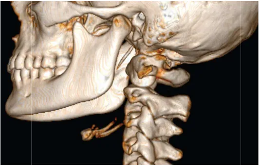
Figure: Volume Rendered CT of Neck with reconstruction showing SP
4
5 Stylopharyngeus
Arises from medial aspect base of styloid process. It slopes down
across the Internal Carotid artery, in front of which it crosses the lower
border of the superior constrictor and passes down inside the middle
constrictor to be inserted with palatopharyngeus into the posterior border
of thyroid lamina and the lateral aspect of epiglottis. The
glossopharyngeal nerve curls round the posterior border of muscle from
medial to lateral and supplies it. It helps lift the larynx during swallowing
and phonation.
Stylohyoid
Arises from posterolateral surface of base of styloid process. Its
tendon is perforated by posterior belly of Digastric tendon. Gets inserted
to the junction of body and greater cornu of hyoid bone. Innervation by
facial nerve. its actions are: pulls hyoid upwards and backwards and with
other hyoid muscles it fixes the hyoid bone.
Styloglossus
It is the shortest of the three muscles. It arises from tip and adjacent
part of the anterolateral surface of the styloid process as well as from the
upper end of stylohyoid ligament. It passes downwards and
forwardsin-between the internal and external branches of Carotid arteryand is
6
hyoglossus. It is supplied by the hypoglossal nerve. During swallowing it
pulls the tongue upwards and backwards.
Thus each of these muscles has a different nerve supply. They all act
significantly during swallowing.
Stylohyoid Ligament
Extends from the tip of the styloid process to lesser cornua of
hyoid bone.
Stylomandibular Ligament
Is an accessory ligament for the temperomandibular joint. It
represents the thickened part of deep cervical fascia which separates the
parotid and submandibular salivary glands. It is attached above the lateral
surface of the styloid process and below the angle and posterior border of
the ramus of the mandible.
The external carotid artery is intimately related to the muscles
attached to the styloid process. Running deep to the stylohyoid and hence
posterior belly of diagastric, it lies superficial to the stylopharyngeus and
the styloglossus en route to the parotid gland. Conversely the
retromandibular vein runs superficially to all elements of the styloid
7
The styloid apparatus (process and muscles) lies lateral to the
carotid sheath. The external carotid artery passes between the muscles of
the stylohyoid apparatus. It runs deep to digastricand stylohyoid, but
superficial to stylopharyngeus, to enter the parotid gland. The
retromandibular vein, on the other hand passes down from the parotid
gland superficial to stylohyoid and digastric.
Glossopharyngeal nerve:
The glossopharyngeal nerve has sensory, motor and autonomic
components. The sensory ganglion cells lie in the superior and inferior
ganglia of the nerve,
The central processes pass to the nucleus of tractus solitaries,
convening the taste sensation and to the nucleus of spinal tract of the
trigeminal nerve, conveying somatic sensation. The motor nucleus lies in
the upper part of the nucleus ambiguous, which receives bilateral
supranuclear innervation from corticobulbar fibres. This nucleus supplies
the stylopharyngeus. The autonomic parasympathetic fibres arise in the
inferior salivatory nucleus. These fibres are carried in the lesser petrosal
nerve via tympanic branch to the otic ganglion. The postganglionic fibres
are distributed to the parotid gland via auriculotemporal nerve.
The glossopharyngeal nerve emerges from the brainstem in line
8
It descends between jugular vein and carotid artery, picking up
sympathetic fibres from carotid plexus as it loops forwards medially to
reach the soft tissues of the oropharynx, posterior tongue and palate. In its
course it gives off lesser petrosal nerve conveying the secretomotor fibres
for the parotid gland to the otic ganglion. An important nerve, the carotid
branch conveys chemoreceptor and stretch reflex information,
respectively from the carotid body and sinus centrally for respiratory and
circulatory reflex function. The final branches of glossopharyngeal nerve
are the pharyngeal,tonsillar, and lingual branches, conveying general
sensation and taste sensation from the posterior third of the tongue and
oropharynx.
Branches :
• Tympanic nerve: also known as Jacobson’s nerve, this branch supplies
the tympanic plexus and is the reason for referred otalgia secondary to
pain affecting the oropharynx
• Lesser petrosal nerve; this branch provides post-ganglionic
secretomotor supply to the parotid gland and passes through the
infratemporal fossa
• Carotid sinus and carotid body branches: these branches are also
called as Herring nerve, take afferent nerves from the chemoreceptor
in the carotid body and baroreceptors from the carotid sinus.
• Pharyngeal branches: these branches join the fibres from the vagus
on the posterior aspect of middle
from the plexus perforate the muscle fibres of the middle constrictor
and innervate the mucosa of oropharynx. The tonsillar branch
anastomoses with lesser palatine nerves to supply mucous membrane
of the palatine
swallowing dysfunction
• Lingual branches: these provide common sensation, taste and
secretomotor supply to posterior third of tongue.
9
on the posterior aspect of middle constrictor muscle. Small nerves
from the plexus perforate the muscle fibres of the middle constrictor
and innervate the mucosa of oropharynx. The tonsillar branch
anastomoses with lesser palatine nerves to supply mucous membrane
of the palatine tonsil. Injury to the pharyngeal plexus may lead to
swallowing dysfunction
Lingual branches: these provide common sensation, taste and
secretomotor supply to posterior third of tongue.
constrictor muscle. Small nerves
from the plexus perforate the muscle fibres of the middle constrictor
and innervate the mucosa of oropharynx. The tonsillar branch
anastomoses with lesser palatine nerves to supply mucous membrane
to the pharyngeal plexus may lead to
10
Figure: Axial Cuts of neck showing Glossopharygeal nerve
Sandev and Klara Soker give possible embryological basis of
styloid process pathology as
1) Tympanohyal part- base of styloid process
2) Stylohyal part- larger part of styloid process
3) Ceratohyal part- precursor of stylohyoid ligament
4) Hypohyal part- development precedes the small horn of hyoid bone
It is believed that the ceratohyal part of second branchial arch
contains small parts of embryological cartilage that may or may not at
later stage mature into bone.
11
Another possibility is mineralisation of the ligament as a result of
ageing and degenerative processes.
EAGLE’S SYNDROME
Pietro Marchetti is credited with the first observation of an
elongated Styloid Process. Watt W. Eagle, an Otorhinolaryngologist, first
described about elongated Styloid process and its association with
craniofacial pain in 1937, presenting his paper “Elongated styloid
processes, report of 2 cases”.
The symptoms were mainly localised in the pharynx similar to
chronic pharyngitis. According to Eagle’s report it was possible for pain
to radiate to middle ear or to the mastoid region. Associated more often
with foreign body sensation in the pharynx with difficulty in swallowing.
On a brief note the following were the two cases reported by Eagle,
Case 1: a 31 year old female who had pain over left neck for 8 years
which radiated to left middle ear and mastoid region. Symptoms appeared
after tonsillectomy. On surgical removal of 2 cm left SP, pain vanished.
Case 2: a 56 year old male who had difficulty in swallowing for 20
years, whose physician advocated against tonsillectomy as his elongated
styloid, would create problems post tonsillectomy. He underwent
tonsillectomy with excision of 7cm elongated SP, which relieved his
12
Eagle reported that the normal styloid projection was ~ 2.5 cm in
length and any process longer than ~ 2.5 cm might be considered
elongated. The incidence was approximately 4%. Of which only 4%
present with symptoms. Kaufman et al, Chandler et al, Ferrario et al, Zaki
et al and Palesy et al reported the average length of styloid process to be
about 2 to 3 cm.
The incidence of Eagle’s syndrome is therefore estimated to be
about 0.16%
The actual cause of elongation of styloid process or the
calcification of the stylohyoid ligament is unclear. But several theories
have been proposed.
It may be entirely genetically determined with partial or complete
calcification of stylohyoid ligament and growth of osseous tissue at the
insertion of stylohyoid ligament.
It has also been suggested that it may be caused by trauma and
there are suggestions to association with early onset of menopause.
Eagle’s syndrome is more common in women compared to men.
Patients are usually older than 30 years; very rarely younger.
A wide variety of symptoms have been attributed to elongated
styloid process
• Cervicofacial pain
13 • Foreign body sensation in throat
• Pain on changing head position
• Cervical pain
• Pain on swallowing
Eagle described two forms based on the symptoms
First group consists of patients suffering from pharyngeal pain
localized in tonsillar fossa that sometimes radiated to hyoid bone. The
symptoms are attributed to scarring around the styloid tip following
tonsillectomy
Second group consists of patients who had not undergone
tonsillectomy. Pain and foreign body sensation are the most common
symptoms.
Steinmann proposed various theories to explain ossification
a) Theory of reactive hyperplasia- trauma can cause ossification at the
end of styloid process down the length of styloid ligament contains
remnants of its connective tissue and fibrocartilagionus origins, the
potential for ossification remains
b) Theory of reactive metaplasia- an abnormal post-traumatic healing
14
c) Theory of anatomic variance- the early elongation of styloid
process and ossification of styloid ligament are anatomical
variations that occur without recognisable trauma.
In some mammals it is normal to find an ossified chain in place of
this ligament; in humans it is uncommon (but not rare) anatomic variant.
In the human embryo, the epihyal cartilage is resorbed and its fibrous
sheath persists as the stylohyoid ligament. Failure of this step leaves
cartilage that may ossify. Instead of a single structure there may be a
chain of 3 or 4 bony parts, reflecting the multiple cartilages of the
embryo. The width of the structures, length and the degree of ossification
are highly variable. Ossification when it occurs is usually (but not
always) bilateral.
Several mechanisms for the pain if Eagle’s syndrome have been
proposed.
These include
1. Compression of the neural elements, glossopharyngeal nerve,
lower branch of the trigeminal nerve and/or chorda tympani by
the elongated styloid process
2. Fracture of the ossified stylohyoid ligament followed by
proliferation of granulation tissue that causes pressure on
15
3. Impingement on the carotid vessels by the styloid process
producing irritation of the sympathetic nerves in the arterial
sheath
4. Degenerative and inflammatory changes in the tendinous
portion of stylohyoid insertion a condition called insertion
tendinosis
5. Irritation of pharyngeal mucosa by direct compression by the SP
6. Stretching and fibrosis involving the fifth, seventh, ninth and
tenth cranial nerves in the post tonsillectomy period.
Stylalgia - pain in elongated styloid process may be due to
formation of a stiff bony clasp which hampers the elastic movement of
stylohyoid ligament during contraction of pharyngeal muscles, the pain
being secondary to irritation of sympathetic plexus around carotid arteries
or sinus branch of glossopharyngeal nerve.
Elongated styloid process may cause pseudoarthrosis between
styloid process and stylohyoid ligament causing intermittent locking or
fixation.
Normal length of styloid process varies greatly although in
majority of subjects it is 20-30 mm long. It is considered elongated when
it is longer than 30mm. incidence being 4% of general population.
16
Earlier study has reported average length of styloid process to be 1.52cm
on the left and 1.59cm on the right in Indian subjects.
The cause of onset of pain in subjects previously free of symptoms
is unknown but several mechanisms have been studied upon including
rheumatic styloiditis caused by pharyngeal infections, trauma,
tonsillectomy and involutional changes associated with ageing
(degenerative cervical discopathy may shorten the cervical spine and alter
the direction of the styloid process).
A styloid process within the normal length is usually not palpable.
The diagnosis of Eagle’s syndrome must be confirmed by good history
taking and physical examination. It should be confirmed by imaging.
There can be several variations in the styloid chain; including variations
• in the length of the process,
• thickness of the segments,
• angle and direction of the deviation and
• the degree of ossification.
The differential diagnosis of this condition includes
1. cranial nerve neuralgias (e.g., trigeminal, glossopharyngeal,
sphenopalatine, superior laryngeal, and primary geniculate
neuralgias,
2. temperomandibular joint disease,
3. chronic pharyngotonsillitis,
17
5. tumors in the oropharynx.
Unlu and colleagues assessed elongation of the styloid apparatus
(styloid process and ligament) in degenerative or inflammatory diseases
such as ankylosingspondylitis, psoriatic arthropathy, and cervical
spondyloarthrosis, in which cervical spine involvement can be seen. The
authors concluded that an elongated styloid process might be another
manifestation of enthesopathy in the cervical spine.
PAINFUL SYNDROME OF OROFACIAL REGION
Several syndromes are in sync with the symptomatology of
EAGLE’s syndrome and are often studied as orofacial syndromes with
differential diagnosis with Eagle’s syndrome.
This group of diseases is
1. Costen syndrome
2. Trotter’s syndrome
3. Myofacial painful syndrome
4. Trigeminal neuralgia
5. Glossopharyngeal neuralgia
6. Carotidynia
7. Hyoid bone syndrome
18
Costen syndrome: (Temperomandibular joint dysfunction)
Manifests with several symptoms that can be divided into
auricular, articular and cranial. The joint is sensitive to palpation with
pain and crepitation. Hearing is poorer with buzzing in the ears. Dizziness
and headache around the eyes crown and back of the head. One
explanation for this condition is that these changes are preceded by loss
of posterior or all teeth during which bite drops and mandible moves
distally pressing the joint (glavicom0 discus articularis and posterior part
of the joint chamber. The pressure causes the disk to deform and shift, so
that it no longer protects the arch and posterior part of the joint chamber
from nerve pressure. This causes irritation from auriculotemporal nerve
which causes headache especially occipital.
In 1958, Freese gave a new hypothesis for Costen syndrome. He
based the explanation on trigger centres. Namely, he considered that
hearing disorders are caused by a “trigger” point in the masseter,
dizziness a “trigger” point in the sternocleidomastoid and pain in the
tongue and pharynx spasm of the geniohyoid, digastric and pterygoid
19 Trotter’s syndrome:
Also known as Morgagni’s sinus syndrome. Comprises of three
symptoms occur in Nasopharyngeal carcinoma as a result of tumour site
and spread. It manifests with neuralgic pains in the lower jaw radiating to
ear, hearing loss and blocked sensation in the ear and palatal asymmetry (
due to levator veli palatine involved dueto tumor invasion) and trismus (
due to invasion into the pterygoid muscles.
Painful Myofascial Syndrome:
Syndrome manifests with muscular spasm, restricted mobility and
sensitivity. Associated with pain in muscular – facial structure of
masticatory, neck and back muscles. The concerned muscles are sensitive
to touch and pressure which the masticatory impulses pass into CNS and
return in the form of pain. Hence “Trigger” zones here are the muscles
and are called as ‘zones of impact’. Patients are conscious of zones of
impact. The disorder gets complicated by occurrence of new trigger zones
under the influence of previous ‘trigger’ zones, thus creating a ‘vicious
cycle.’ It is five times more common in post-menopausal women.
Etiological features include malocclusion or poor deltopectoral posture
20
Chronic fascial pain may be helped by applying local heat,
ultrasound therapy and often massage to relieve trigger point tenderness.
Clinical features of a myofascial trigger point
• Focal point tenderness
• Reproduction of pain on trigger point palpation
• Pseudoweakness of the involved muscle
• Referred pain
• Limited range of motion following approximately 5 seconds of
sustained trigger point pressure.
Pain caused by tumor:
Although tumors rarely present with facial pain, nearly 80% of
patients with head and neck cancers experience facial pain related to their
tumor or treatment. Constant or progressive dull or gnawing pain
particularly if associated with other suspicious symptoms or neurological
signs should alert the treating clinician. A past history of malignancy may
arise the possibility of metachronous tumors or metastasis. A thorough
examination of the head and neck and upper aero digestive tract along
with appropriate radiological imaging is mandatory to exclude the
21
natural history of growth such as neuroma and pain may have been
present for several years.
Trigeminal neuralgia:
Trigeminal neuralgia is one of the most frequent painful syndromes
of facial region ad is probably most painful condition known. In 95% of
subjects it involves the lower two major divisions (maxillary and
mandibular). It occurs in the form of sudden excruciating, ‘boring’ pain,
often described as electric shock sensation, lasts less than one minute.
The pain is usually described in 2 characteristic distributions. First
runs from lower canine tooth along the lower jawto just in front of the ear
and sometimes round into the upper jaw (i.e. involves the mandibular and
maxillary divisions of trigeminal nerve). The second less frequent type
runsfrom the upper incisor or canine, up the side or inside the nose and
encircles the eye, involving both maxillary and ophthalmic divisions of
trigeminal nerve.
It is probably this spread over two divisions that makes simple
surgical section at peripheral branches unsuccessful in managing the
22
Although it is claimed that transient sensory deficit may follow a
spasm of pain any evidence of sensory loss, impaired corneal reflex or
trigeminal motor weakness should invalidate the diagnosis.
Initial attacks are generally mild and infrequentwith remissions
lasting for months. Over time remissions are shorter with severe attacks
until they culminate in daily episodes for months. As a rule it is unilateral
and never passes to the other side. The pain occurs by activating a
specific trigger point which is stimulated by mastication, speech
movements of lips and tongue, yawning, touching the face, sudden
movements of head and walking, noise or strong light.the pain never
occurs during sleep.the attacks are more frequent on the right with
women affected more than men.
Etiology is unknown and there lies a persisting question of origin
of pain – central or peripheral disturbance.
Although trigeminal neuralgia may complicate multiple sclerosis, it
is very rare as presenting symptom of this disease. Treatment of
trigeminal neuralgia is directed toward prophylactic treatment most
commonly using the antiseizure medication carbamazepine. Other
medications include gabapentin, baclofen, phenytoin, sodium valproate,
23
Extracranial peripheral denervation involves treatment at trigger points,
which can be temporary using local anesthetics or permanent using
alcohol, freezing, and heating. Surgical radiofrequency ablation involves
destruction of the Gasserian ganglion by radiologic guidance of a device
inserted into the cheek and through the foramen ovale into the Gasserian
ganglion. The usage of glycerol injection called glycerol trigeminal
rhizotomy is effective but has a high recurrence rate. Microvascular
decompression is utilized to treat vascular loop compression of the
trigeminal nerve by suboccipital retro mastoid craniectomy. Other
procedures utilized include gamma knife radiosurgery and botulinum
toxin injections.
Glossopharyngeal neuralgia:
It is a rare condition that presents at about one tenth the frequency
of trigeminal neuralgia. It consists of excruciating severe pain in the
palate, throat and external auditory canal, locations demonstrating the
somatic sensory distribution of glossopharyngeal nerve. The pain has the
typical burning, electric shock quality of neuralgia and is triggered
mainly by swallowing. The incidence of underlying lesions inside the
skull is very much higher than in trigeminal neuralgia.
Both phenytoin and carbamazepine may control the pain, but MRI
24
exploration is necessary if all medical treatment fails. Peripheral
glossopharyngeal section has little to commend it and can seriously
25
CLINICAL EVALUATION
Elongated styloid process is commonly identified by screening
patients by suspicion. A palpable styloid process is considered elongated.
Palpation of styloid is done by palpating over the tonsillar fossa. Very
commonly the pain associated with Eagle’s syndrome is due to
neurogenic compression on Glossopharyngeal nerve. This can be
aggravated on touch over the tonsil.
Lidocaine pain relief test is another modality to evaluate styloid
process clinically. patients were administered the lidocaine infiltration
test. In this test 3 cc of 2 % lidocaine was infiltrated into the tonsillar
fossa on the side where the patient had complaint of pain and where the
styloid process was palpable. After 15 min the patient was asked to point
out the intensity of pain felt. Reduction in intensity to less than one-third
of the original pain levels after injection was taken as relief.
Incorrect diagnosis can occur by mistaking pterygoid hamulus for
26
RADIOGRAPHIC FINDING
Elongated Styloid process can be typically identified by
radiographic studies.
Common imaging modalities include
1. Orthopantomogram Digital
2. CT neck and CT Temporal bone with 3D reconstruction of styloid
3. Cone beam CT of styloid
4. Conventional lateral and anteroposterior (AP) views of head and
neck radiograph
5. Towne’s view
6. MRI
ORTHOPANTOMOGRAM
Panoramic radiography is useful for detection of an elongated
styloid process and / or ossification of stylohyoid ligaments in patients
with or without symptoms and can thus help avoid misinterpretation of
the symptoms as tonsillar pain or pain of dental, pharyngeal, or muscular
origin.
Panoromic radiography is usually performed in a dentist’s office.
The typical use of this modality is to visualize the temperomandibular
27
mandibular fractures, temperomandibular joint pathologies and styloid
process examination and its relation to surrounding structures.
Panoramic radiography of jaws and styloid process helps overcome
limitations of conventional intraoral radiography. The radiation dose to
the patient is 10 times less than that of the full mouth intraoral
radiography survey. Further it allows overall coverage of entire dental
and surrounding architecture. There can, however be up to 25% distortion
and measurement may therefore difficult. This is a very useful tool when
patient has trismus. The mandibular ramus, styloid process,
temperomandibular joint, superior maxillary sinus and floor of mouth
structures are visualized. These structures cannot be seen in intraoral
radiography. The panoramic view is limited when compared with
conventional intra oral radiography by necessity for careful technique,
sensitivity to patient motion and relatively poor spatial resolution.
Rotational panoramic radiographs are obtained by rotating a
slender x-ray beam in a horizontal axis that is positioned extra orally. The
principle is very similar to that of conventional; x-ray tomography. With
this technique the focal spot of x –ray tube anode acts as the dimension of
projection. To eliminate scatter and artefact, the x-ray beam is angled
beneath the occipital condyles of the skull approximately - 4°to -7°. The
28
rotate around the patient’s face during the exposure. The film is exposed
through a narrow opening in the cassette that is usually flexible. These
films are very technique sensitive. If the patient is properly positioned
certain structures will be out of focal trough (blurred). Even in properly
positioned patients, the midline structures may be flattened and spread
out or may be projected as a double image.
The normal anatomic structures seen in panoramic radiography are
listed below
1. Mandibular condyles
2. Ramus of the mandible
3. Cervical spine
4. Maxillary sinus
5. Inferior turbinate
6. Inferior alveolar canal
7. Mental foramen
8. Nasal fossae
9. Dentition
10.Sigmoid notch
11.Mandibular coronoid process
12.Developing tooth bud
13.Hyoid bone
29
Due to the medial angulation of the styloid process and
superimposition of other skeletal structures, some errors may occur when
measuring the length of the styloid. Further imaging studies are required
to correlate the symptoms with an elongated styloid process as well as
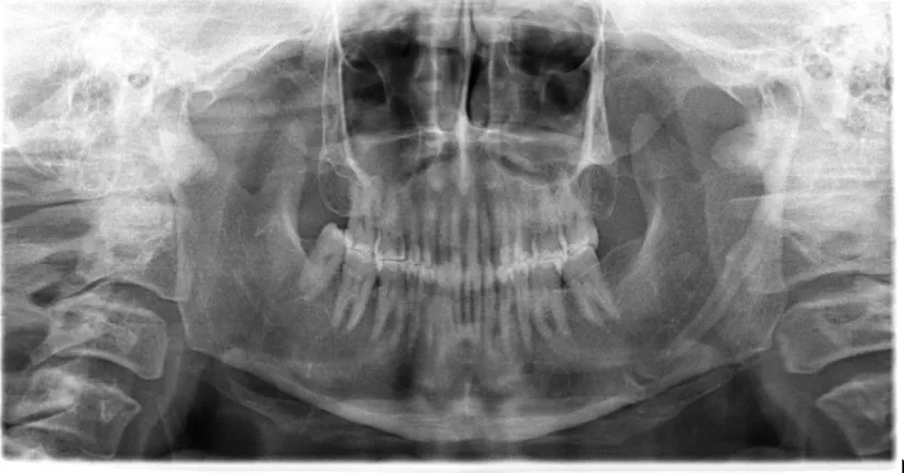
[image:41.595.112.523.248.463.2]with the type and pattern of elongation of the styloid process.
Figure: Digital OPG of Bilateral elongated Styloid process with
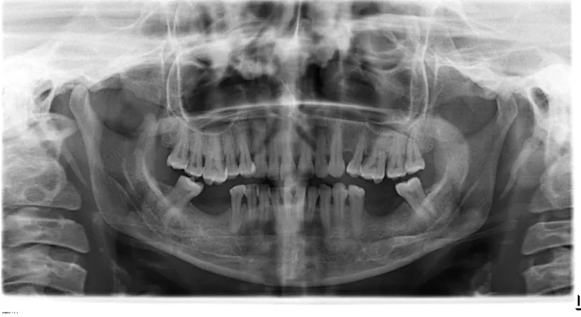
30
Figure: Bilateral elongated styloid process by OPG Digital
CT NECKand TEMPORALBONE: 3D reconstruction
Computer tomography depends on the same interaction of x-ray
photons and atoms within matter as standard radiography. But penetrating
photons do not fall on a photographic cassette. Instead are captured
photoelectric detectors and converted to electrical impulse. Further X-ray
tube is rotated around the patient so that a fan shaped X ray beam passes
through the patient in a discrete plane from 1-10 mm thick. The stream of
varying electrical signals produced in the photoelectric detected is
converted into digital data and mathematically reconstructed into an
image corresponding to the densities in the selected plane of that patient,
rendered as a fine checkerboard of pixels (picture elements)
corresponding voxels (volume elements), tiny discrete volumes of tissue
31
instant viewing, fed to a laser printer for hard copy rendition on film.
Subsequently recorded on magnetic tape, magnetic disc or optical disc for
archiving.
The system has several important advantages:
1. Scale of density differences to identity the difference in x ray
absorption between water, fat, muscle, dense fibrous tissue, blood
clot and tumor tissue as well as major difference between air and
bone
2. Elimination of tissues put aside the selected plane
3. Easy production of coronal, axial views. Allowing 3D
reconstruction.
The densities are presented in a varying range and hence cannot be
presented in a same film.
For styloid process imaging bone window settings is selected. Such
images resemble plain radiographic tomograms without blurring of
smearing effect.
A scout/pilot/guide image is similar to image acquired in digital
radiograph which helps in assessing the film with regards to field under
study and the thickness of planes.
With recent advances in technology computer generated images are
32
another. They are not as fine as originally obtained images because one
dimension of new pixels is determined by thickness of original slices
which is nearly always larger than the dimensions of originally acquired
pixels. Despite this disadvantage, reconstructions are frequently used for
allowing spatial orientation of structure under study to allow for relation
to surrounding structures.
With advent of helical (spiral) CT images are acquired faster
rendering motion artefacts to minimum and allowing for smoother
reconstruction by giving ability to select thinner sections
Accurate determination using two-dimensional radiographic
procedures can be difficult due to projection geometry considerations.
Panoramic radiography may distort the dimensions of styloid process and
magnification of the radiographic image may vary with the angulations of
the process itself.Therefore, a simple measurement in millimetres is not a
suitable criterion due to the radiologic factors involved.
The true character of bilateral asymmetrical malformations may be
evaluated with 3D CT as overlap of structures is not encountered.
Operator related errors are also minimized. Despite this certain
limitations exist- slight movement may result in degradation of images
and higher radiation dose may be required depending on the number of
33
Thus 3-dimensional CT is the best modality to view the length of
the styloid process and provides accurate estimation of length. The
anteroposterior view shows the presence or absence of bilateral
involvement and deviation. Langlai’s states that ‘short styloid
processes,deeply inserted into the petrous part of the temporal bone,
dissimulated by the vaginal crest, are generally invisible in standard
radiographs’. Such misreading of short styloid process could beconfirmed
in the 3D reconstruction of styloid process with CT Neck and/ or
Temporal bone.
Computed tomography scans can define the length, angulation, and
anatomic relationship of the styloid process. On the basis of the location
of palpation of the styloid process in the tonsillar fossa, it was classified
by Ravinder Verma into 3 grades, which provide information on the size
of the styloid process:
a. grade I- the tip of the styloid process is palpable in theupper pole of
tonsillar fossa;
b. grade II- the tip of the styloid process is palpable in the middle of
tonsillar fossa;
c. grade III- the tip of the styloid process is palpable in the lower pole
34
In Verma’s study, the majority of the patients were in grade II or III.
There are many methods of measuring the length of styloid
process. Goldstein and Scopp suggested the following criteria for
radiographic evaluation of ESP.
1. If the area of stylomandibular ligament is indistinct, it is said that
the area is not observable.
2. If the radio-opacity of a styloid process is less than one third of the
length of the ramus of the mandible, it is arbitrarily considered as
within the normal range.
3. If the radio-opacity is more than one third but not down to the
angle of mandible, the elongated SP is ‘partially calcified’
4. If the radio-opacity is touching or at the level of the angle of the
[image:46.595.108.568.506.689.2]mandible, the elongated SP is ‘completely calcified’.
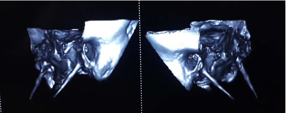
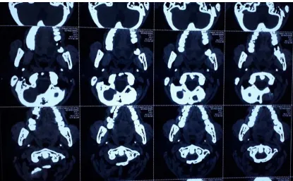
35
Figure: Axial cuts of Neck with temporal bone show styloid process.
CONE BEAM CT:
CBCT technique consists of the use of a round or rectangular cone
shaped X- ray beam with a single 360° scan where the X-ray source and a
reciprocating array of detector simultaneously move around the patient’s
head, which is stabilized with a head holder. Single projection images,
known as “basis” images, are acquired at certain degree intervals, which
are similar to lateral cephalometric radiographic images, each slightly
offset from one another. The series of such basis projection images is
referred to as the projection data, on which software programs
incorporating sophisticated algorithms are applied to generate a 3D
volumetric data set, which can be used to provide primary reconstruction
36
The fundamental difference between CBCT and spiral CT is that
CBCT utilizes a cone shaped beam and an area detector and that captures
a full volume of image in a single rotation where patient movement is not
required. On the other hand, spiral CT makes use of a narrowly
collimated, fan-shaped X-ray beam and a linear group of detectors. Here,
the patient has to be moved continuously along the gantry while the X-ray
beam rotates around the patient. In most of the cases conventional CT has
not been widely utilized due to the high radiation dosage, the cost of the
procedure, proximity to a CT centre and commonly unfamiliarity with
interpreting CT results. The use of CBCT can negate these concerns and
allow for improved diagnosis and patient safety. On comparison with CT,
CBCT has high accuracy and sensitivity and can capture the maxilla and
mandible in a single rotation of the X-ray source.
Cone beam CT is recommended as dose- sparing technique
compared to standard CT for common oral and maxillofacial radiographic
imaging task. Advantages are:
I. Lower radiation dose
II. Individualized over-lap free reconstructions
III. Data can be imported and exported for other applications
Surgical and non-surgical modalities exist as the treatment options
37
Trans pharyngeal infiltration of steroids under local anaesthesia can
[image:49.595.111.514.147.420.2] [image:49.595.155.486.476.694.2]relieve the symptoms, but styloidectomy is the treatment of choice.
Figure: CBCT Sagittal section for styloid process left
38 CONVENTIONAL RADIOGRAPHS:
Conventional radiographs anteroposterior and lateral views of neck
and Towne’s view are routinely used by otolaryngologist to screen for
elongated styloid after clinical evaluation.
Towne view is an angled AP radiograph of the skull, used
to evaluate the condylar and styloid process relation. The patient
positioned with nuchalridge against the image detector. The
infraorbitomeatal line perpendicular to the image receptor.The centring
pointbeam travels 30 degrees caudad to the orbitomeatal line. Is useful to
evaluate skull fractures and the length of styloid. After mapping the first
ear canal and the Frankfurt line. The patient was positioned with the
Frankfurt plane parallel to the floor (horizontal) and the median sagittal
plane perpendicular to the floor, and was asked to occlude in maximum
intercuspation (MI). The cephalometric tracing was performed as follows:
a line was drawn from the ear canal to the styloid process apex and a
second horizontal line was drawn perpendicular to the Frankfurt line and
passing through the lowest point of the external auditory canal, forming
an angle
The reverse Towne view helps in detecting the angulation of
styloid process. patient was positioned facing the x-ray equipment with
39
a horizontal line was drawn passing at base of the skull. A second line
was drawn perpendicular to the horizontal line passing through tip of the
styloid process, and a third line was drawn along the styloid process from
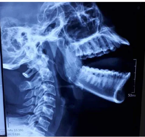
[image:51.595.206.444.225.451.2]the base of the skull to the apex of the styloid process.
Figure: Conventional Radiograph showing elongated styloid process
MAGNETIC RESONANCE IMAGING:
The process of deriving images from weak signals given up by
spinning hydrogen nuclei (protons) after they have been placed in a
strong magnetic field and then irradiated with radiofrequency pulses is
called nuclear magnetic resonance imaging. This has been to shortened to
magnetic resonance imaging. Abbreviated to MRI. MRI thus is based on
electromagnetic radiation. The gray scale in MRI is due to difference in
40
Amount of hydrogen in various substances and tissues under
constructiondiffers only slightly but chemical binding and hence nuclear
spin properties differ significantly. For instance, the protons in hydrogen
atoms bound to carbon in fat respond differently when compared to
protons in hydrogen atoms bound to oxygen in water molecules, which in
turn respond differently than same hydrogen atoms in water bound to
large macromolecules of protein. The weak signals emanating from
relaxing hydrogen protons are spatially coded and mathematically
reconstructed into tomographic images.
MRI in styloid process is mainly to relate to spatial relationship
with the glossopharyngeal nerve and the carotid complex.
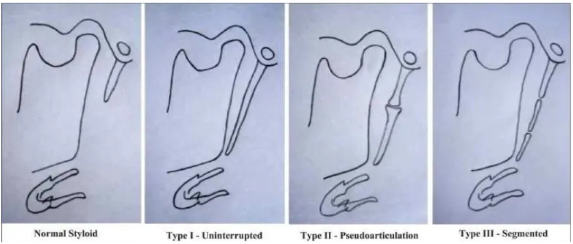
According to Langlais and co-workers radiographic finding of an
elongated styloid process and calcified ligaments of stylohyoid
connections can be divided to two criteria
A)Morphological criteria
a. Elongated styloid process
b. Pseudo articulated styloid process
c. Segmented styloid process
B) Criteria determined by means of calcification (4 types)
41
b. Partially calcified styloid process
c. Nodular type calcification
[image:53.595.111.529.231.409.2]d. Completely ossified styloid process
Figure:Langlais classification
The diagnosis can be made by radiographs or CT. Physical
examination may demonstrate a palpable styloid process in the tonsillar
fossa and palpation of the tip may exacerbate the existing symptoms. CT
provides precise relationship of an elongated styloid process or
42
Eagle syndrome. Three-dimensional volume rendering reconstruction
image from CT demonstrates ossified stylohyoid ligaments with
articulations, extending from the skull base to the hyoid bilaterally.
TREATMENT MODALITIES
First line in any disorder being non-surgical therapy- patient can be
given a trial of analgesic. Most common being NSAIDS and TCAs The
patient related outcome measures for non-surgical therapies when
compared to surgical therapies are relatively poor and seldom
ameliorating.
Conservative management is represented by the medical therapy
which can be further divided into first-line analgesics such as
43
of a combination of anticonvulsants, antidepressants, local injection. Also
a stellate ganglion block added to medical therapy can result in near
complete resolution of symptoms. The most effective treatment of pain
associated with Eagle’s syndrome is the association of gabapentin,
tramadol, and acetaminophen, associated with local injections of
mepivacaine/dexamethasone. The surgical approach turns out to be a
definitive treatment and is associated with a better quality of life
NON SURGICAL TREATMENT FOR ELONGATED STYLOID
PROCESS
Oral medication is usually started with
1. Gabapentin 300 mg/day
2. Tianeptine 1.5 mg/day
3. Tramadol hydrochloride 37.5 mg/day
4. Acetaminophen 325 mg 3 tablets/d
5. Pregabalin 75 mg/day
6. Amitriptyline 10 mg/day
7. 1 ml of Triamcinolone 10 mg combined with 0.3% Mepivacaine 3
ml was injected once in the tonsils and tender areas
8. Stellate ganglion block can be done once a week for 4 weeks- On
44
PARACETAMOL: (ACETAMINOPHEN)-
De-ethylated active metabolite of phenacetin with central analgesic
action by raising the pain threshold and weak antiflammatory action.
Poor inhibitor of PG synthesis in peripheral tissues though an active COX
inhibitive in the brain. Primarily metabolised by conjugation with
glucouronic acid and sulphate. Good absorption orally though only 1/4rth
protein bound. Half-life is 2-3 hrs with effective oral dosage for 3-5
hours.
TIANEPTINE:
Atypical Antidepressant reported to increase 5-HT uptake having
for use in psychomatic pain disorders. It is not stimulative or sedative.
Efficient in anxiodepressive states.
TRAMADOL:
Centrally acting analgesic relieves pain by opioid as well as
additional mechanism. Affinity for µ opioid receptor is low, though κ and
δ is very low. It inhibits reuptake of NA and 5-HT, thus activating
monoaminergic spinal inhibition of pain. Oral bioavailability is good.
Half- life of 5 hours and the effect lasts for 4-6 hours. It has very little
45
GABAPENTIN:
Is a lipophilic GABA derivative crossing the brain and enhances
GABA release but does not act as agonist at GABAA receptor.
Considered as a first line drug in neuralgic pains especially post herpetic
neuralgia, diabetic neuropathy and migraine prophylaxis. Well absorbed
orally. Excreted unchanged in urine. Half- life is 6 hours.
PREGABALIN:
Developed as a successor to Gabapentin, it is a Gabapentinoid.
Used commonly for musculoskeletal pain and off label use for
neuropathic pain. Oral absorption is good with good
bioavailabilitythough onset on action takes a long as a week. Its half -life
is 6 hours.
AMITRIPTYLINE:
Is a tricyclic antidepressant inhibiting NA and 5-HT reuptake into
neurons. On long term intake it desensitizes presynaptic α2, 5-HT1A and
5-HT1D auto receptors and induces other adaptive changes in number and
sensitivity of pre and post synaptic NA and 5-HT receptors as well as in
amine turnover in brain, net effect is enhanced nor-adrenergic and
46
Metabolized in liver. Plasma half-life varies from 16-24 hours. It is
commonly used in neuropathic pain.
STELLATE GANGLION BLOCK:
SGB may be a therapeutic option forfacial congestion for patients
with Eagle’s syndrome.
Stellate ganglion lies anterior to Longus colli muscle which lies
anterior to transverse process of C7 vertebra. The ganglion parse is made
of fused portion of C7 and T1 sympathetic ganglia. The ganglion lies
anteromedial to vertebral artery, medial to common carotid artery and
jugular vein and lateral to trachea and oesophagus.
Patient is placed supine with neck neutral position. Using
ultrasound guidance, junction of C7 transverse process with body is
identified and using 22 gauge 3/4rth needle is inserted and after aspiration
of nil confirmed, a composed solution of anaesthetic drug like lidocaine
or steroid is injected in trajectory fashion.
If no image guidance is used – blind technique, the
sternocleidomastoid muscle serves as a landmark. Medial end of muscle
identified at inferior margin of cricoid cartilage at level of C7 vertebrae
and depressed over the bony structure underlying. Then after palpating
47
fashion to hitch against the bony landmark of junction of transverse
process with its body and infiltered.
The adverse effect being permanent Horner’s syndrome.
PERISTYLOID BLOCK:
Patient is positioned supine with a slight head tilt to opposite side,
and the area over the mastoid process was scanned using a linear array
transducer. The scanning sequence should start from the mastoid process
in an axial view and proceeded caudally in a slight oblique direction to
capture an axial view of the styloid process. The styloid process can be
identified approximately 2 cm from the skin. Then the transducer is
rotated to obtain a longitudinal view of the styloid process. This provides
a view of the styloid process as an elongated hyper echoic line with an
acoustic shadow beneath. Colour flow Doppler can be used to identify the
vascular structures. Following the guide scan, the area should be prepped.
The elongated styloid process should be traced from the mastoid process
using ultrasound guidance. The internal jugular vein and internal carotid
artery can be identified just deep and slightly posterior to the styloid
process. A 1.5-inch, 25-gauge needle is directed in an out-of-plane
approach to the styloid process. Following contact with the process,
hydro localization with 0.2 mL of normal saline can be used to ensure
48
of 0.25% bupivacaine and 5 mg of dexamethasone through the extension
tubing after negative aspiration. Patient should be monitored for 24 hours.
SURGICAL MEASURES FOR ELONGATED STYLOID
PROCESS:
When unsatisfactory improvement is achieved with medication we
consider the following surgical interventions
1. Intraoral resection of styloid process (trans oral and extratonsillar)
2. Trans cervical resection of elongated styloid process
3. Microvascular decompression of glossopharyngeal nerve
Patients are to be evaluated in terms of the following
1. Section with shooting craniofacial pain
2. Precipitant of shooting craniofacial pain
3. Length of the styloid process on the affected and non-affected sides
4. Presence of trigger point and of a rigid funicular process in the
tonsillar fossa by palpation
5. Presence of vascular contact with glossopharyngeal nerve (as
evidenced by MRI)
49
Based on the above the method of surgery for patient is chosen and
followed up post operatively with patient related outcome measures.
TRANSORAL RESECTION OF STYLOID PROCESS:
Following tonsillectomy on the affected side, the white structure of
styloid process should be identified by elevating the connective tissue
around the styloid process. With mucosal elevators bluntly and gently
uncover the styloid process without damaging the branches of the
pharyngeal artery and vein and the glossopharyngeal nerve itself, which
surrounded the styloid process. Manipulation under microscope or
endoscope can be helpful. After uncovering the process, it should be
grasped with forceps, fractured bluntly and removed. Suturing the
superior pharyngeal constrictor can be done with nylon sutures.
Ring curette can be used in placed of periosteal elevator after
detaching the ligament to bluntly strip off the attachments from tip to
base of styloid process. Kerrison punch 4mm forceps can be used to
nibble the exposed styloid from its base.
Extratonsillar and trans oral approach to styloid is tedious and
involves dissection more laterally to anterior pillars and identification of
capsule of tonsil to trace the styloid process laterally to it and using the
same principle of dissecting it off its attachments and fracturing it out.
Advantage being tonsil are manintained and avoids the need for suturing
50
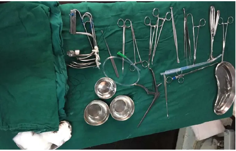
[image:62.595.124.512.142.391.2]Figure: Instrument tray for tonsillo-styloidectomy
Figure: skeletonization of styloid process
[image:63.595.216.452.368.569.2]51
Figure: tonsillectomy done
Figure: Fracturing of skeletonized styloid process
Figure: Post resection of styloid process
[image:64.595.210.421.405.600.2]52
Figure: Fracturing of skeletonized styloid process
using Kerrison’s punch
Figure: Post resection of styloid process
Figure: Fracturing of skeletonized styloid process
53
Figure: Excised Elongated Styloid Process
TRANSCERVICAL RESECTION OF STYLOID:
Under general anaesthesia, a bolster placed under the shoulders,
with the head in slight hyperextension, turned towards the opposite side.
A submandibulectomy skin incision, about 4 cm long, performed 4 cm
below the inferior border of the mandible to avoid the marginal
mandibular branch of the facial nerve, between the anterior edge of the
sternocleidomastoid muscle and the hyoid bone. The platysma should be
incised- together with the superficial layer of the cervical fascia to
provide access to the submandibular space. The submandibular gland in
the submandibular space should then be retracted with a Dautrey
retractor. The styloid process can be identified with a finger by sliding
along the styloglossus muscle superiorly, posteriorly and laterally, and
the stylohyoid ligament should be grasped with forceps and sectioned in
54
detachment using a periosteal elevator in order to progressively disinsert
the styloid muscles, and then fractured by a valgus procedure with a
finger. The styloid process should be carefully removed; taking care to
completely release the last muscle attachments with the periosteal
elevator. A suction drain can be inserted, and the platysma and skin then
be closed. Postoperative management consisted of analgesics and wound
dressings. Drainage to be removed at 48 hours and the patient can be
discharged by the second postoperative day.
An open incision provides better exposure of the operative site,
without interference by mouth opening and the depth of the field via the
trans oral approach. It is therefore often the only solution in the presence
of an atypical course of the styloid process, or an extremely long styloid
process. The disadvantages reported are the neck scar, the risk of injury to
the marginal mandibular branch of the facial nerve, and longer operating
time. Certain techniques have thescar reduced to 4 cm and hidden in a
skin-fold of the neck. The risk of facial nerve injury is negligible with an
incision-situated 4 cm below the inferior border of the mandible. The
operating time can be reduced to as low as 30 minutes, due to a reduced
dissection phase. The neurovascular risk is low, mainly consisting of the
55
Figure: Schematic view of styloidectomy via a right trans cervical
approach after skin incision, incision of the platysma muscle and opening
of the submandibular space. The submandibular gland is retracted
superiorly and anteriorly. Demonstration of the stylohyoid ligament,
styloglossus muscle and tip of the styloid process
MICROVASCULAR DECOMPRESSION OF
GLOSSOPHARYNGEAL NERVE
Exploration of cerebellopontine angle was performed through a
retromastoid craniotomy. Compressive arteries are dissected from the
nerve and brain stem and maintained far from the root of entry zone by
interposing small pieces of Teflon between the offending artery and brain
[image:67.595.216.434.68.234.2]56
DIFFERENTIAL TREATMENT ALGORISM:
Presence Of Typical Symptoms
Exclusion Of Head And Neck Tumour Or Inflammator
Lesion
Presence Of Trigger Point In
Tonsillar Fossa
3D CT Presence Of Styloid Process
>25mm In Length 3 Months
Medication
3 Months Medication
Improved Followed For 1 Year And Recurs Go For
Resction
No Improvement Transoral Resection Of
Styloid
No Improvement With Transoral Resection Of Styloid - DO MRI and Go For Microvascular
Decompression Of Glossopharyngeal Nerve
MRI Improved Follow Up For