JOURNALOFVIROLOGY,JUlY1987,p.2182-2191 0022-538X/87/072182-10$02.00/0
CopyrightC) 1987, American Society forMicrobiology
Tissue-Specific
Expression of
Human
Provirus
ERV3
mRNA
in
Human
Placenta: Two of the Three
ERV3 mRNAs
Contain
Human
Cellular
Sequences
NOBUYUKIKATO,1'tSUSAN PFEIFER-OHLSSON,2't MIEKO KATO,' ERIK LARSSON,3 JANRYDNERT,2 ROLFOHLSSON,2tAND MAURICE
COHEN',*
BRI BasicResearch Program, National CancerInstitute Frederick CancerResearch Facility, Frederick, Maryland 217011; Department of Oncology, University of Umea, Umea, Sweden2; andDepartmentofPathology, Uppsala
University, Uppsala, Sweden3
Received 23 December1986/Accepted 24 March 1987
Threepolyadenylated RNAs, 9, 7.3, and3.5kilobaseslong,ofahumanendogenousretrovirus, ERV3,are abundantinhumanplacental chorion, representingabout0.03 to 0.05% of thetotal mRNA.Wecharacterized the structure of these mRNAsbyNorthern blot and Sl nucleasemappinganalyses. We found that all three RNAsweresplicedmRNAs that lacked 5.9 kilobases ofproviralsequence,includingthegaggeneandmostof thepolgene.In contrasttothetranscriptionpattern usual for otherretroviruses,thetranscription patternof the ERV3 provirus did not include a genome-length mRNA. All three of the ERV3 mRNAs initiated transcriptionatthesamepointin the 5' longterminalrepeat(LTR)andcontained identicalsplice junctionsin theprovirus.The3.5-kilobaseRNAwas atypical subgenomic proviral mRNA,with itspolyadenylationsitein the3' LTR.The twolargerERV3mRNAs, however,extendedthroughthepolyadenylationsite in the 3'LTR andweresplicedatasecondpositionapproximately370 nucleotidesdownstream fromthe 3'LTR. Thisfinding suggeststhat when the ERV3 retrovirusintegratedatthisgenomiclocusinanancestor ofhumans,itintegrated withinor adjacenttoacellulargene.
Human DNA contains multiple copies of endogenous retroviral sequences related to mouseor primate retroviral
sequences. Several human endogenous provirus families
have beenidentified, and clones of each family have been isolated from human recombinant DNA libraries (4, 5, 8, 14-16, 18, 22, 23). Although three reports described RNA expression ofa human type C-related provirus family (11, 27, 28), thesereports could notidentifythe expressedgene
locusbecausetheprovirusis present in 50to 100copiesper
haploidgenome. Wepreviously describedthe structureofa full-length human endogenous retrovirus, ERV3, which is presentinasinglecopyperhaploidgenomeand islocated in chromosome 7 (21, 22). The nucleotide and encoded amino acid sequences of ERV3 were found to be significantly relatedtobutdistinct from those of Moloney murine leuke-mia virus (Mo-MuLV), baboon endogenous virus, and the
humantype C-related provirus family described previously (16). We recently reported the isolation ofanERV3proviral
cDNA clone from a human fetal liver library (6). In the
presentstudy,wedemonstrate thatERV3 mRNAs of 9, 7.3,
and 3.5kilobases (kb)areexpressed in the chorionic villi of
human first-trimester andfull-term placenta. Northern blot andS1 nuclease analyses revealed that these RNAs areall spliced,env-containing forms that have initiatedatthesame
site in the 5' long terminal repeat (LTR) and are missing
identical sequences from the gag andpolgenes. Although
the 3.5-kb RNA is a typical subgenomic env-containing
mRNAthatterminates inthe 3' LTR withapoly(A)tail, the
* Corresponding author.
t Present address: National Cancer Center Research Institute,
Tsukiji5-chome,Chuo-ku, Tokyo,Japan.
tPresent address: Centre for Biotechnology, Karolinska
Insti-tute, Huddinge, Sweden.
9- and 7.3-kb ERV3 mRNAs have read throughthe3' LTR andarespliced againatasitedownstream from the 3' LTR.
MATERIALS ANDMETHODS
Tissues. First-trimester human placentas were obtained from routine elective abortions by vacuum suction at the DepartmentofGynecology,UmeaCounty Hospital, Umea, Sweden. Gestational agewas determinedby known date of conception orestimated from the date of the last menstrual period. In all cases gestational age was also evaluated by ultrasound investigation and fromcrown-rump length
mea-surements.After immediatewashingof the abortus, placen-tal chorionic villi and wholeembryos wereisolated undera dissecting microscope. Full-term humanplacentas were
ob-tained from Frederick Memorial Hospital, Frederick, Md. Tissues were immediately frozen in liquid nitrogen and
storedat -70°Cuntil needed.
RNA preparation. RNA was isolatedfrom first-trimester placentaltissuesbyhomogenization in guanidinium
isothio-cyanate, CsCl gradient centrifugation, phenol-chloroform extraction, and ethanol precipitation (24). Total placental RNA fromtermplacentawasprepared byamodification of
thelithiumchloride-ureaprocedure (1)asfollows. Placental
tissuewashomogenizedinasolutioncontaining 10 volumes
of 3 M lithium chloride-6 M urea, 50 mM Tris (pH 7.4), 5 mM EDTA, 0.1 M mercaptoethanol, and 0.1% (wt/vol) Sarkosyl byaPolytron homogenizerand then placedonice overnight. After centrifugation at 8,000 rpm for 90 min at
4°C,thepelletwas quickly suspended inasolution contain-ing10 mM Tris(pH 7.5),1 mMEDTA, 0.5%(wt/vol) sodium dodecyl sulfate (SDS), and 200 ,ug of proteinase K per ml.
The RNA was treated with phenol and precipitated with
ethanol. After centrifugation the RNA was dissolved in 10
mMTris(pH7.5)-i mMEDTA-0.5%(wt/vol) SDS, divided intoportions,andstoredin70% ethanolat-20°C until used.
2182
Vol. 61, No. 7
on November 10, 2019 by guest
http://jvi.asm.org/
Northern blot hybridization. Total placental RNA (10 ,ug) was heated at 65°C for 10 min in 20
[LI
of 50% (vol/vol) formamide (Fluka)-2.2 M formaldehyde (Fisher Scien-tific)-20 mM MOPS (morpholinepropanesulfonic acid, pH 7.0)-S5mMsodium acetate-1 mM EDTA-0.2% (wt/vol) SDS and subjected to electrophoresis in a 0.8% agarose gel containing 2.2 M formaldehyde for 15 h at 40 V in buffer containing 20 mM MOPS (pH 7.0), 5 mM sodium acetate, and 1 mM EDTA. RNA was transferred overnight to a nitrocellulose membrane in 20x SSC (lx SSC is 0.15 M NaCI plus 15 mM sodium citrate) (32). The membrane was baked in a vacuum oven at 80°C for 5 h and soaked in a boiled solution of 20 mM Tris (pH 7.8) at room temperature for10min. The membrane was prehybridized for 5 to 7 h at 42°C in 50% (vol/vol) formamide-5x SSC-5x Denhardt solution (1x Denhardt solution is 0.02% [wt/vol] Ficoll, 0.02% [wt/vol] polyvinylpyrrolidone, 0.02% [wt/vol] bovine serum albumin)-80 ,ug of denatured yeast tRNA per ml-60 ,ugof denatured salmon sperm DNA per ml-0.1 % (wt/vol) SDS. Nick-translated32P-labeled
DNA fragment (specific activity2 x 108to3 x 108dpm/,Lg) was denatured by boiling and added tothe hybridization solution, which was identical to the prehybridization solution except that the buffer con-tained0.9MNaCl, 50 mM sodium phosphate (pH 7.7), and 10% (wt/vol) dextran sulfate. The membrane-bound RNAs were annealedwith probe (1.3 x10⁶
dpm/ml) for about 20 h at42°C. Afterhybridization, the membrane was washed two timesfor 10 min each at room temperature in 2x SSC-0.1% SDS and twotimes for 1 h each at680Cin 1x SSC-0. 1% SDS or0.1x SSC-0.1% SDS. Membranes were briefly dried and exposed to Kodak X-AR-5film at -70°C with an intensifying screen. As molecular length markers, the RNA ladder (Bethesda Research Laboratories) was utilized. ERV3 DNA fragments used as hybridization probes were isolated from agarosegels by electrophoresis and binding to NA-45 mem-branes (Schleicher & Schuell) (35) and labeled with[a-32P]dCTP
(3,000 Ci/mmol;Amersham Inc.) by nick transla-tion.Si
mapping.TheSi
mappingwasperformedaspreviously described(9),withsomemodifications. Total human placen-tal RNA (25p,g)or yeast tRNA(25 ,ug) as anegative control wasmixed well with32P-end-labeled
DNAfragments (50,000 cpm [about 0.5 ng]) in 10 ,ul ofhybridization solution(80% [vol/vol] formamide, 0.4 M NaCl, 40 mM HEPES [N-2-hydroxyethylpiperazine-N'-2-ethanesulfonic acid] buffer [pH 6.4], 1 mM EDTA) and heated at 850C for 10 min, immediately transferred toasuitable temperature estimated fromthe G+C contentof the DNA probe, andincubatedat that temperature overnight. This wasfollowed by the addi-tion of 0.3 ml of a 0°C solution containing SI nuclease (BoehringerMannheim) (200 to 400U/ml)in 0.28 MNaCl-50 mM sodiumacetate (pH 4.6)-4.5mMzincacetate-20 ,ugof denatured salmon sperm DNA per ml andincubatedat370C for 30 min unless otherwise specified. The reactions were terminatedby additionof 50l.1
ofasolutioncontaining4 M ammonium sulfate and 0.1 M EDTA. Nucleic acids were extracted and collected by ethanolprecipitation. Samples
weresubjected to electrophoresis inan 8% polyacrylamide sequencing gel. Alternatively, protected fragments were
denatured by glyoxal and subjected to electrophoresis in a 2% agarosegel containing5% (wt/vol) glycerol.
RESULTS
Hybridization analysis ofERV3 transcriptsfrom placenta. High levels ofERV3
env-containing
RNAs were firstiden-Embryos Placentas
P5 CDo k) M cl q1 - XCOo 16 en et) N4 es Coo coXCY) co 0b r_ co co X cn X m N cs
_9 kb
rW
P -7.3kb#
to>
- -3.5kb.FIG. 1. Northern blot analysis of ERV3 mRNAs in human placentas. Total cellular RNA was isolated from placental chorionic villi (placentas) and embryos of individual first-trimester human placentas of8 to 11 weeks gestational age(placentas 84, 87, 88, 90, 85, 93,and73) orfromthe chorionic villi of twinfull-termplacentas (36 weeks plus4days)(placentas 22A and 22B). The hybridization probe was the ERV3 pBR322 subclone pHP 1.7, containing frag-mentj (Fig.2).
tified in human first-trimester placental tissues (4.6 to 11 weeks gestational age) by dot blot hybridization analysis (Pfeifer-Ohlsson etal., unpublished results). Because ERV3 isa single-copy human provirus, ERV3 env probes detecta unique fragment in Southern blots washed at moderate stringency(22). InNorthernblot analysis of RNAs isolated fromthechorionicvilli and embryosofhuman first-trimester abortuses, hybridization withanERV3 envprobe revealed a distinctive difference intranscriptionpattern(Fig. 1). Three ERV3 env-containing mRNAs of 9, 7.3, and 3.5 kb were detected in chorionic tissue; the 3.5-kb RNAwas the most abundant. The 9-kb RNA was most abundant in embryos fromtheseabortuses,with less of the 3.5-kb and almost none of the 7.3-kb species. RNA isolated from term placental chorion of twins (Fig. 1, lanes 22A and 22B) revealed a transcription pattern identical to that of first-trimester chorionic villi.
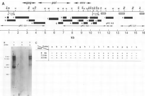
To identify regions of the provirus expressed in ERV3 mRNAs,weusedisolatedfragmentsfrom the ERV3 lambda cloneasprobesinNorthern
blot
hybridizationtototal RNA of full-term chorionic villi. The map locations of all probes usedareshown inFig.2A.Probesb,c,i, j, k,1,m, n, o,and presulted ina stronghybridization signalto all three ERV3 RNAs evenafter a high-stringency wash(0.ix
SSC, 680C) (Fig.2B, lane1). Probes d, e,f,g,and hfailedtodetect any of the ERV3 mRNAs (Fig. 2B, lane 2). These results suggestedthat the9-,7.3-, and3.5-kb RNAspecies wereall spliced RNAs. Because probes c and i detected the three ERV3 RNAs, whereas probes d and h were negative, we anticipatedthat all three RNAs would have the samesplice
junctions,asplicedonorsite inregioncanda
splice
acceptor site inregioni(regionhexcluded). Probes a, r, and s, whichoriginated
from outside the ERV3provirus,
did not detect the 9- or7.3-kb (or 3.5-kb) RNAs,suggesting
that the two [image:2.612.332.528.73.215.2]longRNAs were notsimply
readthrough
products.Probe q, however, detected the 9- and 7.3-kb mRNAs but not the 3.5-kb mRNA(Fig.
2B,lane3).All threeof the ERV3 RNAs were found to be concentrated in thepoly(A)-containing
[poly(A)+] RNAfraction
(Fig. 3),
indicating
that the 9- and 7.3-kb RNAswerenotunprocessed
precursors of the 3.5-kb species. In this and otherexperiments
(unpublished),
the 9-kb RNA appeared as adoublet. Becauseprobe
rdid noton November 10, 2019 by guest
http://jvi.asm.org/
2184 KATO ET AL.
*gag
.- a*po/
-
Ven
v-,.-C cm 0. r- EnZ._. n *-.-t lm
m x mccEDZI em oa- I 4>C<am<mmmI I C:cc c : m a 4
'I~~~ ~ II11 I L 1II 1 1
5'-LTR 3'-LTR
a c f h m o r s
b d 1 n q
e g k m p pRI 0.8
pRI 5.0- pRI 4.8 - - ---pRI 2.7
1
2
3
4
5
6
7
8
9
10
11
12
13
14
15
16kb
B 1 2 3
probe o h q
9kb-7 kb
lBs~~~~~~~~~~~~~~~~~~~~~~~~~~ 2803S .
It
3.5 kt)--- _
1f5S .1
C
Probe RNAA
9 kb
7.3 kb
3.5 kb 1
a b c d e f g h j k m n o p q r s
_ + +.___ _ _ + + + + + + + + +
_-+ +.--- - - + + + + + + + + +
+.__
_-FIG. 2. Northern blot analysis of ERV3 mRNAs. (A) Map location of the probes in a human endogenous retroviral clone, ERV3.
Fragmentsa(BanI-MnlI, 0.1 kb), b(BanI-XmaI, 0.7 kb), c(XmaI-BglII, 1.4 kb), d (BglII-HpaI, 0.6 kb), ande (NruI-EcoRI, 1.3 kb)were
isolated frompRI5.0 plasmid DNA(22). Fragments f (BamHI-BamHI, 1.0 kb) andg(BamHI-EcoRI, 0.5 kb)were isolated from theERV3
lambda clone. Fragments h (EcoRI-PstI, 0.3 kb), i (EcoRI-HindIIl, 1.0 kb), j (HindIII-PstI, 1.7 kb), k (HindIII-AhaIII, 0.8 kb), (AhaIII-AhaIII, 0.6 kb),m(KpnI-PstI, 0.4 kb),n(AhaIII-BaIl, 0.8 kb),o(BstNI-BstNI, 0.5 kb),p,(Hinfl-HhaI, 0.3 kb),andq(BaI-Hinfl, 0.3kb)wereisolated frompRI4.8 plasmid DNA. Fragmentsr(EcoRI-EcoRI, 0.8 kb) ands(BglII-BglII,0.8kb)wereobtained frompRI0.8 andpRI2.7 plasmid DNAs, respectively. Bars with cross-hatching represent regions that were strongly detected afterhybridization with
32P-labeled total human DNA, indicating that they contained highly repeated humansequences. Abbreviations: Ah, AhaIII; Av, AvaI; B, BamHI; Bg,BglII;Bl, BalI; Bn,BanI; Bs, BstNI; H, HindIlI; Hh, HhaI; Hi, Hinfl; Hp, HpaI; K, KpnI; M,MnIl;N, NruI; P, PstI;R, EcoRI; X, XmaI. (B) Northern blot hybridization of human total placental RNA. Finalwashing conditionwas0.1x SSCat68°C (seeMaterials and
Methods for additionaldetails). Lanes: 1, fragmento asprobe; 2,fragmenthasprobe; 3, fragmentqasprobe. (C) Summaryof Northern blot
analysis. +,Positive hybridization with the indicated probe.
hybridize to the ERV3 RNAs, these results suggested that the 9- and 7.3-kb RNAs either contained a second splice donor site or were polyadenylated in the 3'-flanking
se-quence nearregionq.
Initiation site of ERV3 mRNAs. We used S1 nuclease mapping to define the structure of ERV3 mRNAs which
were estimatedby the Northern blot analysis. To determine
wherethe three ERV3 mRNAsinitiated, we initially useda 1.6-kb EcoRI-XmaI fragment as an SI nuclease hybridiza-tion probe (Fig. 4A, bottom). In. this experiment we could
not distinguish between protected probe and traces of re-maining undigested probe (not shown). Therefore, we re-peated the analysis, usingas probea 2.0-kb fragment
com-posed of the 1.6-kb EcoRI-XmaI region plus 0.4 kb of plasmid sequence (Fig. 4A, bottom). The fragment was 5'-endlabeled atthe XmaI site. The predicted site of ERV3 transcription initiation is 171 base pairs (bp) upstream from the XmaI restriction site (21; unpublished results). Ifanyof the ERV3 env-containing RNAs had initiated transcription upstream from the LTR, we would have expected to find protected fragments of 1.6 kb or less in addition to the
1 2 3
9
7.3
kb-:.
3.5
kb-A
J. VIROL.
on November 10, 2019 by guest
http://jvi.asm.org/
[image:3.577.55.545.59.382.2]HUMAN PROVIRAL TRANSCRIPTS CONTAIN GENOMIC SEQUENCES 2185 A
ml 1 gofAw
B
2 3
U
.1 MS? Ml
*:s
wo
kb 2.03 *-1.90 1.58
-1.33
83
O -.62
_ _ .56
_ -.53
_ow: -.40 --.31 -.24
*--.13
c 0 5 -LTR
U5P
ao 4,U
... .J
* 1600 ..._. ---*. 2000
171. 170
< V V
+ A
M 1 2 3 4 M (.3 F- C'0 NIts
624 _
529 _0
406
-311 am
219 _
203 _w
192 ft
182 _
162 *
149 f
:1
-*-. < ,. <,pr
sw .s.
_ | |n g
:S wt
EcBi
_ _
,- ... w.
r >.
_.
*'s:::
_ :-e
-X -X
|
t-_"F
_ _
_
.
@s
.6 '
...MF
_ :.
,s 45mP
.;
...-
t-* -* 4"
_ _
_
5.. 4
'::r Z4@t
_ _s
.os. w
z i
Sw
*Sreb
S, !t
..,.j
Sys,;'. e
.y'
l#:
FIG. 4. S1nucleasemapping analysisof ERV3transcriptionalinitiation site.pRI5.0plasmidDNAwasdigestedwithX,nal, dephosphoryl-atedby bacterialalkaline phosphatase, and labeled atthe 5' end with[y-32P]ATP(3,000Ci/mmol)andpolynucleotide kinase. The DNAwas
recut withBamnHI, releasing a2.0-kbfragment(XmanI-BatwtHI) containing0.4kbofplasmidsequence. The labeledfragmentswereisolated
byagarosegel electrophoresis. Thick baratbottomcorrespondstolengthofprotectedfragmentin lanes 2 and4.AllS1 nucleasedigestsused 400Uofenzyme.(A)S1nuclease-resistantfragmentsweredetectedbyelectrophoresisin a2%agarosegel. Lanes: 1,yeast tRNAannealed
at52'C; 2, humanfull-term placentalRNAannealedat52'C; 3, yeasttRNAannealedat55'C;4,humanfull-termplacentalRNAannealed
at55'C. LanesMl, 32P5'-end-labeled Hindlll-EcoRIfragments ofDNAaslength markers: laneM2. 32P3'-end-labeledMspI fragmentsof pBR322 DNA as length markers. (B) S1 nuclease-resistant fragments were detected by electrophoresis in an 8% polyacrylamide gel. Sequencingladderswere runforaccuratefragmentsizemeasurement(17).Lanes: 1,yeast tRNAannealedat52'C; 2.placentalRNAannealed
at52'C;3, yeast tRNA annealedat55'C;4. placental RNAannealed at55'C;M. 32P3'-end-labeledMspI fragmentsofpBR322 DNAused
as length markers.
171-nucleotide (nt) fragment. Using electrophoresis under FIG. 3. Northern blot analysis after selection by oligo(dT)- conditions that clearly separated single-stranded DNA frag-cellulose. Poly(A)+ RNA wasisolated by onecycle ofchromatog- ments of 2.0 and 1.6 kb, we found protected fragments of raphy on oligo(dT)-cellulose (2). Lanes: 1, total RNA (10 F±g); 2. only 2.0 kb and approximately 170 nt (Fig. 4A). To obtain the poly(A)' RNA(5 ,ug); 3, poly(A)- RNA(10 ,ug). Autoradiographic precise nucleotide sequence at which transcription began,
exposure, 8 h. the
protected
DNAfragment
wasanalyzed by
electrophore-VOL. 61, 1987
ow-Awl' 4m
on November 10, 2019 by guest
http://jvi.asm.org/
[image:4.612.121.476.71.548.2]2186 KATO ET AL.
sis in an acrylamide sequencing gel. Following electropho-resis, two Si nuclease-resistant fragmentsof170and 171 nt were detected (Fig. 4B). The 171-nt fragment corresponded in length to a fragment beginning at the predicted first nucleotide of region R in the 5' LTR (21). The 170-nt fragment may be the result of overdigestion with Si
nucle-ase. We then separated the 9- and 7.3-kb RNAs from the 3.5-kb RNA and repeated the Si nuclease mapping with the fractionated placental RNAs. As before, two Si nuclease-resistant fragments of 170 and 171 nt were detected in both RNA pools (data not shown). From these results we con-clude that the three ERV3 mRNAs initiated at the same site in the 5' LTR.
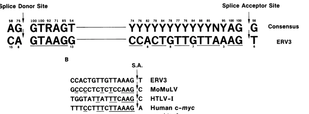
Splice donor site of the three ERV3 mRNAs. Hybridization analysis suggested the existence of a splice donor site ofthe ERV3 primary transcripts in region c (Fig. 2A). To deter-mine the splice donor site, we used the
XmaI-BglII
fragment (1.4 kb) as anS1 nuclease mapping probe (Fig.SA).
The fragment was 3'-end labeled at theXmaI site of the noncod-ing strand and annealed to total full-term placental RNA. After Si nuclease digestion for 30 min, a major protected fragment of 154 nt was detected (Fig.SA, lane 2). Shorter bands in lane 4 of Fig. SA are probably the result of overdigestionwithS1 nuclease. From a comparison with the ERV3 nucleotide sequence in this region (see sequence ladder of noncoding strand, Fig.SA), we inferred that the splice donor site sequence is CAI
GTAAGG(arrowdenotes splice point). This sequence was a close match to that of the consensus splice donor site, AGI
GTA/GAGT (19). This site was located 224 nt downstream from the end of the 5' LTR. Since the XmaI-BglII fragment (probe c in Fig. 2) hybridized to all three ERV3 mRNAs, we predict that this site was used as the splice donor in the three ERV3 mRNAs. Splice acceptor site of ERV3 mRNAs. The results of our Northern blot analysis showed that the splice acceptor site ofall three ERV3 transcripts could reside in region i between the PstI and HindIll sites because the three RNAs were detected with probe i but not probe h (Fig. 2A). Thus, we used twofragments from this region as probes in S1 nuclease mapping of the splice acceptor site (Fig.SB, bottom). First, we used as probe a 0.51-kb DdeI-NcoI fragment labeled at the 5' end of the DdeI site. An S1 nuclease-resistant DNA fragment ofabout 355 nt was detected (not shown), indicat-ing that the spliceacceptor site was located upstream from a site previously suggested from the nucleotide sequence (6). Todetermine the nucleotide sequence of the splice acceptor site, we used as probe a 0.65-kb HindIII-PstI fragment labeled at the 5' end of theHindlIl
site. In this case, anS1 nuclease-resistant band of 190 nt was detected by polyacryl-amide gel electrophoresis (Fig. SB). This length exactly corresponded to that estimated from the DdeI-NcoI probe experiment. From the nucleotide sequence of this region (6), wepredicted that the splice acceptor site of the three ERV3 mRNAs is CCACTGTTGTTAAAGI
T, which is located precisely 190 nt upstream from the labeled HindIII end. The splice acceptor site was located in the 3' portion of the pol gene 469 ntupstream from the putative start of the env gene product (6).Poly(A) addition site of 3.5-kb mRNA and second splice donor site of 9- and 7.3-kb mRNAs. As shown in Fig.
2C,
probe p (Hinfl-HhaI) hybridized to the 9-, 7.3-, and 3.5-kb ERV3 mRNAs, but probe q
(BalI-Hinfl)
hybridized to only the 9- and 7.3-kb mRNAs. Thus, we anticipated that the 3.5-kb mRNA wouldterminate in the 3' LTR. Furthermore, an ERV3 cDNA clone obtained from a human second-trimester fetal liver library terminated at the estimated end ofregion R in the 3' LTR, 18 nt downstream from the AATAAA signal sequence (6). Because all three ERV3 mRNAscontained the same transcriptional initiation, splice donor, and splice acceptor sites (see above), and because probe r (located 1 to 2 kb downstreamfrom the 3' LTR) did notdetect the 9-and 7.3-kbmRNAs,wepredicted thatthese two RNAs would contain a second splice donor site near region q. Therefore, wechose the 1.2-kb
HhaI-EcoRI
frag-ment and 1.1-kb
BalI-EcoRI
fragment asS1 nuclease map-ping probes foridentifying the poly(A) addition site and the postulated second splice donor site (Fig.6C).
To increase the sensitivity ofthe assay, we usedpoly(A)+
RNA.Results of the analysis confirmed our prediction. The
HhaI-EcoRI
probe revealed afragment of about 530 nt plus several small bands (Fig. 6A and C). The latter included major bandsof 132, 131, and 130 nt and minor bandsof128 and 126 nt. The nucleotide sequence and termini ofthese protected fragments are indicated in Fig. 6D (21). These probably represent poly(A) addition sites since they were located from 14 to 20 nt downstream from the AATAAA polyadenylation signal sequence. Furthermore, the se-quence of the 130-nt band corresponded to the poly(A) addition site in the ERV3 cDNA clone (6). With theBalI-EcoRI
probe, anS1 nuclease-resistant band of about 430 nt was detected (Fig.6B).
Smaller protected fragments were not observed with this probebecause theBalI
site is only31 nt distant from the polyadenylation site (6), and hybrids of such a length were probably too unstable to be detected under the conditions used. As shown in Fig. 6C, the end of the 430-nt protected fragment exactly coincided with thatof the 530-nt fragment observed with theHhaI-EcoRI
probe. This result thus defines the position of a probable second splice donor site for the 9- and 7.3-kb mRNAs, even though the protected DNA fragment was not mapped to the precise nucleotide position. The DNA sequence of the ERV3 lambda clone revealed two potential splice donor sites in this region (unpublished data). The alternative possibility, that this position represents the polyadenylation siteof the 9- and 7.3-kb mRNAs, is inconsistent not only with the hybridiza-tion and S1 nuclease data (see above) but also with the absence of an AATAAA signal sequence in the adjacent, upstream sequence.DISCUSSION
A human endogenous provirus, ERV3, which has a long open reading frame in the env region (6) is highly expressed in placental chorion throughout gestation as mRNAs of 9, 7.3, and 3.5 kb. ERV3 is present as a stable, single-copy provirus in the DNA of apparently all humans (over 20 individuals have been tested [22; N. Kato and M. Cohen, unpublished results]). This contrasts with other human pro-viruses, which are moderately or highly repetitive in human DNA (5, 8, 14-16, 23). Thus, we were able to investigate the detailed structure of the ERV3 mRNAs by using Northern blot hybridization and S1 nuclease mapping analysis.
In this report we characterized the structure of the ERV3 subgenomic 3.5-kb mRNA, including the initiation site, splice sites, and poly(A) addition site. We have determined that the 9- and 7.3-kb mRNAs are unusual in that they contain the same initiation and splice sites as the 3.5-kb mRNA but extend through the 3' LTR and are spliced again about 370 nt downstream from the LTR. The probable structures of the three ERV3 mRNAs are summarized in Fig. 7.
A 5.9-kb gag-pol region was missing from all three ERV3 mRNAs, probably removed from the primary transcript by J. VIROL.
on November 10, 2019 by guest
http://jvi.asm.org/
1 2 3 41 M
.... ...
...
ig5.'' 'E...
4L...
B
Nts
622
527
M 1 2 3 4 5 6 7 8
,,.' A*
404
404
309
309
242 238
..
217
#0 160 201
0 147
190
180
123
160
110
147
... -po
-0
UJ g. z :
I . I
aZ
aM
I
-- 650
190
123 S355510
E
1400
*
--154 _
FIG. 5. S1 nuclease mapping analysis of ERV3 splice donorandacceptorsites.(A)Splicedonor site. pRI5.0plasmid DNAwasdigested
withXmaIand labeledatthe 3' endwith[a-32P]dCTP(3,000Ci/mmol)byusing theKlenowfragment of DNA polymeraseIand thenrecut
withBgII.The 1.4-kb fragment probe(XmaI-BglII) wasisolatedbyagarosegelelectrophoresis. Hybridizationwasdoneat63°C.Thick bar
atbottomcorrespondstolength of major protected fragment in lane 2.S1 nuclease-resistant fragmentsweredetected by 8% polyacrylamide
gel electrophoresis. Sequencing ladder of the sameXmaI end-labeled fragment was runtoprovide the DNA sequencearound the splice
junction. Lanes: 1,yeasttRNA, 400 U ofS1 nucleaseat37°C, 30min; 2, placental RNA, 400 U ofS1 nucleaseat37°C,30min;3,yeasttRNA,
400UofS1nucleaseat37°C, 90min; 4, placental RNA,400Uof S1 nucleaseat37°C, 90 min;M,32P-end-labeledMspIfragments of pBR322
DNAas length markers. (B) Splice acceptorsite. S1 nuclease mapping analysis of the ERV3 splice acceptorsite. pSP65 plasmid DNA
containing PstI-ScaI fragment (-100to1109 [6]),subcloned from pRI4.8 plasmid,wasdigested withHindlll, dephosphorylated, and labeled
atthe5' endasdescribedinlegendtoFig. 4,and thenrecutwithPstI.ThelabeledHindIII-PstI 0.65-kb fragmentwasisolated byagarose
gelelectrophoresis. Thick baratbottomcorresponds tolength of major protected band. S1 nuclease-resistant fragmentsweredetected by electrophoresisina8%polyacrylamide gel.Lanes:1,yeasttRNAannealedat54°C, 200 U ofS1 nuclease; 2, placentalRNAannealedat54°C,
200 UofS1 nuclease; 3,yeasttRNAannealedat54°C, 400 U ofS1 nuclease; 4, placentalRNAannealedat54°C,400 U ofS1 nuclease; 5,
yeasttRNAannealedat59°C,200U ofS1 nuclease; 6, placentalRNAannealedat59°C,200 U ofS1 nuclease; 7, yeasttRNAannealedat
59°C,400 U ofS1 nuclease; 8, placentalRNAannealedat59°C,400 U ofS1 nuclease; M,32P 5'-end-labeledMspI fragmentsofpBR322DNA usedaslength markers.
A M Nts
622 527
.#' 242
" 238
"I 217
.r 201 r'0 190
..180
..*
-..-gag-.-...-an
on November 10, 2019 by guest
http://jvi.asm.org/
[image:6.612.126.478.77.574.2]J. VIROL. 2188 KATO ET AL.
A
Nt..
F *0
*,,,- 4w
-;!! "_
-a Sb 4.
....
::
1.,t
!.
.'1-i _ iig
to,ul
U
C
S
- d ..a.SF
4w
I;-
-g} U 3 R L.. _ .
3 -LTR US
530
-132 130 128.126.i
n:r
13 v) Ln
---1200
_....
...--- 1
430
D
I2 128'3 313 '32
A AT A A A A CC TT CCTGTTG CACCCA'GCTQGATCT CT
FIG. 6. Si nuclease mapping analysisof the ERV3 polyadenylation site and secondsplicedonor site.The HhaI-EcoRI 1.2-kbprobewas
obtainedasfollows. pRI4.8 plasmidDNAwasdigestedwith HhaIand labeled at the 3' end with [a-32P]dGTP (3,000 Ci/mmol) by usingT4
DNApolymerase. After recleavagewithEcoRI,the Hhal-EcoRlfragmentwasisolatedbyagarosegel electrophoresis.TheBalI-EcoRI 1.1-kb
probewasobtained similarly.ThepRI4.8 plasmidDNAwasdigestedwithBalI and labeledatthe 3' end with the KlenowfragmentofDNA
polymeraseI and [c-32P]dCTP(3,000Ci/mmol). AfterrecleavagewithEcoRI,theBaIl-EcoRl 1.1-kbfragmentwas isolatedbyagarosegel electrophoresis. (A) Si nuclease-resistantfragmentswiththe Hhal-EcoRI probeweredetectedby electrophoresisinan8%polyacrylamide gel.Sequencing ladderswere runforaccuratefragmentsizemeasurement(17).Lanes:1,yeasttRNAannealedat55°C,400 U ofS1nuclease;
2,totalplacental RNAannealedat55°C,400 U ofS1 nuclease; 3, placental poly(A)+ RNA(10 pig)annealedat55°C,400 U ofS1 nuclease;
M,32P3'-end-labeledMspI fragmentsofpBR322DNAas size markers.(B) S1 nuclease-resistantfragmentwith theBallI-EcoRI probewas
detected by electrophoresis in an 8% polyacrylamide gel. Lanes and markers same as in panel A. (C) Map of the probes and S1
nuclease-resistant fragments. Thick bars representlengthsofprotected fragments. (D) ERV3 sequence surrounding the poly(A)addition
site(s).Thearrows indicate thepositionsofpolyadenylation.
* .
I
1, 1
.0..F'
:. :iT.!
Am
45
..IAIL
7 ..
1!#.
0
f.1":
IAk
4
j
on November 10, 2019 by guest
http://jvi.asm.org/
[image:7.577.89.507.85.595.2]1 kb 2 3 8 9 10 11 12
7 RNU E
I., x co ~~~~~~~~~~0.zc
I
I~~~~~~~~~~~~~~~~~~~~~~~~~~~~~~~~~~~~~~~~~~~~~~~~~~~~~~~~~~~~~~~~
IWI~ ~ ~ ~ ~~
II~ ~ ~ ~ ~ ~
3.5kbRNAI
171 ts154Ns4 9 Nts[
I
9 kbRNA
7.3 kbRNAI
7:RU5 - o
I xIiII
PA SD
_. _y
ORF (1944Nts) -1 polyA
132-130Nts
ORF(1944 Nts) 530 Nts poy
FIG. 7. Schematicstructural presentation of the three ERV3 mRNAs. Each line with a star represents an end-labeledS1nuclease mapping probe; blackboxes indicate protected regions. ORF, Open reading frame; TI, transcription initiation site; SD, splice donor site; SA, splice acceptorsite; PA,poly(A)addition site.
splicing (Fig. 5). Figure 8A shows the proviral sequences bounding the splice site of the ERV3 mRNAs. The intron sequenceof the splice donor site closely matched that of the consensus sequence (19), although the exonended with the rare dinucleotide CA. In addition, the intron sequence adjacent to the ERV3 splice acceptor site was unique. In contrasttotheusualpyrimidine in the position 3 nt upstream fromthesplice acceptor site, ERV3 contained an adenosine. Anadenosine at this position has been noted in only 3.0% of thepublished sequences (44of 1,448 cases; data not shown) and in severaldifferent species. Interestingly, an adenosine in this position has also been found in the splice acceptor sequence of Mo-MuLV (31), human T-cell lymphotropic virus type I HTLV-I (29), and human c-myc intron 2 (26). The boundary sequence of human c-myc intron 2 is most homologoustothe ERV3 splice acceptor site sequence (Fig. 8B). In the c-myc genes of chicken (34) and mouse (3), however, the c-myc splice acceptor sequence contains a cytosine at this position, and there is no evidence that
changes in this nucleotide affect the specificity or rate of RNAprocessing.
The absence of an ERV3 genome-length mRNA is un-usual. In contrast to the usual complete splicing of eucary-otic gene introns (10), processing of retroviral mRNAs is normallyincomplete, resulting in cytoplasmic accumulation ofagenome-lengthmRNA andasplicedsubgenomicmRNA (33). ERV3 isatypicalbecause the gag-polintron was absent from all three mRNAs. This highly efficient ERV3 splice could be due either to the presence of a positive-acting cellularfactor such as a small nuclearribonucleoprotein or totertiaryfolding oftheprimarytranscript thatwouldallow exceptionalaccesstothesplicingapparatusof the cell. Such possibilitieswere suggested forsplicing of the P-element in Drosophila melanogaster(13).
An alternative explanation for the complete splicing of ERV3transcriptsis thatanegative controlelement hasbeen lost. The normal incomplete processing of retroviral tran-scripts may be due to the presence ofafactor that
specifi-A
Splice
DonorSite
Splice Acceptor
Site58 75 100100 92 71 85 54
AG;
GTRAGT-CA
GTAAGG-15 8 13
74 76 82 78 84 78 77 76 84 88 85 95 100 100 56
YYYYYYYYYYYNYAG
G
CCACTGTTGTTAAAG T
8 8 7 .3 6
B
S.A.
CCACTGTTGTTAAAG T ERV3
GCCCCTCTCTCCAAG
C MoMuLVTGGTATTATTTCAAG
C
HTLV-ITTTCCTTTCTTAAAG
A Human c-myc [image:8.612.56.547.67.196.2]Intron2
FIG. 8. Comparisonof the ERV3 splice junctionsequences withthose of othergenes. (A) Underlining: homologiesbetween theERV3 sequenceand that of theconsensussplice junctionsequence of othergenes(26,29, 31).Numbers above and belowsequencesindicate the
percentageof the known splice junctions containingthat nucleotide at thatparticular position. (B)Underlining: spliceacceptorsite (S.A.)
homology between ERV3and othergenes.
Consensus
ERV3
on November 10, 2019 by guest
http://jvi.asm.org/
[image:8.612.60.566.497.685.2]2190 KATO ET AL.
callyinhibits retroviralsplicing.Theabsence of suchafactor could explain whymammalian cells transformed with avian
sarcoma virus expressed predominately a spliced src
mRNA, whereas permissive avian cells expressed mainly the genome-length mRNA (25). From the experiments de-scribed in this report, it is not possible to decide whether
splicing of the ERV3 gag-pol intron is controlled by a positive or a negative regulatory element, or whether the highly efficient splicing is simply a result ofproviral muta-tions that have optimized splice site recognition.
We havepreciselydetermined thepoly(A)addition site of the3.5-kbRNAspecies.InourSi nucleaseanalysis (Fig. 6), three majorSi nuclease-resistantfragmentswere observed,
theshortestof which(130 nt) mappedtothesamelocationas
the poly(A) addition site found in a cDNA clone isolated
fromalibrary made from RNA ofasecond-trimester human fetus(6). This sitewas 18ntdownstream fromanAATAAA signalsequence. Thetwoothermajorbandswereequivalent
to positions 19 and 20 nt downstream from the AATAAA
sequence. Although multiple poly(A) addition sites may actually be present in the 3.5-kb ERV3 mRNA, the several Si nuclease-resistant fragments may have originated from artifacts of the Si nucleasedigestion. Analysis of additional cDNA clones should resolve this question.
Poly(A) addition site analysis of the ERV3 mRNAs re-vealed a second splice donor site in the 9- and 7.3-kb
mRNAs about 530ntdownstream from theHhaI-labeled end and370ntdownstream from the 3' LTR(Fig. 6A andC). A second Si nucleaseprobe revealed afragment that mapped
to the identical location (Fig. 6B). This site cannot be the
polyadenylation site of the 9- and 7.3-kb mRNAs because bothspeciesinitiatedatthesamepositionasthe3.5-kbRNA andlacked the same 5.9-kbregion, aresult ofsplicing in the
gag-polgenes. Thus, processing of the ERV3 primary
tran-scriptis different from that ofthe endogenousavian
retrovi-rus, ev-1, that produces mRNAs polyadenylated 100 to 200 ntdownstream of the normal poly(A) addition site in the 3'
flanking region (7). The ERV3 mRNAs, therefore, should contain an additional 5.5 or 3.8 kb of human sequence, respectively (thedifference between9or7.3 kb and 3.5 kb). If, inahuman ancestor, the ERV3 provirus integratedin the
coding sequence of an existing gene, the two large ERV3 mRNAs might be expected to contain several exons in the
remaining sequence. Alternatively, a new splice donor site
mighthave been fortuitously generated after integration. Recently, novel processing of retroviral transcripts has been reported in two cases in which retroviruses have integrated within or adjacent to a cellular oncogene: avian
leukosis virus (ALV)-induced erythroblastosis (12, 20) and Mo-MuLV-inducedtumors(30). In the formercase, integra-tion of ALVinto the c-erbB locus resulted in expressionof different, spliced c-erbB mRNAs. One of these initiated in the 5' LTRof theprovirusand linked ALVgagsequencesto the c-erbB gene, bypassing the 3' LTR. Another mRNA which initiated in the 5' LTR containedadoublesplice,from
gagintoenvandfromenvintoc-erbB, again bypassingthe3' LTR (12, 20). In Mo-MuLV tumors, novel c-myb mRNAs initiatinginthe 5' LTR of Mo-MuLVaresplicedbetween the gaggeneandexon 1of the c-myb gene,removing pol, env,
and the3' LTR(30). The 9- and7.3-kbmRNAsof ERV3are
different from these examples because although the ERV3 mRNAs were linked to cellular sequences, the 3' LTRwas
notbypassed viaa splice.
Thisstudy demonstrates that ERV3transcription is
regu-lated at two levels. ERV3 expression is controlled at the level oftranscriptional initiation ormRNA stability: ERV3
mRNAis
significantly
moreabundant in the chorion than in theembryo
(Fig. 1). Secondly,
ERV3expression
isregulated
at the level of RNAprocessing
in two distinct ways: the three ERV3 mRNAs lacked sequencesinthegag-pol
region
so thatno
genome-length
mRNAwasdetected,
and expres-sion of the 9-kb and 7.3-kb mRNAs wasregulated
in theplacenta
because chorionic villiexpressed
aboutequal
levels of theseRNAs,
whereasembryos
expressed
the 9-kb mRNA but almost none of the 7.3-kb form(Fig. 1). Although
the ERV3 9-kb mRNA was detected in many normal humantissues,
the 7.3-kb mRNA was observedprimarily
in theplacental
chorion(N.
Kato etal., unpublished
results).
Because the twoRNAs areapparently
identical within theirproviral
portions, they
mustdiffer in their cellular domainsas a consequence of alternative
splicing
or alternativepoly(A)
addition site selection.Analysis
of cDNA and ge-nomic clones downstreamfrom theprovirus
should resolve the differences and make clear thesignificance
of ERV3 mRNAs thatcontain human cellular sequences.ACKNOWLEDGMENTS
Wethank J. Cleveland, F. Propost, E. Brownell,andS.
Hughes
for criticalreadingofthe
manuscript.
Researchwassponsored,inpart,bytheNational Cancer Institute under contract NO1-CO-23909 with Bionetics Research, Inc. Re-search was also sponsored by theJapanese Overseas Cancer Fel-low(ship)oftheFoundation for Promotion of Cancer Research.
LITERATURECITED
1. Auffray, C., and F. Rougeon. 1980. Purification of mouse
immunoglobulin heavy-chain messenger RNAs from total myelomatumorRNA. Eur. J. Biochem. 107:303-314. 2. Aviv, H.,and P. Leder. 1972. Purificationof
biologically
activeglobinmessenger RNAby
chromatography
onoligothymidylic
acid-cellulose. Proc. Natl. Acad. Sci. USA 69:1408-1412. 3. Bernard, O., S. Cory, S. Gerondakis, E. Webb, and J. M.
Adams. 1983. Sequence ofthe murine andhumancellularmyc oncogenes andtwo modes of myc
transcription resulting
from chromosome translocation in Blymphoid
tumors. EMBO J. 2:2375-2383.4. Bonner, T. I., C. O'Connell, and M. Cohen. 1982. Cloned endogenousretroviralsequencesfromhuman DNA. Proc. Natl. Acad. Sci. USA79:4709-4713.
5. Callahan, R., W. Drohan, S. Tronick, and J. Schlom. 1982. Detection andcloningof human DNAsequencesrelatedtothe
mouse mammary tumor virus genome. Proc. Natl. Acad. Sci. USA79:5503-5507.
6. Cohen, M., M. Powers,C. O'Connell, and N. Kato. 1985. The nucleotide sequence ofthe envgenefromthe human provirus ERV3 and isolation and characterization ofan
ERV3-specific
cDNA. Virology147:449-458.
7. Conklin,K.F.,J.M.Coffin,H. L.Robinson,M.Groudine,and R. Eisenman. 1982. Role of methylation in the induced and spontaneous expression ofthe avian endogenous virus ev-1: DNA structureand geneproducts. Mol. Cell. Biol. 2:638-652.
8. Deen, K.C., and R. W.Sweet. 1986. Murine mammarytumor
virus pol-related sequences in human DNA: characterization and sequencecomparison with thecompletemurine mammary
tumorviruspolgene. J. Virol.57:422-432.
9. Favaloro,J., R. Treisman,and R. Kamen. 1980. Transcription maps of polyoma virus-specific RNA: analysis by
two-dimensional nuclease S1 gel mapping. Methods Enzymol.
65:718-749.
10. Flint, J. 1984. Processing of mRNA precursors in eukaryotic
cells, p. 151-179. In D.Apirion(ed.),ProcessingofRNA.CRC
Press,BocaRaton, Fla.
11. Gattoni-Celli, S., K. Kirsch, S. Kalled, and K. J. Isselbacher. 1986. Expression oftype C-related endogenous retroviral
se-quences in human colon tumors and colon cancer cell lines. Proc.Natl. Acad. Sci. USA 83:6127-6131.
J. VIROL.
on November 10, 2019 by guest
http://jvi.asm.org/
12. Goodwin, R. G., F. M. Rottman, T. Callaghan, H.-J. Kung,
P. A. Maroney, and T. W. Nilsen. 1986. c-erbB activation in avian leukosis virus-induced erythroblastosis: multiple epider-mal growth factorreceptormRNAsaregenerated byalternative
RNAprocessing. Mol. Cell. Biol. 6:3128-3133.
13. Laski, F. A., D. C. Rio, and G. M. Rubin. 1986. Tissue specificity ofDrosophila P element transposition is regulatedat
the level of mRNA splicing. Cell 44:7-19.
14. Maeda, N. 1985. Nucleotide sequence of the haptoglobin and
haptoglobin-relatedgenepair: the haptoglobin-relatedgene
con-tainsaretrovirus-like element. J. Biol. Chem. 260:6698-6709.
15. Mager, D. L., and P. S. Henthorn. 1984. Identification of a
retrovirus-like repetitive element in human DNA. Proc. Natl. Acad. Sci. USA81:7510-7514.
16. Martin, M. A., T. Bryan, S. Rasheed, and A. S. Khan. 1981. Identification and cloning of endogenous retroviral sequences
present in human DNA. Proc. Natl. Acad. Sci. USA
78:4892-4896.
17. Maxam, A., and W. Gilbert.1980.Sequencing end-labeled DNA with base-specific chemical cleavages. Methods Enzymol. 65:499-560.
18. May, F. E. B., andB. R. Westley. 1986. Structure ofa human
retroviralsequence relatedtomouse mammarytumorvirus. J. Virol. 60:743-749.
19. Mount, S. M. 1982. Acatalogue of splicejunction sequences.
Nucleic Acids Res. 10:459-472.
20. Nilsen, T. W., P. A. Maroney, R. G. Goodwin, F. M. Rottman, L.B. Crittenden, M. A. Raines, and H.-J. Kung. 1985. c-erbB activation inALV-induced erythroblastosis: novel RNA
proc-essing andpromoterinsertion result inexpression ofan
amino-truncated EGFreceptor. Cell 41:719-726.
21. O'Connell, C. D., and M. Cohen. 1984. The LTRsequencesofa
novel human endogenous retrovirus. Science 226:1204-1206. 22. O'Connell,C. D., S. J. O'Brien, W. G. Nash, and M. Cohen.
1984.ERV3,afull-length human endogenous provirus:
chromo-somal localization and evolutionary relationships. Virology 138:225-235.
23. Ono, M. 1986. Molecular cloning and long terminal repeat
sequences of human endogenous retrovirus genes related to
typesAand Bretrovirusgenes.J. Virol.58:937-944.
24. Pfeifer-Ohlsson, S., A. S.Goustin, J. Rydnert, T. Wahlstrom,L.
Bjersing, D. Stehelin, and R. Ohlsson. 1984.Spatial and temporal
pattern of cellular myc oncogene expression in developing human placenta: implications forembryonic cell proliferation. Cell 38:585-596.
25. Quintrell, N., S. H. Hughes, H. E. Varmus, and J. M. Bishop. 1980. Structure of viral DNA and RNA in mammalian cells infected with avian sarcoma virus. J. Mol. Biol. 143:363-393.
26. Rabbitts, T. H., P. H. Hamlyn, and R. Baer. 1983. Altered nucleotide sequences of atranslocated c-myc gene inBurkitt's lymphoma.Nature (London) 306:760-765.
27. Rabson, A. B., Y. Hamagishi, P. E.Steele, M. Tykocinski, and M. A. Martin. 1985. Characterization of human endogenous retroviral envelopeRNAtranscripts. J. Virol.56:176-182. 28. Rabson, A. B., P. E. Steele, C. F. Garon, and M. A. Martin.
1983. mRNAtranscripts relatedtofull-length endogenous ret-roviralDNA in human cells. Nature (London)306:604-607. 29. Seiki, M., S. Hattori, Y. Hirayama, and M. Yoshida. 1983.
Human adult T-cell leukemia virus: complete nucleotide se-quence of theprovirusgenomeintegratedinleukemia cellDNA. Proc. Natl. Acad. Sci. USA80:3618-3622.
30. Shen-ong, G. L. C., H. C. Morse III, M. Potter, and J. F.
Mushinski. 1986. Two modes of c-myb activation in virus-inducedmouse myeloidtumors. Mol. Cell. Biol. 6:380-392. 31. Shinnick, T. M., R. A. Lerner, and J. G. Sutcliffe. 1981.
Nucleotide sequenceof Moloney murineleukaemiavirus. Na-ture(London)293:543-548.
32. Thomas, P. S. 1980. Hybridization of denaturedRNAand small DNAfragments transferredtonitrocellulose.Proc. Natl.Acad. Sci. USA 77:5201-5205.
33. Varmus, H., and R. Swanstrom. 1984. Replication of retrovi-ruses, p. 369-512. In R. Weiss, N. Teich, H. Varmus, and J. Coffin(ed.), RNA tumorviruses. Cold Spring Harbor Labora-tory,Cold Spring Harbor,N.Y.
34. Watson, D. K., E. P. Reddy, P. H. Duesberg, and T. S. Papas. 1983. Nucleotidesequenceanalysis ofthechickenc-myc gene reveals homologousanduniquecoding regions by comparison with the transforming gene of avian myelocytomatosis virus MC29,gag-myc. Proc. Natl. Acad. Sci. USA80:2146-2150. 35. Winberg, G., and M.-L.Hammarskjold.1980.Isolation ofDNA
fromagarosegelsusing DEAE-paper.Applicationtorestriction site mapping of adenovirus type 16 DNA. Nucleic Acids Res. 8:253-264.