0022-538X/86/110376-09$02.00/0
Copyright © 1986, AmericanSocietyforMicrobiology
Encephalomyocarditis
Virus
3C Protease: Efficient Cell-Free
Expression from Clones Which
Link Viral 5'
Noncoding
Sequences
to
the P3
Region
GRIFFITH D. PARKS,* GREGORY M. DUKE, ANDANN C. PALMENBERG
Biophysics Laboratoryof the Graduate School andDepartment ofBiochemistry oftheCollege of Agriculture andLife Sciences, University of Wisconsin, Madison, Wisconsin 53706
Received 14 April 1986/Accepted 15 July 1986
All picornaviral peptides are derived by progressive posttranslational cleavage of a giant precursor polyprotein. Translation ofencephalomyocarditis virus (EMC) RNA in rabbit reticulocyte extractsproduces active viral peptides, including protease 3C, which is responsible for many cleavage reactions within the processing cascade. DNAplasmids containing 5'noncodingsequences of EMC linkedtootherportionsof the viral genome were constructed and transcribed into RNA. Like virion RNA, the clone-derived transcripts directed efficient protein translation in vitro. The5'-linkedconstructionsmay representexamplesofageneral method for cell-free expression of any cloned gene segment. One construction produced a self-cleaving P3 region precursor, which contained active 3C protease. A genetically engineered insertion within the 3C sequences eliminatedendogenousself-cleavage activitywithoutalteringtheabilityof the P3peptideto serveas substrate in bimolecular reactions with added3C. Anotherplasmidencodingthe L-VPOportionof thecapsid region was used todemonstrate that scission between the leader peptide (L) and capsid protein VPO can be catalyzed by3C. Theenzymeresponsiblefor this step waspreviouslyunidentified. Arapidpurificationscheme for isolation of 3C from EMC-infected HeLa cells is alsopresented.
Encephalomyocarditis virus (EMC) is a member ofthe cardioviral subgroup of the picornavirus family. All picornaviral genomesencodea single, largeprecursor poly-protein which represents most of the theoretical coding
capacity of the RNA molecule (22). The polyprotein is processed in a series ofproteolytic cleavage steps to yield mature virion capsid proteins and other nonstructural pro-teins (Fig. 1). At least three proteolytic activities are in-volvedin these cleavages(15).
The first proteolytic event within the polyprotein takes place while the peptide is stillnascent on aribosome(12, 28). Recent experiments with poliovirus suggest that viral pep-tide 2A plays a role in this step (P1-P2 cleavage) (15). However, the primary cleavage event for EMC is not completely analogous, because it occurs at a different site withinthe polyprotein (2A-2B) and may be catalyzed by a peptide other than 2A. The last step in the proteolytic cascade is maturation processing ofVPO to VP4 and VP2 (13). An agent responsible for this event has never been
isolated, but crystallographic data implicate a mono-molecular, autocatalyticcleavage mechanism (21).
Most of the remaining polyprotein cleavages are carried out by the viral 3C protease (10, 17, 18). This enzyme is capable ofmono- and bimolecularreactions within its pre-cursorsubstrates (3ABCDto 3AB and 3C and 3D) (11, 19) and is also responsible for several processing steps within capsidprecursor peptides(P1 toVPOand VP3 and VP1) (9, 18). Although not all of the proteolytic sites within the EMC polyprotein have been precisely identified, those known to be cleaved by 3C occur only between glutamine-glycine or glutamine-serine amino acid pairs which are flanked by proline residues (17).
* Correspondingauthor.
Becauseofitsimportancein the viralproteolytic process-ing scheme, we have directed our attention towards the 3C protease, focusing on the enzyme from EMC. Cardioviral RNA translates withunusually high efficiencyinreticulocyte extracts, producing active 3C capable of processing reac-tions indistinguishable from those observed during viral infection of cells (28). We have taken advantage of this translationalactivity andconstructed DNAplasmidswhich linkEMC 5' noncodingsequences to other segments of the viral genome. Like virion RNA, the clones efficiently ex-pressed viralpeptides in cell-free reactions. One construc-tion, encoding the P3 region of EMC, produced active 3C protease invitro. We haveused this plasmidtoexaminethe effects ofanengineered insertion into the putative active site of 3C.
Wealsoreport a newprocedure forthe rapid purification of the 3Cproteasefrom EMC-infected HeLacells. Purified enzyme andplasmid-derived 3Chavebeentestedforactivity on various segments ofthe EMC genome. Using a clone which expressed the leader and VPO peptides,weshow that scission of the leaderprotein from its capsid precursor can becatalyzed by 3C.
MATERIALS AND METHODS
Nomenclature. EMC peptides are named by the L-4-3-4 conventions (24), except that traditional designations are usedforthevirioncapsid proteinsVPO,VP4, VP2, VP3, and VP1 (1AB, 1A, 1B, 1C, and 1D, respectively). Previously, peptidesL-Pl-2A,P3, 3CD, 3D, 3C, and 3AB were referred to asAl,C, D, E, p22, and H, respectively. Peptide elis a charge-alteredform of VPO peculiarto cell-free translation samples programmed with EMC RNA (16). The "large" fragmentof RNA includes allheteropolymeric sequences 3'
376
on November 10, 2019 by guest
http://jvi.asm.org/
poly C 4- VPO
*Ccc
I
wvp41
VP2m VP31
VP1vPg
N -14
P1
I I gr I PM POL - m
polyA
P2
nL
lABCOl 2ABC I 3ABCD1ABC 2A I2BC 3CD
protease?
1AB 3C4'.2B 2C2C 33D
,#protease polymerase
r. .r pr
/ 11--z;1capsid proteins
30i
FIG. 1. Structureofthepicornaviralgenome.ViralRNA, encoded peptides, and their proteinprecursors areillustratedschematically.
gr denotesthe portion of thegenomeassociated with guanidine resistance, and VPg is the viral protein covalently linkedtothe5'end ofthe
viral RNA (7).
A Purflictlonfraction
1 2 3 4
3CD
3D-EI 6
3D
-YPO- _
SP2.
VP2 - .
VP3-_
_40 3C - |
2A
...
....
B
Activityof fractionsS mock 1 2 3 4
L-PI-2A
PI-2A
-Pi
" . wEP..
*,&
-e-YPO
-VPo
-VPI
-VP2
VP3
-L/2A
-3JW- *
21-
VP4-FIG. 2. Isolation of protease 3C from EMC-infected HeLa cells. (A) Purification fractions. Equal amounts (about 60,000 cpm) of
[3H]leucine-labeled proteinfrompurificationfractionsFl, F2, F3,andF4(lanes1through4,respectively)werefractionatedby polyacryamide gel electrophoresis and visualized by fluorography. The arrow indicates the position of the 3C peptide. (B) Activity offractions.
[35S]methionine-labeled capsid precursor L-P1-2A, produced byin vitro translation (lane S), was incubated for5 h at 30°C with 5 ,ul of
mock-infectedFlfraction(lanemock)or5 p1lofpurificationfractionsFlthroughF4(lanes1through4,respectively)and thensubjectedto
gelelectrophoresisand visualizedbyautoradiography. Lane V contains EMCvirionproteinsasmarkers.
3'
jbb-
ip-I I I I
5!
.4
-viral RNAgenome_01
C"Oaw.. .0
gm
on November 10, 2019 by guest
http://jvi.asm.org/
[image:2.612.74.553.70.231.2] [image:2.612.145.474.307.671.2]to the poly(C) tract. The "small" fragment includes all sequences 5' to the poly(C) tract (4, 5, 25).
Purification of 3C protease. The infection and growth of EMCinHeLa cells has been describedpreviously (23). Cells (2.4 x 10⁹) were infected (10 PFU/cell) with EMC and labeled with [3H]leucine (Amersham; 131
Ci/mmol)
at 0.01 mCi/ml from 3 to 4 h postinfection. The cells were washed once with phosphate-buffered saline (26) and then swollen for 5 min in 25 ml of ice-cold buffer A (10 mM Tris hydrochloride, pH 7.8, 0.1% 2-mercaptoethanol) before Dounce homogenization. Cellular debris was removed by centrifugation (5min at 5,000 rpm and then10min
at 10,000 rpm, Sorval type SS34 rotor,5°C).
The resulting supernatant (Fl fraction, 19.1 ml) was made 10 mM in EDTA and centrifuged for 2 h at 39,000 rpm in a typeSW41
rotor at5°C.
The fraction 2supernatant (F2, 19.3 ml) was mixed with 10 ml (packed volume) of buffer A-equilibrated car-boxymethyl-Sephadex C-50 (Pharmacia Fine Chemicals) at 22°C for 15 min. After being washed with 60 ml of buffer A andthen with 60 ml of buffer B (buffer A containing 80 mM NaCl), the beads were transferred to a column (gel bed 1.5 by 4.5cm) and further washed with 400 mlof buffer B. The protein was eluted at 0.3 ml/h with buffer C (buffer A containing 0.35 M NaCl). The3H-containing
fractions were pooled and concentrated with aCentricon-10
(Amicon) microconcentrator (F3 fraction, 440,u).
A portion of the F3 sample (300,lI)
was injected into a gel filtration high-pres-sure liquid chromatography (HPLC) column(TSK-G3000SW, 7.5 by 300 mm; Toyo Soda Manufacturing Co., Ltd.) flowing at 0.5 ml/min with buffer D (50 mM sodium
phosphate, pH 7.2, 0.05% 2-mercaptoethanol, 0.1 M
NaCI).
Fractions containing radiolabeled 3C were pooled, concen-trated byCentricon-10 filtration, and stored at -
70°C
as theF4fraction (220,ul).
Construction of plasmid DNAs. Restriction enzymes were
purchased from New England Biolabs. DNA manipulations were done by standard methods (14). All transformations to
ampicillin resistance were performed with Escherichia coli
HB101. Construction of DNA plasmids
pEST10
and pE3T11 will be described in detail elsewhere. Briefly, EMC RNA was copied into double-stranded DNA. EcoRI DNA linkers (New England Biolabs) were ligated to the ends of thedouble-stranded DNA, and the resulting material was in-serted into theEcoRI site of the transcription vector pSPT18
(PharmaciaFine Chemicals). Plasmid pSPT18 is a derivative of plasmid pUC18 containing T7 transcriptional promoter
sequences and a polylinker cloning site. The EMC-derived DNA within pEST10 is approximately 7,500 base pairs in length and encodes the EMC RNA sequences which extend from the 3' side of the viral poly(C) tract (near base 320)
through the poly(A) sequence at the extreme 3' end of the
genome. Plasmid pE3T11 contains EMC sequences originat-ing from within the poly(C) tract and extending 3' (about 2,300 bases) into the region encoding viral peptide VP3.
Toconstruct plasmid pESP3, DNA (3.5
ptg)
from plasmid pEM3 (17) was digested withNruI
and MluI. The resulting2,242-base-pair fragment, encoding nearly all of the P3 region of EMC, was isolated by preparative agarose gel
electrophoresis. The purified fragment was then ligated (T4 DNA ligase; Promega Biotec) into 0.5
,g
ofpEST10
DNA whichhadbeen previously cut withBalI
andMlul.
Samples ofthereaction mixture were used to transform bacteria, andcolonieswere screened by restriction enzyme analysis (KpnI and PstI) for plasmids containing the appropriate EMC-derived segments. One representative clone was selected anddesignated pESP3.
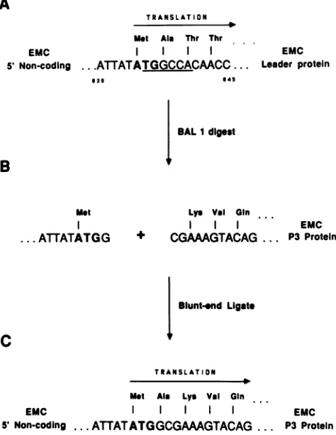
A
TRANSLATION
Met Ala Thr Thr
EMC
5' Non-coding ..
.ATATATGGCCACMCC
...029 45
EMC Leader protein
BALI digest
B
met
I
...ATTATATGG
Lys Val
Gin
EMC + CGAAAGTACAG ... P3 Protein
Bluntend Ligate
c
TRANSLATION
Met Aia Lys Val Gin
EMC
5' Non-coding
...
ATTATATGGCGAAAGTACAG
... P3EMCProteinFIG.
3. Construction ofplasmid
pE5P3.
The strategy used tolink5'
noncoding sequences
of EMC to asegmentderived fromtheP3
region
of thegenome
is outlined.(A)
Sequence of EMC at the start of thepolyprotein
readingframe. TheBalI
restriction sequenceis
underlined,
and the AUG
codon which initiates translation is boldfaced.(B)
5'noncoding segment
afterBalI
digestion and also the5'sequence
of the P3segment
produced
byNruI
digestion. (C)Completed
constructionresulting
from blunt-end ligation of thesegments,
including the reconstructed reading frame.To construct
pESP3c,
purified
plasmid
DNAfrompESP3
(6
,ug)
wasdigested
tocompletion
withBglII
and thenreacted with 9 U of
DNApolymerase
Klenow
fragment(Bethesda
ResearchLaboratories)
inthepresence
of dCTP,dGTP, dATP,
and dTTP(final
concentrations, 1 mM each)for 30
min
at20°C.
Thesample
was extracted with an equalvolume of
phenol-chloroform
(1:1)
and
precipitated
withethanol. The
resulting pellet
wassuspended
in buffer (22pAl)
containing
ClaI
linker
fragments
(2.5
,ug;
New EnglandBiolabs,
catalog
no.1001)
which had been previouslyphosphorylated
(14).
The reaction mixture was incubatedwith 40
U of T4 DNA
ligase
for 3
hat
20°C
and thenprecipitated
with
ethanol. Excess free linker
fragments wereremoved
by gel
filtration(Sephacryl
S-300;
Pharmacia FineChemicals),
and the
plasmid
DNA was
digested
with Clal.Following purification
by agarose
gel
electrophoresis,
theplasmid
DNA was
redigested
with
Clal,
extracted
withphenol-chloroform, precipitated
with
ethanol,
and
treatedwith T4 DNA
ligase.
A
sample
of the resulting
DNA wasused to transform bacteria.
Colonies were screened
for thepresence
of a
unique
Clal site within the plasmid
DNA.Clone
pESP3c
was selected asrepresentative
of this con-struction.To construct
plasmid pE5LVPO,
DNA from
plasmidpE3T11
(1
,ug)
was
digested
with
XbaI
to completion.
Afteron November 10, 2019 by guest
http://jvi.asm.org/
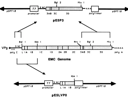
[image:3.612.318.555.70.376.2]Ogi 2
.4 T7mote
promotear
[image:4.612.93.524.64.393.2]MIu 1
...
...
... A A
3AB 3C 3D
AAI
polylinker pSPT18
pE5P3
Nru 1 Bgl 2
NIS
I
_ 5'N C 1- ...' .'.-'..''..'.. | 1
c~~ ~...~ ~ ~ ~
Ij-.-..
~ ~ ~I..
| 1'-A
A A Apoly C L IA 1B IC ID 2A 25 2C 3AB 3C 3D poly A
'
pSPTI8L-EMC Genome
Xba 1
J
_
|
_~~~~~~~~~~~~~~I.
moter L 1A 15 polyli
pE5LVPO
FIG. 4. Keyelements ofplasmidspE5P3 and pE5LVPO. The EMC genomicsegmentsincluded within plasmidspE5P3 andpE5LVPOare
illustratedschematically. Importantrestriction sitesused in the constructionsareshown. Open boxes (T7promoterandpolylinker)represent
sequencesfrom theparentalvector(pSPT18). The5'noncodingsequences(5'NC)contained within thetwoplasmidswerederivedfrom the
segmentextendingfromthe 3' sideof the EMC poly(C)tractuptotheAUGcodonatthebeginning of the polyprotein readingframe.Stippled
areasdenote viralcodingsequenceswithintheplasmids.
extraction with phenol-chloroform and precipitation with ethanol, the DNA wasreacted with T4 DNA ligase, and a
portion of the mixturewasused totransform bacteria. The resulting colonies were screened for the size of the EMC
segment. One representative isolate was chosen and
desig-natedpE5LVP0.
In vitro transcription of plasmid DNA. Purified plasmid DNA (14) was linearized by digestion with restriction
en-zyme BamHI (clones pE5P3 and pE5P3c) or XbaI (clone
pE5LVP0). After extraction with phenol-chloroform and precipitation with ethanol, the samples were suspended in
water. Typically, about 1 ,ug of linear plasmid DNA was
transcribedinreactions(25 ,ul)withT7 RNApolymeraseas
specified by the enzyme manufacturer (Bethesda Research Laboratories), except that the ribonucleotides and dithio-threitol were increasedto 1 mM and 25 mM, respectively. RNase inhibitor (RNasin; Promega Biotec) was also in-cluded (1.5 U/pAl). After incubation at 37°C for 1 h, the samples were extracted with phenol-chloroform, precipi-tated withethanol, dried undervacuum, and suspendedin water(10 Pld;estimated concentration, 1 ,ug/,ul).
Cell-free translation.In vitrotranslationreactions in
retic-ulocyte extracts were carried outexactly as described pre-viously (28). Typically, 3 to 5 jil ofplasmid transcription product (see above) was used to direct cell-free protein synthesis reactions (30 jil) radiolabeledwith [35S]methionine
(specific activity, 1,100 Ci/mmol; final concentration, 1 ,uCi/l). After 40 min at 30°C, reactions were stopped by
addition of pancreatic RNase and cycloheximide (to 0.3 mg/ml each). EMC virion RNA wasused to program
reac-tions for the synthesis of radiolabeled capsid precursor
protein L-P1-2A and also for synthesis of the complete polyprotein as described previously (18, 28). Nonradiolab-eled reaction products to be used forprotease assayswere
synthesized in lysate mixtures lacking [35S]methionine but supplementedwith 100 jiM unlabeled methionine.
Gel electrophoresis. The method foranalytical polyacryl-amide slabgel electrophoresishas beendescribedpreviously (18). Urea (5 M) was included in the gels to aid in the separation of EMC-specific proteins. Proteinbands labeled with[35S]methionine werevisualized byautoradiographyof the driedgels. Protein bandslabeled with [3H]leucine were
visualized by fluorography (2). 35S-labeled virion proteins usedasmarkerswereproducedasdescribedpreviously (23).
RESULTS
Purification of the EMC 3C protease from infected HeLa cells. EMC-infected HeLa cells were labeled with
[3H]leucine to facilitate detection of viral peptides during purification.Clarified supernatantfromlysedcellswasused
Bal I
VPg
Xba Mlu 1
A
nLiD
r pSPT 18p PTIS -Wp,
on November 10, 2019 by guest
http://jvi.asm.org/
as
starting
material for 3C proteasepreparations (Fl
frac-tion).
Afterhigh-speed centrifugation,
theresulting
superna-tant,F2,
was mixed withcarboxymethyl-Sephadex.
Boundprotein
was elutedby
ahigh-salt
wash (F3 fraction) and further fractionatedbyHPLCgelfiltration.Samplescontain-ing3H-labeled 3Cpeptidewerepooledandconcentrated(F4
fraction).
Figure
2A shows the 3H-labeledproteins
present in Fl,F2, F3,
and F4.Approximately equal
amounts ofradioac-tivity
wereapplied
toeachsample
lane. Theintensity
ofthe 3C band relative to those of other viralproteins
increasedthroughout
thepurification scheme,
with thegreatestenrich-ment in thecation
exchange chromatography
step(F3
frac-tion).
HPLC of the F3 fraction resultedina 3C preparationwhich was
substantially
free of other viralproteins (lane
4,Fig. 2A).
Thegel
depicted
inFig
2A wasreexposed
for alonger
time (10days),
but 3H-labeled bands other than 3Cwere still not detectable. Coomassie blue
staining
ofsimilarpreparations
revealed smallquantities
ofunlabeled cellularprotein
in the F4samples.
Based on densitometric tracingsof
fluorograms,
we estimatethatatleasta 100-foldpurifica-tion of
peptide
3C was achieved between the Fl and F4samples (not shown).
Fractions Fl
through
F4 wereassayed
for theability
tocleave EMC
capsid
precursor L-P1-2A. The[35S]me-thionine-labeled substrate was
synthesized
in alimited cell-free translationreaction directedby
EMC virionRNA(18).An
autoradiogram showing
thepeptides produced
after reaction of the substrate withenzymefractions ispresented
in
Fig.
2B. The substrate(lane S)
wasunchanged by
incu-bation withextractfrom uninfectedHeLacells(mock lane).
However,
incubation withsamples
from any of the four enzyme fractions resulted incleavage
of the substrate intomature
capsid proteins (VPO, VP3,
and VP1) and theirprocessing
intermediates(P1-2A, P1,and1ABC).
PeptidesL(leader), 2A,
and el were also evident. Thesepeptide
profiles
areexactly
asexpected
fornormalprocessing
ofthe L-P1-2Aprecursorby
the 3C protease (9, 18, 27).During typical
cell-freeprotease assays,it is oftendifficult to detectVPO andVP3,
astheseproteins
represent the last3C-catalyzed
cleavage
step in thecapsid region processing
cascade
(12, 18).
Therefore,
theVPOandVP3 bands inFig.
2Bareindicative ofa
high
level of3C proteaseactivity
in the enzyme fractions.Although
we do not yet have a method whichprecisely quantifies
the relativespecific activityof 3C enzymesamples,
we conclude that our rapidisolation pro-cedureyields preparations
ofactive, radiochemically
pure 3C protease. Purified F4samples retained cleavage activityafter storageat -
70°C
(not shown).Cloning
andexpressionoftheP3regionof EMC.The RNA ofEMC,
unlike that ofpoliovirus
or rhinovirus, translates withunusually high efficiencyincell-free reactions(28). We believe this property to be a function of the 5' noncodingregion
ofthe genome, specifically the segment extending fromthe 3'side ofthepoly(C)tractuptotheAUGinitiation codon for thepolyprotein (4, 5).Theabilityof clone-derivedtranscripts
to serve as mRNA in cell-free reactions can bedestroyed by
alterations or deletions in these sequences(manuscript
inpreparation).The
complete
nucleotide sequenceofEMC has now been determined(17; unpublisheddata).Thefortuitousposition of aBalIrestriction enzyme recognitionsequence (UGGCCA) as part of the polyprotein translation initiation codonsug-gested
ageneral
methodforthecell-freeexpression of viralcoding
sequencesby linking
themtothe EMC 5' noncodingregion.
Wehaveusedthistechnique
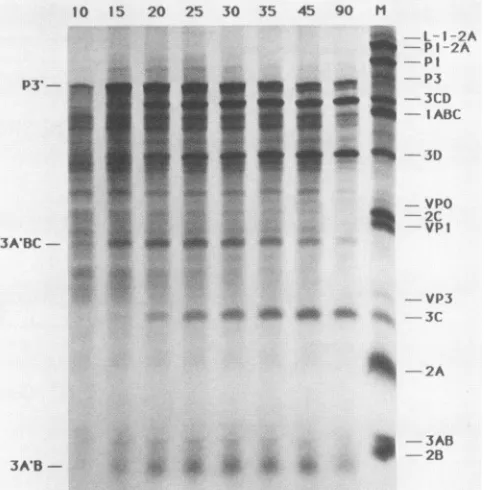
toexpress thepeptides10 15 20 25 30 35 45 90 M
ILI 2A
PI ?A
P3' P3
p3- - P3CD___
IABCL
3ABC
-2A
3A'B- * * s
--3 AB .4 ._-2B
FIG. 5. Translational time course of pE5P3-derived RNA in reticulocyte extracts. Plasmid DNA was transcribed invitro, and the products were used to program cell-free protein synthesis. Samples (2 ,ul) were removedat the indicated times (in minutes), made2% in sodiumdodecyl sulfate and 1% in2-mercaptoethanol, and heatedto90°C (5 min). Acetoneprecipitation, electrophoresis, and autoradiographyof the sampleswere as described previously (18). The marker lane (M)representsastandard in vitrotranslation sample programmed with EMCvirionRNA.Thedesignations P3', 3A'BC,and 3A'Bdenotepeptidescontainingthe truncatedformof EMC protein3A,asdescribedin thetext.
from the P3regionof the EMC genome invitro, includingan active 3Cprotease (Fig. 3).
As a result of the sequencing work (17), bacterial DNA
plasmids containing portions ofthe EMC genome became available. Construction pEST1O, containing the large
frag-ment ofEMC, was usedas the sourceof 5' noncodingand
plasmid vector sequences. Clone pEM3wasusedto supply the P3regionsegmentbecause theviral sequences contained within this clonewere specificallydetermined (17). Plasmid DNA from pEM3 was digested with restriction enzymes NruIandMlul.Thegel-purified fragment(encompassingthe P3
region)
wasligatedintopE5T1Owhich had beencutwith BalIand Mlul. Linkageof thesetwofragments coupledthe normal EMC polyprotein initiation codon into the same translationalreadingframe thatproducestheP3protein. Thekey elements of the resulting clone, pE5P3 (EMC, 5'
noncoding, P3), areillustrated in Fig. 4.
RNAtranscribed invitrofrom pE5P3 was used todirect translationreactionsinreticulocyteextracts. Atypical time
courseofprotein synthesisis shown in Fig. 5. After 10 min
of incubation, a large peptide (P3') was evident, with an
electrophoretic mobilityslightly fasterthan the authentic P3 markerpeptide. The smaller size of P3' relative to P3 was
expected, as cleavage with NruI during the cloning proce-dureresulted inaDNAfragmentmissing33codonsfrom the 5' end of the P3 region (amino end of the peptide; refer to
Fig. 4).
Withincreasingtime ofincubation,otherbands (3CD,3D,
3A'BC, 3C, and 3A'B) also appeared in the translation
on November 10, 2019 by guest
http://jvi.asm.org/
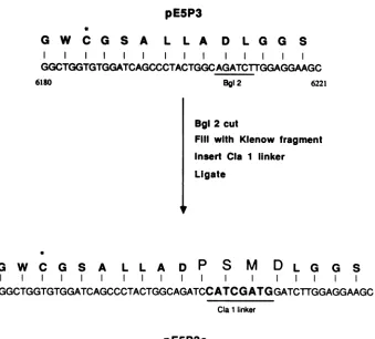
[image:5.612.315.557.69.314.2]pE5P3
*
G W
C G S A
L
L A
D
L G G
S
I II
GGCTGGTGTGGATCAGCCCTACTGGCAGATCTTGGAGGAAGC
6180 Bgl2 6221
Bgl 2 cut
Fill with Klenow fragment Insert CIa 1 linker
Ligate
G
WC
G
S
A L L A DP
S
M
D
L G G S...GGCTGGTGTGGATCAGCCCTACTGGCAGATCCATCGATGGATCTTGGAGGAAGC...
Cla1linker
pE5P3c
FIG.6.Construction of plasmid pE5P3c. The procedures used to insert 12 bases into the 3C region of clonepE5P3 are outlined. The BgIII restriction sequence and the sequence of the insertedClaIlinker areunderlined. Cys-159 is denoted with an asterisk(*). Amino acid residues uniquetoclone pE5P3c (PSMD) are in boldface type.
samples. The overall peptide profile is characteristic of normal P3region processingcatalyzed by viral protease 3C (12, 18, 19). This result indicates that the 3C sequences contained within P3' were active when expressed in vitro. Peptides 3A'BC and 3A'B werelike P3' in that they repre-sented slightly shorter versions of their normal processing counterparts. Tryptic profiles (not shown) of clone-derived 3D and 3D isolated from infected cells were indistinguish-able, confirming thatcorrectprocessing had indeed occurred and that the normal viral translational reading frame was expressed in vitro.Theseresults arethefirstdemonstration that the P3 peptide of EMC is capable of autocatalytic
processing in the absence ofpeptides fromtheL,P1, andP2
regionsof the viralgenome.
Inactivation of cloned 3C protease by anengineered inser-tion.EMC 3Cproteasebelongstotheclassof thiolenzymes (8, 20). Sequence homology with other picornaviral
proteases suggests that the catalytically active residues within3C includeCys-159 and His-177(EMC 3Chasatotal of 205residues) (1). Totesttheeffects ofgenetic
manipula-tion on this region, 12 nucleotides were inserted into a unique Bglllrestriction site within the 3CregionofpE5P3to formpE5P3c. As aresult ofthisinsertion, plasmid pE5P3c
contained four extra codons (Pro, Ser, Met, and Asp)
between the sequences encoding Cys-159 andHis-177 (Fig.
6).
In vitro transcription and translation of pE5P3c DNA produced a peptide (P3'c) whichwas similar in sizetothat expressed bythe parentalclone (Fig. 7, lane 1). However,
unlike P3', the P3'cpeptideshowed noevidence of sponta-neous processing even after prolonged incubation (lane 2).
Addition of active 3C protease from exogenous sources resulted inthe normalappearanceof 3CD, 3D, 3C, 3A'BC, and 3A'B. The processing occurred after the addition of enzymeisolatedfrom infected cells(F4fraction,lane 5) orof proteasefroma cell-freetranslation of virion RNA(lane 3) or pE5P3-derived RNA (lane 4). The data show that the four-codon insertion inactivatedtheproteolytic ability of the endogenous 3CsequenceswithinP3'c, but didnotaffectthe
capacity ofP3'c to serve as substrate in bimolecular reac-tions with added3C enzyme.
Cleavageof the leaderpeptideiscatalyzedbyprotease3C. Theproteaseresponsible forthefirst cleavageeventwithin theEMCcapsidregion (L-P1-2AtoLandP1-2A) hasnever beenspecifically identified(3, 27). Usingacloned construc-tion, pE5LVPO (EMC, 5' noncoding, Leader, VPO), which expressedapeptidecontainingthissite,wetestedtheability
ofvarious 3C enzyme preparations to catalyze scission of theleader protein fromits precursor.
ClonepE5LVPOcontainsa5' noncodingsegmentof EMC and extends 3' from the polyprotein initiation site through
theleaderandVPO coding regions,stoppingjust short(two
aminoacids)oftheVPO-VP3cleavagesite(Fig.4).Cell-free transcription and translation ofplasmid-derived RNA pro-duced an L-VPO peptide (Fig. 8, lane 1) which was stable after incubationin reticulocyte extracts (lane 2). However,
additionofeither FlorF4protease frominfectedcells(lanes
7 and 8) or 3C expressed from pE5P3 (lane 5) resulted in cleavage of L-VP0 intoa leader-sizedpeptide (L) and VPO (1AB and el). The L-VPO peptide was not affected by
incubation with extracts containing brome mosaic virus translationproducts(lane 3),EMCcapsidprecursorprotein
on November 10, 2019 by guest
http://jvi.asm.org/
[image:6.612.140.479.62.368.2]382 PARKS ET AL.
(lane4), ortheP3'c peptide (lane 6).The VPO derived from thecloned peptide(L-VPO) containedthe same methionine-labeledtryptic fragmentsasauthenticVPOproducedin vivo (not shown).
Theseexperiments show that L-VPOcleavageiscatalyzed by viral protease 3C and not, as often speculated, by a "host" orother reticulocyte enzyme (17, 27, 28). Further-more,this result is evidence that clone-derived 3C protease (pE5P3) can catalyze a capsid region processing step in a mannersimilar tothatof theenzyme isolated frominfected HeLa cells.
DISCUSSION
Among picornavirus RNAs, the genome of EMC is un-usual in its ability to direct high levels of viral protein synthesis in reticulocyte extracts (28). Past experiments have suggested thisproperty might reside primarily within the 5'noncoding region of thegenome(833bases),between thepoly(C)tract(bases 149through 291) and the polyprotein initiation codon (base 834) (5; unpublished observations).
Our engineered constructions support this idea. Plasmids pE5P3 and pE5P3c contained only 480 5' noncoding viral
1 2 5 4 5 m
PJ.c
~~~~~~~~~-
- p: CI)IABC
3D
....VPO
iC-VP' 3A'BC
! ---
I--
~~~~VP3
3C* -2A
[image:7.612.308.565.69.312.2]3AB-3AB
FIG. 7.Expression and cleavage of P3'c protein. Plasmid DNA frompE5P3cwastranscribed in vitro, and the products were used to program [35S]methionine-labeled protein synthesis in reticulocyte lysate.After40 min of incubation at 30°C, the reaction was stopped byaddition of cycloheximide and RNase, and a sample (2,ul)was saved (lane 1). Similar samples were mixed with buffer (2
pl
of phosphate-buffered saline, lane 2), fraction F4 protease (5,ul, lane 5),alysate sample which had translated EMC virion RNA (1 ,ul, lane 3),or alysate sample which had translatedpE5P3-derivedRNA (1 ,ul,lane 4).After further incubation at 30°C (12 to 15 h), the sampleswerefractionated by gel electrophoresis and visualized by autora-diography. The marker lane (M) represents a standard in vitro translation sample programmed with EMC virion RNA.
1 2 3 4 5 6 .7 5 U
IA
-. 2
-ID
-IC
-fb-3C
L
-FIG. 8. Expression and cleavage of L-VPO protein. Plasmid pE5LVPOwastranscribedand translated asdescribedforplasmid pE5P3c in the legend to Fig. 7. A portion (2 RI) of the [35S]methionine-labeled samplewassaved(lane 1). Similarsamples
were mixed with 1 to 3 ,ul of untranslated lysate (lane 2), lysate which had translated brome mosaic virus RNA (lane 3), lysate containing EMC capsid precursor proteins (lane 4),lysate which had translatedpE5P3-derived RNA (lane5),lysate which had translated pE5P3c-derived RNA (lane 6), fraction Fl protease (lane 7), or
fraction F4 protease (lane 8). Sampleswereincubated(30°C, 3 h) and thenanalyzed by gelelectrophoresis andautoradiography.Lane
Mrepresentspeptidesproducedduringcell-free translationofEMC virion RNA.
bases, derived from thegenomic segmentimmediately 5' to the polyprotein initiation codon. Sequences from the small fragment, poly(C), L,P1, and P2regionswereabsent. Yet, like virion RNA, the clone-derived transcripts were effi-ciently translated in vitro. While ourexperiments were not intended to precisely define the particular viral sequences required for efficient protein synthesis, it is clear that the clonescontain all theelementsnecessaryforcell-free trans-lation.
Unlike most eucaryotic mRNAs, translational activity directedby picornavirusRNAsdoesnotrequirea 5'capping group (7). It is interesting that our plasmid-derived tran-scripts likewise retained this viral property and expressed protein effectively in vitro without treatment in specific cappingreactions.
Based on ourresults, we propose that the 5'-linked viral constructions represent examples of a general method for cell-free expression of any coding segment from the EMC genome. By taking advantage of the Ball restriction se-quence at the EMC polyprotein initiation codon, it should also bepossibletoconstruct5'-linkedhybridplasmidswhich actively express coding sequences from different viruses (e.g.,polio-orrhinovirus)orfrom other exogenous sources. The EMC P3 region sequences within clones pE5P3 and pE5P3c were correctly expressedin vitro intopeptidesP3' andP3'c. The P3' protein contained an active 3Cprotease, as evidenced by the typical P3 region processing profile J. VIROL.
L-VPO d
L/ 2A
.4b:
on November 10, 2019 by guest
http://jvi.asm.org/
[image:7.612.81.278.304.610.2]during translation reactions (12, 18, 19). Thecodingsegment within pE5P3 was derived from pEM3, a clone which was isolated and examined as part of the EMC sequencing
project (17). Thus, the in vitro activity shows that the published version of the 3C sequenceis that ofacompetent viral protease.
Expression and processing ofP3'demonstratedthat pep-tidesfrom otherregions of the EMC genome (L, P1, orP2) are not required in any way for proper cleavage ofthe P3
region. The spontaneous nature of the P3' cascade supports previous contentions that the endogenous 3C protease is capableof monomolecularautocatalysis (11, 19). Thatis,3C sequences within the precursor molecule (P3, 3ABC, or 3CD) release themselves by catalyzing self-cleavage reac-tions. Our experiments with P3'c, which contains an inser-tion within the putative active site of 3C, strengthen this argument, asinactivation of theendogenousenzymeseemed to preclude self-cleavage. The P3'c peptide, however, was still functional as a substrate in bimolecular reactions catalyzed by added 3C.
The results with pE5P3 and pE5P3c provide further evi-dence that the P3 region processing cascade canbe carried outbytwo independent,autocatalytic pathways,witheither monomolecularorbimolecularreaction mechanisms. Previ-ously, the two pathways were distinguishable only by dilu-tion processing experiments (19). We can now use P3'c to study exclusively the bimolecularP3 regionreactions inthe absence ofany monomolecular cleavage. Furthermore, we can use pE5P3 as aconvenient invitro source of active 3C protease free of viral peptides from the L, P1, and P2 regions.
We have also developed a rapid purification scheme for the isolation of 3C from EMC-infected HeLa cells. In the past, enzymatic activitywaspartially purifiedfromcell-free translation samples programmed with virion RNA
(18).
Alternatively, 3C was isolated from virus-infected Krebs-2 cells by lengthy chromatographic steps after denaturation with urea (8). Our method is based on cation exchange and takes advantage of the high positive charge onthe EMC 3C
peptide (6). The procedure results in stable
preparations
of highly activeproteasefreeof other detectable viralproteins.
The purified 3C andpE5P3-derivedenzymeswere
equally
effective in processing reactions with substrate
prepared
from clone pE5LVP0. This result demonstrates that 3C protease cancatalyze the scission of the leader
protein
from its precursor, the first step within the EMCcapsid
region
cascade(3,27). Thiseventhad
previously
beenattributedtoan unidentified hostprotease(17, 27, 28). TheL-VPO cleav-age probably occurs between the
glutamine-glycine
amino acidpairatposition67-68ofthepolyprotein (17).
This istheonlyEMC 3C site thus far identified which is notflanked
by
a proline residue. Future
mutagenesis
experiments
withpE5LVPO and other clones will be directed toward
deter-miningthe effects offlankingsequences on protease recog-nition of thecleavage sites.
ACKNOWLEDGMENTS
Wethank Roland Rueckert and PaulKaesbergforhelpful discus-sions.
Thiswork is supported byPublic Health ServicegrantAI-17331 from the National Institutes of HealthtoA.C.P. G.D.P. issupported
by American CancerSociety grantMV34. G.M.D.was supported
byNational Institutesof Health traininggrantGM07215 in Molec-ularandCellularBiology.
LITERATURECITED
1. Argos,P., G. Kamer,M.J.H. Nicklin,andE.Wimmer. 1984.
Similarityin gene organizationandhomologybetweenproteins
of animal picornaviruses and a plant comovirus suggest com-mon ancestry of these virus families. Nucleic Acids Res. 12:7251-7267.
2. Bonner, W.M., and R. A. Laskey. 1974. A film detection method for tritium-labelledproteins andnucleic acids in
poly-acrylamide gels. Eur.J. Biochem. 46:83-88.
3. Campbell, E.A., and R.J. Jackson. 1983. Processing of the
encephalomyocarditisviruscapsidprecursorprotein studied in rabbit reticulocyte lysates incubated with
N-formyl-[35S]methionine-tRNAflet.
J.Virol.45:439-441.4. Chumakov, K. M., and V. I. Agol. 1976.
Poly(C)
sequence is located near the 5' end of encephalomyelitis virus RNA. Biochem. Biophys.Res.Commun. 71:551-557.5. Chumakov, K. M., N. V. Chichkova, and V. I. Agol. 1979. 5'-Terminal sequence ofencephalomyocarditisvirus RNA: lo-calization of the poly C tract and role in translation. Dokl. Biochem. 246:209-212.
6. Churchill, M.A., and R.J. Radloff. 1981. Two-dimensional
electrophoretic analysisof
encephalomyocarditis
viralproteins.
J. Virol. 37:1103-1106.7. Golini, F., B. L. Semler,A.J. Dorner, and E.Wimmer. 1980. Protein-linked RNA of poliovirus is competent to form an
initiation complex of translation in vitro. Nature (London)
287:600-603.
8. Gorbalenya,A. E.,and Y.V.Svitkin. 1983.
Encephalomyocar-ditis virus protease: purification and role of the SH groupsinprocessingof the precursorof structural
proteins.
Biochemistry (U.S.S.R.)48:385-395.9. Gorbalenya, A. E., Y. V. Svitkin, and V. I. Agol. 1981.
Proteolytic activity of the nonstructural
polypeptide
p22 ofencephalomyocarditisvirus.Biochem.
Biophys.
Res.Commun. 98:952-960.10. Hanecak, R., B. L. Semler, C. W.Anderson,and E. Wimmer.
1982. Proteolytic processing of poliovirus
polypeptides:
anti-bodies to polypeptide P3-7c inhibit cleavage atglutamine-glycinepairs. Proc. Natl.Acad. Sci. USA 79:3973-3977. 11. Hanecak, R.,B.L. Semler,H. Ariga,C.W. Anderson,and E.
Wimmer. 1984. Expression of a cloned gene segment of
poliovirus in E. coli: evidence forautocatalytic
production
of theviralproteinase. Cell 37:1063-1073.12. Jackson, R.J. 1986. Adetailed kinetic analysisof the in vitro
synthesis and
processing
ofencephalomyocarditis
virusprod-ucts. Virology 149:114-127.
13. Jacobson, M. F., J. Asso, and D. Baltimore. 1970. Further evidenceontheformation of
poliovirus
proteins.J. Mol. Biol. 49:657-669.14. Maniatis, T., E. F.Fritsch, andJ. Sambrook. 1982. Molecular
cloning: alaboratorymanual. Cold
Spring
HarborLaboratory,ColdSpringHarbor, N.Y.
15. Nicklin, M.J.H.,H. Toyoda,M. G.
Murray,
and E.Wimmer. 1986. Proteolytic processing in the replication of polio and relatedviruses. Biotechnology4:33-42.16. Palmenberg, A. C. 1982. In vitro synthesis and assembly of
picornaviral capsid intermediate structures. J. Virol. 44:
900-906.
17. Palmenberg, A. C., E. M. Kirby, M. R. Janda, N. L. Drake, G.M. Duke,K. F.Potratz,and M.S. Collett. 1984. The nucle-otide anddeduced amino acidsequencesofthe
encephalomyo-carditis viral polyprotein coding region. Nucleic Acids Res.
12:2969-2985.
18. Palmenberg,A.C.,M. A.
Pallansch,
and R. R. Rueckert. 1979.Protease requiredforprocessing
picornaviral
coat proteinre-sides in the viralreplicasegene. J. Virol. 32:770-778.
19. Palmenberg, A.C., and R. R. Rueckert. 1982. Evidence for intramolecular self-cleavage of picornaviral replicase
precur-sors.J. Virol. 41:244-249.
20. Pelham, H. R. B. 1978. Translation of
encephalomyocarditis
virus RNA in vitro yields an activeproteolytic processing
enzyme. Eur.J. Biochem.85:457-461.
on November 10, 2019 by guest
http://jvi.asm.org/
21. Rossmann, M. G., E. Arnold, J. W. Erickson, E. A. Frankenberger, J. P. Griffith, H.-J. Hecht, J. E. Johnson, G. Kamer, M. Luo, A. G. Mosser, R. R. Rueckert, B. Sherry,and G. Vriend. 1985. Structure ofahumancommoncold virus and
functional relationship to other picornaviruses. Nature
(Lon-don) 317:145-153.
22. Rueckert, R. R. 1985. Picornaviruses and theirreplication, p.
705-736. In B. N. Fields (ed.), Virology. Raven Press, New
York.
23. Rueckert, R. R., and M. A. Pallansch. 1981. Preparation and characterization of encephalomyocarditis virus. Methods Enzymol. 78:315-325.
24. Rueckert, R. R., andE. Wimmer. 1984. Systematic nomencla-tureofpicornaviralproteins.J. Virol. 50:957-959.
25. Sangar, D. V., D.N. Black, D. J. Rowlands, T. J. R. Harris,
andF. Brown. 1980. Location oftheinitiation site forprotein synthesis on foot-and-mouth disease virus RNA by in vitro
translation of definedfragments of the RNA. J. Virol.
33:59-68.
26. Sherry, B., and R. R. Rueckert. 1985. Evidence foratleasttwo dominant neutralization antigens on human rhinovirus 14. J. Virol. 53:137-143.
27. Shih, C. T., and D. S. Shih.1981.Cleavageof thecapsid protein
precursors ofencephalomyocarditisvirus inrabbitreticulocyte
lysates. J. Virol. 40:942-945.
28. Shih, D.S., C. T. Shih, D. Zimmern, R. R. Rueckert, and P. Kaesberg. 1979. Translation of encephalomyocarditis virus in reticulocyte lysates: kineticanalysis of the formation of virion
proteins and a protein required for processing. J. Virol.
30:472-480.
on November 10, 2019 by guest
http://jvi.asm.org/

![FIG. 8.pE5LVPOpE5P3cwerewhichfractioncontainingtranslatedpE5P3c-derivedvirion[35S]methionine-labeledandM represents Expression and cleavage of L-VPO protein](https://thumb-us.123doks.com/thumbv2/123dok_us/1370553.90407/7.612.81.278.304.610/cwerewhichfractioncontainingtranslatedpe-derivedvirion-methionine-labeledandm-represents-expression-cleavage-protein.webp)
