Journal of Inflammation Research
Dove
press
R E V I E W
open access to scientific and medical research
Open Access Full Text Article
Immune-regulating effects of exercise on cigarette
smoke-induced inflammation
Ashkan Madani1
Katharina Alack2
Manuel Jonas Richter3,4
Karsten Krüger1
1Department of Exercise and Health, Institute of Sports Science, Leibniz University Hannover, Germany; 2Department of Sports Medicine, University of Giessen, Germany;
3Department of Internal Medicine,
Justus Liebig University Giessen, Universities of Giessen and Marburg Lung Center (UGMLC), Germany;
4German Center for Lung Research
(DZL), Giessen, Germany
Abstract: Long-term cigarette smoking (LTCS) represents an important risk factor for cardiac infarction and stroke and the central risk factor for the development of a bronchial carcinoma, smoking-associated interstitial lung fibrosis, and chronic obstructive pulmonary disease. The pathophysiologic development of these diseases is suggested to be promoted by chronic and progressive inflammation. Cigarette smoking induces repetitive inflammatory insults followed by a chronic and progressive activation of the immune system. In the pulmonary system of cigarette smokers, oxidative stress, cellular damage, and a chronic activation of pattern recogni-tion receptors are described which are followed by the translocarecogni-tion of the NF-kB, the release of pro-inflammatory cytokines, chemokines, matrix metalloproteases, and damage-associated molecular patterns. In parallel, smoke pollutants cross directly through the alveolus–capillary interface and spread through the systemic bloodstream targeting different organs. Consequently, LTCS induces a systemic low-grade inflammation and increased oxidative stress in the vascular system. In blood, these processes promote an increased coagulation and endothelial dysfunc-tion. In muscle tissue, inflammatory processes activate catabolic signaling pathways followed by muscle wasting and sarcopenia. In brain, several characteristics of neuroinflammation were described. Regular exercise training has been shown to be an effective nonpharmacological treatment strategy in smoke-induced pulmonary diseases. It is well established that exercise training exerts immune-regulating effects by activating anti-inflammatory signaling pathways. In this regard, the release of myokines from contracting skeletal muscle, the elevations of cortisol and adrenalin, the reduced expression of Toll-like receptors, and the increased mobilization of immune-regulating leukocyte subtypes might be of vital importance. Exercise training also increases the local and systemic antioxidative capacity and several compensatory mechanisms in tissues such as an increased anabolic signaling in muscle or an increased compliance of the vascular system. Accordingly, regular exercise training seems to protect long-term smokers against some important negative local and systemic consequences of smoking. Data suggest that it seems to be important to start exercise training as early as possible.
Keywords: physical activity, pulmonary system, muscle wasting, lymphocytes, tobacco, airway epithelial cells
Introduction
Tobacco use is the most significant preventable cause of morbidity and mortality, with ~5 million deaths caused by direct tobacco use and >600,000 deaths due to secondhand smoke worldwide every year. Cigarette smoking (CS) is the most common form of
tobacco consumption in most countries.1 Due to the well-known detrimental effects
of long-term cigarette smoking (LTCS) on health, many countries have implemented
Correspondence: Karsten Krüger Department Exercise and Health, Leibniz University Hannover, Am Moritzwinkel 6, 30167 Hannover, Germany
Email [email protected]
Journal name: Journal of Inflammation Research Article Designation: REVIEW
Year: 2018 Volume: 11
Running head verso: Madani et al
Running head recto: Exercise and cigarette smoke-induced inflammation DOI: http://dx.doi.org/10.2147/JIR.S141149
Journal of Inflammation Research downloaded from https://www.dovepress.com/ by 118.70.13.36 on 24-Aug-2020
For personal use only.
This article was published in the following Dove Press journal:
Dovepress
Madani et al
intensified tobacco control efforts which resulted in a reduced prevalence of daily smoking since 1980. However, in many countries, the number of smokers is actually increasing, and there are preliminary indications that global prevalence among men will increase further in the next years.1,2
LTCS represents an important risk factor for cardiac infarction and stroke and the central risk factor for the development of a bronchial carcinoma, smoking-associated interstitial lung fibrosis, and chronic obstructive pulmonary disease (COPD). About 20% of smokers develop a COPD which is actually ranking as the fifth most common cause
of mortality worldwide.2 Also, smoking cessation does not
reverse the progression of COPD in patients, indicating that smoking is an important cause, but not the only driver of disease progression in COPD patients. COPD is character-ized not only by the destruction of lung tissue but also by a systemic inflammation. It is suggested that a sustained systemic inflammation develops during LTCS, resulting in COPD and its comorbidities such as muscle wasting, vas-cular diseases, heart diseases, and stroke.3,4 The purpose of this review was to summarize the current knowledge about cigarette smoke-induced inflammation. Studies about the immunological effects of acute smoking, LTCS, secondhand CS, and COPD patients were included. In order to describe the molecular mechanisms of smoke-induced inflammation, in vitro studies and animal studies of smoke exposure were also included. The purpose of the second part of the review was to describe the current knowledge of the immune-regulating systemic and local potentials of regular exercise training after smoke-induced inflammation.
Methods
We searched various electronic databases such as PubMed, Web of Sciences, and Cochrane Library for English language
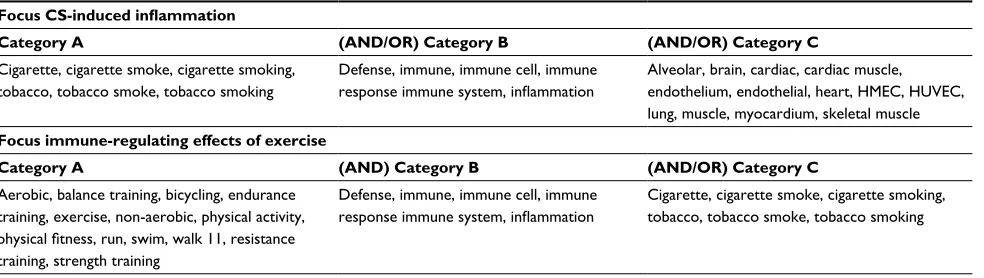
articles without any date restriction. Our review focused on the effects of CS-induced inflammation on different organs (such as brain, lung, heart, muscle, etc) as well as on immune-regulating effects of exercise which may coun-teract CS-induced inflammation. Search terms on PubMed (abstract and/or title) as shown in Table 1 were used. After careful review of titles and abstracts, it was decided whether the full-text will further be analyzed and consequently con-sidered in this review.
From CS to inflammation
The mechanisms of initiation and persistence of cigarette smoke (CS)-induced inflammation are not completely under-stood. CS comprises about 4,000 chemicals, including several carcinogens. Toxicologic studies have revealed a multitude
of immunomodulatory chemicals and gas.5 Consequently,
LTCS results in repetitive inflammatory insults leading to a chronic and progressive activation of the immune system accompanied by an abnormal inflammatory response of the airways to various noxious gases and particles.6,7 On the one hand, smoke pollutants cross through the alveolus–capillary interface and spread directly through the systemic blood-stream targeting different organs.7 At this point, they might be recognized by receptors of the innate immune system
which initiate inflammatory signaling cascades via NF-κB
activation.5 On the other hand, inflammatory processes are
suggested to originate in the pulmonary system. Here, toxic substances disturb the barrier function of the respiratory epithelium and impact both innate and adaptive host defense mechanisms. This primarily local inflammatory processes spillover into the circulation leading to inflammatory and degenerative processes in other organs and tissues. Thus, inflammation is suggested to be the main driver of the central comorbidities.7,8
Table 1 Search terms on PubMed Focus CS-induced inflammation
Category A (AND/OR) Category B (AND/OR) Category C
Cigarette, cigarette smoke, cigarette smoking, tobacco, tobacco smoke, tobacco smoking
Defense, immune, immune cell, immune response immune system, inflammation
Alveolar, brain, cardiac, cardiac muscle, endothelium, endothelial, heart, HMEC, HUVEC, lung, muscle, myocardium, skeletal muscle
Focus immune-regulating effects of exercise
Category A (AND) Category B (AND/OR) Category C
Aerobic, balance training, bicycling, endurance training, exercise, non-aerobic, physical activity, physical fitness, run, swim, walk 11, resistance training, strength training
Defense, immune, immune cell, immune response immune system, inflammation
Cigarette, cigarette smoke, cigarette smoking, tobacco, tobacco smoke, tobacco smoking
Abbreviations: HMEC, human microvascular endothelial cell; HUVEC, human umbilical vein endothelial cell.
Journal of Inflammation Research downloaded from https://www.dovepress.com/ by 118.70.13.36 on 24-Aug-2020
Dovepress Exercise and cigarette smoke-induced inflammation
Effects of CS on inflammatory processes
in respiratory tract and lungs
CS comprises various components that damage the pulmonary epithelium. LTCS has been shown to injure the cell membranes and alter the mucosal permeability.9 Cellular damage is fol-lowed by a compromised immune status, allowing opportunistic pathogens to cause infections that might amplify the inflam-matory processes. Furthermore, components of the innate and adaptive immune system are chronically activated. Analysis of bronchoalveolar lavage fluid and breath condensate provides evidence that even acute exposure to cigarette smoke results in oxidative stress and tissue damage as suggested by increased products of lipid peroxidation and degradation products of extracellular matrix proteins.5 In bronchoalveolar lavage (BAL) of long-term smokers, an increase of interleukin (IL)-1β, IL-6, IL-8, monocyte chemoattractant protein-1 (MCP-1),
macro-phage inflammatory protein (MIP)-1α, regulated on
activa-tion, normal T-cell expressed and secreted (RANTES), tumor necrosis factor (TNF)-α, IL-12 (p40), and IL-17 was found.10,11 Recent data suggest that airway epithelial cells (AECs) which represent a first line of defense against inhaled toxicants have altered inflammatory signaling in response to CS exposure. These cells were shown to upregulate cytokine expression and expression of matrix metalloproteases (MMPs) via extracellular signal-regulated kinase (ERK) signaling and increased p38 activation. Furthermore, AECs show characteristics of cellu-lar damage and cell death consequently leading to the release of damage-associated molecular patterns (DAMPs) into the extracellular space. DAMPs target pattern recognition recep-tors such as Toll-like receptor (TLR). TLRs are found on both immune and epithelial cells throughout the pulmonary system. TLRs recognize patterns of bacteria, fungi, and viruses, and the levels of TLR4 are elevated in cigarette smokers with COPD. After TLR activation, the NF-kB pathway is induced followed by the secretion of a variety of pro-inflammatory cytokines.12 In particular, MMP-9 and -12, surfactant protein D, and IL-1, IL-6, IL-8, and IL-17 have been found in higher quantities in the
lungs of long-term smokers with the ongoing inflammation.10
In parallel, immune cells, macrophages, neutrophils, dendritic cells, and lymphocytes migrate into the pulmonary system.11–13 Alveolar macrophages might play a key role in the pathogenesis of inflammation in lungs. These cells produce increased levels of MMPs, such as MMP-1, MMP-2, MMP-9, MMP-12, and
MMP-14, after smoke exposure (Figure 1).10
Specific role of lymphocytes
Currently, it is discussed that lymphocytes might play a cru-cial role in inflammatory pathogenesis. Specifically, CD8+ T
lymphocytes have been shown to be dramatically increased in the lungs of heavy smokers accompanied by a shift toward a type 1 profile. This immune cell subtype produces large amounts of interferon-γ and releases perforins and granzyme. Also B lymphocytes are activated, and it is suggested that their antigen-specific responses could turn against self-epitopes, partly because of impaired tolerance. In parallel, smoke expo-sure led to an accumulation of forkhead-box-protein (FOX)P31 T-regulatory cells (Tregs) in lungs of mice which might par-ticipate in controlling inflammatory processes.14 Accordingly, LTCS seems to alter the pulmonary immune equilibrium which turns into a chronic activated, immunosuppressed condition.15
Effects of LTCS on systemic inflammation
Every smoked cigarette seems to elicit a slight increase of oxidative stress and inflammation in blood indicated by an increase of thiobarbituric acid-reactive substances, neu-trophil elastase, leukotrienes, and neuneu-trophils after acute
CS in humans.5 Chronically, most studies agree that LCTS
induces an increase in the numbers of circulating neutrophils, macrophages, and lymphocytes. These cells show several inflammatory characteristics such as expression of activation markers and adhesion molecules which might mediate the migration into the bronchoalveolar system or other tissues.8,16 On the molecular levels, LTCS induces a systemic low-grade inflammation characterized by chronically elevated levels of various markers for inflammation, tissue deterioration, and
coagulation, such as C-reactive protein (CRP), TNF-α, von
Willebrand factor (vWF), tissue inhibitor of metalloprotein-ases-1, factor VII, and fibrinogen. Blood is suggested to be a transit way for transfer and spreading these molecules which target other organs and tissues.17,18
Effects of CS on the vascular endothelium
The particulate phase of CS consists of lipophilic com-ponents, which can pass the lipid bilayer of respiratory membranes; therefore, the damage is not limited to the lung tissue as it can also affect the vascular system.19 The integ-rity of endothelial cells (ECs) is essential, since it preserves vascular homeostasis, allows continuous adjustment of vas-cular tone and maintenance of blood fluidity, and regulates leukocyte traffic.20 Components of CS are toxic for ECs, and LTCS can lead to dysfunction of ECs,21 an early hallmark of atherosclerosis.22 Endothelial dysfunction is characterized by an imbalance of vasoconstrictors and vasodilators, aber-rant interaction between endothelial and immune cells, and
higher expression of adhesion molecules.23 Dysfunctional
ECs express lower levels of prostacyclin, thrombomodulin,
Journal of Inflammation Research downloaded from https://www.dovepress.com/ by 118.70.13.36 on 24-Aug-2020
Dovepress
Madani et al
tissue plasminogen activator (tPA), and NO while expres-sion levels of endothelin-1, angiotensin II, plasminogen
activator inhibitor-1 (PAI-1), and vWF are increased.20
Therefore, CS favors inflammatory processes in ECs and is a huge risk factor for the development of atherosclerosis
and cardiovascular diseases (CVDs).22 In vitro studies in
ECs demonstrate that CS induces cell injury in a dose- and
time-dependent manner24 which can lead to apoptosis,25
autophagic cell death,26 and necrosis.27 Different mechanisms are responsible for the induction of apoptosis in ECs induced by CS. One study showed that aqueous filtrates of CS lead to mitochondrial membrane depolarization, representing an early step in the apoptotic pathway.22 The negative influence of CS on apoptotic-related genes has also been reported. For
example, CS decreases p53 and Bcl-2 expression,28 disrupts
the vascular endothelial growth factor (VEGF), and fluid shear stress-mediated VEGFR2/phosphoinositide 3-kinase
(PI3K) signaling pathway29 and reduces the cytochrome-c
oxidase II expression through aberrant DNA methylation.25
Vascular damage through excessive apoptosis was also shown to be initiated by a p53-independent caspase-3 activating
pathway.30 EC injury may also be mediated through protein
carbonylation which is caused by reactive species in CS.31
Recruitment of leukocytes to the inflammation site happens by cytokine signaling, MMP-1 and MMP-9 upregulation, and
through cell adhesion of immune cells to ECs.32
Potential mechanisms of atherogenesis
The underlying mechanism of atherogenesis of ECs induced
by CS is not fully understood yet.33 So far, CS-induced
inflammation-related responses have been described in experimental studies in vitro. CS led to phosphorylation of various mitogen-activated protein kinases (MAPK), like
p38,34 c-Jun N-terminal kinase (JNK), and ERK.34,35 The
expression levels of osteopontin,35 E-selectin, intercellular cell adhesion molecule-1 (ICAM-1),36 and IL-837 were also induced by CS. Furthermore, CS induced nicotinamide adenine dinucleotide phosphate (NAD(P)H)-oxidase-derived
Figure 1 Illustration about cigarette-induced induction of oxidative stress and inflammation in AECs.
Abbreviations: AEC, airway epithelial cell; TLR4, Toll-like receptor-4; DAMP, damage-associated molecular pattern; MMP, matrix metalloprotease; ROS, reactive oxygen species; IL, interleukin; TNF, tumor necrosis factor; IFN, interferon.
Noxious gases, particles
ROS
Damage to AECs
Release of DAMPs
TLR4
Alveolar macrophages
Metalloproteases (MMP-9, MMP-12…) Cytokines (IL-1, IL-6,
IL-8, IL-I7, TNF-α…) IFN-γ
Perforins Granzyme Leukocytes (T cells [CD8 T cells], dendritic cells, neutrophils)
p38MAPK
NfB
Migration into lung tissue
AECs
Journal of Inflammation Research downloaded from https://www.dovepress.com/ by 118.70.13.36 on 24-Aug-2020
Dovepress Exercise and cigarette smoke-induced inflammation
H2O2 generation,38 upregulated fractalkine (CX3CL1),
increased IL-13Rα2 through the activation of protein kinase A-cAMP response element-binding protein (PKA-CREB)
pathway,39 increased cyclooxygenase-2 (COX-2) expression
through nuclear β-catenin accumulation due to the activation of epidermal growth factor receptor (EGFR)/Akt/glycogen synthase kinase-3β pathways.40 One cell culture study dem-onstrated that ECs treated with CS for 72 hours expressed only minor differences in various cytokines on mRNA level.41 CS promotes endothelial dysfunction as well by impairing endothelium-dependent relaxation, presumably through
sup-pression of NO production42 and CS-low-density lipoprotein
(CS-LDL).43 Cell culture studies proved that exposure of
ECs to CS impaired the VEGF-induced EC migration44,45
and tube formation, explaining the negative effect of CS on vessel growth and endothelial function.45
Effect of smoking on the cardiac tissue
Persistent inflammation is an important factor in the develop-ment of CVD.46 Since CS promotes inflammation47 and injures the cardiovascular system chronically, it is not surprising that the risk for CVD is twice as high in smokers than in
non-smokers.48 Toxic effects of CS on the myocardium have been
proved experimentally as well as clinically,15 but whether
smoking is a direct or indirect cause of CVD still needs to
be proved.49 It is somehow remarkable that even secondhand
smoke has the ability to increase the risk for CVD to as
high as 30%.48,50–54 Secondhand smoking combined with an
unhealthy lifestyle was shown to reduce the ability of the heart
adapt sensitively to sidestream smoke in a murine model.48
Furthermore, in nonsmoking humans, secondhand smoking increased WBC count immediately as well as CRP levels 18 h after exposure. Both of these are markers for inflammation
and have been linked to a higher incidence of CVD.50
The situation for smokers is worse. Active smoking increases cardiac afterload, promotes a pro-thrombotic sta-tus, reduces fibrinolysis, changes the profile of circulating lipids,48 promotes neutrophil infiltration in the myocardium,55 alters T-cell function,56 and causes DNA adducts in the
myo-cardium.57 CS leads to the production of reactive oxygen
species (ROS) which initiate ROS-sensitive signal transduc-tion pathways, such as MAPKs, and various transcriptransduc-tion factors, including NF-kB15 resulting in an aberrant cytokine
profile.58–60 Gene analysis of the hearts of mice revealed
an upregulation of the xenobiotic-metabolizing enzyme cytochrome P-450 1A1 and a downregulation of PAI-1,
representing a key gene involved in fibrinolysis.61 Taken
together, all mentioned factors are suggested to increase the
risk for several diseases of the cardiovascular system also in
human smokers.62
Effects of LTCS on muscle tissue
Human smokers tend to have a lower BMI while central or abdominal obesity seems to be increased. Thus, the weight loss associated with tobacco smoking may be due to loss of lean mass rather than fat.63 It is suggested that inflam-mation and oxidation of proteins are two main contributors to the development of skeletal muscle loss and dysfunction
observed in LTCS and COPD patients.63 Structurally, LTCS
leads to a reduced percentage of type I fiber, a lower muscle fiber cross-sectional area, an increased glycolytic enzymatic activity, and decreased muscle oxidative activity.8,63,64 Mice chronically exposed to cigarette smoke tend to a reduced muscle capillary to fiber ratio along with decreased VEGF, lowered endothelial and neuronal nitrite oxide synthase activ-ities in muscle vessels, and increased inflammatory activity
indicated by an increased mRNA expression of TNF-α and
IL-1β.8,64,65 The role of numerous cell signaling pathways in the development of skeletal muscle atrophy, a key element of muscle dysfunction in long-term cigarette smokers and COPD patients, has been investigated.65 In general, atrophy occurs when protein degradation exceeds protein synthesis. With regard to protein degradation, the ubiquitin proteasome system (UPS) seems to have an important role during LTCS. In smoke-exposed mice, an increased ubiquitination of target proteins was demonstrated, which was indicated by the increased activities of the E3 ubiquitin ligases atrogin-1
and muscle RING finger protein-1 (MuRF1).66 In parallel,
key factors that induce protein synthesis such as insulin-like growth factor 1 (IGF-1), are reduced followed by a lower activation of anabolic signaling pathways such as protein kinase B (Akt) and rapamycin (mTOR) pathways. All the mentioned pathways interact with inflammatory signaling
molecules such as TNF-α turning protein balance toward an
enhanced degradation leading to muscle wasting.63,66
Effects of LTCS on brain inflammation
Atherosclerosis and vascular brain lesions share similar pathological features such as oxidative stress and increased inflammation.67 Oxidative stress, for example, plays a deci-sive role in the pathogenesis of ischemic brain injury.68 It is not surprising that direct and secondhand CS are associated with various cerebrovascular-related diseases,32 in particular, smoking is a risk factor for stroke.69
Likewise to the effects of CS on endothelial cells,70 higher expression of VEGF, ICAM-1, IL-8, and nuclear factor
Journal of Inflammation Research downloaded from https://www.dovepress.com/ by 118.70.13.36 on 24-Aug-2020
Dovepress
Madani et al
(erythroid-derived 2)-like 2 was also observed in cultured
brain ECs.71 Furthermore, it was shown that CS extracts
induced heme oxygenase-1 (HO-1) expression mediated by
phosphatidylcholine phospholipase C/protein kinase Cδ/
NADPH oxidase-dependent platelet-derived growth factor
receptor (PDGFR)/PI3K/Akt pathway.19 Higher HO-1
expres-sion was shown to exacerbate early brain injury during intra-cerebral hemorrhagic stroke.72 Endothelin-1 levels decreased in rat brains exposed to CS, implying that endothelin-1 may
contribute to the hemodynamic response to chronic CS.73
Effects on brain ECs
Moreover, animal experiments proved that CS negatively affected endothelial tight junctions71 and downregulated the activity of Na-K-2Cl cotransporter in brain ECs. The latter could possibly contribute to an increase in extracellular K+.
Therefore, CS may exacerbate ischemic cellular damage and hinder recovery from ischemic damage. In addition, accumulation of extracellular fluid K+ is a risk factor for
cellular edema in astrocytes and neurons and could impair neuronal conduction after stroke.74 Increased blood viscos-ity due to CS impairs the blood flow and risks the integrviscos-ity
of the brain microvasculature.32 On top of everything, CS
negatively affects the viability of the blood–brain barrier (BBB). Taken together, CS and hemodynamic impairments contribute synergistically to vascular inflammation and BBB damage.32 Inflammation of brain cells due to CS was also con-firmed in various in vivo studies using mouse and rat models. Inflammation and cell death processes in the brain are often characterized by alterations of the neuroproteome.73–75 Mice exposed to secondhand CS, showed higher levels of ROS, induction of lipid peroxidation, activation of the transcription factors NF-kB and activator protein-1, as well as activation of MAPK, including JNK, ERK, and p38, and COX-2 in
various regions of the brain.69 Furthermore, secondhand CS
altered enzymatic antioxidant defenses by reducing superox-ide dismutase as well as catalase and increasing glutathione S-transferase activity in rat brains. Moreover, rats exposed to secondhand CS showed increased proteolytic degradation
of αII-spectrin through caspase-3 and dephosphorylation of
phosphoprotein enriched in astrocytes-15, both indicating apoptotic cell death.75
Immune-regulating effects of
exercise training
Exercise training has been shown to be an effective non-pharmacological treatment strategy in pulmonary diseases and systemic lung diseases. Furthermore, regular exercise
has been shown to increase patients’ strength, endurance capacity, quality-of-life scores, and symptoms of fatigue and dyspnea.76 Thus, the beneficial effects of exercise training in pulmonary rehabilitation are well established. Recent studies provided evidence that regular and moderate exercise exerts protective effects against smoke-induced lung disease due to its anti-inflammatory effects.64,77
Anti-inflammatory effects of exercise
Exercise training exerts its immune-regulating effects
by activating anti-inflammatory signaling pathways.78,79
Contracting skeletal muscle produces and secretes the anti-inflammatory myokine IL-6 during an acute bout of exercise, which evokes a subsequent rise in circulating levels of IL-6 followed by an ensuing increase in systemic levels of the
anti-inflammatory cytokines IL-10 and IL-1RA.77–79 IL-10,
which is mainly produced by Tregs, reduces tissue dam-age caused by inflammation and is known to diminish the
adaptive immune response.80–82 Complementarily, IL-1RA
is capable to limit the effects of the pro-inflammatory cyto-kine IL-1β and therefore serves as an important contributor
to exercise-induced anti-inflammatory state.79 Besides,
exercise-induced systemic elevations of cortisol, adrenalin, and IL-6 inhibit the secretion of pro-inflammatory TNF-α by monocytes.78,82,83 Moreover, after an acute bout of strenuous prolonged exercise, a reduced expression of TLRs on mono-cytes can be observed, which results in subsequent inhibition of pro-inflammatory cytokines and promotes the expression of costimulatory molecules and major histocompatibility
complex.83,84 CD14lowCD16+ monocytes are characterized
by heightened TLR-4 expression and thereby associated with pro-inflammatory properties.85 Regular exercise lowers
the ratio of pro-inflammatory monocytes (CD14lowCD16+)
to classical monocytes (CD14hiCD16–).86 Chronic exercise training also increases Treg cell numbers in circulation. In detail, athletes participating in sports where aerobic capacity is a prominent factor for performance outcome seem to have increased Treg counts.87,88
Specific role of exercise during obesity
In case of obesity, exercise training stimulates anti-inflam-matory signaling via a reduction in visceral fat mass, which is accompanied by a decrease in the production of several
pro-inflammatory adipokines (eg, TNF-α, leptin,
retinol-binding protein) and higher levels of adiponectin, which has anti-inflammatory effects and functions as an insulin
sensitizer.89 Current mouse and rat model studies indicate
that acute bouts of exercise and exercise training stimulate
Journal of Inflammation Research downloaded from https://www.dovepress.com/ by 118.70.13.36 on 24-Aug-2020
Dovepress Exercise and cigarette smoke-induced inflammation
phenotypic switching from M1-type macrophages producing TNF, IL-6, and nitric oxide toward M2-type macrophages, which release arginase and anti-inflammatory cytokines.90,91 In addition, after exercise training, a reduced tissue expres-sion of ICAM-1, which is involved in the adheexpres-sion of inflam-matory cells to endothelium and conveys interactions of T cells with target cells, and an inhibition of pro-inflammatory
M1-type macrophage migration in adipose tissue occur.91
Effects of exercise on pulmonary system
after LTCS
Experimental animal studies demonstrated that aerobic exercise after CS exposure or asthma induction reduces lung inflammation92 and remodeling.93,94
In particular, exercise was shown to increase Th1 response and suppress Th2 cytokine levels in lungs of smoke-exposed mice.95 In parallel, exercise increased antioxidant defense and reduced oxidative stress markers.96,97 In CS-exposed mice, it was shown that prior exercise training significantly reduced bronchoalveolar capillary permeability, inflammatory cell infiltration, epithelial thickening, expression of proliferat-ing cell nuclear antigen, mucin 2, cytokines, chemokines, adhesion molecules, and activation of NF-κB.98 These data proved an important preventive effect of exercise training for smoke-induced inflammation in lung tissue.
Effects of exercise on CS-induced
inflammation in blood
Regular exercise training has been shown to lower the levels of several inflammatory, chemoattractive, and coagulative factors in the blood of smoke-exposed mice. However, some human studies and clinical trials also demonstrated that due to dyspnea, COPD patients have restricted activity levels and muscle wasting, a markedly impaired exercise capacity.99,100 Therefore, some of these patients develop a kind of exercise intolerance. In this regard, it seems to be important to start exercise programs carefully, because acute and intensive bouts of exercise are known to induce a systemic immunologic response and oxidative stress, which might force inflammation in patients. However, a pro-inflammatory effect of exercise was only shown in muscle-wasted COPD patients after acute and intensive bouts of exercise. Interestingly, this effect was partially blunted by short-term supplementary oxygen.100,101 In general, longer periods of regular exercise training show
a decrease of many inflammatory cytokines such as TNF-α,
IL-2, IL-4, and CRP in COPD patients.102 Similarly, in murine models, a reduced expression of cell surface markers on cir-culating immune cells such as vascular adhesion molecule-1
(VCAM-1), ICAM-1, and CD62L was shown after regular treadmill running. Also several other inflammatory cytokines
such as IL-1α, MCP-3, MIP-1β, MIP-1α, and CD40L were
shown to decrease in smoke-exposed mice after training.64 In addition, regular endurance exercise has been shown to have favorable effects on blood coagulation by affecting fibrinolysis via decreasing vWF and factor VII.103
Effects of exercise on endothelium after
LTCS
The effects of CS and exercise on the inflammation of ECs have been well established in the literature. However, to our knowledge, no study has investigated the effects of exercise toward CS-induced inflammation in blood vessels. Therefore, it can only be hypothesized how exercise possibly ameliorates CS-induced inflammation in vessel walls by its effects on inflammation in general.
Endurance exercise training promotes
endothelium-dependent vasodilation104–108 which is related to a shear
stress-induced and Akt-dependent phosphorylation of endothelial NOS, resulting in NO activation.107 Furthermore, regular physical activity reduces oxidative stress,108 inflam-mation,109,110 and promotes LDL oxidation.19 It has also been demonstrated that exercise training has a positive impact on inflammatory markers. Regular physical activity reduced the levels of circulating adhesion molecules111 like soluble
intercellular adhesion molecule-1,112,113 soluble vascular
adhesion molecule-1 (sVCAM-1),112 soluble P-selectin,
and circulating CRP.109 Prolonged exercise sessions may
increase cell adhesion molecules like P-selectin,113 E-selectin, ICAM-1, and VCAM-1 first, but the endothelium recovers
rapidly afterwards.114 It is noteworthy that the potential
influence of exercise training on inflammation, circulating biomarkers, and anti-oxidative capacity depends on exercise capacity.115–117 For example, heart failure patients showed reduced endothelial response toward exercise. Indeed, plasma levels of vWF and tPA remained unaffected after exercise while their values increased in healthy subjects.118,119 Therefore, endothelial dysfunction and chronic inflammation probably impair exercise capacity.120 It is therefore crucial that exercise interventions in smokers should be considered as soon as possible since its benefits may decline with the progression of possible diseases.
Effects of exercise on cardiac tissue of
smokers
Smoking and physical inactivity are two avoidable risk
factors for CVD.121 As endothelial dysfunction can lead to
Journal of Inflammation Research downloaded from https://www.dovepress.com/ by 118.70.13.36 on 24-Aug-2020
Dovepress
Madani et al
cardiac dysfunction,122,123 it is reasonable to assume that protective effects of exercise toward endothelium in (non-) smokers, which are mentioned above, might also be cardio-protective. The decreasing risks for CVDs through moderate exercise training are in part mediated through inducing anti- inflammatory factors.124–127 Nonetheless, as it was mentioned for vessel walls, studies examining anti-inflammatory effects of exercise in CS-induced inflammation of the myocardium remain poor.
Short-time swimming exercise in CS-exposed Wistar rat could attenuate the impact of CS to the cardiovascular
system compared to the control group.47 Another study with
young women demonstrated that even secondhand smoking had a negative influence on exercise capacity due to reduced
values of VO2max and exercise duration and an increased
R-to-R value.129
Exercise intensity rather than duration has a more powerful impact on physiological adaptations regarding inflammation and oxidative stress,128 like it was described for vessel walls. Even if high-risk patients with severe coronary artery disease or heart failure could benefit the most from more intensive exercise training like high-intensity interval training, its safety has not been properly established.130 For example, exercise intensity might be a critical factor for the development of exercise-induced hypertension. Increased exercise intensity could trigger more endothelial responses in the absence of inflammatory markers. Therefore, exercise intervention plans should always have to be appropriate to each condition.23
Effects of exercise on muscle wasting
after LTCS
Exercise is able to reverse sarcopenia and muscle wasting in LTCS by different pathways. On the one hand, a decrease of systemic inflammation and inflammatory mediators in
muscle such as TNF-α and IL-1β might indirectly reduce
the activation of catabolic pathways and increase anabolic signals.64 In this regard, exercise has been shown to decrease for FoxO1 phosphorylation and reduce the expression of atrogin-1 and MuRF-1 in skeletal muscle of smoke-exposed mice. Consequently, exercise training abrogates the expres-sion of protein catabolic E3 ligases, which are considered key factors in myofibrillar protein breakdown via the UPS. On the other hand, in particular, resistance training is also known to directly increase IGF-1 signaling followed by the
activation of the Akt–mTOR–pathway.131,132 The reduction
of catabolic and stimulation of anabolic signaling attenuate or reverse muscle wasting after smoke exposure. Endurance
training was also shown to increase metabolic capacities of muscles by increased expression of genes involved in fatty acid transport into the mitochondrial matrix. Similarly, glu-cose uptake was optimized after regular exercise training.64 The differentiated effects of exercise training on muscle tissue might also depend on the mode of exercise. While endurance training more efficiently addresses type I fibers and oxidative metabolism, strength or resistance training mainly affects type II fibers, induces hypertrophy, and increases strength capabilities. However, most pulmonary rehabilitation pro-grams include both endurance and resistance exercise to maximize gains from both modalities. Alternatively, it has been shown that combined training program which includes both resistance and endurance exercise modalities increases
strength and endurance in COPD patients.133
Effects of exercise on brain after LCTS
Inflammation and vascular-induced abnormalities in the brain are two conditions associated with stroke and other neurovascular diseases, which can be protected by regular
physical activity in humans.134 In humans and animals,
exercise upregulates brain neurotrophin and brain-derived neurotophin factor (BDNF), which is an important factor of neuronal function, growth, and survival. BDNF increases
the brain’s resistance to damage and degeneration.135 The
immediate response of the brain to acute exercise produces
only marginal changes of inflammatory mediators.136 On the
other hand, regular physical activity improves the overall immune condition in the brain.137 Murine studies proved that pro-inflammatory cytokines impair the IGF-1 signal trans-duction in neurons. Peripheral IGF-1 is essential in glucose metabolism and cerebrovascular function. One mechanism by which the negative effects of inflammation are counter-acted by exercise is the restoration of IGF-1 signaling.137 In a study with mice, endurance and strength training decreased
most of the inflammatory factors, such as IL-1α, IL-2, and
IL-18. Interestingly, NF-κB and COX-2 protein levels were
significantly increased probably due to circulating IL-6 after training. The increased expression of COX-2 and microsomal prostaglandin E synthase, an enzyme downstream of COX-2, were independent of peripheral inflammation.138 Exercise improved oxidative stress and inflammation directly at the brain of old high-fat-fed ApoE−/− mice, reaffirming the neu-roprotective effects of exercise in a model of mice with vas-cular brain lesions.138 On the other hand, aged mice, training above the lactate threshold showed increased levels of brain
PGC-1α, mTOR, and phospho-mTOR protein levels, as well
as citrate synthase mRNA levels.139 A similar relationship has
Journal of Inflammation Research downloaded from https://www.dovepress.com/ by 118.70.13.36 on 24-Aug-2020
Dovepress Exercise and cigarette smoke-induced inflammation
been confirmed in young mice.140 In addition, for FOXO-3
translocated from the nucleus to the cytoplasm, predicting
an increased and facilitated VEGF-A expression.141 Another
study using rats in a traumatic brain injury model showed that aerobic exercise training enhances the endogenous anti-inflammatory response (IL-10), inhibits the infiltration of neutrophils, and attenuates BBB breakdown as well as pro-inflammatory cytokines (IL-1β, TNFα).141
Neuroprotective effects of exercise
Neuroprotective and anti-inflammatory effects of exercise on the brain have also been shown in humans. Moderate intensity interval training in Parkinson’s disease patients attenuated inflammation by decreasing circulating sVCAM-1 and serum
TNF-α and increasing serum BDNF levels.142
Accordingly, exercise training has the potential to be a new therapeutic approach to control acute inflammation.143 These effects remain to be proven in response to CS-induced inflam-mation. So far, only one study has investigated the effects of exercise training on CS-exposed brain oxidative stress.144 Mice exposed to CS showed decreased levels of BDNF and higher immobility in a forced swim test. Exercise was able to prevent oxidative damage, but surprisingly, it could neither reverse
the decrease of BDNF nor it was able to prevent CS-induced depressive-like behavior.144 These results clearly show that molecular effects of exercise on CS-induced inflammation at the brain needs to be investigated in future research projects.
Conclusion
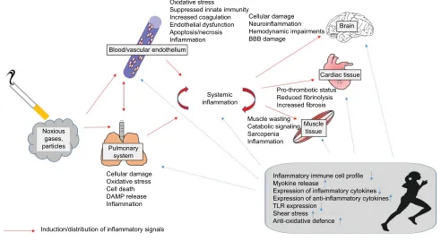
Taken together, LTCS induces local and systemic inflam-matory processes which might be mediated directly by pol-lutant particles and a spillover of inflammatory signals to other tissues. These inflammatory processes might induce or amplify signals of tissue degradation and catabolic pro-cesses. Exercise training has been shown to prevent and even reverse inflammatory processes leading to reduced tissue degradation and catabolic processes. In parallel, exercise elicits anabolic signals leading to an increased functional capacity. Accordingly, regular exercise training seems to protect long-term smokers against some important nega-tive local and systemic consequences of smoking. In this regard, the immune- regulating properties of exercise might have relevance (Figure 2). It has to be considered that many molecular findings from smoking or exercise effects on tis-sues were obtained from animal studies, and this knowledge has to be only carefully transferred to humans.
Figure 2 Overview about the distribution of inflammatory signals induced by tobacco smoking from the pulmonary system to blood, brain, cardiac tissue, and muscle and the immune-regulating effects of regular exercise training.
Abbreviations: BBB, blood–brain barrier; TLR, Toll-like receptor; DAMP, damage-associated molecular pattern.
Blood/vascular endothelium Oxidative stress
Suppressed innate immunity Increased coagulation Endothelial dysfunction Apoptosis/necrosis Inflammation
Cellular damage Neuroinflammation Hemodynamic impairments BBB damage
Brain
Cardiac tissue
Muscle tissue Pro-thrombotic status Reduced fibrinolysis Increased fibrosis Muscle wasting
Catabolic signaling Sarcopenia Inflammation
Inflammatory immune cell profile Myokine release
Expression of inflammatory cytokines Expression of anti-inflammatory cytokines TLR expression
Shear stress Anti-oxidative defence Systemic
inflammation
Pulmonary system
Cellular damage Oxidative stress Cell death DAMP release Inflammation
Induction/distribution of inflammatory signals
Immune-regulating/anti-inflammatory effects of exercise training Noxious
gases, particles
Journal of Inflammation Research downloaded from https://www.dovepress.com/ by 118.70.13.36 on 24-Aug-2020
Dovepress
Madani et al
More studies are needed to discover the mechanisms how exercise affects inflammation in smokers. Specifically, the effects of exercise on inflammation in various organs in smokers have to be confirmed. From a clinical point of view, data suggest that it seems to be important to start exercise training as early as possible for smokers. In this regard, the progressive increase of certain inflammatory signals might be important predictors of the necessity of starting a regular exercise training program.
Disclosure
The authors report no conflicts of interest in this work.
References
1. Marie Ng, Freeman MK, Fleming TD, et al. Smoking prevalence and cig-arette consumption in 187 countries, 1980-2012. JAMA. 2014;311(2): 183–192.
2. Wakefield MA, Germain D, Durkin SJ. How does increasingly plainer cigarette packaging influence adult smokers’ perceptions about brand image? An experimental study. Tob Control. 2008;17(6):416–421. 3. Edwards R. The problem of tobacco smoking. BMJ. 2004;328:217–219. 4. Foschino Barbaro MP, Carpagnano GE, Spanevello A, Cagnazzo
MG, Barnes PJ. Inflammation, oxidative stress and systemic effects in mild chronic obstructive pulmonary disease. Int J Immunopathol
Pharmacol. 2007;20(4):753–763.
5. van der Vaart H, Postma DS, Timens W, Ten Hacken NH. Acute effects of cigarette smoke on inflammation and oxidative stress: a review. 2004;59:713–721.
6. Pauwels RA, Buist AS, Ma P, Jenkins CR, Hurd SS. Global strategy for the diagnosis, management, and prevention of chronic obstructive pulmonary disease: National Heart, Lung, and Blood Institute and World Health Organization Global Initiative for Chronic Obstruc-tive Lung Disease (GOLD): execuObstruc-tive summary. Respir Care. 2001;46(8):798–825.
7. Zhang J, Liu Y, Shi J, Larson DF, Watson RR. Side-stream cigarette smoke induces dose-response in systemic inflammatory cytokine production and oxidative stress. Exp Biol Med.2002;227(9):823–829. 8. Krüger K, Dischereit G, Seimetz M, Wilhelm J, Weissmann N, Mooren FC. Time course of cigarette smoke-induced changes of systemic inflammation and muscle structure. Am J Physiol Lung Cell Mol
Physiol. 2015;309(2):L119–L128.
9. Rusznak C, Mills PR, Devalia JL, Sapsford RJ, Davies RK, Loze-wicz, S. Effect of cigarette smoke on the permeability and IL-1b and sICAM-1 release from cultured human bronchial epithelial cells of never-smokers, smokers, and patients with chronic obstructive pul-monary disease. Am J Respir Cell Mol Biol. 2000;23:530–536. 10. Crotty Alexander LE, Shin S, Hwang JH. Inflammatory diseases of
the lung induced by conventional cigarette smoke: a review. Chest. 2015;148(5):1307–1322.
11. Kou YR, Kwong K, Lee LY. Airway inflammation and hypersen-sitivity induced by chronic smoking. Respir Physiol Neurobiol. 2011;178:395–405.
12. Pouwels SD, Hesse L, Faiz A, et al. Susceptibility for cigarette smoke-induced DAMP release and DAMP-smoke-induced inflammation in COPD.
Am J Physiol Lung Cell Mol Physiol. 2016;311(5):L881–L892.
13. Tang GJ, Wang HY, Wang JY, et al. Novel role of AMP-activated protein kinase signaling in cigarette smoke induction of IL-8 in human lung epithelial cells and lung inflammation in mice. Free Radic Biol Med. 2011;50(11):1492–1502.
14. Botelho FM, Gaschler GJ, Kianpour S, et al. Innate immune processes are sufficient for driving cigarette smoke-induced inflammation in mice. Am J Respir Cell Mol Biol. 2010;42(4):394–403.
15. Kaplan A, Abidi E, Ghali R, Booz GW, Kobeissy F, Zouein FA. Functional, cellular, and molecular remodeling of the heart under influence of oxidative cigarette tobacco smoke. Oxid Med Cell Longev. 2017;2017:3759186.
16. Majo J, Ghezzo H, Cosio MG. Lymphocyte population and apoptosis in the lungs of smokers and their relation to emphysema. Eur Respir J. 2001;17:946–953.
17. Wannamethee SG, Lowe GD, Shaper AG, Rumley A, Lennon L, Whincup PH. Associations between cigarette smoking, pipe/cigar smoking, and smoking cessation, and haemostatic and inflamma-tory markers for cardiovascular disease. Eur Heart J. 2005;26(17): 1765–1773.
18. Liu J, Liang Q, Frost-Pineda K, et al. Relationship between biomarkers of cigarette smoke exposure and biomarkers of inflammation, oxida-tive stress, and platelet activation in adult cigarette smokers. Cancer
Epidemiol Biomarkers Prev. 2011;20(8):1760–1769.
19. Shih RH, Cheng SE, Hsiao LD, Kou YR, Yang CM. Cigarette smoke extract upregulates heme oxygenase-1 via PKC/NADPH oxidase/ ROS/PDGFR/PI3K/Akt pathway in mouse brain endothelial cells.
J Neuroinflammation. 2011;8:104.
20. Ribeiro F, Alves AJ, Duarte JA, Oliveira J. Is exercise training an effective therapy targeting endothelial dysfunction and vascular wall inflammation? Int J Cardiol. 2010;141(3):214–221.
21. Unverdorben M, der Bijl A, Potgieter L, Liang Q, Meyer BH, Roethig HJ. Effects of levels of cigarette smoke exposure on symptom-limited spiroergometry. Prev Cardiol. 2007;10(2):83–91.
22. Vayssier-Taussat M, Camilli T, Aron Y, et al. Effects of tobacco smoke and benzo[a]pyrene on human endothelial cell and monocyte stress responses. Am J Physiol Heart Circ Physiol. 2001;280(3):H1293–H1300.
23. Jee H, Park J, Oh JG, Lee YH, Shin KA, Kim YJ. Effect of a prolonged endurance marathon on vascular endothelial and inflammation mark-ers in runnmark-ers with exercise-induced hypertension. Am J Phys Med
Rehabil. 2013;92(6):513–522.
24. Hoshino S, Yoshida M, Inoue K, et al. Cigarette smoke extract induces endothelial cell injury via JNK pathway. Biochem Biophys Res
Com-mun. 2005;329(1):58–63.
25. Yang M, Chen P, Peng H, et al. Cigarette smoke extract induces aber-rant cytochrome-c oxidase subunit II methylation and apoptosis in human umbilical vascular endothelial cells. Am J Physiol Cell Physiol. 2015;308(5):C378–C384.
26. Csordas A, Kreutmayer S, Ploner C, et al. Cigarette smoke extract induces prolonged endoplasmic reticulum stress and autophagic cell death in human umbilical vein endothelial cells. Cardiovasc Res. 2011;92(1):141–148.
27. Messner B, Frotschnig S, Steinacher-Nigisch A, et al. Apoptosis and necrosis: two different outcomes of cigarette smoke condensate-induced endothelial cell death. Cell Death Dis. 2012;3(11):e424. 28. Yang YM, Liu GT. Damaging effect of cigarette smoke extract on
primary cultured human umbilical vein endothelial cells and its mechanism. Biomed Environ Sci. 2004;17(2):121–134.
29. Edirisinghe I, Rahman I. Cigarette smoke-mediated oxidative stress, shear stress, and endothelial dysfunction: role of VEGFR2. Ann N Y
Acad Sci. 2010;1203:66–72.
30. Wang J, Wilcken DE, Wang XL. Cigarette smoke activates caspase-3 to induce apoptosis of human umbilical venous endothelial cells. Mol
Genet Metab. 2001;72(1):82–88.
31. Gornati R, Colombo G, Clerici M, et al. Protein carbonylation in human endothelial cells exposed to cigarette smoke extract. Toxicol
Lett. 2013;218(2):118–128.
32. Mazzone P, Tierney W, Hossain M, Puvenna V, Janigro D, Cucullo L. Pathophysiological impact of cigarette smoke exposure on the cere-brovascular system with a focus on the blood-brain barrier: expanding the awareness of smoking toxicity in an underappreciated area. Int J
Environ Res Public Health. 2010;7(12):4111–4126.
33. Giunzioni I, Bonomo A, Bishop E, Castiglioni S, Corsini A, Bellosta S. Cigarette smoke condensate affects monocyte interaction with endothelium. Atherosclerosis. 2014;234(2):383–390.
Journal of Inflammation Research downloaded from https://www.dovepress.com/ by 118.70.13.36 on 24-Aug-2020
Dovepress Exercise and cigarette smoke-induced inflammation
34. Low B, Liang M, Fu J. p38 mitogen-activated protein kinase medi-ates sidestream cigarette smoke-induced endothelial permeability.
J Pharmacol Sci. 2007;104(3):225–231.
35. Bishop E, Theophilus EH, Fearon IM. In vitro and clinical studies examin-ing the expression of osteopontin in cigarette smoke-exposed endothelial cells and cigarette smokers. BMC Cardiovasc Disord. 2012;12:75. 36. Chen HW, Lii CK, Ku HJ, Wang TS. Cigarette smoke extract induces
expression of cell adhesion molecules in HUVEC via actin filament reorganization. Environ Mol Mutagen. 2009;50(2):96–104. 37. Wang H, Ye Y, Zhu M, Cho C. Increased interleukin-8 expression by
cigarette smoke extract in endothelial cells. Environ Toxicol
Pharma-col. 2000;9(1–2):19–23.
38. Orosz Z, Csiszar A, Labinskyy N, et al. Cigarette smoke-induced proinflammatory alterations in the endothelial phenotype: role of NAD(P)H oxidase activation. Am J Physiol Heart Circ Physiol. 2007;292(1):H130–H139.
39. Rius C, Company C, Piqueras L, et al. Critical role of fractalkine (CX3CL1) in cigarette smoke-induced mononuclear cell adhesion to the arterial endothelium. Thorax. 2013;68(2):177–186.
40. Meng M, Liao H, Zhang B, et al. Cigarette smoke extracts induce overexpression of the proto-oncogenic gene interleukin-13 receptor alpha2 through activation of the PKA-CREB signaling pathway to trigger malignant transformation of lung vascular endothelial cells and angiogenesis. Cell Signal. 2017;31:15–25.
41. Barbieri SS, Weksler BB. Tobacco smoke cooperates with interleukin-1beta to alter beta-catenin trafficking in vascular endothelium resulting in increased permeability and induction of cyclooxygenase-2 expres-sion in vitro and in vivo. FASEB J. 2007;21(8):1831–1843. 42. Allam E, Delacruz K, Ghoneima A, Sun J, Windsor LJ. Effects of
tobacco on cytokine expression from human endothelial cells. Oral
Dis. 2013;19(7):660–665.
43. Ota Y, Kugiyama K, Sugiyama S, et al. Impairment of endothelium-dependent relaxation of rabbit aortas by cigarette smoke extract – role of free radicals and attenuation by captopril. Atherosclerosis. 1997;131(2):195–202.
44. Kagota S, Yamaguchi Y, Shinozuka K, Kwon YM, Kunitomo M. Ciga-rette smoke-modified low density lipoprotein impairs endothelium-dependent relaxation in isolated rabbit arteries. Gen Pharmacol. 1996;27(3):477–481.
45. Snajdar RM, Busuttil SJ, Averbook A, Graham DJ. Inhibition of endothelial cell migration by cigarette smoke condensate. J Surg Res. 2001;96(1):10–16.
46. Bjornstad HH, Bruvik J, Bjornstad AB, Hjellestad BL, Damas JK, Aukrust P. Exercise training decreases plasma levels of soluble CD40 ligand and P-selectin in patients with chronic heart failure. Eur J
Cardiovasc Prev Rehabil. 2008;15(1):43–48.
47. de Sa FG, da Mota GR, Sant’Ana PG, da Cunha MR, Marocolo M, Castardeli E. Training and cardiovascular responses from cigarette smoke exposure. Chin J Physiol. 2014;57(6):315–319.
48. Tilton SC, Karin NJ, Webb-Robertson BJ, et al. Impaired transcriptional response of the murine heart to cigarette smoke in the setting of high fat diet and obesity. Chem Res Toxicol. 2013;26(7):1034–1042. 49. Stallones RA. The association between tobacco smoking and coronary
heart disease. Int J Epidemiol. 2015;44(3):735–743.
50. Zhang J, Fang SC, Mittleman MA, Christiani DC, Cavallari JM. Secondhand tobacco smoke exposure and heart rate variability and inflammation among non-smoking construction workers: a repeated measures study. Environ Health. 2013;12(1):83.
51. Blackburn H. Environmental tobacco smoke exposure was associated with an increased risk of ischemic heart disease. Evid Based
Cardio-vasc Med. 1998;2(2):43–44.
52. Japuntich SJ, Eilers MA, Shenhav S, et al. Secondhand tobacco smoke exposure among hospitalized nonsmokers with coronary heart disease.
JAMA Intern Med. 2015;175(1):133–136.
53. Vandivier RW. Learning to act on secondhand tobacco smoke expo-sure to limit risk for coronary heart disease. JAMA Intern Med. 2015;175(1):136.
54. Enstrom JE, Kabat GC. Environmental tobacco smoke and coronary heart disease mortality in the United States – a meta-analysis and critique. Inhal Toxicol. 2006;18(3):199–210.
55. Das A, Dey N, Ghosh A, Das S, Chattopadhyay DJ, Chatterjee IB. Molecular and cellular mechanisms of cigarette smoke-induced myo-cardial injury: prevention by vitamin C. PLoS One. 2012;7(9):e44151. 56. Leone A. Biochemical markers of cardiovascular damage from tobacco
smoke. Curr Pharm Des. 2005;11(17):2199–2208.
57. Izzotti A, D’Agostini F, Balansky R, et al. Exposure of mice to cigarette smoke and/or light causes DNA alterations in heart and aorta. Mutat
Res. 2008;644(1–2):38–42.
58. Zhou X, Li C, Xu W, Chen J. Trimetazidine protects against smoking-induced left ventricular remodeling via attenuating oxidative stress, apoptosis, and inflammation. PLoS One. 2012;7(7):e40424. 59. Zhou X, Li C, Xu W, Chen J. Protective effects of valsartan against
cigarette smoke-induced left ventricular systolic dysfunction in rats.
Int J Cardiol. 2013;167(3):677–680.
60. Khanna AK, Xu J, Mehra MR. Antioxidant N-acetyl cysteine reverses cigarette smoke-induced myocardial infarction by inhibiting inflamma-tion and oxidative stress in a rat model. Lab Investig. 2012;92:224–235. 61. Halappanavar S, Stampfli MR, Berndt-Weis L, Williams A, Douglas
GR, Yauk CL. Toxicogenomic analysis of mainstream tobacco smoke-exposed mice reveals repression of plasminogen activator inhibitor-1 gene in heart. Inhal Toxicol. 2009;21(1):78–85.
62. McCall MR, van den Berg JJ, Kuypers FA, et al. Modification of LCAT activity and HDL structure. New links between cigarette smoke and coronary heart disease risk. Arterioscler Thromb. 1994;14(2):248–253. 63. Gea J, Agustí A, Roca J. Pathophysiology of muscle dysfunction in
COPD. J Appl Physiol (1985). 2013;114(9):1222–1234.
64. Krüger K, Seimetz S, Ringseis R, et al. Exercise training reverses inflammation and muscle wasting after smoke exposure. Am J Physiol
Regul Integr Comp Physiol. 2017 Epub Nov 1.
65. Basic VT, Tadele E, Elmabsout AA, et al. Exposure to cigarette smoke induces overexpression of von Hippel-Lindau tumor suppres-sor in mouse skeletal muscle. Am J Physiol Lung Cell Mol Physiol. 2012;303(6):L519–L527.
66. Gomes MD, Lecker SH, Jagoe RT, Navon A, Goldberg AL. Atrogin-1, a muscle531 specific F-box protein highly expressed during muscle atrophy. Proc Natl Acad Sci U S A. 2001;98(25):14440–14445. 67. Dutta P, Courties G, Wei Y, et al. Myocardial infarction accelerates
atherosclerosis. Nature. 2012;487(7407):325–329.
68. Hafezi-Moghadam A, Thomas KL, Wagner DD. ApoE deficiency leads to a progressive age-dependent blood-brain barrier leakage. Am
J Physiol Cell Physiol. 2007;292(4):C1256–C1262.
69. Manna SK, Rangasamy T, Wise K, et al. Long term environmental tobacco smoke activates nuclear transcription factor-kappa B, activa-tor protein-1, and stress responsive kinases in mouse brain. Biochem
Pharmacol. 2006;71(11):1602–1609.
70. Seo SB, Choe ES, Kim KS, Shim SM. The effect of tobacco smoke exposure on the generation of reactive oxygen species and cellular membrane damage using co-culture model of blood brain barrier with astrocytes. Toxicol Ind Health. 2017;33(6):530–536.
71. Prasad S, Sajja RK, Park JH, Naik P, Kaisar MA, Cucullo L. Impact of cigarette smoke extract and hyperglycemic conditions on blood-brain barrier endothelial cells. Fluids Barriers CNS. 2015;12:18. 72. Wang J, Dore S. Heme oxygenase-1 exacerbates early brain injury
after intracerebral haemorrhage. Brain. 2007;130(Pt 6):1643–1652. 73. Ohno N, Tanaka T, Kita T, et al. Changes of brain endothelin levels and
peripheral endothelin receptors by chronic cigarette smoke in sponta-neously hypertensive rats. J Pharmacol Sci. 2004;94(3):287–296. 74. Paulson JR, Roder KE, McAfee G, Allen DD, Van der Schyf CJ,
Abbruscato TJ. Tobacco smoke chemicals attenuate brain-to-blood potassium transport mediated by the Na,K,2Cl-cotransporter during hypoxia-reoxygenation. J Pharmacol Exp Ther. 2006;316(1):248–254. 75. Fuller BF, Gold MS, Wang KK, Ottens AK. Effects of environmental
tobacco smoke on adult rat brain biochemistry. J Mol Neurosci. 2010; 41(1):165–171.
Journal of Inflammation Research downloaded from https://www.dovepress.com/ by 118.70.13.36 on 24-Aug-2020
Dovepress
Madani et al
76. McCarthy B, Casey D, Devane D, Murphy K, Murphy E, Lacasse Y. Pulmonary rehabilitation for chronic obstructive pulmonary disease.
Cochrane Database Syst Rev. 2015;2:CD003793.
77. Pedersen BK, Febbraio MA. Muscle as an endocrine organ: focus on muscle-derived interleukin-6. Physiol Rev. 2008;88(4):1379–1406. 78. Gleeson M, Bishop NC, Stensel DJ, Lindley MR, Mastana SS,
Nimmo MA. The anti-inflammatory effects of exercise: mechanisms and implications for the prevention and treatment of disease. Nat Rev
Immunol. 2011;11(9):607–615.
79. Pedersen BK. Anti-inflammatory effects of exercise: role in diabetes and cardiovascular disease. Eur J Clin Invest. 2017;47(8):600–611. 80. Maynard CL, Weaver CT. Diversity in the contribution of IL 10 to
cell-mediated immune regulation. Immunol Rev. 2008;226:219–233. 81. Moore KW, de Waal Malefyt R, Coffman RL, O’Garra A. Interleukin 10 and the interleukin 10 receptor. Annu Rev Immunol. 2001;19:683–765. 82. Bergmann M, Gornikiewicz A, Sautner T, et al. Attenuation of
catecholamine-induced immunosuppression in whole blood from patients with sepsis. Shock. 1999;12(6):421–427.
83. Gleeson M, McFarlin BK, Flynn MG. Exercise and Toll-like receptors.
Exerc Immunol Rev. 2006;12:34–53.
84. Oliveira M, Gleeson M. The influence of prolonged cycling on mono-cyte Toll-like receptor 2 and 4 expression in healthy men. Eur J Appl
Physiol. 2010;109(2):251–257.
85. Skinner NA, MacIsaac CM, Hamilton JA, VisvanathanK. Regula-tion of Toll-like receptor (TLR)2 and TLR4 on CD14dimCD16+
monocytes in response to sepsis-related antigens. Clin Exp Immunol. 2005;141(2):270–278.
86. Timmerman KL, Flynn MG, Coen PM, Markofski MM, Pence PB. Exercise training-induced lowering of inflammatory (CD14+CD16+) monocytes: a role in the anti-inflammatory influence of exercise?
Leukoc Biol. 2008;84(5):1271–1278.
87. Shaw DM, Merien F, Braakhuis A, Dulson D. T-cells and their cytokine production: the anti-inflammatory and immunosuppressive effects of strenuous exercise. Cytokine. 2017; pii:S1043-4666(17)30292-2. 88. Weinhold M, Shimabukuro-Vornhagen A, Franke A, et al. Physical
exercise modulates the homeostasis of human regulatory T cells.
J Allergy Clin Immunol. 2016;137(5):1607–1610.e8.
89. Krüger K. Inflammation during obesity – pathophysiological con-cepts and effects of physical activity. Dtsch Z Sportmed. 2017;68: 163–169.
90. Oliveira AG, Araujo TG, Carvalho BM, Guadagnini D, Rocha GZ, Bagarolli RA. Acute exercise induces a phenotypic switch in adipose tissue macrophage polarization in diet-induced obese rats. Obesity. 2013;21:2545–2556.
91. Kawanishi N, Yano H, Yokogawa Y, Suzuki K. Exercise training inhibits inflammation in adipose tissue via both suppression of macrophage infiltration and acceleration of phenotypic switching from M1 to M2 macrophages in high-fat-diet-induced obese mice. Exerc Immunol
Rev. 2010;16:105–118.
92. Lowder T, Dugger K, Deshane J, Estell K, Schwiebert LM. Repeated bouts of aerobic exercise enhance regulatory T cell responses in a murine asthma model. Brain Behav Immun. 2010;24(1):153–159. 93. Pastva A, Estell K, Schoeb TR, Atkinson TP, Schwiebert LM. Aerobic
exercise attenuates airway inflammatory responses in a mouse model of atopic asthma. J Immunol. 2004;172(7):4520–4526.
94. Dugger KJ, Chrisman T, Jones B, et al. Moderate aerobic exercise alters migration patterns of antigen specific T helper cells within an asthmatic lung. Brain Behav Immun. 2013;34:67–78.
95. Lakier Smith L. Overtraining, excessive exercise, and altered immu-nity: is this a T helper-1 versus T helper-2 lymphocyte response? Sports
Med. 2003;33(5):347–364.
96. Onur E, Kabaroğlu C, Günay O, et al. The beneficial effects of physical exercise on antioxidant status in asthmatic children. Allergol
Immu-nopathol (Madr). 2011;39(2):90–95.
97. Menegali BT, Nesi RT, Souza PS, et al. The effects of physical exercise on the cigarette smoke-induced pulmonary oxidative response. Pulm
Pharmacol Ther. 2009;22(6):567–573.
98. Yu BU, Liao YW, Su KH, et al. Prior exercise training alleviates the lung inflammation induced by subsequent exposure to environmental cigarette smoke. Acta Physiol (Oxf). 2012;205(4):532–554. 99. Rennard SI, Drummond MB. Early chronic obstructive
pulmo-nary disease: definition, assessment, and prevention. Lancet. 2015;385(9979):1778–1788.
100. Saey D, Debigare R, LeBlanc P, et al. Contractile leg fatigue after cycle exercise: a factor limiting exercise in patients with chronic obstructive pulmonary disease. Am J Respir Crit Care Med. 2003;168(4):425–430. 101. Van Helvoort HA, Heijdra YF, Thijs HM, Vina J, Wanten GJ, Dekhui-jzen PN. Exercise-induced systemic effects in muscle-wasted patients with COPD. Med Sci Sports Exerc. 2006;38(9):1543–1552. 102. Abd El-Kader SM, Al-Jiffri OH, Al-Shreef FM. Plasma
inflamma-tory biomarkers response to aerobic versus resisted exercise training for chronic obstructive pulmonary disease patients. Afr Health Sci. 2016;16(2):507–515.
103. El-Sayed MS, Sale C, Jones PG, Chester M. Blood hemostasis in exercise and training. Med Sci Sports Exerc. 2000;32(5):918–925. 104. Clarkson P, Montgomery HE, Mullen MJ, et al. Exercise training
enhances endothelial function in young men. J Am Coll Cardiol. 1999;33(5):1379–1385.
105. Kelly AS, Wetzsteon RJ, Kaiser DR, Steinberger J, Bank AJ, Dengel DR. Inflammation, insulin, and endothelial function in overweight children and adolescents: the role of exercise. J Pediatr. 2004;145(6):731–736. 106. Hambrecht R, Wolf A, Gielen S, et al. Effect of exercise on coronary
endothelial function in patients with coronary artery disease. N Engl
J Med. 2000;342(7):454–460.
107. Hambrecht R, Adams V, Erbs S, et al. Regular physical activity improves endothelial function in patients with coronary artery disease by increasing phosphorylation of endothelial nitric oxide synthase.
Circulation. 2003;107(25):3152–3158.
108. Laufs U, Wassmann S, Czech T, et al. Physical inactivity increases oxidative stress, endothelial dysfunction, and atherosclerosis.
Arte-rioscler Thromb Vasc Biol. 2005;25(4):809–814.
109. Morikawa Y, Mizuno Y, Harada E, et al. Aerobic interval exercise training in the afternoon reduces attacks of coronary spastic angina in conjunction with improvement in endothelial function, oxidative stress, and inflammation. Coron Artery Dis. 2013;24(3):177–182. 110. Babbitt DM, Diaz KM, Feairheller DL, et al. Endothelial activation
microparticles and inflammation status improve with exercise training in African Americans. Int J Hypertens. 2013;2013:538017. 111. Zoppini G, Targher G, Zamboni C, et al. Effects of moderate-intensity
exercise training on plasma biomarkers of inflammation and endo-thelial dysfunction in older patients with type 2 diabetes. Nutr Metab
Cardiovasc Dis. 2006;16(8):543–549.
112. Wegge JK, Roberts CK, Ngo TH, Barnard RJ. Effect of diet and exercise intervention on inflammatory and adhesion molecules in postmenopausal women on hormone replacement therapy and at risk for coronary artery disease. Metabolism. 2004;53(3):377–381. 113. Adamopoulos S, Parissis J, Kroupis C, et al. Physical training reduces
peripheral markers of inflammation in patients with chronic heart failure. Eur Heart J. 2001;22(9):791–797.
114. Walther C, Mobius-Winkler S, Linke A, et al. Regular exercise training compared with percutaneous intervention leads to a reduction of inflam-matory markers and cardiovascular events in patients with coronary artery disease. Eur J Cardiovasc Prev Rehabil. 2008;15(1):107–112. 115. Nielsen HG, Lyberg T. Long-distance running modulates the expression
of leucocyte and endothelial adhesion molecules. Scand J Immunol. 2004;60(4):356–362.
116. Bartzeliotou AI, Margeli AP, Tsironi M, et al. Circulating levels of adhesion molecules and markers of endothelial activation in acute inflammation induced by prolonged brisk exercise. Clin Biochem. 2007;40(11):765–770.
117. Wisloff U, Stoylen A, Loennechen JP, et al. Superior cardiovascu-lar effect of aerobic interval training versus moderate continuous training in heart failure patients: a randomized study. Circulation. 2007;115(24):3086–3094.
Journal of Inflammation Research downloaded from https://www.dovepress.com/ by 118.70.13.36 on 24-Aug-2020