REVIEW OF LUNG NODULES IDENTIFICATION
RULES EXTRACTION WITH NEURAL FUZZY
NETWORK
Lakhwinder singh
1, Rashwinder singh
2, Navneet kaur Mavi
3 1,2,3University college of Engineering, Punjabi University, Patiala
ABSTRACT
Image processing is widely used in various medical areas to perform image improvement in detection and treatment at early stages of diseases, particularly in several cancers and tumors such as breast cancer, lung cancer, etc. The image segmentation sometimes generates wrong outcomes that result in false detection of probable cancerous sections from the background images. Lung cancer is one of most severe and widespread cancer in the world. Therefore, the early detection and treatment of lung cancer are important to avoid advanced stages of cancer and death. This paper focuses on the methods and techniques used by other authors for Image segmentation of different types of images. This paper introduces the concept of Image processing and its various techniques, from which Image segmentation is used to segment the images in order identify the patterns from the images.
Index Terms: Image processing, Image segmentation,Neural Network, Image Enhancement, lungs
.
I.
INTRODUCTION
Lung cancer is the most severe disease in the world. The death rate because of lung cancer is high as compared to all other types of cancers. Lung cancer has smallest possibility of survival after diagnosis and results into high death rates every year. If the symptoms of lung cancer can be detected at an early stage, then it can be treated with high chances of survival.
Lung cancer is divided into two categories which are: non-small cell and small-cell lung cancer. Nowadays, Computed Tomography is considered as the most effective technique to detect and diagnose the lung cancer. However, many types of research are focusing on the Image processing techniques for detecting the cancerous lung sections from the images.
The cancerous lung sections can be detected using various image segmentation and image enhancement techniques of image processing. [16]
1.1.
Image Processing
by some other technique of three-dimensional signals which lieon Z-axis, and it is applicable in optical and analog image processing techniques. [5]
Image processing includes and lies upon following three steps-
It imports the image with the help of optical scanner which is done in digital photography.
It also helps to analyze and deploying the images which mainly includes data compression and other recognizing patterns in the satellite photographs.
The last step of image processing is the result which comes out by altering the image which is based on image investigation.
1.2.
Insightsinto image processing
Image processing is mainly used to convert the normal pictures into digital images. It is done with the
techniques of processing in which image is analyzed and manipulatedproperly. The acquirement of images is
basically done on the capturing images of something or object. [16]
The image processing also converts the image into digitalization mode. It is only possible with the help of
computer devices. Digitalization operation can be done with the help of ascanner or by avideo camera. The first
step in image processing is to digitalization of image and then in next step image processing will be executed to
the imaging procedure. [16]
1.3.
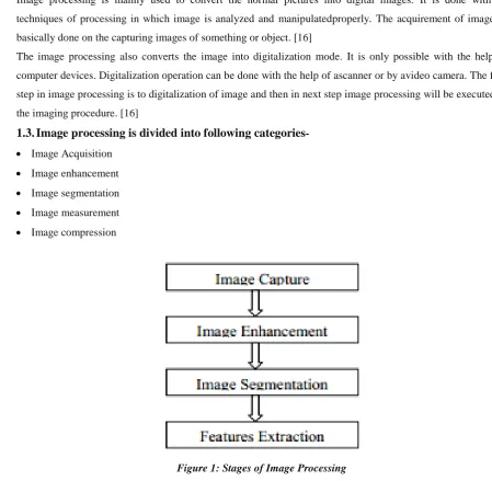
Image processing is divided into following categories-
Image Acquisition Image enhancement Image segmentation Image measurement Image compression
Figure 1: Stages of Image Processing
There are various techniques in which image processing will be applied to images. The first one is image
compression it is the simplest way and easily used by users. It mainly used to reduce the size of the image so
that it takes fewer storage data to save in the computer system. Sometimes, we wantto send the images to other
The second one is image enhancement techniques which are used to detect the mistakes in the images and
corrected it with the help of digitalization. The measurement extraction will be used to obtain the necessary
information from the image. The last technique of image processing is image segmentation which helps to
change the depiction of an image into meaning full information which is easy to analyze by theuser. It also sets
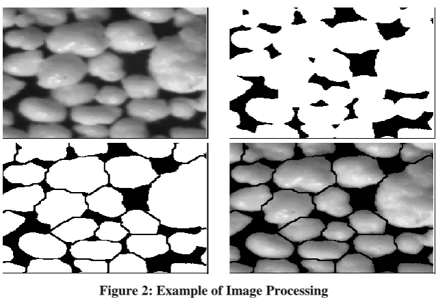
the image boundaries and objects in the image processing techniques. [16] Below images are some examples of
image enhancement and image measurement. The examples describe the procurement of image in gray scale
which means the pixel in theimage lies up to 0 to 255. The 0 mainly represents the black pixel, and 255
represents the white pixel. It also helps to convert the black and white images into color images.
Figure 2: Example of Image Processing
The above images representthe numerous examples of image processing categories in which the images can be
categorized according to the needs and requirements.
1.4.
Purpose of Image Processing
The main purpose of image processing is explained below:
The image processing is used for the visualization. It helps to visualize that objects which are not visible in
the image.
It creates the new features in the image to develop into better quality by the improving and renovation fo
image.
It also helps to recover the images.
The image processing helps to measure the pattern to fully optimizes the images. It also helps to distinguish the images according to their size and pattern.
1.5.
Types of Image Processing
There are two types of themethods which are used in the image processing. The first one is analog and second
one is digital image processing. The analog method of image processing is used on the hard copies of images
like printouts and photographs. Image analyst used several types of clarification techniques to visualize the
images. The image processing is the valuable tool in which various techniques of visualization isused, so the
analyst applies their skills and the personal knowledge to conduct the images according to their pattern and size.
helps to manage the raw data from sensors of thesatellite which sometimes contains many absences. So, to get
out from these problems, it should undergo through various processes of digital imaging. It mainly uses three
digital techniques which are pre- processing, improvement and display information abstraction.[4]
1.6.
Applications of Image Processing
There are many applications which are processed under digital image processing:
Remote sensing Medical field
Image refining and renovation
Transmission and encrypting of images. Video and audio processing
Minuscule imaging Machine robot vision Color image processing Pattern appreciation Others
1.7.
Image Processing Techniques
Image refining and renovation
Image refining and renovation technique help to process the images into high quality of pictures which are
basically captured from modern cameras. It helps to manipulate the images into their desired results. The image
refining is done with the help of Photoshop in which various features have been processed to create the high
definition images. It mainly includes Zooming, blurring, and sharpening of pictures. It converts the gray scale
images into color images by detecting edges and pattern in the processing technique.
Medical field
The image processing is also done in themedical field.
It is used in the X- ray imaging In Gamma ray imaging
Medical CT – Scan process to operate the body functions PET scanning
UV imaging
Remote Sensing
The image processing technique is also applicable in the remote sensing of various objects like to scan the
parameter of theearth. It is done with the help of satellite which obtains the necessary information to be used in
further proceedings of optimization. The main use of remote sensing is to detect the damages in the substructure
of theearth due to the occurrence of anearthquake. It is a very beneficial and time-consuming technique which
helps to examine the damages that are not possible by the human eye. [16]
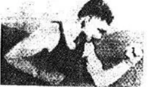
Transmission and Encoding
The transmission of theimage is done the with the wire The picture which is sent from this technique is shown
Figure 3: Took three hours to reach its destination.
But now in the modern generation, there are many techniques have been discovered like the live video and
CCTV footage which sends the picture from one place to other within a few seconds. This technique is mainly
focused on transmission and encoding into various formats. It helps to encode the high-low bandwidth photos
so that it can be easily shared on the internet.
II.
LITERATURE REVIEW
Li Zhang described an automated method to quantify and segment the volume of cystoid macular edema (CME)
with the help of macular hole (MH) for abnormal retina used in 3D OCT images. In this proposed framework, there are three parts such as a) preprocessing which also includes flattering, MH, denoising, intraretinal layers segmentation and vessel silhouettes exclusion;b) coarse segmentation which includes AdaBoost classifier that is used for the constrained and seeds area for the Graph Cut; c) and last one is fine segmentation which includes the algorithm of graph cut that helps in refining the segmentation results. Automated method evaluated the 3D OCT images from the 18 different patients with MH and CMEs. From the results, it was concluded that for the CME volume segmentation, the accuracy rate (ACC), false positive volume fraction (FPVF), and positive volume fraction (TPVF) are 99.7%, 1.7%, and 84.6% respectively. [1]
De Oliveira, J.P.S presented the segmentation of the infrared images which is a new technology used to detect
the breast diseases in early stages. In the first stage of cancer, it promotes the process of the vascularization in the affected area that modifies the local temperature and increasing the blood flow in the human body. The infrared radiations that are emitted by the human body can be captured with the help of athermal camera which is used for measuring the temperature of the human body and shows the results in the form of animage. With thermography, suspicious regions can be detected in the patients of any age and also in the cases of thedense breast in which detection of abnormality cannot be accomplished by another method. Thermal images are also used in the development of computer-aided diagnosis (CAD) systems which allow the executions of exams with the help of techniques that follow the proper protocols and routines which already occur in the exam of mammography. Mammography is used for the analysis by the doctors. In this paper, automatic detection of the regions of interest (ROI) is compared with the performed segmentation which provides a methodology used for automatic segmentation of the thermal images of thelateral breast. Various groups truth was generated for the evaluation of results on the internet that allows verification of the correct results. At last, the results of the proposed method for 328 images wasused, and theresult shows the average value of accuracy. [2]
Ayas, S. demonstrated about microscopic image segmentation which is based on Firefly Algorithm for the
diseases which is diagnosed by the laboratory technicians. In the method of the microscopy diagnosis including hand-eye control, the misdiagnosis rate is too high. In the microscopic imaging, the diseases were diagnosed with the help of computer-aided automatic diagnosis method. In the automatic diagnosis method, the robustness is depending on accurate segmentation of the microscopic images. Image segmentation method was used to solve the various problems by identifying a special solution. Firefly algorithm which is based on the swarm intelligence used in microscopic imaging to segment the images. An optimal threshold value in the proposed method was used to determine the grey-level microscopic images which are based on the Firefly algorithm. With the help of the optimal threshold value, the microscopic images were converted to the binary format. At last, the results of the segmentation were compared with the result of expert-guided segmentation, and it was concluded from the results that performance ratio of segmentation is 96% which was obtained from the Firefly algorithm that is based on swarm intelligence. [3]
Garg, N presented the segmentation which is based on the histogram method used in the image contrast
enhancement. Histogram equalization method was used to process the digital images for image enhancement and normalization. But they do not give the true result always. So, in this research,segmented histogram technique was used for contrast enhancement of images by scaling the discrete wavelet transform coefficient. Its advantage is that they preserve the color consistency to improve the contrast of the images. This method also reduced the contaminated noise in the images with the help of wavelet shrinkage adaptively. Image contrast enhancement was based on the wavelet transform that is simple and computational efficient because these estimations are performed in the form of compressed wavelet domain and estimated coefficients which are scaled linearly. The proposed technique was used to improve the local and global contrast of images. They gave the better enhancement of the visual quality; Absolute mean brightness error, peak signal to noise ratio, entropy and standard deviation value as compared to the PME technique. [4]
Banerjee developed a gray level image thresholding with the help of Particle Swarm Optimization Algorithm.
Using Bilevel Kapur's entropy function, the images were segmented which were maximized by PSO.Various entropy functions were used for the process of segmentation. In this research, the threshold values were verified with the help of histogram of the test images and observed the visual representation. Image segmentation method was used to understand the system of theimage that divided the image into multiple disjoint regions which are based on the homogeneity. Image segmentation technique is highly problem-specific and used to decrease the complexity of the problem. The main objective of this research was to find the optimized threshold value for the image segmentation with the help of particle swarm optimization algorithm. [5]
Arun, G implemented the split classification method which was used to detect the presence of Haemorrhage
area which resulted in less error. Splat classification method basically used to detect the presence of the Haemorrhage which is based on the pixel distribution on the retinal images. [6]
M.Airaksinenestimated the noise robust of the voice source with the help of thedeep neural network. In the
process of the analysis of the speech production, the information regarding voice source was obtained non-invasively by glottal inverse filtering methods. The current state-of-the-art glottal inverse filtering methods were able to produce the high-quality estimates in the suitable conditions such as low noise, but their performance in the non-ideal conditions deteriorated because there is a need for thenoise-sensitive parameter. In this research, an approach was developed forrobust noise estimation of the voice source which created amapping with the help of deep neural network with the features of robust of low-level and the desired reference. With the help of the GIF method, a time-domain glottal flow was computed. The result concluded that the proposed method outperforms the QCP method with SNRs i.e. less than 50-20 Db, but in the IAIF method, there were very low SNRs. [7]
K. A. Faroukintroduced an artificial neural network meta model for systems in series with buffers under the
imperfect repair. Reliability and availability are deteriorated as the number of components increases. For which redundancy is used in order to remove such type of problems. Line production and liquid supply chains provide the buffers between the components of the system as economical solution which eliminate the problem of availability and reliability from the system of multi-components which are connected in the series. The rated capacity of the components can be enhanced by the sufficient margin which provide the buffers with necessary and sufficient qualities in order to keep the system uninterrupted in case of the failure of the components. Simulations models are used in such types of models but they are very slow when they are used in optimal allocation for components maintainability and reliability. For such types of optimal problems, an artificial neural network is used due to its fast output and capability of modelling the non-linear model. [8]
M.J. Evangelineexplained the method for segmentation which was used in the 2D MRI acquisitions of kidney
images. This type of the segmentation promotes the times course of avoxel in and out of the kidney. From the result, it was found that this segmentation driven has the great potential DCEMRI model which driven the segmentation of the kidney. Magnetic Resonance Imaging of kidney needs proper segmentation and correction to enable the estimation of glomerular filtration rate with the help of pharmacokinetic modeling. Pharmacokinetic and segmentation modeling are applied sequentially as the separate processing model. A 2D segmentation model was used to demonstrate the model in the numerical experiment which is used to normalize gradient and Mahalanobis distance from the time course of the segmented regions for supervised segmentation. This resulted in the correction of the kidney images. [9]
D. W. Shattuck presented a three-stage sequence of the technique which was used to classify and identify the
brain tissues in T1-weighted MRI of the human head. It removed the non-brain tissues with the help of anisotropic diffusion filtering, mathematical morphology, and edge detection. This method was used in the low-levels and provided the information at the voxel level related to the contents of the image of the tissue. The method was also validatedwith the real human data, and it was concluded that it outperformed on the several methods. [10]
D. H. Laidlaw presented the new algorithm that identified the distribution the various material types in the
allowed the mixtures of materials which treat the voxels as the regions and this technique decreased the errors as compared to the other techniques which created along the boundaries between the materials and used for creating the accurate model of geometric. They have the potential to measure the volume more accurately a classifies the low-resolution and noisy data. In this research, two methods were used.. The distribution was represented with the help of the histogram over the region of the voxel. The chosen size was matched to the space of the samples because space is related to the minimum feature size which represented the reconstructed continuous function. [11]
D. L. Collins designed and constructed the realistic digital brain phantom. After the implementation and
conception of the medical image processing algorithm, it is essential to ensure all the requirement set which are forth at the initial stage of design. A comprehensive validate the simulated data which are evaluated on the real data to establish the ground truth with the help of vivo data. the experiments which are performed on the simulated data control the evaluation over awide range of conditions such as intensity artifacts, thelevel of noise, contract, and geometric distortion. Parallel pipe and ellipsoids do not reflect the complexity of brain anatomy. So, they present the high-resolution, realistic, and digital phantom of the human brain which is made up of ten volumetric data sets which define the spatial distribution of the various tissues in which the voxel intensity is directly proportional to the fraction of tissue with the voxel. [12]
V. M. Catterson investigated about artificial neural networks for the automated diagnosis of the defects which
causes partial discharge. This research discussed the use of the deep neural networks for diagnosis of PD data. The data was captured from the defect samples in oil with the help of UHF sample. The effects on the rectified linear unit activation function and diagnosis of a number of thelayerhavebeen explored in this research. From the results, it was found that the accuracy of the diagnosis is increased to 86% from 72% as compared to shallow networks with sigmoid activation function. [13]
B. Cunhadescribed modeling techniques in scheduling system with the help of artificial neural network in three
steps. In thefirst step, the method established the information capture techniques and the database design to clear which information classify the user. Then, themathematical structure was constructed which classified the users such as Bayes networks. Network training allowed the system to create the precision classifications for its users. In the final step, way to use the classification was decided by the mathematical classifier. [14]
D. Chakraborty described the error minimizing method with the help of conventional back propagation
algorithm which is used for training feed-forward neural network that suffers from problems such as local minima trap and slow convergence. In this research, gradient-free optimization was used for the error minimization that avoids the local minima. For this, they introduced the concept of hybrid algorithm integration and biology-inspired flower pollination algorithm. The gravitational search algorithm is a meta-heuristic optimization and based on theNewtonian law of gravity, and the mass interaction whereas flower pollination algorithm is based on the pollination characteristics of flower plant. The results showed that that hybrid FP-GSA outperforms for GSA and FPA has better outperforms training in FNNs. [15]
III. FINDINGS
Author Determine Method/Tech
nique
Results
N. Garg, A. Angra, P. Sengar (2015)
Image Enhancement using Scaling the Discrete Wavelet coefficients
Segmented Histogram Equalization
Improve the local and global contrast of images.
Better enhancement of the visual quality, Absolute mean brightness error, peak signal to noise ratio, entropy, and standard deviation.
S. Banerjee, N.D. Jana (2015)
Searched the optimized threshold value for Image
Segmentation
Bilevel Kapur's entropy function and Swarm Optimization Algorithm
Develops a gray level image thresholding.Reduced complexity of the problem
Arun, N Sasirekha (2015)
Detection of Retinal Hemorrhage in Color Fundus Image
Splat Feature Segmentation
Detected the presence of the Hemorrhage
M. Airaksinen, T. Raitio, P. Alku (2015)
Noise Robust Estimation of the voice source
Deep Neural Network
Time-domain glottal flow was computed
K. A. Farouk, M. Younes, M.N. Fors (2015)
Determine the output
parameters of the artificial neural
network as the meta model
Nonlinear Regression
The adequacy of the ANN evidenced thehigh values of the coefficient of determination i.e.R2 >94% indetermining bo and R2 > 97%
M.J.Evangelin,L.P
.Suresh (2015)
Segmentation
of 2D
MRIacquisitio ns for kidney image
Pharmacokine tic modelling
D. W. Shattuck, et al. (2001)
A three-stage sequence of techniquesfor identifying and
classifying the brain tissues
Partial Volume Model
Used in the low-levels. Provided the information at the voxel level related to the contents of the image of the tissue
D. H. Laidlaw, et al. (1998) Partial-Volume Bayesian Classification of Material Mixtures in MR Volume Data
VoxelHistogr ams
Decreased the errors as compared to the other techniques used for creating the accurate model of geometric
D. L. Collins, et al.(1998)
Design and Construction of a Realistic Digital Brain Phantom
Three -dimensional digital brain phantom
Used to drive simulators for different modalities,
Test intramodality registration algorithms
V. M. Catterson, B. Sheng (2015)
Use of deep neural networksfor PD diagnosis Artificial neural networks
Improved in speech and image recognitiontasks.
B. Cunha, A. Madureira, J. P. Pereira (2015)
Scheduling System with Artificial Neural Networks
User Modeling
Presents developers with multiple options to advantage of various technologies to model their users
D. Chakraborty, S. Saha, S. Maity (2015)
Gradient free optimization
Hybrid FP-GSA outperforms for GSA and FPA has better outperforms training in FNNs.
IV. CONCLUSION
This paper provides a brief insight into Image processing and its various techniques. We discussed various
methods and techniques of Image processing which are used for performing segmentation on images. For this
purpose, previous researches were analyzed to select the best and suitable technique for segmentation of images
images to detect the cancerous section. Therefore, it is required to propose a new segmentation method to
segment the probable cancerous section accurately.
REFERENCES
[1]. LiZhang,WeifangZhu, Fei Shi, Haoyu Chen, Xinjian Chen, “Automated segmentation of intraretinal cystoid macularedema for retinal 3D OCT images with macular hole”, Biomedical Imaging (ISBI), 2015 IEEE 12th InternationalSymposium,pp.1494 – 1497.
[2]. De Oliveira, J. S., Conci, A., Perez, M. G., &Andaluz, V. H. ( 2015). Segmentation of infrared images: A new technology for early detection of breast diseases. 2015 IEEE International Conference on Industrial Technology (ICIT), 1765-1771. doi:10.1109/icit.2015.7125353
[3]. Ayas, S., Dogan, H., Gedikli, E., &Ekinci, M. ( 2015). Microscopic image segmentation based on firefly algorithm for detection of tuberculosis bacteria.2015 23rd Signal Processing and Communications Applications Conference (SIU), 1-4. doi:10.1109/siu.2015.7129962
[4]. Garg, N., Angra, A., &Sengar, P. (2015). Image enhancement by scaling the discrete wavelet coefficients based on segmentation. 2015 2nd International Conference on Signal Processing and Integrated Networks (SPIN), 520-525. doi:10.1109/spin.2015.7095374
[5]. Banerjee, S., & Jana, N. D. (2015). Bi-levelkapurs entropy based image segmentation using particle swarm optimization.Proceedings of the 2015 Third International Conference on Computer, Communication, Control and Information Technology (C3IT), 1-4. doi:10.1109/c3it.2015.7060212
[6]. Arun G, &Sasirekha, N. (2015). Detection of retinal hemorrhage in color fundus image using splat feature segmentation. 2015 International Conference on Innovations in Information, Embedded and Communication Systems (ICIIECS), 1-5. doi:10.1109/iciiecs.2015.7192928
[7]. Airaksinen, M., Raitio, T., &Alku, P. ( 2015). Noise robust estimation of the voice source using a deep neural network. 2015 IEEE International Conference on Acoustics, Speech and Signal Processing (ICASSP), 5137-5141. doi:10.1109/icassp.2015.7178950
[8]. Farouk, K. A., Younes, M., & Nashat Fors, M. ( 2015). An Artificial Neural Network meta-model for theavailability of systems in series with buffers under imperfect repair. 2015 International Conference on Industrial Engineering and Operations Management (IEOM), 1-7. doi:10.1109/ieom.2015.7228095 [9]. Evangelin, M. J., & Suresh, L. P. ( 2015). Segmentation driven image application to 2D-MRI of the
kidney.2015 International Conference on Circuits, Power and Computing Technologies [ICCPCT-2015], 1-5. doi:10.1109/iccpct.2015.7159484
[10]. Shattuck, D. W., Sandor-Leahy, S. R., Schaper, K. A., Rottenberg, D. A., & Leahy, R. M. ( 2001). Magnetic Resonance Image Tissue Classification Using a Partial Volume Model. NeuroImage, 13(5), 856-876. doi:10.1006/nimg.2000.0730
[12]. Collins, D., Zijdenbos, A., Kollokian, V., Sled, J., Kabani, N., Holmes, C., & Evans, A. ( 1998). Design and construction of a realistic digital brain phantom. IEEE Transactions on Medical Imaging, 17(3), 463-468. doi:10.1109/42.712135
[13]. Catterson, V. M., & Sheng, B. ( 2015). Deep neural networks for understanding and diagnosing partial discharge data. 2015 IEEE Electrical Insulation Conference (EIC), 1-4. doi:10.1109/icacact.2014.7223616 [14]. Cunha, B., Madureira, A., & Pereira, J. P. ( 2015). User modeling in scheduling system with artificial neural networks.2015 10th Iberian Conference on Information Systems and Technologies (CISTI), 1-6. doi:10.1109/cisti.2015.7170449
[15]. Chakraborty, D., Saha, S., & Maity, S. (2015). Training feedforward neural networks using hybrid flower pollination-gravitational search algorithm.2015 International Conference on Futuristic Trends on Computational Analysis and Knowledge Management (ABLAZE), 261-266. doi:10.1109/ablaze.2015.7155008
[16]. Chaudhary, A., & Singh, S. S. (2012). Lung Cancer Detection on CT Images by Using Image Processing.