IJEDR1603097
International Journal of Engineering Development and Research (www.ijedr.org)593
Segmentation of Brain Tumor from MRI Using Skull
Stripping and Neural Network
1Dimple Kapoor, 2R. Kashyap 1Student, 2HOD ECE
1Rayat insititude of Engineering and Information technology, Punjab,India
Abstract - Brain tumor is an alarming disease if not noticed on time. Several researchers have done their researches in this field to discover some new methods of brain tumor detection. In this paper, we present a method of brain tumor detection from MRI images. Segmentation is done by using Self-Organizing Map (SOM) and Neural network (NN). Stationary Wavelet Transform (SWT) is used to extract the features from an input image before the training process for segmentation. We proposed a new skull stripping algorithm for the purpose of effective skull stripping. We used BRAINIX medical images as a dataset for our method. The proposed method performs better than the methods discussed in the literature. It is easy to implement and robust.
Keywords - Brain Tumor, Neural Network (NN), Segmentation, Self-Organizing Map (SOM), Stationary Wavelet Transform (SWT).
I.INTRODUCTION
Image segmentation is the process of isolating an image into number of sections which are known as segments. It is used to partioned an image on the basis of region of interest (ROI). It is utilized to take out a section from an image. Segmentation of image is one of the essential and demanding steps in the digital image processing. It has also its application in the field of medical imaging and is also used to detect brain tumor from magnetic resonance images (MRI). Brain tumor detection is an application of MRI. There are so many imaging techniques which are employed to study Tumors such as Computer Tomography (CT), PET, MRI, Single photon emission computer tomography etc.
Brain tumor is a dreadful disease. A brain tumor occurs when abnormal cells forms inside the brain.Brain tumor may also referred as intracranial neoplasm. When the set of abnormal cells which starts in the brain, then the tumor take place in the brain. Brain tumor have almost 120 kinds that makes complicated treatment. There are mainly two kinds of brain tumor namely cancerous tumor and benign tumors. A number of different imaging techniques are developed to study Tumors such as Computed Tomography (CT), Positron emission tomography (PET), Magnetic Resonance Imaging (MRI), Single photon emission computer tomography etc. Currently, CT and MRI are the most widely used techniques because of their High resolution images ability. Magnetic Resonance tomography is a medical imaging technique used by radiologists to visualize the internal structure of human body in detail. MRI can create more detailed images of human body than possible with X-rays.
II.LITERATURE SURVEY
IJEDR1603097
International Journal of Engineering Development and Research (www.ijedr.org)594
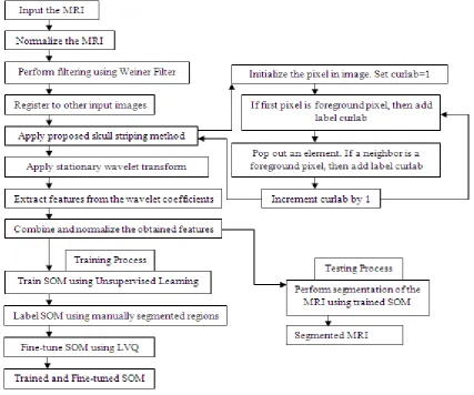
Fig. 1 Flowchart of Segmentation and Proposed Skull Stripping AlgorithmNormalization and Noise Removal
It is one of the preprocessing step in which image can be converted into normalized form. Basically, by normalization means, the process of removing the noise, distortion, illumination etc from the image. The major purpose of normalization is to get a normalized image by elimination the noises which are produces within the environment in which the image is taken.
An input image can be normalized within the intensity range of [0 1] by dividing the intensity values of the pixels by the minimum and maximum value of the range. The equation for normalization is as:
𝐼𝑛=
𝐼 max (𝐼)
IJEDR1603097
International Journal of Engineering Development and Research (www.ijedr.org)595
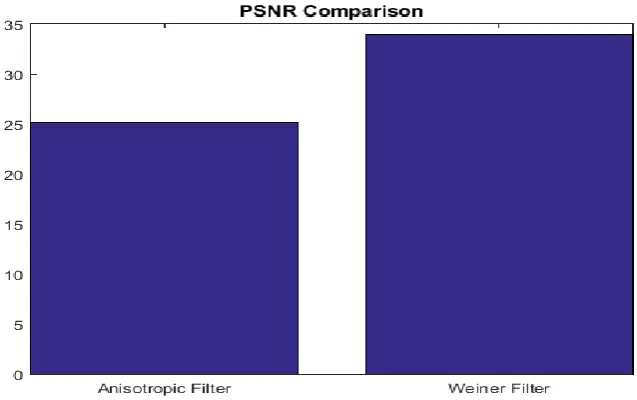
Fig. 2 Comparison between Anisotropic Filter and Weiner Filter on the basis of their PSNRThresholding
Thresholding is the common and significant approach for pixel based segmentation. This method is used to produce a binary image from the grayscale image. In segmentation method, let us assume a threshold value x. then for generating a binary image, the pixels of an image having intensity value less than threshold value are converted to black and the pixels having intensity value greater than threshold value are converted to white color pixels. In order to calculate the threshold value, Otsu’s method has been used. This calculated threshold value is converted to binary image. It is as follows:
𝜎𝑤2(𝑡) = 𝑤0(𝑡)𝜎02(𝑡) + 𝑤1(𝑡)𝜎02(𝑡)
Find maximum value of 𝜎𝑤2(𝑡) where,
𝑤0(𝑡)= ∑ 𝑝(𝑖)
𝑡−1
𝑖=0
𝑤1(𝑡)= ∑ 𝑝(𝑖)
𝐿−1
𝑖=𝑡
Skull Stripping
Skull stripping is one of the most significant steps during brain tumor segmentation. It eliminates skull, fat, skin and the regions of brain which are not required. Additionally some extra portion from the brain is also being removed to simplify the segmentation process. Here in this paper, we are using blob detection and labeling method for the purpose of skull striping.
Fig. 3 Efficiency of the original and proposed method
IJEDR1603097
International Journal of Engineering Development and Research (www.ijedr.org)596
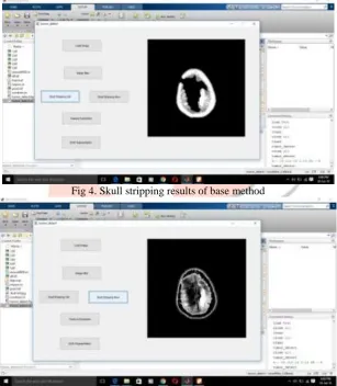
Segmentation is the process of isolating an image into number of sections which are known as segments. It is used to extract the region of interest from an image. In order to detect the tumor region, self-organizing map is used. The self-organizing map is a kind of artificial neural network. It is trained using unsupervised learning and Learning Vector Quantization (LVQ) is used to fine tune the SOM network.Fig 4. Skull stripping results of base method
IJEDR1603097
International Journal of Engineering Development and Research (www.ijedr.org)597
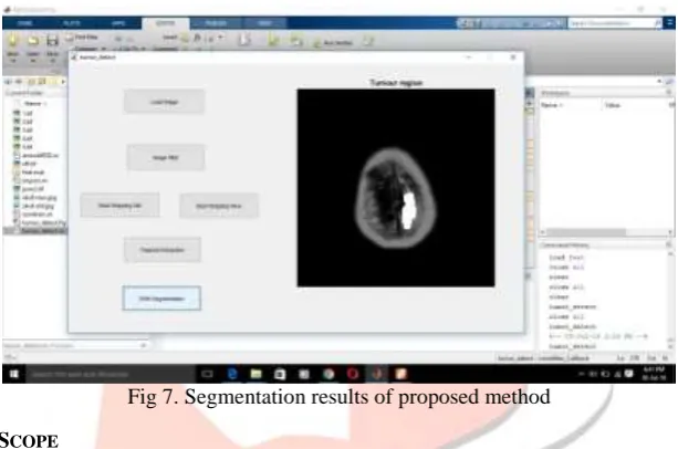
Fig 6. Segmentation results of base methodFig 7. Segmentation results of proposed method
IV.CONCLUSION AND FUTURE SCOPE
In this paper, we proposed a brain tumor segmentation algorithm for skull stripping along with weiner noise filtering that segments the brain and skull and detects the tumor region. We used weiner filter for noise removal that improves the PSNR. We also proposed a new skull stripping algorithm based on connected component analysis and labeling method. According to the proposed skull stripping algorithm, each closed portion in an input MRI image is considered as a connected component. Then starting from the outer side, we labelled each connected component moving inward. This method effectively performs skull stripping as compared to the method of morphological operations using erosion and dilation.
We implemented our method on BRAINIX medical image dataset using MATLAB. Our method performs better than other methods as discussed in the literature. It is also concluded that weiner filter performs better than anisotropic filter. The proposed method effectively strips out the skull portion resulting in better segmentation. In future, the work is required to further improve the efficacy of the skull stripping method as there are still some traces (very less) of skull’s outer region remains in the image. Though this is the outer region and does not affect much as we can see in the results, we still need to clear it out in order to get better skull stripping.
In future, the work is required to further improve the efficacy of the skull stripping method as there are still some traces (very less) of skull’s outer region remains in the image. Though this is the outer region and does not affect much as we can see in the results, we still need to clear it out in order to get better skull stripping.
V.REFERENCES
[1] Clark, Matthew C., et al. "Automatic tumor segmentation using knowledge-based techniques." Medical Imaging, IEEE Transactions on 17.2 (1998): 187-201.
[2] He, Shijuan, et al. "Research on MRI brain segmentation algorithm with the application in model-based EEG/MEG." IEEE
transactions on magnetics37.5 (2001): 3741-3744.
[3] Fan, Yong, Tianzi Jiang, and David J. Evans. "Volumetric segmentation of brain images using parallel genetic algorithms." Medical Imaging, IEEE Transactions on 21.8 (2002): 904-909.
[4] Van Leemput, Koen, et al. "A unifying framework for partial volume segmentation of brain MR images." Medical Imaging,
IJEDR1603097
International Journal of Engineering Development and Research (www.ijedr.org)598
segmentation." Signal Processing Letters, IEEE 22.5 (2015): 573-577.
[11]Damodharan, Selvaraj, and Dhanasekaran Raghavan. "Combining tissue segmentation and neural network for brain tumor detection." Int. Arab J. Inf. Technol. 12.1 (2015): 42-52.
[12]Demirhan, Ayse, Mustafa Toru, and Inan Guler. "Segmentation of tumor and edema along with healthy tissues of brain using wavelets and neural networks." Biomedical and Health Informatics, IEEE Journal of 19.4 (2015): 1451-1458.
[13]Zhao, Guangjun, et al. "Segmenting Brain Tissues from Chinese Visible Human Dataset by Deep-Learned Features with Stacked Autoencoder."BioMed research international 2016 (2016).
[14]Gonzalez, Rafael C., Richard E. Woods, and Steven L. Eddins. "Digital image processing using MATLAB." (2004).
[15]Woods, Rafael C. Gonzalez Richard E. "Digital image processing." (2002).
[16]http://www.bioss.ac.uk/people/chris/ch4.pdf