Functional analysis of a homeobox-containing
gene expressed during early
Xenopus laevis
development
Margarida Trindade
Thesis submitted to the University College London for the
degree of Doctor of Philosophy
All rights reserved
INFORMATION TO ALL USERS
The quality of this reproduction is dependent upon the quality of the copy submitted.
In the unlikely event that the author did not send a complete manuscript and there are missing pages, these will be noted. Also, if material had to be removed,
a note will indicate the deletion.
uest.
ProQuest 10013326
Published by ProQuest LLC(2016). Copyright of the Dissertation is held by the Author.
All rights reserved.
This work is protected against unauthorized copying under Title 17, United States Code. Microform Edition © ProQuest LLC.
ProQuest LLC
789 East Eisenhower Parkway P.O. Box 1346
Abstract
Xom is a homeobox-containing gene expressed during early stages of Xenopus
laevis development which is involved in the specification of ventral tissues. Expression
of Xom is induced as an immediate-early response to Bone Morphogenetic Protein-4, a
member of the Transforming Growth Factor-P family. Moreover, Xom contains a novel
homeodomain, which might affect its DNA binding specificity.
This thesis describes a functional analysis of Xom. First, a preferred Xom DNA
binding site was determined and the ability of Xom to bind potential binding sequences
was tested in a series of in vitro assays. Together, these results showed that the sequence
CTAATT(A/G) is critical for Xom to bind DNA, but that binding is greatly enhanced by
the presence of an ATTA motif 6 or 7 nucleotides downstream of the core TA AT. A cell
culture assay further demonstrated that Xom interacts with the selected sequence in vivo.
Second, the ability of Xom to regulate transcription was analysed. Xom was
shown to behave as a transcriptional repressor in Xenopus embryos and its repressing
activity was mapped to N-terminal and C-terminal regions flanking the homeodomain.
X om ’s transcriptional repressing activity, together with its ventral expression pattern and
ventralising activity in the early Xenopus embryo, suggested that Xom could function by
down-regulating the expression of genes that are required for dorsal development in
X enopus. C onsistent with this suggestion, over-expression o f Xom RNA, or of a
dominant-negative version, indicated that Xom regulates the expression of goosecoid, a
homeobox-containing gene expressed in the organizer capable of partially mimicking
the activity of the organizer.
Finally, to test whether Xom acts by repressing goosecoid transcription directly,
reporter constructs containing a goosecoid prom oter fragm ent containing or lacking
point mutations in potential Xom binding sites were co-injected with different effector
RNAs into X en o pus embryos. These experiments suggested that at least part of the
ability of Xom to repress goosecoid is direct, and identified a possible site to which Xom
Time has come to write what is the trickiest, and surely the most requested, part
of my thesis. Time to thank those who really did it.
I am grateful to Antonio Coutinho and Alexandre Quintanilha for the creation of
the Programa Gulbenkian de Doutoramento em Biologia e M edicina (PGDBM), which
has financed me throughout the PhD and allow ed me to spend an exciting year of
tutorial courses based at the Gulbenkian Institute in Lisbon. I am also grateful to
Eduardo Crespo, Professor at the Faculdade de Ciências in Lisbon, for his continuous
interest in my work, to my PGDBM colleagues for the wonderful year we spent together
and to Greta and Maria José Marinho for being there for us.
I am very grateful to my supervisor Jim Smith for the opportunity given to work
in such a stimulating and culturally diverse environm ent as the NIM R at Mill Hill.
Thanks for guidance and support throughout these years and for giving me some
freedom to learn from my mistakes. I truly appreciate it. Thanks also for the ultra-fast
reading of my thesis!
When I first arrived at Mill Hill, not yet sure in which country I was, Raj Ladher
was the person who guided me both in the intricate secrets o f Mill Hill and in the
discovery of the city of London. Thank you for all the fun. I w on’t forget The Beauty of
the whole mounts! Masa Tada (or shall I say M aster M asa?) taught me most of what I
know about Developmental Biology. Thanks for the infinite patience and friendship.
Thanks to you I now know that there is always a better experiment to do! A big thanks
goes to Joshua Brickman who contributed enormously to the final part of my thesis, by
introducing me to the ways of transcription and lecturing me endlessly on how to present
my work. And to think that everything started with salsa... Thanks Niall Armes for
making me think in molecules (lots of them, all together, making complexes, shaking in
the air, separating, fusing, spinning, getting obscene conformations ! !... U ff, I am tired.
Are you sure they all obey quantum mechanics?). L et’s try seriously now: thanks! With
Mike Jones I had the opportunity to go to the Xenopus course at the Gulbenkian Institute
advice, and this also applies to Frank Colon and Yasu Saka, but in particular to Branko
Latinkic who encouraged me not to let the goosecoid promoter escape! I am grateful to
Lynne Fairclough for all the technical support and four almost four years of sharing a
laboratory bay without friction (I am sure it was W alter who took that!). Thanks to
Surrendra Kotecha and to Tim Mohun for the help with the binding site selection. Thank
you to all the Photographies staff, which made the figures of my thesis. My sincere
admiration goes to Jacky Smith who provides the unnoticeable everything to keep the
lab going and is always willing to help. A big thanks also goes to Leonor Saude for
keeping me in touch with life in the Institute during the writing up period and for top
quality occasional post(wo)man services! Finally, many thanks to all other past and
present members of the laboratory (and adjacent laboratories) with whom I had exciting
discussions, exchanged ideas, reagents, or many times, purely entertaining gossip.
My thoughts also go to all my friends in London who transformed my life out of
the lab in a very pleasant time. Perhaps without noticing, they greatly contributed to my
mental sanity. To list them all would be too long and surely incomplete. However, a few
lines are needed just to acknowlege my fantastic Medresco House ex-neighbours (Ana,
Pedro, Josh, Leonor, Jacinto, Maria, Sergio), Sofia, Rui (by the way, I won!). Ana Paula,
Pedro, Andreas, Miguel, Kathy, Raj, Salvatore, CP, etc., etc., etc.
Thanks to my all my friends which came from far to enjoy the comfort of my
B&B (I am sorry, Julia, I thought you should know), but only to those that did not forget
the chouriços and the bacalhau! Thanks also to those that never came, but who I am sure
will regret it for the rest of their lives.
My family has been exceptional during this period of my life, as in all others.
Their support, encouragement and optimism are infinite. Obrigada màe, pai, avos. Ana,
Zé e André. A special thanks goes to Lounès for making the best of our life together.
Table of contents
Abstract 2
Acknowledgements 3
List of Figures 12
List of Tables 14
CHAPTER 1 15
INTRODUCTION 16
Early Xenopus development 17
Axis specifîcation 21
Germ layer specification 22
Fibroblast Growth Factors (FGFs) 25
Transforming Growth Factor-P (TGF-|3) factors 26
Activin 27
Vg-1 28
T-box family: VegT 28
Dorso-ventral axis specification 30
Dorsal specification: the Nieuwkoop centre 31
Molecular players: P-catenin and upstream pathway components 32
P-catenin in the nuclei 37
The Spemann organizer 38
Ventral specification 39
Bmp antagonists 43
Chordin(Sog)/Bmp(Dpp) antagonism 43
Noggin and Follistatin 44
The astacin family of metalloproteases 45
Bmp receptors 46
Intracellular Bmp signalling: Smads 51
Smads transduce Bmp signals from the membrane to the nucleus 51
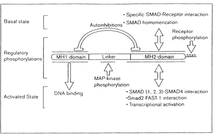
Smad protein domains and their functions 55
Smad-mediated regulation of the Bmp signalling 56
Other mediators of Bmp signalling 58
Bmp signalling in other vertebrate systems; zebrafish and m ouse 59
Potential Bmp response genes 63
Homeodomain-containing proteins 68
Transcription factors 71
This study 74
CHAPTER 2 76
MATERIALS AND METHODS 77
Abbreviations 77
Molecular Biology Techniques 77
Preparation and storage o f competent bacteria 77
Plasmid transformation o f competent bacteria 78
Small scale preparation o f plasmid DNA 79
Medium and large scale preparation o f plasmid DNA 80
Phenol/Chlorophorm extraction 81
Precipitation 81
Restriction digestions 82
Ligation and dephosphorylation reactions 82
5 ’ phosphorylation o f oligonucleotides 82
Agarose gel electrophoresis o f DNA and RNA 83
Purification o f specific DNA fragments from gels 83
Polymerase Chain Reaction (PCR) 83
DNA Sequencing 84
In vitro transcription 84
In vitro protein synthesis 86
Western blot 87
Site-directed Mutagenesis 88
Binding site selection 89
1)Preparation of double stranded R76 90
2)Binding reaction 91
3) Immunoprécipitation 92
4) Elution 93
5) PCR amplification of selected pool of oligonucleotides 93
6) Electrophoretic mobility shift assay (EMSA) 94
7) Cold PCR 95
8) Cloning the final pool of selected oligonucleotides 95 9) Sequencing of final pool of selected oligonucleotides 96
10) Analysis of sequences 96
Electrophoretic Mobility Shift Assays (EMSA) 96
Plasmid Constructs 98
For RNA injections into Xenopus embryos and/or EMSA 98
For expression o f recombinant protein 99
Luciferase-based vectors 100
Obtaining Xenopus embryos 102
Microinjection o f Xenopus embryos 102
Animal cap dissection 103
Luciferase assays on animal cap or whole embryo lysates 103
Whole mount in situ hybridization 103
Whole mount antibody staining 105
RNAse Protection 107
Photography 109
Cell Culture 110
Handling culture cells 110
Lipofectamine-based transient transfection method 111
Luc if erase assays 112
Preparation o f protein extracts fo r Western blot analysis 113
Immunocytochemistry: Russell’s protocol 113
Statistical Analysis 114
t- te s t 114
Formulation of Frequently Used Solutions 115
Formulation of Frequently Used Bacterial Growth Media 116
CHAPTER 3 117
DNA binding properties of Xom 118
INTRODUCTION 118
RESULTS 122
Determination of the binding preference of Xom 122
In vitro analysis 126
Cell culture analysis 132
DISCUSSION 133
Xom binding specificity 135
Does Xom form dimers? 136
The requirement for antibodies to detect Xom-DNA complexes in EMSA gels is bypassed by
XomVP16 138
The homeodomain is necessary but perhaps not sufficient for DNA binding 138
Future prospects 139
CHAPTER 4 141
Role of Xom in regulation of transcription 142
INTRODUCTION 142
RESULTS 143
Xom is a transcriptional repressor 143
Xom is present in the nucleus 144
DISCUSSION 148
Xom repression activity is context-dependent 148
Structural basis for a role of Xom in transcriptional repression 149 Xom does not repress transcription by competition for access to DNA 152
CHAPTER 5 153
Function of Xom in the Xenopus embryo 154
INTRODUCTION 154
RESULTS 156
Mis-expression of Xom and PV. 1 in the embryo 156
Morphological and histological phenotypes 156
M olecular marker analysis 161
Dominant-negative approaches 164
Xom^2‘^’’ 164
XomVP16 167
DISCUSSION 169
Differential regulation of XFKHl by Xom and Xvent-1 (P V .l) 171
Xom down-regulates goosecoid expression 172
CHAPTER 6 174
Analysis of the effect of Xom on the gsc promoter 175
INTRODUCTION 175
RESULTS 176
Xom represses activin-activated gsc promoter in reporter studies 176
Analysis of point mutations in the promoter 180
DISCUSSION 183
Xom represses goosecoid transcription 184
Activation of goosecoid transcription 186
Other proteins involved in repressing gsc transcription 187
CHAPTER 7 189
General Discussion 190
Bmps instructs cells to become ventral 190
Bmp response genes mediate different subsets of Bmp functions 190
Xom (and Xvent-1) act downstream of Bmp-2/4 192
Xom mediates Bmp function by repressing goosecoid 192
Are there any other Xom targets? 193
List of Figures
Fig. 1.1
Life cycle of the African claw-toed frog Xenopus laevis.Fig. 1.2
Cell movements during Xenopus laevis gastrulation.Fig. 1.3
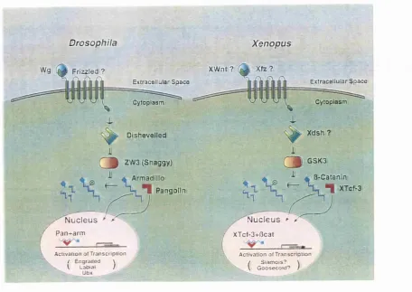
A comparison of the Drosophila and Xenopus WgAVnt signal transduction, pathway.Fig. 1.4
TGF-P family signalling pathway.Fig. 1.5
Smad protein domains and their functions.Fig. 1.6
Amino acid sequence alignment of the Xvent family of genes.Fig. 1.7
Structure of the Antennapedia homeodomain bound to DNA.Fig. 3.1
Amino acid sequence alignment of Xom and X vent-1 homeodomains showing potentially important residues for the recognition of a TA AT core motif.Fig. 3.2
In vitro translated proteins used as source of DNA binding activity in binding site selection analysis and/or in electrophoretic mobility shift assays.Fig. 3.3
Schematic representation of the consensus binding sequence derived from a PCR-based target site selection analysis (Pollock and Treisman, 1990).Fig. 3.4
Electrophoretic mobility shift assays (EMSA) define Xom DNA binding site.Fig. 3.5
Summary of the results of in vitro binding analyses presented on Fig. 3.4.Fig. 3.6
X om V Plb binds Xom consensus binding sequence in electrophoretic mobility shift assays.Fig. 3.7
X om V Plô requires the Xom consensus sequence to activate transcription.Fig. 4.1
Xom represses transcription in Xenopus embryos.Fig. 4.2
The C-terminal region of Xom mediates repression of transcription in cell culture.Fig 4.3
Subcellular localisation of Xom-HA and Xom^^’^^-HA in cultured cells.Fig. 5.2
Over-expression of Xom causes down-regulation of goosecoid expression inXenopus embryos and in activin-induced animal caps.
Fig. 5.3
Xom^^’^^ over-expression causes partial secondary axis formation and ectopic muscle formation.Fig. 5.4
Over-expression of X om V P lô causes ectopic activation of goosecoid.Fig 6.1
Time course of Xom repressing effect on activin-induced activation of thegoosecoid promoter.
Fig. 6.2
Xom represses activin-induced activation of the goosecoid promoter.List of Tables
Table 1.1
Mammalian TGF-P receptors and its homologues in Xenopus laevis.Table 1.2
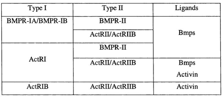
Bmp and activin ligands and respective type I and type II receptors.Table 1.3
Zebrafish mutations affecting dorsal/ventral axis specification.Table 1.4
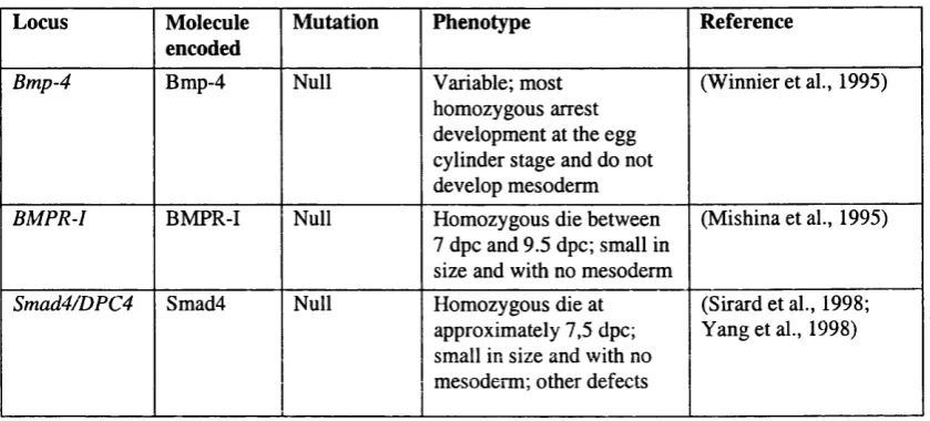
Mouse targeted mutations in some locus coding for Bmp signalling pathway components.Table 2.1
Constructs used as templates for in vitro transcription.Table 2.2
Constructs used as templates for RNAase protection probes.Table 5.1
Morphological phenotypes observed after five independent dorsal injections of Xom or P V .l in Xenopus embryo.Table 5.2
W hole mount antibody staining using MZ15 antibody.Table 6.1
Analysis of point mutations in the goosecoid promoter.Chapter 1, Introduction
INTRODUCTION
The general body plan of all vertebrate embryos becomes established during
gastrulation, a stage in development during which extensive morphogenetic movements
lead to the correct positioning of three distinct cell layers - the germ layers, from which
all tissues of the adult will originate. Amongst different vertebrate species, the earliest
phases of development occur by different processes. However, for a period starting
during gastrulation and preceding the acquisition of species-specific morphological
traits, the embryos present high morphological resem blance. This period, called the
phylotypic stage (Slack et al., 1993), highlights the phylogenetic invariability of
vertebrate developm ent, in particular o f the m echanism s that lead to and control
gastrulation.
The amphibian Xenopus laevis has been used extensively as a model to study
early vertebrate development. It lays large eggs and the embryos are easily accessible
since amphibian development occurs outside the mother. In addition, Xenopus embryos
are easy to culture and dissect and its development has been very well characterised
from early to later stages of differentiation. Although it is unsuitable for genetic analysis
because of its long generation time (18 months) and pseudo tetraploidy (Kobel and Du
Pasquier, 1986), many of these disadvantages can be partially overcome by techniques
involving expression of gain-of-function or dominant-negative constructs in the embryo.
In this thesis the Xenopus embryo was used as a biological model to assess the
role of a homeobox-containing gene, Xom, which is involved in the early patterning of
the X enopus embryo. This introduction reviews classical and recent embryological
experiments in Xenopus, together with a vast amount of molecular data generated only
in the last two decades, that represent the basis for current models of early patterning in
the vertebrate embryo. Xom is expressed ventrally in the gastrula embryo and acts as a
potential Bmp response (Ladher et al., 1996). Em phasis will therefore be given to
signals involved in establishing ventral phenotypes in the Xenopus embryo, in particular
Early
Xenopus
development
X en o p u s development starts with fertilisation of the mature egg (Fig. 1.1).
Fertilisation is im mediately follow ed by a period o f rapid and synchronous cell
divisions. At this stage, cell division is uncoupled from cell growth and within several
hours a compact cluster of cells known as the morula is formed, with approximately the
same volume as the uncleaved egg. By this time a cavity begins to form in the centre of
the embryo - the blastocoel - which separate the large vegetal blastomeres from the
smaller animal blastomeres and the embryo is called a blastula (Gilbert, 1994). Until this
stage, development has occurred at the expense of maternal gene products. However, by
blastula stage (stage 8, Fig. 1.1), a coordinated group of changes occurs, including the
onset of zygotic expression and the breakdown of cleavage synchrony, which are
collectively called the mid-blastula transition (Yasuda and Schubiger, 1992). They
prepare the embryo for gastrulation, which will result in the correct positioning of the
three primary germ layers, ectoderm, mesoderm and endoderm. Briefly, the ectoderm
will generate the epidermis and neural tissue of the embryo, the mesoderm will originate
the notochord, somites, muscle and lateral plate m esoderm al derivatives (blood,
pronephros) and the endoderm will originate the embryo gut and parts o f the head (Dale
and Slack, 1987).
The first indication of gastrulation is the local invagination of a particular set of
presumptive endodermal cells, the bottle cells, at a precise place in the marginal zone of
the embryo (Fig. 1.2). This can be visualised from the exterior by the appearance of the
dorsal blastopore lip in the marginal zone, on the future dorsal side of the embryo.
Marginal cells then involute through this blastopore lip: the most dorsal cells involute
first at the most dorsal position and the most ventral cells involute last at progressively
more lateral and ventral positions, eventually leading to the formation of a circular
blastopore in the vegetal pole of the embryo (Gilbert, 1994). This involution movement
of the marginal cells displaces and reduces the blastocoel and begins the formation of
another cavity, the archenteron (Fig. 1.2). The migration of mesodermal precursors
inside the embryo involves a narrowing (convergence) and a lengthening (extension) of
Chapter 1, Introduction
of dorsal mesodermal structures such as notochord and muscle. There is also a temporal
correlation between involution time and the position of the tissue along the anterior-
posterior axis. The first cells to move as a result of involution movements (the anterior
endoderm and the prechordal m esoderm ) will originate anterior structures, and,
conversely, the later cells to involute will contribute to more posterior structures. During
involution of marginal zone cells, ectodermal tissue expands to take its place through
movements known as epiboly, involving cell division and the integration of previously
independent cell layers (Gilbert, 1994).
The formation of neural tissue also starts during gastrulation, when the midline
ectodermal cells become elongated to form the neural plate and then invaginate and fuse
at their edges to form a hollow tube beneath the ectoderm, the neural tube (Gilbert,
1994). The induction and patterning of the neural tissue continues throughout neurula
stages. Neural induction occurs by signalling events between the dorsal ectoderm and
the involuting dorsal mesoderm (reviewed by Kessler and Melton, 1994). Two modes of
signalling have been suggested to operate: a vertical signal from involuting
chordamesoderm (presumptive notochord) to the overlying ectoderm, and a planar signal
spreading horizontally within the same plane from the mesoderm to the ectoderm. The
type of neural tissue induced reflects the antero-posterior character of the inducing
mesoderm, as was proposed initially by Otto M angold in 1933 (reviewed in Kessler and
M elton, 1994). At the end of gastrulation the embryo is a polarised structure, with
defined antero-posterior and dorso-ventral axes and many precursors of adult tissues
A d u lt (sta ge 66)
(stage 1)
Cleavage M etam o rph osis
Ï
T a ilb ud e m b ryo (stage 26)
////////
&
O rgan og en esis
B la stu la
(stag e 8)
F re e -sw im m in g ta d p o le (stage 45) J ]
4 c : ^ (at 2 5 ° C ) / / D a y s H o u r s ' 11 a f t e r a f t e r
, f e r t i l i z a t i o n f e r t i l i z a t i o n j
Stage 10 (section)
r
N eurula
(stage 16) (dorsal view)
G astru la tio n G astru la
(section)
\
(stage 12)
Chapter 1, Introduction
Animal pole (AP)
Blastocoel
Superficial
j cells
Deep cells
Vegetal pole
Dorsal I blastopore
Blastocoel' displaced
I
Archenteron
\ ^ Mesoderm
Endoderm
Dorsal mesoderm
D o r s a l l i p o f
blastopoi Endoderm (D) Ectoderm Anterior endomesoderm Ectoderm ^Archenteron Notochord Dorsal '/ blastopore ' A lip
Lateral blastopore , / lip
! Yolk plug
Ventral blastopore lip Mesenchyme Ventral mesoderm Ectoderm Notochord Dorsal blastopore Up
Axis specification
The first evidence for axis organizer activity within the Xenopus embryo came
with the discovery of Spemann’s organizer by Spemann and M angold in 1924 (reviewed
by Smith, 1989). The Spemann’s organizer corresponds to the dorsal marginal region of
a gastrula embryo, which when transplanted into the ventral region of a host gastrula
generates the formation of two complete dorsal axes, with both the graft and the host
cells contributing to the ectopic axis. Thus, this region is able to induce the
differentiation of most tissues of the embryo and to place them in their correct positions
according the anterior-to-posterior and ventral-to-dorsal axes of the embryo. Conversely,
early organizer removal leads to the development of an embryo lacking axial structures
(a ‘Bauchstiick’ or belly piece) (Stewart and Gerhart, 1990). Thus, the organizer is both
sufficient and necessary for axial developm ent and acts by recruiting (organising)
neighbouring cells.
An earlier-acting organiser region was originally identified in experiments
performed initially by Nieuwkoop, but later revisited by other authors. In this work, the
most dorsal vegetal blastomeres of a blastula stage embryo were transplanted to the
ventral side of a normal embryo. The embryo receiving the explant formed a complete
secondary axis (suggesting that an organizer was formed) without the contribution of the
dorsal blastomeres themselves to the tissues of the secondary axis (Gimlich and Gerhart,
1984). This experiment was a clear demonstration of induction since the signalling cell
instructs the neighbouring cells to change their fate but does not itself participate in the
differentiation of dorsal mesoderm. This organizer-inducing dorsal vegetal region of the
blastula was named the Nieuwkoop centre (Harland and Gerhart, 1997).
Taken together, these pioneering experim ents not only dem onstrated the
existence of embryonic induction, but also laid the groundwork for subsequent work to
understand how the embryonic axes are specified.
O ther experiments in which vegetal blastomeres at the 16-32 cell stage were
juxtaposed with animal pole explants showed that dorsal vegetal blastomeres were able
Chapter 1, Introduction
and head endoderm (Boterenbrood and N ieuwkoop, 1973). Similarly, intermediate
vegetal blastomeres induced intermediate types of mesodermal derivatives (muscle) and
ventral vegetal blastom eres induced ventral m esoderm derivatives (blood and
mesenchyme) in the animal pole explants (Boterenbrood and Nieuwkoop, 1973). These
results revealed the existence of a dorsal to ventral gradient o f activity inducing
progressively more ventral mesodermal fates.
One later interpretation of these experiments, together with results from other
grafting experiments, evoked the existence of three qualitatively different signals to
explain axis formation in the Xenopus embryo (Smith, 1989). In this model, two signals
would emanate from dorsal and ventral vegetal blastomeres to divide the marginal zone
into two distinct territories: the future Spemann's organizer dorsally, and the remainder
of the mesoderm, ventrally and laterally. A third signal from the organizer region would
then impose more dorsal and intermediate fates on the neighbouring ventral mesoderm
during gastrula stages. This signal was therefore called the dorsalisation signal (Smith
and Slack, 1983).
The three-signal model was originally focused on the induction and patterning of
mesoderm, however many of the molecular candidates for inducers of mesoderm also
have a role in endoderm induction (see below). To remain relevant, the three-signal
model needs to be reformulated to include a generic meso-endodermal signal on which a
dorsal-modifying signal is superimposed (Harland and Gerhart, 1997). Next, I review
evidence supporting this model.
Germ layer specification
Germ layer specification seems to occur independently of axis specification in
the embryo. This idea first emerged from experiments that prevent dorsal axis formation
using ultraviolet light (UV) radiation (see later), thus allowing the uncoupling of the two
events. Embryos irradiated with UV, either at the oocyte or fertilised egg stage, develop
as cylindrical masses with no dorsal structures but containing the three primary germ
layers - endoderm on the inside, mesoderm in between, and ectoderm on the outside
The view that germ layer specification occurs via a generic meso-endodermal
inducer, produced by vegetal blastomeres, was initially suggested by Nieuwkoop and
colleagues (reviewed in Harland and Gerhart, 1997). Evidence supporting the idea came
from comparisons between the fate map of cleavage stage Xenopus embryos, which
shows the normal fate of a particular region of the embryo, and the specification map,
which shows the fate of explants cultured in isolation (Slack, 1994). The fate map of a
Xenopus blastula indicates that mesoderm is formed on the marginal zone of the embryo.
However, animal hemispheres from blastula-staged em bryos cultured in isolation
generated epidermis, while vegetal explants, cultured under the same conditions, either
did not generate any recognisable tissue or generated posterior endodermal tissue. These
experiments suggested that contact between animal and vegetal explants was necessary
to generate mesoderm in the marginal zone of the blastula embryo (Slack, 1994). When
animal explants were grafted onto vegetal explants, both mesoderm and pharyngeal
endoderm were induced from animal cells (Harland and Gerhart, 1997). This result
clearly dem onstrated the existence of mesoderm induction (and also of endoderm
induction), w hich Nieuw koop nam ed m eso-endoderm induction. The mesoderm
component of this induction has been the main focus of attention for several years due to
the lack of endodermal molecular markers, but this has changed recently.
Attempts to determine when meso-endoderm induction occurs involved grafting
progressively older animal caps onto early vegetal inducing cells. These experiments
showed that animal caps lose the competence to respond to vegetal signals at early
gastrula stages (Gurdon et al., 1985; Jones and W oodland, 1987). Conversely,
combining progressively younger vegetal blastomeres with early gastrula animal caps,
which have only a brief period of com petence left, showed that vegetal cells are
signalling as soon as it is feasible to isolate them, at the 16 to 32 cell stage (Jones and
W oodland, 1987). Since zygotic transcription only starts after the m id-blastula
transition, one implication of the latter result is that meso-endoderm induction relies on
maternal material (mRNA or protein) deposited in the oocyte (Harland and Gerhart,
1997).
This idea has been challenged by m ore recent experim ents involving
Chapter 1, Introduction
and lateral mesoderm marker {Xwnt-8), which showed that vegetal masses do not release
mesoderm-inducing signals until after the m id-blastula transition. Although based on
only two m olecular markers this experim ent raises the possibility that mesoderm-
inducing genes may be acting zygotically (Wylie et al., 1996). More extensive marker
analysis should be performed to elucidate this issue.
Together, these results suggest that meso-endoderm formation is controlled by
processes involving induction, thus cell-to-cell communication. However, at least one
study contradicts this idea and suggests that germ layer specification may occur cell-
autonomously in response to determinants asym m etrically laid along the animal to
vegetal axis of the Xenopus oocyte. In particular, experiments in which embryos were
dissociated show that the most dorsal and the most ventral equatorial blastomeres initiate
expression of dorsal and ventral mesodermal genes, respectively, even in the absence of
cell-to-cell contact (Lemaire and Gurdon, 1994). However, the same genes are not
activated in embryos where cell-to-cell signalling by some members of the Transforming
Growth Factor family (TGF-(3; see later) is inhibited (Hemmati-Brivanlou and Melton,
1992). Although this result supports the idea that meso-endoderm requires cell surface
receptors and, thus, occurs by induction, it does not rule out the possibility that germ
layers are established cell-autonomously. This could occur if the activity of localised
determinants required autocrine signalling from members of the TGF-P family.
M ost candidates for endogenous meso-endoderm-inducing factors belong either
to the Fibroblast Growth Factor (FGF) or to the TGF-P families of growth factors (for
reviews see Isaacs, 1997; Kingsley, 1994; Massague, 1998). However, to date none of
the candidate factors fulfils three general criteria that have been used to judge potential
candidates for endogenous meso-endoderm inducers (Heasman, 1997). These criteria
include localisation of maternal RNA and/or protein to the vegetal hemisphere of the
embryo, ectopic induction of mesoderm and endoderm if mis-expressed, and inhibition
of this induction by using dominant-negative approaches in the embryo (Slack, 1994).
However, recently a transcription factor from the T-box family, named VegT (Zhang
and King, 1996), also known as Xombi (Lustig et al., 1996), Antipodean (Stennard et al.,
autonomous component of the pathway leading to meso-endoderm induction in the
embryo. In the next sections I will present some of the best known candidate meso-
endoderm inducers, and discuss the relevance of VegT for germ layer specification.
F ibroblast Growth F actors (FGFs)
Basic FGF (bFGF) was the first purified m olecule able to induce ventral
mesodermal tissues from isolated animal pole tissue (Kimelman and Kirschner, 1987;
Slack et al., 1987). FGF activity was detected in an animal cap assay. This assay has
been widely used to detect mesoderm-inducing activity and consists in explanting the
animal pole region of the Xenopus blastula followed by culture in medium containing
the factor to be assayed. The untreated cap will round up and eventually form epidermis,
whereas an induced cap forms mesoderm, which can be detected either by histological
criteria or by the expression of mesoderm marker genes. The typical mesoderm-induced
caps m orphology corresponds to elongation of the explants, thought to mimic
gastrulation-like movements of involuting presumptive mesodermal cells (Symes and
Smith, 1987).
Models to explain the role of FGF in early Xenopus development have, however,
been extensively modified since its discovery and the current view supports the idea that
FGFs may be involved in maintenance, rather than in induction, of dorsal and ventral
types of mesoderm.
Four members of the FGF family (bFGF, FGF-3, eFGF and FGF-9) have been
identified in the X en o p us embryo (for a review see Isaacs, 1997). Their expression
patterns do not support an early role in mesoderm induction in vivo because their
expression does not commence until after the blastula stage (Isaacs, 1997). Moreover,
bFGF is unlikely to be a secreted factor in vivo because its lacks a secretory signal
sequence (Kimelman et al., 1988). FLowever, studies in which the activity of the FGF
receptor at late blastula and gastrula stages was assayed by the activity of M AP kinase,
an intracellular mediator of FGF signalling (LaBonne and W hitman, 1997), indicated
that slightly higher levels of activity in the vegetal pole. This is consistent with a role for
C hapter 1, Introduction
The main evidence for a role of FGFs in mesoderm formation, both of ventral
and dorsal character, comes from interference with FGF signalling. In particular, over
expression of a truncated dominant-negative FG F receptor, by mRNA injection in
Xenopus embryos, resulted in blockage of most kinds of mesoderm formation, including
the dorsal m esoderm derivatives m uscle and notochord. Only parts of the head
mesoderm derivatives developed in the absence of FGF signalling (Amaya et al., 1991;
Amaya et al., 1993; Kroll and Amaya, 1996). Furtherm ore, experim ents involving
interference with FGF signalling downstream of the receptor have also suggested that
the FGF signal transduction pathway is required for production or maintenance of most
of the m esodenn (LaBonne et al., 1995; Umbhauer et al., 1995; W hitman and Melton,
1992). Experiments in which transient expression of mesodermal genes (such as Xbra) is
induced after injection of embryos with truncated FGF receptor favours the idea that
FGFs are necessary for mesoderm maintenance rather than mesoderm induction (Isaacs
et al., 1994; Schulte-Merker and Smith, 1995).
Interestingly, inhibition of FGF signalling prevents continued expression of
mesodermal markers such as Xbra in response to the secreted factor activin (see below),
suggesting that FGFs may be involved in the m aintenance of an activin-like meso-
endoderm inducing signal (LaBonne and Whitman, 1994).
T ransform ing Growth Factor-p (TGF-P) fa c to rs
Activin, a member of the TGF-p class of growth factors, was first implicated in
mesoderm formation by the discovery that the mesoderm-inducing activity present in the
supernatant of a Xenopus cell line (Smith, 1987) was due to a homologue of activin A
(Asashima et al., 1990; Smith et al., 1990). Around the same time activin B, another
activin isotype, was also shown to be a potent mesoderm inducer in animal cap assays
(Thomsen et al., 1990). Recently, activin was also shown to have endoderm inducing
properties (Gamer and Wright, 1995; Henry et al., 1996).
Several other members of the TGF-P superfamily have been shown to be potent
inducers of a full range of endodermal and mesodermal tissues. These include Vg-1
al., 1995; Joseph and Melton, 1997; Smith et al., 1995). The X enopus nodal-related
genes are unlikely to be involved in the early events of m eso-endoderm induction
because they start to be expressed only after the onset of zygotic transcription. They will
be discussed briefly later, in the context o f V egT inducing properties. Bone
morphogenetic proteins (Bmps) are involved in inducing ventral mesoderm and will be
considered in more detail later.
Activin
The distribution of activin mRNA and protein has been analysed in the Xenopus
embryo. Transcripts of activin A and B have been found in the follicle cells around
Xenopus oocytes, but not in oocytes or fertilised eggs, and then later in blastula stages
(for activin B) or late gastrula stages (for activin A) (Dohrmann et al., 1993; Thomsen et
al., 1990). Despite the absence of maternal activin transcripts, three forms of activin
protein (A, AB and B) are present in early Xenopus embryos (stage 1 to 5) at least in part
as a com plex with follistatin, an activin-binding protein (Fukui et al., 1994).
Furthermore, activin D, a recently described isotype which is a less potent mesoderm
inducer than activin A or B, is expressed during early Xenopus development (Oda et al.,
1995). Taken together, these results suggest that activin stored in the egg and the
embryo, and perhaps activin D during cleavage stages, may have a role in mesoderm
induction in the Xenopus early embryo. However, it is less clear what is the role for
follistatin in the complex with activin. Fukui and collaborators (1994) suggest that
follistatin may have a dynamic regulatory role in modulating activin’s activity through
out developmental changes (Fukui et al., 1994).
Over-expression of follistatin in the embryo does not however block mesoderm
induction (Schulte-M erker et al., 1994). This argues against a role for activin as an
endogenous mesoderm inducer. Interestingly, although truncated versions of activin
receptors also block signalling by other TGF-P family members, and therefore lack
specificity (Schulte-Merker et al., 1994), a secreted version of a type II receptor does
show specificity for activin and expression of this construct causes defects in mesoderm
Chapter 1, Introduction
Vg-1
There is extensive evidence for the involvement of Vg-1 in mesoderm induction.
Vg-1 is maternally expressed and localised to the vegetal pole of Xenopus oocytes and
cleavage stage embryos (Mowry and M elton, 1992; Rebagliati et al., 1985). As a
member of the TGF-6 family, Vg-1 is expected to form disulphide-linked dimers that are
subsequently cleaved to release a mature C-terminal peptide as a secreted active dimer
(Kingsley, 1994). The Vg-1 precursor protein is abundantly detected in the early
Xenopus embryo but the cleaved mature form has not been found (Dale et al., 1993;
Thomsen and Melton, 1993). An artificially-created mature Vg-1, constructed by fusing
the N-terminal pro-region and the cleavage site o f a Bmp to the C-terminal mature
region of Vg-1, induced dorsal (but not ventral) mesoderm in animal caps (Dale et al.,
1993; Thomsen and Melton, 1993). The fact that ventral mesodermal tissue, such as
blood, was not induced in this experiment suggests that additional factors are required
during normal development to induce mesoderm around all the circumference of the
embryo. Another artificially created mature version of Vg-1, constructed by using an
activin B instead of a Bmp pro-region, corroborates these results (Kessler and Melton,
1995). More recently, studies using a dominant-negative Vg-1 ligand suggested that Vg-
1 is essential for the specification of meso-endodermal fates in vivo (Joseph and Melton,
1998). However, the possibility that this dominant-negative also interferes with derriere,
a novel TGF-beta family member that is closely related to V gl (Sun et al., 1999), cannot
be excluded.
T-box family: VegT
VegT belongs to the T-box fam ily of genes, o f which the only amphibian
member known until recently was the pan-mesodermal marker Xbra (Smith et al., 1991).
Members of this family are putative transcription factors that contain a conserved DNA-
binding sequence - the T-box (Muller and Herrmann, 1997; for a review see Smith,
1999). V egT was cloned simultaneously by many groups (Horb and Thomsen, 1997;
Lustig et al., 1996; Stennard et al., 1996; Zhang and King, 1996) and it appears first as a
maternal mRNA localised to the vegetal hemisphere of the oocyte and early embryo.
gastrula, and then more laterally and ventrally as gastrulation proceeds. Ectopic
expression studies show that VegT can induce both mesoderm and endoderm (Horb and
Thomsen, 1997; Lustig et al., 1996; Stennard et al., 1996; Zhang and King, 1996),
whereas a dominant-negative construct consisting of V eg T fused to the engrailed
repressor inhibits mesoderm formation and severely disrupts normal development (Horb
and Thomsen, 1997).
A major breakthrough in the understanding o f germ layer formation came from
studies which allowed the separation between the maternal and zygotic functions of
VegT (Zhang et al., 1998). This was done by injecting antisense VegT oligonucleotides
into Xenopus oocytes. The oligonucleotides hybridise to endogenous VegT RNA, which
is then cleaved by endogenous RNase H (Zhang et al., 1998). Surprisingly, maternal
VegT mRNA was shown to be im portant for germ layer form ation in general, as
embryos presented a shift in the fate map towards the vegetal pole. In particular, in
yggT-depleted embryos the marginal zone generated exclusively ectodermal derivatives
(epidermis and neural tissue) whereas the vegetal pole generated both ectodermal and
mesodermal derivatives, but no endoderm (Zhang et al., 1998). W hen the mesoderm-
inducing properties of the VegT vegetal masses were tested by recombining vegetal and
animal explants, V^gT-depleted vegetal masses were unable to secrete a mesoderm-
inducing signal, although very weak induction of the mesodermal marker Xbra was still
observed. VegT-depleted animal caps could still be induced to form mesoderm by
untreated vegetal tissue (Zhang et al., 1998). These results show that VegT is essential
for the release of the mesoderm-inducing signal, but is not required to receive it
(Kimelman and Griffin, 1998).
Several models have been proposed to explain these findings. The simplest
model is to assume a morphogenetic gradient of VegT protein. At high levels in the
vegetal mass, VegT activates endoderm and represses mesoderm, whereas at low levels
in the m arginal zone, it activates m esoderm (Zhang et al., 1998). If V e g T were
incom pletely depleted in the vegetal hem isphere, this model would explain why
depletion of maternal VegT shifts the pattern of primary germ layer derivatives toward
Chapter 1, Introduction
mRNA is uniformly distributed in the vegetal hemisphere of the embryo.
Another model assumes that VegT is only required in the vegetal mass to specify
endoderm and to generate mesoderm-inducing signals. Here, the lack of mesoderm
formation in the marginal zone of Va^T-depleted embryos would be a consequence of
the lack of mesoderm-inducing signals produced by the vegetal mass (Zhang et al.,
1998). However, to explain why mesoderm forms ectopically in the vegetal pole of
VegT-depleted embryos, a weak mesoderm-inducer of unknown identity must also exist
vegetally, with effects that are too weak to be detected when maternal V egT is present
(Kimelman and Griffin, 1998). Consistent with this is the observation of very low levels
of Xbra in the marginal zone of Vé^gT-depleted embryos and in animal caps conjugated
with VggT-depleted vegetal tissue (Kimelman and Griffin, 1998).
W hile VegT may have a role in meso-endoderm induction its identity as a
transcription factor indicates that it must exert its effects after the onset of zygotic
transcription. One possibility is that VegT activates the transcription of a TGF-P signal
or processes the release of an existing signal, such as Vg-1. In this respect, the nodal-
related genes Xnr-1/2 (Jones et al., 1995) are possible targets of VegT, as is the
Vg-1-related zygotically-expressed TGF-P family member derriere (Sun et al., 1999).
Finally, the fact that a transcription factor such as VegT is involved in meso-
endoderm induction may explain the results of experiments with dissociated embryos
suggesting that germ layers form cell-autonomously (Lemaire and Gurdon, 1994). Thus,
maternal VggT RNA may act cell-autonomously to mediate the induction of mesodermal
markers in cells from dissociated embryos.
Dorso-ventral axis specification
Understanding how dorsal specification occurs in the embryo starts with the
search for the earliest signs of dorsal determinant activity, which will lead to the
formation of the Nieuwkoop centre and later, Spem ann’s organizer. I next review
experiments that led to the discovery of the molecular players involved in establishing
the Nieuwkoop centre and Spem ann’s organizer activities. Later, I analyse how the
antagonises dorsalising signals.
D orsal specification: th e Nieuwkoop c e n tre
Embryos irradiated vegetally with ultra-violet light (UV) prior to fertilisation (in
the oocyte) reveal the existence of at least one oocyte component necessary for dorsal
specification, which is localised to the cortex of the vegetal pole before maturation
(reviewed by Elinson and Pasceri, 1989). Although this oocyte determinant has not been
characterised molecularly, its vegetal localisation was confirmed by cytoplasmic transfer
experiments (Holowacz and Elinson, 1995). This vegetal activity is irreversibly lost once
it is disrupted with UV irradiation (Sive, 1993).
Fertilisation triggers dorsal development, as the future dorsal side of the embryo
always arises opposite the point of sperm entry in the oocyte. This point defines the axis
of cortical rotation, a process taking place within 15 minutes of fertilisation during
which the cortex of the egg rotates in relation to the inner cytoplasmic mass of the egg
(Vincent and Gerhart, 1987). This rotation involves the establishment of a parallel array
of microtubules in the vegetal hemisphere between the cortex and the inner cytoplasm,
presumably to provide the tracks upon which the cortex moves. When the formation of
microtubules is disrupted by vegetal UV irradiation of the fertilised egg (as opposed to
the oocyte), cortical rotation does not occur and the embryo lacks a dorsal-ventral axis
(for a review see Gerhart et al., 1989). However, the effects of UV irradiation at this
stage are reversible as cortical rotation can be artificially induced by applying
centrifugal force during the first cell cycle (Gerhart et al., 1989). This procedure mimics
cortex/inner cytoplasm movements occurring during cortical rotation.
The microtubule-rich zone formed in the vegetal pole of Xenopus embryos after
cortical rotation is a transport zone (Rowning et al., 1997). Therefore, the cortex rotation
may be a device to align microtubules into a single parallel array used for efficient
transport of a maternal determinant from the vegetal pole to the location of the future
Nieuwkoop centre (Rowning et al., 1997). Experiments supporting this idea include
inhibition of axis formation by ablations of vegetal cytoplasm adjacent to the cortex of
Chapter I, Introduction
transplants of vegetal cytoplasm or the cortex o f the egg induce secondary axis
formation (Fujisue et al., 1993; Kageura, 1997; Yuge et ah, 1990).
After cortical rotation, the axis inducing activity of the cortex was found broadly
distributed over the dorsal side of the embryo, reaching as far as the upper animal
hemisphere (Kageura, 1997). In experiments in which pairs of dorsal blastomeres from
32-cell stage embryos were transplanted into UV-ventralised embryos or into the vegetal
side of a normal embryo, the highest (primary or secondary) axis forming activity was
observed in the most dorsal vegetal cells (tier 4) (Gallagher et al., 1991; Gimlich, 1986;
Kageura, 1990). These cells will normally populate the endoderm just below the dorsal
blastopore lip (Bauer et al., 1994). However, a substantial amount of activity was also
found in dorsal cells which go on to populate the dorsal blastopore lip (tier 3). Some
activity was even detected in cells from the dorsal animal part of the embryo (tier 1 and
2). The region of strongest axis inducing activity, in the dorsal vegetal blastomeres,
corresponds to the Nieuwkoop vegetal organising centre (Gerhart et al., 1989). However,
the broad distribution of the dorsalising activity at blastula stages (Gallagher et al., 1991;
Gimlich, 1986; Kageura, 1997; Kageura, 1990) indicates that cells giving rise to the
Spemann organizer may also be able to contribute to dorsal cell fate through self-
induction. Thus, although the Nieuw koop centre is activated before Spem ann’s
organizer, the two signalling centres may physically overlap (Kimelman et al., 1992).
Molecular players: p-catenin and upstream pathway components
Several strategies have been used to identify the m olecular nature of the
dorsalising activity of the Nieuwkoop centre, including over-expression studies and
rescue o f U V -ventralised embryos. Secreted m olecules able to induce com plete
secondary axes include Xwnt-8 (Christian et al., 1991; Smith and Harland, 1991),
*Xwnt-8b (Cui et al., 1995), Noggin (Smith and Harland, 1992), Chordin (Sasai et al.,
1994), X n rl and 2 (Jones et al., 1995) and m odified *V g-l (Thomsen and Melton,
1993). However, there is little evidence that any of these molecules is a true dorsal
determ inant (reviewed by Harland and Gerhart, 1997; Heasman, 1997; Moon and
Kimelman, 1998). Most of these molecules are absent or only weakly expressed during
ectopically, may reflect an ability to trigger a dorsal pathway in early pluripotent
blastomeres which would not normally be activated in undisturbed embryos. Dominant-
negative constructs with high specificity for the desired endogenous molecule are
necessary to elucidate this subject further.
A large body of evidence implicates the Wnt pathway in the specification of the
dorsal axis (Fig. 1.3). The components of this signalling pathway have best been
established in D rosophila (for a review see Cadigan and Nusse, 1997). Briefly, the
wingless (wg) ligand is likely to bind and activate the Frizzled receptor, which
subsequently activates a cytoplasmic protein. Dishevelled. D ishevelled causes the
repression of Zeste White-3 (ZW3 or Shaggy) by an unknown mechanism. Although
there may be several intervening proteins, ZW3 is a kinase that represses the
accumulation of Armadillo by promoting its degradation. In the presence of active Wg
signalling, ZW3 is repressed and Armadillo accumulates. As it accumulates, it is likely
to form complexes with the HMG box transcription factor Pangolin, thus leading to the
regulation of target genes.
p-catenin (the vertebrate homologue of Armadillo) is present in the Xenopus egg
and early embryo (DeMarais and Moon, 1992; Fagotto and Gumbiner, 1994) and its
over-expression in Xenopus embryos is sufficient to induce a complete secondary axis
(Guger and G um biner, 1995). More importantly, depletion of P-catenin maternal
transcripts resulted in the inhibition of dorsal specification (Heasman et al., 1994).
P-c a ten in depleted em bryos develop w ithout dorsal structures, including somites,
notochord and neural tubes, and resemble the most severe cases of UV-ventralised
embryos (Heasman et al., 1994).
P-catenin protein has an interesting spatial and temporal distribution in the
embryo. Starting with the first round of division, the dorsal side of the embryo becomes
progressively enriched in cytoplasmic P-catenin. By the 16 to 32-cell stage, P-catenin
translocates to the nuclei of dorsal blastomeres and remains nuclear until after the onset
of zygotic transcription, but it disappears before gastrulation starts (Larabell et al., 1997;
Schneider et al., 1996). These results support the idea that P-catenin is involved in dorsal
Chapter 1, Introduction
Experim ents examining GSK-3, the vertebrate homologue of ZW3 (Fig. 1.3;
Siegfried et al., 1992) also support the involvement of this kinase in dorsal specification.
A dominant-negative form of GSK3 causes an increase in P-catenin ventrally (Larabell
et al., 1997) and induces ectopic axis formation in Xenopus (Dominguez et al., 1995; He
et al., 1995; Pierce and Kimelman, 1995), but it is unable to rescue P-catenin-deficient
embryos (Wylie et al., 1996). Conversely, over-expression of GSK3 reduces P-catenin
levels dorsally and ventralises embryos (He et al., 1995; Larabell et al., 1997). This
defines GSK3 as a negative regulator of p-catenin, as its counterpart ZW3 is to
Armadillo in Drosophila.
In vitro evidence suggests that GSK3 functions by directly phosphorylating p-
catenin at a specific amino terminal site (Yost et al., 1996). When this site is left intact,
and active GSK3 is present, p-catenin is targeted to the ubiquitin proteasom e
degradation pathway; however if this site is mutated and GSK3 is present, P-catenin is
not degraded (Aberle et al., 1997). Taken together, these results led to the proposal of a
model in which a lower activity of GSK3 on the dorsal side of the embryo would result
in dorso-ventral asymmetries in p-catenin phosphorylation and, hence, stability (for a
review see Moon and Kimelman, 1998). It remains to be determined how the levels or
activity of GSK3 become asymmetric, since G SK 3 RNA was found ubiquitously
distributed in the early embryo (Dominguez et al., 1995; Pierce and Kimelman, 1995).
One candidate to regulate levels of GSK3 is GBP, an inhibitor of GSK3 that prevents
G SK3-dependent phosphorylation and increases P-catenin levels (Yost et al., 1998).
However, GBP does not seem to be localised asymmetrically in the early embryo either.
There are several other proteins, such as Axin and the tumour suppressor protein APC
(adenomatous polyposis coli), that form a multiprotein complex with P-catenin and
GSK3 and may play important roles in regulating P-catenin or GSK3 activity locally
(Ikeda et al., 1998; Rubinfeld et al., 1995; Sakanaka et al., 1998; Zeng et al., 1997).
By analogy to the Drosophila Wg pathway, maternal Xenopus Wnts could act
upstream of P-catenin to control its dorsal accumulation. The observation that Xwnt-8 or
Xwnt-Sb over-expression generates complete secondary axis (Cui et al., 1995; Smith and
stages (Christian et al., 1991; Smith and Harland, 1991) and, although X w nt-8b is
present at the right time to control the pathway leading to dorsal p-catenin accumulation,
dominant-negative experiments with several W nt signalling components argue that this
is not the case. In particular, ectopic expression of dominant-negative mutants of Wnt,
Frizzled or D ishevelled, do not block endogenous axis formation in X enopus even
though they inhibit ectopic axis formation in response to mis-expression of a Wnt ligand
(Deardorff et al., 1998; Hoppler et al., 1996; Moon and Kimelman, 1998; Sokol, 1996).
These results raise the possibility that the Wnt pathway could be activated independently
Chapter 1, Introduction
Drosophila X enopus
X W n t? Frizzled ?
Extracellular S p ace Extracellular S p ace
Cytoplasm Cytoplasm
Xdsh ? Dishevelled
ZW3 (S h ag g y ):
V
%Pangolin
/-N u c le u s
/
P a n + a r m( _Æ_
\ A c t i v a t i o n of T r a n s c r i p t i o n / E n g r a i l e d \
V L a b i a l )
- .. U b x
( 2 0 GSK3
Æ fîrC atenini ^ ■ [ XHCfr3
N u c l e u s > X
r
\ X T c f-3 + fic a tA c t i v a t i o n of T r a n s c r i p t i o n ■ \ / S i a m o i s ? \\ G o o s e c o i d ? /
p-catenin in the nuclei
p-catenin nuclear localisation occurs via interaction with members o f the
LEF/Tcf class of high mobility architectural (HGM box) transcription factors (Behrens
et al., 1996; M olenaar et al., 1996). The X en o p u s homologue of Tcf-3, XTcf-3, is
expressed m aternally but is not localised within the early embryo and, when over
expressed, causes translocation of P-catenin to the nucleus (M olenaar et al., 1996).
However, XTcf-3 does not cause axis duplication when over-expressed into the embryo
(Molenaar et al., 1996). This might suggest that p-catenin, but not XTcf-3, acts as a
limiting factor (Molenaar et al., 1996). Further studies demonstrate that XTcf-3 is not
only a transporter for p-catenin but also acts in transcriptional regulation. XTcf-3/p-
catenin forms complexes on target DNA (Behrens et al., 1996; H uber et al., 1996)
which, unlike XTcf-3 alone, are able to activate a reporter construct containing Tcf/LEF
binding sites upstream of a minimal promoter (Molenaar et al., 1996). In the absence of
P-catenin, Tcf factors may act as transcriptional repressors through interaction with
members of the Groucho family of transcriptional repressors (Roose et al., 1998).
Candidates for genes directly regulated by P-catenin include the related
homeobox containing genes siamois and twin (Lemaire et al., 1995; Laurent et al., 1997)
and a gene encoding the TGF-P family member, Xnr-3 (Smith et al., 1995). Siamois is
expressed in dorsal vegetal and equatorial cells, before the expression of organizer genes
such as goosecoid, and is necessary for the formation of the organizer and the embryonic
axis (Fan and Sokol, 1997; Kessler, 1997; Lemaire et al., 1995). Expression of siamois is
induced in animal pole explants by components of the W nt signalling pathway (Camac
et al., 1996; Yang-Snyder et al., 1996), including P-catenin (Brannon and Kimelman,
1996; Fagotto et al., 1997). Analysis of the siam ois promoter revealed that it contains
three LEF/Tcf-3 consensus binding sites, and mutation of these sites eliminates p-
catenin inducibility (Brannon et al., 1997). Thus, XTcf-3 is likely to repress siam ois
expression throughout the embryo with the exception of the dorsal region, where p-
catenin interacts with XTcf-3 to activate siamois transcription (reviewed in Moon and
Kimelman, 1998). The promoter of tw in is very similar to that of siamois, as it also