Acetylcholinesterase Activity and
Isozyme Pattern in Normal and
Lithium-treated Developing Chick
Brain.
Anahita Gouri
Master of Science (II)
Literature Survey
In
Partial Fulfillment
Of
M.Sc (Life Sciences)
By
Anahita Gouri
University of Mumbai
Department of Life Sciences
C E R T I F I C A T E
This is to certify that the undersigned candidate Ms.
Anahita Gouri has completed the Literature Survey and
Project Work for Master of Science (II), in Life Sciences
(Neurobiology), for the year 2003-2004.
A C K N O W L E D G E M E N T S
During the course of my thesis work, many individuals have unselfishly
contributed their time and support to help make this project possible. I would
like to extend my sincere gratitude to those who have provided guidance in
every step along the way.
First, I would like to acknowledge the grace of the Divine Providence who
has encouraged me in numerous ways and whose faithfulness is marvelous.
I want to thank Dr. M.C Arunan (Head of the Department of Life Sciences,
Sophia College) for giving me permission to commence this thesis in the
first instance, to do the necessary research work, to use departmental data
and for his stimulating suggestions time and again.
I have furthermore to thank Dr. Medha Rajadhyaksha who gave and
confirmed this permission and encouraged me to go ahead with my thesis. I
am also bound to her, for her stimulating support. Her scientific curiosity,
encouragement, and guidance through out this work have been necessary for
this thesis.
I am deeply indebted to my supervisor Dr. Hemalata Ramachandran whose
help; suggestions and encouragement helped me in all the time of research
for and writing of this thesis.
I am also grateful to Dr. Hema Subramaniam, Dr. Yasmin Khan and Dr.
I thank Lady Tata Memorial Trust for its generous financial support to the
project.
My former colleagues, Ashwati, Nikita, Anagha, Shaila, Shweta, Shachi and
Rashmi from the Department supported me in my research work. I want to
thank them for various bits of knowledge and wisdom, which they impart to
me during various stages.
My classmates Ambika, Kinneri, Prajakta, Sheetal, Shalaka and Ketan for
their profound support and loving friendship, that is long lasting and deeply
treasured.
I want to thank my juniors Dhara, Shoba, Vishal, Nikhil, Hitesh, Lallu and
Pratibha for providing the necessary distraction - the importance of which is
often underestimated and for all the fun-loving moments, which shall grow
into fond memories.
Last but certainly not least, I am forever indebted to the love and caring of
my family. Gratefulness for my family's support, encouragement and
understanding cannot be expressed in words.
C O N T E N T S
Literature Survey: - A Review on Invertebrate and Vertebrate
Acetylcholinesterase
1 Acetylcholinesterase –history, structure, function and molecular
diversity
1.1 Introduction
1.2 Historical outline of Cholinesterases (ChEs)
1.3 Physiological function of Cholinesterases
1.3a Physiological function of Acetylcholinesterase (AChE)
1.3b Physiological function of Butyrylcholinesterase (BChE)
1.4 Structure of Acetylcholinesterase
1.5 Isoenzymes / Isozymes
1.5a Isoenzymes of Acetylcholinesterase
1.6 Conclusion
2 Role of Acetylcholinesterase in Invertebrates
2.1 Introduction
2.2 Acetylcholinesterase in Invertebrates
2.2a Phylum Protozoa
2.2b Metazoa
2.3 Conclusion
3 Role of Acetylcholinesterase in Chordates
3.1 Introduction
3.3 Acetylcholinesterase in Vertebrates
3.3a Chondrichthyes
3.3b Aves
3.3c Reptilia
3.3d Mammalia
3.4 Conclusion
Project Report: - AChE activity and Isozyme Pattern in Normal and
Lithium-treated Developing Chick Brain.
A) Introduction
B) Materials and Methods
C) Results
D) Discussion
E) Future Direction
Bibliography
List Of Tables
1.1 Cholinesterases (ChEs)
1.2 Multiple Forms of Enzymes
List Of Figures
1.1 The Cholinergic Synapse
1.2 Structural Features of Acetylcholinesterase (AChE)
Literature Survey: -
A Review on Invertebrate
and Vertebrate
A B S T R A C T
Chapter 1: -
Acetylcholinesterase- History, function, structure
and molecular diversity
1.1 Introduction: -
In the last 25 years of neuroscience research, the main aspects of the
cholinergic neurotransmission, including proteins responsible for the
synthesis, storage, release and degradation of Acetylcholine (ACh), have
been elucidated. In this chapter, the lytic enzyme of the cholinergic system,
Acetylcholinesterase (AChE), has been discussed with reference to its
history, function, structure and molecular diversity, in order to provide a
general overview and understanding of some of its basic attributes before
discussing its more complex roles.
1.2 Historical outline of Cholinesterases (ChEs)
Dale postulated the existence of ChEs in 1914. The first experimental
evidence came from the observation that the horse serum had a ‘splitting’
activity on ACh. Then, in 1932, Stedman and Colleagues successfully
prepared and studied crude extracts of esterase from whole blood and
showed that serum contains 2 enzymes capable of hydrolyzing ACh. A little
later, Nachmasohn and Lederer (1939) reported esterases not only in blood,
(1940) reported, that the erythrocyte esterase showed optimum substrate
concentration for rate of ACh hydrolysis. They also reported, that the
esterase present in the erythrocytes was similar to the one present in the
brain tissue. Augustinson and Nachmansohn introduced the term
‘Acetylcholinesterase’ in 1949, for the esterase capable of hydrolyzing
Acetylcholine faster than other choline esters. In 1964, the enzyme
commission recommended [Acetylcholine Acetylhydrolase; E.C.3.1.1.7.],
Acetylcholinesterase (AChE) for a ‘true’ and ‘specific’ cholinesterase. The
other cholinesterases were collectively termed as ‘pseudo’ [Acylcholine
Acylhydrolose; E.C.3.1.1.8.]. The pseudocholinesterases included
Butyrylcholinesterase (BChE) [defined by its capacity of hydrolyzing
butyrylcholine faster than any other choline esters] and
Propionylcholinesterase [defined by its capacity of hydrolyzing
propionylcholine faster than any other choline esters]. Hence on the basis of
substrate specificity and inhibitor sensitivity [Table 1], it is possible to
differentiate between the ‘true’ and ‘pseudo’ forms of cholinesterase.
Sequence analysis of AChE and BChE reveals a strong homology [53%
identity] between these two enzymes and mutagenesis of only a few amino
acids can convert AChE into a BChE-like enzyme.
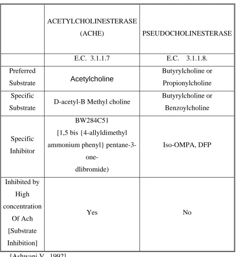
Table 1.1: - Cholinesterases
ACETYLCHOLINESTERASE
(ACHE) PSEUDOCHOLINESTERASE
E.C. 3.1.1.7 E.C. 3.1.1.8.
Preferred
Substrate Acetylcholine
Butyrylcholine or
Propionylcholine
Specific
Substrate D-acetyl-B Methyl choline
Butyrylcholine or
Benzoylcholine
Specific
Inhibitor
BW284C51
[1,5 bis {4-allyldimethyl
ammonium phenyl}
pentane-3-one-
dlibromide)
Iso-OMPA, DFP
Inhibited by
High
concentration
Of Ach
[Substrate
Inhibition]
Yes No
1.3 Physiological function of Cholinesterases:
-1.3a Physiological function of AChE: -
AChE catalyses thehydrolysis of the neurotransmitter ACh in the following ways: -
Reaction: -
[Soreq H and Seidman S., 2001]
As seen in the above AChE promotes ACh hydrolysis by forming an
acetyl-AChE intermediate with the release of choline, and the subsequent
hydrolysis of the intermediate to release acetate. The catalytic turnover rate
of AChE is very high. The choline released is recycled in the pre-synaptic
nerve cell. Thus AChE is a key enzyme for cholinergic transmission and
plays a vital role in the nervous system of both the vertebrates and the
invertebrates and in the neuromuscular junction (NMJ) of vertebrates and
some invertebrates. [Massoulie J., et al., 1999]
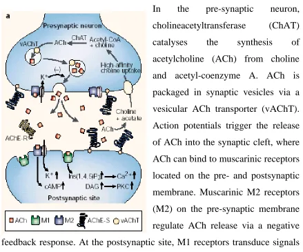
Figure 1.1: - The Cholinergic Synapse
In the pre-synaptic neuron,
cholineacetyltransferase (ChAT)
catalyses the synthesis of
acetylcholine (ACh) from choline
and acetyl-coenzyme A. ACh is
packaged in synaptic vesicles via a
vesicular ACh transporter (vAChT).
Action potentials trigger the release
of ACh into the synaptic cleft, where
ACh can bind to muscarinic receptors
located on the pre- and postsynaptic
membrane. Muscarinic M2 receptors
(M2) on the pre-synaptic membrane
regulate ACh release via a negative
feedback response. At the postsynaptic site, M1 receptors transduce signals
through a pathway involving diacylglycerol (DAG), inositol-1, 4,5-
trisphosphate (Ins (1,4,5) P3) and a Ca2+- dependent protein kinase (PKC).
In the hippocampus, most of the postsynaptic receptors are of the M1
subtype; in the cortex M2 receptors might also be located on the
postsynaptic membrane. ACh is hydrolysed in the synaptic cleft by AChE-S
tetramers, which are indirectly attached to the neuromuscular junction by a
collagen-like tail, or by another structural subunit to brain synapses.
AChE-R monomers would remain soluble within the synaptic cleft. A high affinity
choline-uptake mechanism returns choline to the pre-synaptic neuron. Brain
distribution of AChE includes both acetylcholine releasing and
1.3b Physiological function of BChE: -
BChE is present in non-neuronal tissues like liver, lung, plasma etc. The
function of BChE in higher vertebrates is unclear. Humans lacking BChE
activity do not show any pathology, except patients undergoing surgery
when exposed to succinylcholine as a curarizing agent, fail to recover
breathing at the end of anaesthesia. BChE present as a soluble form, in the
plasma of mammals, is suggested to serve as a safeguard against the
diffusion of ACh into the blood stream and/or against orally ingested toxic
compounds, since this enzyme has a broad range of substrates including
plant alkaloids such as cocaine. [Massoulie J., et al., 2002]
1.4 Structure of AChE:
-The 3-D structure of AChE obtained by X-ray crystallography in Torpedo,
mammals and Drosophila showed that the catalytic domain of the enzyme is
organized as a globular assembly of β sheets and α helices [α/β fold]. Since
it is known, that AChE has a very high catalytic turnover rate, it was very
surprising to find, the ‘active site’ of the enzyme located in a cavity, only
accessible through a deep and narrow ‘catalytic gorge’. The active serine
belongs to the catalytic triad Glu-His-Ser. The substrate ACh is oriented in
the active site by cation-π interaction of its quaternary ammonium group
with a tryptophan group. The polarized distribution of the charged residues
generates an electrostatic dipole, which may attract cationic substrates
towards the active site, but this needs further investigations. Moreover, the
determines its specificity towards different choline esters and also its
sensitivity to active site inhibitors. It is also a possibility, that the peripheral
site located at the entrance of the catalytic gorge serves as the first binding
site for the positively charged substrates, on their way to the active site. The
catalytic domain of AChE consists of N-terminal and C-terminal
sub-domain, which do not interpenetrate but establish a close contact at the
peripheral site. The general stability and flexibility of enzyme depends on
the residues localized in this region. This suggests, that the catalytic and the
substrate specificity of AChE depends on the dynamic movements within
the protein structure, and that the reaction products, exit the active site of the
enzyme via an alternative route, called the ‘back door’ avoiding trafficking
problems with the substrate molecules entering via the catalytic gorge.
[Massoulie J., et al., 2002]
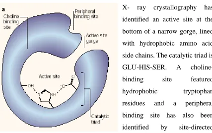
Figure 1.2: - Structural Features of the enzyme AChE.
X- ray crystallography has
identified an active site at the
bottom of a narrow gorge, lined
with hydrophobic amino acid
side chains. The catalytic triad is
GLU-HIS-SER. A
choline-binding site featured
hydrophobic tryptophan
residues and a peripheral
binding site has also been
identified by site-directed
1.5 Isoenzymes / Isozymes:
-They are variable forms of the same enzyme. Isozymes are highly
homologous to each other but contain small differences in amino acid
sequence. The term isozyme is not a precise one and can refer to different
gene products, or different products of the same gene with alternative
splicing. The term "isoenzyme" or "isozyme" should apply only to those
multiple forms of enzymes arising from genetically determined differences
in primary structure and not to those derived by modification of the same
primary sequence. Groups 1 to 3 of Table 1.2 represent enzymes having
isozymes. The term "multiple forms of the enzyme " should be used as a
broad term covering all proteins catalyzing the same reaction and occurring
naturally in a single species, for instance groups 4 to 6 of Table 1.2.
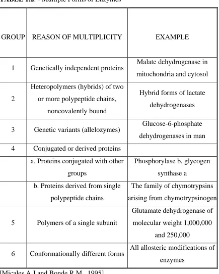
TABLE 1.2: - Multiple Forms of Enzymes
GROUP REASON OF MULTIPLICITY EXAMPLE
1 Genetically independent proteins Malate dehydrogenase in
mitochondria and cytosol
2
Heteropolymers (hybrids) of two
or more polypeptide chains,
noncovalently bound
Hybrid forms of lactate
dehydrogenases
3 Genetic variants (allelozymes) Glucose-6-phosphate
dehydrogenases in man
4 Conjugated or derived proteins
a. Proteins conjugated with other
groups
Phosphorylase b, glycogen
synthase a
b. Proteins derived from single
polypeptide chains
The family of chymotrypsins
arising from chymotrypsinogen
5 Polymers of a single subunit
Glutamate dehydrogenase of
molecular weight 1,000,000
and 250,000
6 Conformationally different forms All allosteric modifications of
enzymes
1.5a Isoenzymes of AChE
AChE exists in a large variety of molecular forms. There are designated as
‘G’ (globular forms) and ‘A’ (asymmetric collagen-tailed forms). All the
molecular forms posses the same catalytic domain but distinct C-terminal
peptides [AChER, AChEH, AChET, AChES]. They are as follows: -
AChER: - where R stands for ‘readthrough’
Readthrough or the ‘R’ transcripts or subunits of type R correspond to the
hypothetical products of ‘readthrough’ transcripts, which retain the
‘intronic’ region that forms the last exon encoding the catalytic domain.
AChER molecular forms remain soluble and monomeric and are found only
in vertebrates. [Massoulie J., et al., 1999, 2002]
AChEH: - where H stands for ‘hydrophobic’.
‘Hydrophobic’ or H transcripts or subunits of type H, are called so, because
the C-terminal regions of these subunits are characterized by hydrophobic
sequences. These hydrophobic regions consist of one or two cysteine
residues, which establish disulphide bonds & result in a dimeric mature
protein. The hydrophobic region majorly corresponds to a signal for a
glycophosphotidyl (GPI) anchor. Hence these molecular forms are
GPI-anchored dimmers and are studied in both vertebrates and invertebrates.
[Massoulie J., et al., 1999, 2002]
AChET: - wherein T stands for ‘Tailed’ forms
AChET is the only type of catalytic subunit that exists in all vertebrates
producing major AChE forms in the brain and the muscle. AChET generates
multiple structures, ranging from monomers, dimmers to collagen- tailed and
hydrophobic tailed forms. These tailed forms are catalytic tetramers
associated with anchoring proteins, which help them attach to the basal
lamina of NMJ or to the cell membrane. The monomeric G1 form is the first
translational product and more complex forms like G2, G4 are sequentially
derived from it. Hence the order of formation and appearance is G1->G2
AChE is subdivided into a secreted form, hydrophobic tailed form and an
incipient collagen tailed asymmetric form. In the collagen tailed forms,
AChET subunits are associated with a specific collagen, ColQ which
contains a short peptide motif, the proline- rich attachment domain [PRAD],
that triggers the formation of different heteromeric forms, which contain
one, two or three catalytic tetramers [A4, A8, A12] from monomers and
dimmers. Forms such as A4 and A8 AChE are intermediates, existing in
appreciable amounts only when the production and breakdown of A12 AChE
is rapid. The collagen-tailed forms are predominant at the neuromuscular
junction (NMJ). The AChET subunits are also found to be associated with a
hydrophobic glycoprotein of about 20 KD. This protein contains a signal
peptide, an extracellular domain, which includes a proline rich motif and an
N-glycosylation site, a transmembrane domain and a cytoplasmic domain.
The proline rich domain present is responsible for the formation of
membrane bound G4 tetramers. This transmembrane glycoprotein, which
constitutes the membrane anchor of AChE, is called the Proline-Rich
Membrane Anchor [PRiMA]. AChET subunits associated with PRiMA
predominantly exist in the brain. Although careful biochemical analysis of
muscle sections have shown, that this form is also located in the
‘peri-junctional’ zone surrounding the NMJ and is physiologically regulated
[increases or decreases] depending on the type of muscle activity/exercise.
Hence it is suggested, that the physiological role of AChET subunits
associated with PRiMA in muscles is to control the diffusion of ACh around
the junction on repetitive stimulation of the muscle. These findings need to
be further investigated. New developments in the intracellular pathways,
AChE transport, mode of association and regulation of the catalytic subunit
of the PRiMA. The AChET subunits are studied in both vertebrates and
invertebrates. [Massoulie J., et al., 1999, 2002]
AChES: - wherein S means ‘Soluble’. These AChES subunits comprise of
soluble monomeric forms of the enzyme and have been studied only in
vertebrates. [Massoulie J., et al., 1999, 2002]
In Vertebrates a single gene encodes for AChE. The following 3 processes
generate this panoply of AChE molecules.
[1] Alternate splicing of the AChE gene.
[2] Oligomerisation of the catalytic subunits.
[3] Associations with non-catalytic subunits.
Vertebrate AChE molecular forms are further discussed in Chapter 3.
Unlike Vertebrates, invertebrates posses variable number of AChE genes.
Moreover the process of alternate splicing has never been suggested for the
generation of invertebrate AChE molecular forms. The molecular forms of
invertebrates and the genes encoding them have been discussed in Chapter 2.
1.6 Conclusion: -
The enzyme history and it’s various attributes like basic structure, functions
and molecular diversity, has enabled biologists in understanding,
interpreting and analyzing the complex role played by this multifaceted
Chapter 2: -
Role of Acetylcholinesterase in Invertebrates
2.1 Introduction: -
AChE in invertebrates, has been and still is the focus of research for various
reasons. In some invertebrates (e.g.: - sea urchins, oysters etc) it’s potential
as a biochemical marker for environmental toxicity has been explored, in
others (e.g.: - Protozoans, Aplysia etc) it’s non-cholinergic functions in
processes like cell-adhesion and neuritogenesis have been elucidated;
whereas the multiplicity and the variable number of genes encoding
invertebrate AChE (e.g.: - four genes in nematodes, one gene in Drosophila)
are being interpreted to derive a phylogeny providing an insight in it’s
evolution and it’s sensitivity towards various antiparasitic drugs and
insecticides being continuously understood in order to prevent infection by
worms and destruction of crops by insects respectively. Hence, in order to
provide a better understanding of all the above functions of invertebrate
AChE, the study of it’s expression, function, structure and molecular
diversity within each phylum was undertaken and explained in this chapter.
2.2 Acetylcholinesterase in Invertebrates: -
2.2a Phylum Protozoa: -
Protozoans are unicellular Eukaryotes, nowoften included in a separate kingdom together with fungal protists and algae.
i) Sarcodina
Acetylcholinesterase [AChE] was detected in Protozoans. Work done on
Dictyostelium discoideum showed its serine esterase to have a strong
sequence similarity with AChE of Torpedo. This led to further investigations
on characterization and possible function of cholinesterases in protozoan. It
appeared that histochemically, biochemically and electrophoretically AChE
activity of amoeboid cells was similar to that of Electrophorus Electricus.
Possibilities of using this Protozoan as an efficient bio-indicator were
investigated. Dictyostelium Discoideum used in bioassays for pre-chemical
screening of both moist environments and fresh waters in relation to
neurotoxic organophosphorus compounds like Basudin, showed a significant
dose dependant inhibition of Propionylcholine but the effect on AChE was
insignificant. [Falugi C., et al 2002]
ii) Ciliophora
Work done on another protozoan Paramecium primourelia indicates the role
of AChE in cell-to-cell adhesion in the process of conjugation. In immature
cells, choline acetyl transferase (ChAT) activity was completely absent. But
AChE was seen in the cytoplasm. The activity in the cytoplasm is due to the
260-Kda molecular form of AChE. This form is found in the membrane
protein fraction of competent mating cell along with the presence of other
protozoan shows similarity to the membrane bound tetrameric form present
in human erythrocytes. By inhibiting AChE activity in competent mating
cells, a significant reduction in mating cell pairing was observed. Hence the
role of AChE in cell-to-cell adhesion for the process of conjugation was
elucidated. [Corrado M.U.D, et al., 2001]
2.2b Metazoa: -
Metazoans are multicellular mitochondrial eukaryotes.i) Phylum: - Echinodermata: Echinderms, are marine deuterostome
organisms’ characterized by tube feet which form part of water vascular
system, thought to have a common ancestry with the chordates.
The cholinergic system is formed by a set of molecules, they are, the signal
molecule ACh [acetylcholine]; the biosynthetic enzyme ChAT
[cholineacetyltransferase E.C.2.3.1.6.]; the lytic enzyme
Acetylcholinesterase [E.C.3.1.1.7.] and two classes of receptors Nicotinic
and Muscarinic. [Harrison K.P., et al 2002]. Detection of the Cholinergic
System in the male gametes of vertebrates and invertebrates lead to
questioning the possible role of this signaling system in regulating
intracellular ion exchange, functional to sperm mobility and its probable
interaction with the egg. When sea urchin eggs (species: - Paracentrotus
lividus) were used for this study, the presence of AChE molecules
performing AChE activity detected by Immunofluorescence suggested a
signaling pattern. [Piomboni P., et al 2001] Furthermore work done on sea
urchin larvae suggested the presence of molecules of the cholinergic system
since early developmental stages. With the progress in development, a
histochemical, immuno-histochemical and confocal laser scanning
microscopy. [Falugi C., et al., 2002]. Like in the Neuromuscular Endplates
of higher organisms, AChE in sea urchin larvae was generally distributed in
motile structures. In Starfish, AChE activity is enhanced with increasing
motility [Semenova M.N., 2000]. Along with other molecules of the
cholinergic system AChE was found to be associated with the ciliary
structures, which modulate ciliary movement for feeding. The pattern in
which the molecules of AChE are present in the larval body wall muscles
and along the arms contributes in understanding its role and organization.
Also a sensitive function for AChE is suggested since it is found localized at
the tip of the spines in the larvae. The mature molecular form of the enzyme
is present since early larval stages as indicated by electrophoretic studies.
[Falugi C., et al., 2001]. In another electrophoretic study on sea urchins,
Strongylotrotus drobachiensis and S. intermedius and their hybrids, two
AChE fractions were found. These fractions differed in their thermostability
and are also developmentally regulated such as the thermo-labile form is
predominant at the Gastrula stage whereas the thermo-stable one
pre-dominates the mid-plutus stage. [Ivanenkov V.V., et al., 1976] Hence in sea
urchins AChE has been detected in sperm cells, eggs, Primary Mesenchye
Cells [suggesting a role in developmental cell interaction] and during larval
growth and metamorphosis. Since several structures with “Embryonic”
cholinergic molecules modulated by AChE activity are present from the
early stages of sea urchin development, which indeed is sensitive to a variety
of pollutants and hence is extensively being used as a model system in
assessing the effects of neurotoxic pesticides. The Effects of Basudin [an
organophosphate compound containing 20% of a thionophosphate Diazinon,
[which compete with the substrate ACh] were studied. Results indicated
concentration dependant AChE inhibition during early development of sea
urchin. These effects revealed at molecular level of AChE activity were
more sensitive and reveal anomalies at chronic exposure concentrations.
[Pesando D., 2003]. Hence determining AChE activity and using this
biomarker as an early warning signal of exposure and/or adverse effects can
also be used in the future to study the spatial and temporal trends in the
quality of coastal waters. [den Besten P.J., et al., 2001]
ii) Phylum: - Platyhelminthes: - Flatworms, acoelmate animals of
uncertain origin.
AChE is very broadly distributed in many species including parasites. It is
present in all helmintic invertebrates, including trematodes and nematodes.
In parasitic studies, the mechanism in which the parasite evades the immune
defense mechanism of the host is puzzling; and hence functional proteins
like AChE that might elicit protective immunity against infection are being
identified. The biological role of AChE in parasites is been constantly
understood in context with the development of antihelmintic drugs. For
instance various AChE inhibitors were shown to significantly decrease the
amplitude of muscle contractions in various parasites, such as F.hepatica and
S.mansoni; and are being used as antihelmintic drugs. Furthermore, levels of
parasitic AChE in the serum, is assayed after immunoprecipitation and is
used to monitor circulating filarial parasites both in human and experimental
Example: - Echinococcus Granulosus
Though little information is available on the neuromuscular system of
cestoda, it was known that ACh exists as an inhibitory neurotransmitter. PG
et.al. (2000) detected the presence of AChE spectrophotometrically and
electrophoretically. It is known that on ingestion by a definitive host
Echinococcus granulosa protoscolices become adults and their interest may
be related to dissemination following cyst rupture. It is hypothesized that
Echinococcus granulosa protoscolices could release AChE in hydrated cyst.
The released enzyme could to pass through the germinal layer and travel to
the host tissues, destroying the host ACh. Hence AChE present in this
tapeworm could be an attractive target for chemotherapeutic or
immunological intervention in hydatid disease. In future, a combination of
compounds could be used, which would inhibit AChE thereby allowing ACh
to accumulate and cause abnormally high levels of inactivation and a total
blockage of the nerve function. Hence AChE activity would be considered
as a possible target in chemotherapy. [Gimenez-Pardo C., et al., 2000]
Example: - Fasciola hepatica
Several putative neurotransmitters have been identified in the primitive
nervous system of parasitic flatworms. But the mechanism of
neurotransmission and kinds of neurotransmitters remain unclear.
Acetylcholine [ACh] has been identified as an inhibitory neurotransmitter
responsible for muscle contractions. Its receptors are the mixed muscarinic
and nicotinic type and hence pharmacologically primitive. The worms are
capable of synthesizing and degrading endogenous ACh. Although this data
throws some light on the role of ACh as a neurotransmitter in the worms, it
is necessary to demonstrate the localization of ACh in the appropriate sites
of the nervous system. To do so, an indirect method was adopted wherein
the lytic enzyme of the cholinergic system, AChE was localized
histochemically and this localization of AChE was then linked with the
functional localization of ACh. Histochemical studies performed on
Trematoda Fasciola hepatica demonstrated AChE activity in the neural cell
bodies and extracellularly in the neuropile of the cerebral ganglia. This
ultrastructural localization of AChE activity provided strong evidence of
ACh as a neurotransmitter. However the patterns and location of AChE in
the nervous system of F.hepatica were found to be very similar to those seen
in higher invertebrates and vertebrates suggesting that the basic mechanisms
of neurotransmission in higher invertebrates and vertebrates are already
present in this primitive phylum. [Sukhdeo S.C., et al., 11988]
Example: - Schistoma
A lot of groups have worked on AChE of Trematoda Blood fluke Schistoma.
By 1994 it was known that AChE was present at all stages of Schistoma Life
Cycle. AChE was found on the parasitic surface and in the muscles.
Comacho and Colleagues [1994] worked on 3 species of Schistoma
{S.mansoni, S.haematobium and S.bovis} and found G2 as the dominant
molecular form, majorly located on the tegument, in all the 3 species. But
they also noticed, that the AChE activity differed from species to species.
They found, at the adult stage, the major difference between species lay in
the relative amounts of AChE activity in their teguments; S.haematobium
teguments carried 20 times and S.bovis 6.9 times the activity present on
S.mansoni teguments. This explained the species-specific susceptibility to
In S.mansoni, AChE was first demonstrated in adult worms by Bueding
[1952] and was partially characterized both histochemically and
biochemically. Early studies also suggest involvement of AChE in the motor
activity of S.mansoni by demonstrating anticholinestrase-induced paralysis
in worms. Schistosomal AChE is the target for several antiparasitic drugs,
including hycanthon, lucanthone, metrifonate and phosphonium compounds.
Extensive biochemical and immunological investigations were carried out in
Ruth Arnon’s laboratory on the enzyme AChE of S.mansoni. On extensive
analysis of these investigations it was shown that the enzyme AChE was
concentrated up to 350 times more in the outer membranes of the worms, in
comparison to the rest of the worm. This AChE present in the outer
membranes of the worm, is probably expressed at the surface of the parasite
attached to the tegumental membrane via a covalently linked GPI anchor,
was suggested to possess a non-cholinergic function and also serve as an
effective immunological target. Furthermore their investigation showed that
AChE in S.mansoni appears in 2 principal molecular forms both globular,
with sedimentation coefficient of ~6.5S and ~8S. The 2 forms of AChE were
found to differ in their heparin binding and immunological specificity. The
8S form binds to heparin, the 6.5S form did not and the 2 forms were
selectively recognized by different monoclonal antibodies. Polyclonal
antibodies raised against S.mansoni AChE purified by affinity
chromatography were specific for the parasitic AChE, reacting with both
molecular forms, but do not recognize AChE from the other species. They
interact with the surface localized enzyme on the intact organism and
produce almost total compliment dependent killing of the parasite
multifaceted activities, which can serve as a suitable candidate for diagnostic
purposes, vaccine development and drug design. [Arnon R., et al., 1999]
The presence of AChE in bacteria, plants, non-excitable tissues of complex
organisms suggests it has a wide range of alternative non-cholinergic
functions. Similarly the AChE on the parasitic surface is known to be
involved in non-cholinergic functions like glucose scavenging from the host
blood stream. The glucose is transported through the membrane with the
help of transporters, but the exact mechanism underlying the link of
non-cholinergic system present on the parasitic surface to glucose transport is not
yet known. AChE on the schistome surface is the target of
organophosphorous drugs like Metrifonate. But this drug is not widely used,
since it cannot differentiate between host AChE and parasitic AChE and thus
is toxic. On elucidating the structure of Parasitic AChE, it was known that
the several functionally important regions of schistome AChE are
sufficiently different from human AChE. Hence, now it is possible to design
drugs and vaccines, which would specifically inhibit only the parasitic
AChE. [Jones A.K., et al., 2002]
iii) Phylum: - Aschelminthes – includes Nematodes, roundworms,
threadworms [some], whipworms, lungworms, hookworms, eelworms, a
pseudocoelamate phylum with both parasitic and free-living representatives,
Class Nematoda: -
Example: - C.elegans
Nematoda is one of the rare phyla, wherein multiple “Ace” genes encoding
for AChE have been identified. For instance in Nematoda C.elegans, four
genes [Ace-1, Ace-2, Ace-3 and Ace-4] encoding for AChE have been
identified. Amongst the four genes, Ace-1 and Ace-2 encode the 2 major
forms of AChE, which show a 35% identity in their coding regions, but
differ in the pharmacological properties and tissue repartition. Ace-1
expression is observed throughout the body wall and vulval muscle cells of
the nematode C.elegans. Moreover, further studies revealed that the
C.terminus of Ace-1 was homologous to that of the T subunits present in
vertebrate AChE’s and that Ace-1 oligomerized into amphiphilic tetramers.
Whereas Ace-2 is exclusively expressed in neurons and has a hydrophilic C
terminus of AChEH type. It associates into glycolipid-anchored dimmers.
Ace-3 and Ace-4 show 54% identity, as they lay in close proximity to each
other on the same chromosome. Ace-3 represents a mere 5% of the total
AChE activity. They are known to hydrolyze the substrate butyrylcholine
faster than acetylcholine. Ace-3 encodes H subunit catalytic subunits and are
resistant to usual cholinesterase inhibitors. The mRNA level of Ace-4 is
extremely low, and hence, even though it is transcribed, no enzyme activity
has been detected for this form of AChE till date. This distinct
tissue-specific expression of Ace-1, Ace-2 and Ace-3 is indicative of the fact, that
these genes are not redundant. [Combes D., et al 2003],[Combes D., et
al.,2001],[Combes D., et al 2000]. The enzyme AChE expressed by these
genes has been recognized in the crude extracts and in the excretion
secretion products of the nematodes. The amount of AChE present varies
variation in the amount of AChE present is responsible for sex-, species-,
stages- and strain specific behavior of the nematode. AChE plays an
important role in the nematode neuromuscular system and in the host
parasite relationship and has been associated with the modulation of several
host mechanisms like gastrointestinal motility, cell membrane permeability,
anti-coagulant processes, glycogenesis, acetate and choline metabolism,
antihelmintic resistance, immune and anti-inflammatory responses. Hence
AChE of the nematode poses as the priority target for several drugs been
tested to protect against nematode infection.[Ros-Moreno R.M., et al., 2002]
iv) Phylum: - Annelida: - Annelids, Segmental Coelamate worms with
Chitonous Bristles.
Example: - Leech
Two forms of AChE, one the “spontaneously-soluble” form i.e. SS-AChE
and the other the “detergent-soluble” form i.e. DS-AChE were studied in
medicinal leech “Hirudo medicinalis”. The 2 forms of AChE were found to
differ in their substrate and inhibitor specificities. The spontaneously soluble
form was recovered easily from the hemolymph and tissues of leech in a low
salt buffer solution and was suggested to represent the hydrophilic
monomeric [G1] form of AChE. Whereas the detergent-soluble form was
extracted from the tissues of leech in low salt buffer solution containing 1%
Triton-X and was suggested to represent an amphiphilic glycolipid anchored
dimeric molecular form of AChE. [Talesa V., et al., 1995] Early studies on
leech suggested that AChE was a key enzyme marker in the leech
contained 3 to 24 folds higher AChE than non-cholinergic neurons.[Wallace
B.G., et al., 1982]
v) Phylum: - Rotifer [-Rotatoria] rotifers, “wheel animals”, named for
rotating ring of cilia; a pseudocoelamate phylum.
In rotifers, AChE activity was localized in the neurons and muscular tissues,
in sensory organs, in all the ciliated cells, secretory cells (sub cerebral, saliva
and pedal glands) and gonad cells (nuclei of the syncytial vitellaruim and
follicular layer, oocytes and eggs)[Raineri M 1984]
vi) Phylum Mollusca: -Molluscs, soft bodied animals, mostly with an
internal or external calcareous shell.
Soluble and membrane bound AChE was detected in the bivalve mollusc
Mytillus gallprovincialis [Pelecypoda: -filibranchia] collected from the
northern Adriatic Sea and in the Benthic mollusc Scaphara inaequivalvis
which was collected in spring 1999 from 3 areas of northern Adriatic Sea.
[V Talesa et.al., 2001,2002]. Amongst the 3 forms of AChE purified in this
mollusc, 2 were the spontaneously soluble forms and one was the membrane
bound form. The two spontaneously soluble forms [A and B] occurred in the
hemolymph and accounted for 80% of the total AChE activity. They
represented the globular tetrameric [12S] and the globular dimeric [6S]
molecular forms of AChE respectively. Whereas the third membrane bound,
detergent soluble form of AChE represents the GP1 anchored amphiphilic
BW284C51 a specific inhibitor for AChE, indicating that they represent
“true” AChE forms. [Talesa V., et al., 2001]
In the gills of oysters, 2 forms of AChE [Form A and Form B] were
detected. Form A is a glycolipid anchored and non-glycosylated molecular
form of AChE sensitive to organophosphorus and carbamate insecticides.
Whereas form B is a hydrophilic and glycosylated molecular form of AChE
insensitive to organophosphorus and carbamate insecticides. Oysters are
widely used as bio-indicators and AChE activity as a Biomarker. Hence
elucidating the sensitivity of each AChE molecular form present in the gills
of oysters would help improving the techniques used to monitor the marine
environment and report accurate results. [Bocquene G., et al., 1997]
AChE is also present in significantly high amounts in the haemolymph of
Aplysia. Haemolymh in mature Aplysia is neurotropic. Hence the possibility
of AChE playing a neurotropic factor in haemolymph was investigated.
When Dopaminergic neurons from Pedal ganglia were cultured in a medium
containing haemolymph for 24 hrs, it was observed, that the neurons were
well attached to the substratum and exhibited multiple neuritis. Similar
observations were reported when the culture medium was supplemented
with AChE purified from the haemolymph. But in medium with no
haemolymph the neurons showed poor attachment to the substratum and
only one or two short neuritis were exhibited, indicating that AChE
circulating in the haemolymph promotes neurite growth of adult neurons.
Similar reduction in neurite growth was observed, when its specific inhibitor
BW284C51 inhibited both, the catalytic and the peripheral site of the
was inhibited by Echothiophate, the enzyme does not loose it’s function as a
neurotropic factor, indicating that only the peripheral site of the enzyme
AChE is responsible for Neurite growth. [Srivatsan M., et al 1997] It is
known that the decrease in the AChE levels signals the onset of aging in
Aplysia. This process coincides with impaired neural function. It was
observed a specific sensory input like chronically applied sensory
stimulation [CSS] could upregulate enzyme activity [Peretz B., et al 1996]
vii) Phylum Arthropoda: -arthropods, 'jointed legged animals'
characterized by segmented bodies and jointed appendages; have gills or
tracheae; easily the largest phylum of all animals & of great economic
importance.
Example: - Insects
Acetylcholinesterase is an important enzyme of the insect nervous system.
For several years it was and still remains to be the priority target of
insecticides. Hence extensive work done in this area led to the bio-chemical
characterization of this enzyme in more than 20 insect species. Moreover,
molecular analysis of its gene structure was also completed in some insect
species like [1] fruit fly [Drosophila melanogaster] [2] yellow fever
mosquito [Aedes aegyypti] [3] Colorado potato beetle [Leptinotarsa
decemlineata], housefly [Musca domestica], green rice leafhopper
[Nephotettix cincticeps] and Austrailian sheep blow fly [Lucilio cuprina].
Recent developments in this area elucidate the 3D structure of the AChE
enzyme in insects. It was then learnt, that although the insect AChE showed
relatively low amino acid sequence identity [36%] to that of vertebrates it
the basis of several other studies in Lepidotera, Diptera, Orthoptera and
Coleoptera it was learnt that AChE in insects exhibited polymorphism and is
majorly present as a glycolipid anchored amphiphilic dimer [G2a], with
variable amounts of hydrophilic dimmer [G2h] and sometimes traces of
hydrophilic and amphiphilic monomers [G1a and G1h] all possessing the
same catalytic properties and belonging to a single gene. These findings
were further confirmed with the detection of a single Ace locus in
Drosophila and with the cloning of a single gene in Anopheles stephensi and
Aedes aegypti. The only exception to this was Culex pipens, wherein
Bourget and Colleagues were successful in detecting not one but two AChEs
[AChE 1 and AChE 2] differing in their substrate and inhibitor specificities.
This finding is under speculation and is being investigated. [Bourguet D., et
al., 1997] [Gao J.R., et al., 2002]
2.3 Conclusion: -
This survey helps understand the cholinergic and non-cholinergic essence of
AChE, a well established enzyme, of the invertebrate nervous system and
muscles; and throws light on it’s high potential in being used as a biomarker
of environmental toxicity. It also builds up a need, to acquire more
knowledge about this interesting multifaceted enzyme in vertebrates
Chapter 3:-
Role of Acetylcholinesterase in Chordates
3.1 Introduction: -
Extensive research conducted on vertebrates (Torpedo, electric eel, chick, rat
etc) for several years has generated a sea of information related to all aspects
concerning the AChE enzyme. Some of these aspects include expression,
structure, cholinergic functions, non-cholinergic functions, gene regulation,
isoforms, implications in neurodegeneration, post translational modifications
etc. A sincere effort has been put in accumulating as much as information
possible and surveying it with reference to each phylum in vertebrates.
3.2 AChE in Chordates: -
Chordates are characterized by having a single dorsal nerve cord, a
notochord and pharyngeal gill slits at some stage of their life cycle.
i) Subphylum Cephalochordata:
Cephalochordate amphioxus possesses two ChE genes of which only one
represents a ‘true’ AChE. Phylogenetic sequence analysis of these genes
indicated that the two genes were the result of a duplication event in the
lineage leading to amphioxus. This duplication event is distinct from the
event that gave rise to AChE gene in vertebrates, because Agnatha
[hagfishes], which represent the most primitive extinct vertebrates, seem to
possess a single AChE gene and exhibit both globular and asymmetric forms
acyl-binding site, typical of other vertebrates. These studies added to our
better understanding on evolution of AChE in Chordates.
[Sutherland D., et al., 1997]
3.3 AChE in Vertebrates: -
Vertebrata: Vertebrates have backbones, group indicates lampreys and all
jawed vertebrates.
3.3a Chondrichthyes: -
Example: - Torpedo
A single AChE gene, in Torpedo, undergoes alternative splicing of its H and
T exons, to generate AChEH and AChET subunits. In addition to AChEH and
AChET, R transcripts [i.e. ‘readthrough’ which are not spliced after the last
catalytic exon, but appear to be mature i.e. polyadenylated] have been
characterized. The GPI anchored dimeric molecular form derived from
AChEH subunit is the only form expressed in the Torpedo skeletal muscle.
This form has also been characterized in other tissues, like the electric
organs [derived from modified muscles] and the electric lobes [which is the
region of the CNS that commands electric discharges]. It is also known that
this form, shows dramatic increase in its level, right from the first electric
discharges, that is from their origin in the Electric Lobes to their entry in the
electric organs. The soluble tetrameric G4 form derived from AChET
subunits is maintained at a uniform level in the electromotor nerves.
Whereas the collagen tailed AChE molecular forms are present in the
embryonic electric organs coincides with the establishment of contacts
between the nerve terminals and the electrocytes. The expression of AChE
molecular forms, in Torpedo, was observed to be tissue-specific suggesting
that polymorphism exhibited by AChE is dependant on a number of factors
including [1] amount of ACh released [2] size of the synaptic space and/or
[3] temporal course of the physiological stimulation [Massolie J., et.al.,
1999]. In developing electric organ of torpedo, earliest AChE expression
was observed at the 35mm stage of the embryo. Both asymmetric as well as
G2 isoforms of AChE were present at this stage. The G1 isoforms of AChE
become prominent later, at the 85mm stage of the embryo, and remains more
abundant than G2 through adulthood. Hence the pattern of expression of
AChE isoforms in a developing electric organ of Torpedo reveals less about
the production and inter-conversion of AChE forms, but nevertheless
indicates maturation of specific structures with which these particular forms
are associated, like the appearance of asymmetric forms might be associated
synaptogenesis and laying down of the basal lamina [Brimijoin S., 1982]
3.3b Aves: -
Birds have feathers, no teeth, modified forelimbs [wings], canregulate their body temperature and have land adapted eggs with shells.
Birds possess a single AChE gene. This gene contains only one exon
encoding C- terminal region of type T and does not contain any other
alternative exons [Massoulie J., et al., 1999] Extensive work been carried out
on AChE of the developing chick brain indicates, that the ganglia of 3.5-4
day old embryo shows the earliest expression of AChE. Further, intense
AChE activity is evident on embryonic day 6, as the structures expressing
like G1 appear first during the early embryonic stages and pose as
biosynthetic precursors of more complex forms like G4, which appears a
little later and gradually increases until maturity. [Salceda R., et al 1992]
Though the hydrophobic G4 form predominates in the brain, the A12 form of
AChE has also been detected in some structures like the mesencephalon
[1%], retina [7%] and the ciliary ganglion [10%]. The latter is a
parasympathetic [cholinergic] structure, which along with A12 AChE [10%]
at the nerve endings also expresses G1 AChE [7%], G2 AChE [43%] in the
cell body and G4 AChE [40%] in the axons of the pre-synaptic and
post-synaptic nerves. This structure-specific expression of AChE isoforms,
elucidated by Courad and Colleagues [1980], suggested structure related
functions for these isoforms. Like for instance, the A12 AChE form is known
to be densely packed with catalytic sites and hence its concentration at the
nerve endings increases efficiency of the cholinergic transmission.
[Brimijoin S., 1983]. Early embryonic expression of AChE is not only
observed in neuronal [e.g.: - ciliary ganglion] but also in non-neuronal
tissues [e.g.: - retinal pigment epithelial cells]. Furthermore, AChE activity
in these tissues; show a parallel increase with development of the chick
brain. These findings strongly suggested some non-cholinergic functions for
AChE apart from its novel role in neurotransmission. In vitro studies on
chick retinal cultures exposed to an AChE inhibitor showed cell size
reduction whereas, studies on an AChE rich medium stimulated neurite
growth. [Tarrao S.A., et al., 2000] In another study, chick retinal pigment
epithelial cells in culture were reported to synthesize AChE and express A12,
G1, G2, and G4 molecular forms of the enzyme. The synthesized AChE either
remained associated to the cell, expressing A12 in a one day old culture but
AChE was released in the growth medium expressing higher levels of G4
AChE in the latter stages and lower levels of G2 and G1 AChE in the earlier
stages. Hence the process of cell differentiation is accompanied by an
increase in the cell-associated as well as released AChE along with an
alteration in their isozyme pattern with respect to the age of the culture. The
released AChE reported higher activity than cell associated AChE and was
implicated in non-cholinergic function of cell differentiation. [Sakeda R., et
al., 1992] These in vivo and in vitro studies in the central nervous system
[CNS] suggested that the developmentally regulated AChE enzyme played a
role in non-cholinergic functions like morphometric processes, cell
differentiation and synaptogenesis along the nervous system. [Tarrao S.A., et
al., 2000] In the peripheral nervous system [PNS], AChE was studied in
dystrophic chicken muscles, wherin it showed increased activity per unit
weight of the tissue and an altered isozyme pattern depending on the severity
of the disease. Avian dystrophy primarily affects fast-twitch glycolytic
fibers. Hence it was observed that the AChE isozyme pattern was
remarkably altered in muscles rich in fast-twitch fibers [e.g.: -pectoral
muscles]. The isozyme patterns of a dystrophic chicken muscle and a
dennervated avian muscle, showed no resemblance, and it was observed, that
a dystrophic muscle just like a normal muscle could respond to a neural
response and loose its asymmetric AChE on denervation. Hence it was
concluded that the avian dystrophy in no ways resembled the process of
denervation. [Brimijoin S., 1988] Studies done on dystrophic chickens, in
order to analyze potential abnormality in the axonal transport of AChE
forms surprisingly yielded negative results wherein despite large increases in
the activity of G1 and G2 forms in limbs and pectoral muscle, no change was
in ligated branchial and sciatic nerves. But experiments done on chickens
with peripheral neuropathy induced by acrylamide revealed specific
abnormalities in the axonal transport of AChE forms. It was observed that in
sciatic nerves, the A12 AChE form was 5 times that of normal and in ligated
nerves, the rate of accumulation of A12 was less than half of normal as well
as the G4 AChE was reduced by 20%. These abnormalities were
accompanied by alteration in the isozyme pattern of AChE, in the hind limb
muscle and resembled effects of denervation. It was hence suggested, that,
the degenerative changes induced in muscles of animals with acrylamide
neuropathy is due to defective fast axonal transport of proteins like AChE.
Hence AChE and its isozymes are also implicated in peripheral
neuropathies. [Brimijoin S., 1983]
3.3c Reptilia:-
reptiles have scales and an amniote egg adapted to survivalout of water, cold blooded mostly, well adapted to life on land, contains
many fossil groups including dinosaurs.
A single AChE gene in the snake Bungarus fasciatus undergoes alternative
splicing its ‘S’ and ‘T’ exons to generate AChES and AChET subunits.
A short, highly charged peptide region, which does not contain any cysteine,
characterizes the C-terminal region encoded by the AChES subunit and
hence the resultant form of AChE is soluble and monomeric. This form is
exclusively present in Venom glands, liver and muscle of elapid snakes.
Venoms of Mambas [e.g.: - the green mamba, Dendroaspis viridus] are an
exception, as they do not contain AChE but contain a peptide toxin
fasciculin that inhibits mammalian AChE with high affinity. The possibility
high levels, has been ruled out, by the fact, that neither AChE itself is toxic,
nor does it reinforce toxicity of venom toxins. Hence the pathological
significance of this form of AChE in venom remains unclear, however it was
suggested that in muscles, the soluble monomeric form of AChE co-exists
with the molecular forms encoded by AChET subunits and participates in the
functional hydrolysis of ACh. [Cousin X., et.al.;1998]
3.3d Mammalia: -
Mammals can regulate their body temperature,generally have hair, bear live young ones and nourish them with milk
produced by mammary glands.
Mammals possess a single AChE gene, which undergoes alternative splicing
of its H and T exons to generate AChEH and AChET subunits. Any defect in
this process of alternate splicing generates R transcripts.
The AChE H subunit generates GPI-anchored dimmers in mammals. This
isoform was also detected in Torpedo and Drosophila. However the H
peptide encoding the H exon in mammals, showed no sequence homology to
the H peptides of Torpedo or Drosophila. Hence even though these H
peptides were found to be functionally equivalent, their exons appeared
independent of each other, during evolution. This isoform of AChE is
expressed in mammalian blood cells [erythrocytes and lymphocytes]
embryonic muscles and liver. In mammalian blood cells, AChE expression
demonstrated by immunochemical and immunocytochemical techniques in
both T and B-lymphocytes, suggested a putative role for the cholinergic
system as regulators of immune function. [Tayebati K.S., et al., 2002].
The AChE T subunit generates amphiphilic monomers [G1
a
] amphiphilic
dimmers [G2
a
], hydrophobic tailed tetramer [G4
a
collagen tailed forms amongst these isoforms, the G1 a
and G2
a
are
intercellularly present biosynthetic precursors of functional hydrophobic
tailed tetramers, soluble tetramers, and collagen- tailed forms of AChE. The
soluble tetrameric form of AChE constitutes the majority of the plasma
AChE [56%] and the cerebro spinal fluid [CSF] AChE [82% in lumbar CSF
and [69%] inventricular CSF] and hence AChE activity measured in these
body fluids in a number of neurological and neuropsychiatric disorders
involving central or peripheral cholinergic neuropathy, reflects the
pathological changes in the structures of the CNS or PNS from where these
enzymes originate. Like for instance AChE and it’s isoforms in the CSF are
prognostic markers for Alzheimer’s disease. [Atack R.J., et al., 1987]. The
hydrophobic tailed tetramers, mainly anchors AChE in the mammalian brain
membranes, where it constitutes 80% - 90% of the total AChE activity and is
apt for efficient cholinergic transmission in the small synaptic spaces of the
Central Nervous System [CNS]. This form of AChE is also implicated in
cell adhesion and neurodegeneration. It was demonstrated, that the
membrane-associated G4 form of AChE expressed in neuroblastoma cells,
consisted of the HNK-1 epitope, which functions as a mediator in cell
adhesion. Hence this form of AChE functions as a cell adhesion molecule
binding laminin and other extra cellular matrix components. These cell
adhesion molecules in the CNS might play an important non-cholinergic role
in long term storage of information as the HNK-1 present on the isoform
might be involved in memory formation. Hence the significant loss of
hydrophobic membrane- associated tetrameric isoforms may lead to
dementia as seen in neurodegenerative diseases like Alzheimer’s. [Johnson
G and Moore W.S., 2001]. The hydrophobic tailed tetramers were also found
the diffusion of AChE around the junction, on repetitive stimulation of the
muscles. Hence regulation of this isoform in the peripheral nervous system
[PNS] depends on the kind of muscle expressing it and the type of activity
exhibited by that muscle. The collagen-tailed isoforms generated by
associations of AChE T subunits with a special collagen called ColQ, is
mainly expressed at the NMJ. The collagen ColQ encoded by a single gene
producing multiple variants in mammals is of great physiological
significance, since the levels of AChE and ColQ multiple variants could
describe the kinds of isoform present in a particular type of muscle and help
the muscle in adapting to various stimulations. For instance, fast muscles in
rat exclusively express A12 isoform, as ColQ is only expressed at the
junction and its level is comparatively low compared to AChE, so all the
three ColQ chains are associated with AChE tetramers, giving rise to the A12
AChE. Whereas slow soleus muscles express both A4 and A8 AChE as ColQ
is expressed uniformly in extra-junctional and junctional regions and AChE
is expressed at a lower level, so significant fractions of ColQ chains are not
occupied by AChE tetramers resulting in A4 and A8 AChE. Hence
collagen-tailed isoforms are controlled by innervations and by the type of muscle.
Moreover, a defect in the ColQ gene, in humans, results in a rare congenital
myasthenia syndrome, classified as CMS-1c, wherin due to lack of
collagen-tailed AChE, abnormal accumulation of AChE at NMJ leads to receptor
desensitization resulting in muscle weakness and fatigability.
The ‘R’ transcripts generating soluble monomers, result from errors during
the process of alternate splicing. The C-terminal peptide of AChER subunits
is called ARP. The ‘R’ transcripts [mRNAs] are expressed in embryonic
tissues during development, in cell cultures and in mouse erythroid cells.
intoxication by anti-cholinesterase drugs, in order to modulate hematopoietic
differentiation and immunological responses. The best evidence for these
isoforms is also derived from experiments using the brain of a stress induced
mice, wherin an increase in the mRNA level was followed by specific
pattern of migration in native gels. These soluble monomers have never been
characterized in vivo and hence lack direct evidence of existence.
[Massoulie J., 2002] [Massoulie J., et al., 1999]
Acetylcholinesterase in mammals is also implicated in morphogenesis and
neurodegeneration [e.g.: - Alzheimer’s disease]. It’s role in insulin-
dependent diabetes mellitus, a neurological disease, was best studied by
Gustavo S.C and Rocio S [1995-2001]. Diabetes mellitus is characterized by
increase in blood glucose level and by other complications, like retinopathy.
AChE activity was reported to significantly increase [100%] in the cerebral
cortex and [55%] in the serum whereas decrease [30%-40%] in the
erythrocytes and [30-40%] in all the molecular forms of the retinal pigment
epithelium [RPE], but did not deviate from its normal activity in the retina,
liver and muscle of a streptozotocin induced diabetic rat. This change in
AChE activity was not evident and significant until 7 days after
streptozotocin treatment. Hence, not only hyperglycemia, but also,
differential regulation of the enzyme in different tissues by insulin was
suggested to affect enzyme activity. Moreover the decrease in activity of
AChE molecular forms in RPE may alter the RPE environment resulting in
abnormalities observed in diabetic rat retinopathy. These changes in AChE
activity reflect impairment in biosynthesis; degradation or insertion into the
plasma membrane as diabetic state is known to cause membrane alterations
affecting kinetic properties of membrane bound AChE. [Sanchez-Chavez G.,
3.4 Conclusion: -
The sea of information generated from the extensive research on
acetylcholinesterase in vertebrates represents “ONE Enzyme – MULTIPLE
Functions” scenario. Acetylcholinesterase, the lytic enzyme of the
cholinergic system, is developmentally regulated, involved in
synaptogenesis, morphogenesis and is also implicated in central nervous
system and peripheral nervous system neuropathy.
On having surveyed acetylcholinesterase in both invertebrates and
vertebrates, it is now possible to make comparisons and derive a phylogeny,
giving an insight into acetyl cholinesterase’s evolution.
Although the information obtained from molecular biology techniques for
invertebrate acetylcholinesterase is limited, it has provided an insight,
indicating, that the invertebrate acetylcholinesterase is not a typical
vertebrate acetylcholinesterase, but exhibits properties (kinetics and
sequence analysis) intermediate to that of vertebrate acetylcholinesterase and
butyrylcholinesterase. This is particularly evident on comparing the
structures of vertebrate and invertebrate cholinesterases. The catalytic gorge
of vertebrate acetylcholinesterase is lined with fourteen aromatic amino
acids. These aromatic amino acids interact with the cholinergic ligands like
acetylcholine, as they are associated with the anionic or choline-binding
subsite of the active site, the acyl-binding pocket and the peripheral anionic
site. Two bulky side chains of phenylalanine residues, present at the
large acyl groups, accommodate small substrates like acetylcholine and
hence are essential in determining the substrate specificity of
cholinesterases. In vertebrate butyrylcholinesterase, six of the fourteen
aromatic amino acids, including both the phenylalanines of the acyl pocket
are replaced with small non-aromatic amino acids. These small non-aromatic
amino acids might play an important role in functionally differentiating
butyrylcholinesterase from acetylcholinesterase. It has been suggested, that
the duplication of the acetylcholinesterase gene and its subsequent
replacement with small non-aromatic amino acids in an ancestor, is
responsible for the emergence of vertebrate butyrylcholinesterase. The
invertebrate acetylcholinesterase on the other hand, posses only a single
phenylalanine residue at the acyl-binding pocket and hence expresses a
deletion, relative to both vertebrate acetylcholinesterase and
butyrylcholinesterase, thereby exhibiting intermediate substrate specificity.
This finding, also suggested, that the insertion of a second phenylalanine
residue in an ancestral invertebrate cholinesterase gene may be responsible
for the origin of vertebrate acetylcholinesterase. [Sutherland D., et.al.,1997]
Further research on acetylcholinesterase of invertebrates and chordates, will
result in better comparisons and a clearer picture on the evolution of
Project Report: -
Acetylcholinesterase [AChE]
Activity and Isozyme Pattern
in Normal and
Lithium-treated Developing Chick
A B S T R A C T
Acetylcholinesterase [AChE], the lytic enzyme of the cholinergic system,
functions in hydrolyzing the neurotransmitter Acetylcholine [ACh] and
hence is used as a marker for cholinergic function. In vertebrates the protein
is synthesized by a single gene and undergoes alternative splicing to give
several isoforms. This enzyme and its isoforms are also involved in
synaptogenesis, modulated by stages of development and differentially
distributed in the brain. Not only is it known to be a marker for the
developing chick brain, but is also implicated in neurodegenerative diseases
like Alzheimer’s Disease. Isozyme Pattern of AChE is suggested to serve as
a useful prognostic marker in neuronal degeneration.
Lithium, a well-known teratogen, has been shown in our laboratory to
induce apoptosis in a developing chick brain. Understanding the dynamics of
AChE isoform pattern in lithium induced neural tissue damage would help
elucidating the role of these isoforms in a degenerating system and add to
our understanding of neurodegenerative diseases.
We have therefore studied activity and isozyme pattern of AChE in
lithium-treated and control 7-day old developing chick brain and report the
A] Introduction: -
AChE, the lytic enzyme of the cholinergic system functions in hydrolyzing
and thus terminating the action of neurotransmitter ACh. Since it plays an
important role in cholinergic transmission, it is used as a marker for
cholinergic functions. This enzyme exhibits polymorphism. In vertebrates
AChE is synthesized by a single gene, which undergoes alternative splicing
to yield several isoforms, which are basically of two types [1] Globular [G]:
- G1, G2 and G4 which are readily extractable in low ionic strength buffer or
tightly bound to membranes, requiring detergent for solubilisation [2]
Asymmetric forms [A]: - A4, A8 and A12. They are collagen tailed, do not
interact with detergent but are released in buffer with high salt concentration
[Sanchez-Chavez G., et al., 1995]. All the molecular forms posses the same
catalytic domain but distinct C-terminal peptides [AChER, AChEH, AChET,
AChES] [Massoulie J., et al., 1999]. They are as follows: -