Treatment of a patient with posterior cortical
atrophy (PCA) with chiropractic manipulation and
Dynamic Neuromuscular Stabilization (DNS):
A case report
Vinicius T. Francio,
DC, MSaRon Boesch,
DCbMichael Tunning,
DC, ATCca Chiropractic Physician, Researcher
OSSO Oklahoma Spine Surgery and Orthopedics – Community Hospital Campus 3110 SW 89th, Suite 200E Oklahoma City, OK 73159
Palmer Center for Chiropractic Research (PCCR) 741 Brady Street, Davenport, IA 52803
b Professor, Faculty Clinician, Dean of Clinics, Palmer Academic Health Center
Palmer College of Chiropractic 741 Brady Street, Davenport, IA 52803
c Assistant Professor, Department of Diagnosis & Radiology
Palmer College of Chiropractic 1000 Brady Street, Davenport, IA 52803
Disclaimers: This paper was written as part of the Research Honors program at Palmer College of Chiropractic, Davenport IA. This original article received the Research Honors Award presented by the Palmer Center for Chiropractic Research, in June 2013.
Principal investigator and corresponding author: Vinicius T. Francio, DC, MS Sources of support: None
Patient provide informed consent.
Conflicts of Interest: The authors declare no conflicts of interest. © JCCA 2015
Objective: Posterior cortical atrophy (PCA) is a
rare progressive neurodegenerative syndrome which unusual symptoms include deficits of balance, bodily orientation, chronic pain syndrome and dysfunctional motor patterns. Current research provides minimal guidance on support, education and recommended evidence-based patient care. This case reports the utilization of chiropractic spinal manipulation, dynamic
Objectif : L’atrophie corticale postérieure (ACP) est un
neuromuscular stabilization (DNS), and other adjunctive procedures along with medical treatment of PCA. Clinical features: A 54-year-old male presented to a chiropractic clinic with non-specific back pain associated with visual disturbances, slight memory loss, and inappropriate cognitive motor control. After physical examination, brain MRI and PET scan, the diagnosis of PCA was recognized.
Intervention and Outcome: Chiropractic spinal manipulation and dynamic neuromuscular stabilization were utilized as adjunctive care to conservative
pharmacological treatment of PCA. Outcome measurements showed a 60% improvement in the patient’s perception of health with restored functional neuromuscular pattern, improvements in locomotion, posture, pain control, mood, tolerance to activities of daily living (ADLs) and overall satisfactory progress in quality of life. Yet, no changes on memory loss progression, visual space orientation, and speech were observed.
Conclusion: PCA is a progressive and debilitating condition. Because of poor awareness of PCA by physicians, patients usually receive incomplete care. Additional efforts must be centered on the musculoskeletal features of PCA, aiming enhancement in quality of life and functional improvements (FI). Adjunctive rehabilitative treatment is considered essential for individuals with cognitive and motor disturbances, and manual medicine procedures may be consider a viable option.
(JCCA 2015; 59(1):37-45)
k e y w o r d s: chiropractic, spinal manipulation,
neurodegenerative disease, physical and rehabilitation medicine
neuromusculaires dynamiques et d’autres procédures d’appoint connexes au traitement médical de l’ACP. Caractéristiques cliniques : Un homme de 54 ans s’est présenté à une clinique de chiropratique avec des douleurs lombaires non précisées, des troubles de la vue, de légères pertes de mémoire et un contrôle cognitif moteur inadéquat. Après un examen clinique, une IRM du cerveau et une tomographie par émission de positons, l’ACP a été diagnostiquée.
Intervention et résultats : On a utilisé des manipulations chiropratiques vertébrales et des
exercices de stabilisation neuromusculaire dynamique à titre de soins d’appoint au traitement pharmacologique conservateur de l’ACP. Selon l’évaluation des résultats, le patient voit une amélioration de sa santé de l’ordre de 60 %, soit le retour des fonctions neuromusculaires, l’amélioration de la locomotion, la posture et l’humeur, une diminution de la douleur, la capacité à réaliser les activités de la vie quotidienne, et une satisfaction d’ensemble de la progression de la qualité de vie. Toutefois, on n’a pas observé d’amélioration sur le plan de la progression de la perte de la mémoire, l’orientation visuelle dans l’espace et la parole.
Conclusion : L’ACP est un trouble progressif et débilitant. Souvent, les patients souffrant d’ACP ne reçoivent pas tous les soins dont ils ont besoin, car les médecins connaissent mal ou ne connaissent pas l’ACP. On doit investir plus d’efforts dans les caractéristiques musculosquelettiques de l’ACP dans le but d’améliorer la qualité de vie et la fonctionnalité. Le traitement d’appoint de réadaptation est considéré comme essentiel aux patients atteints de troubles cognitif et moteur, et il convient de considérer les procédures de médecine manuelle comme étant une option viable.
(JCCA 2015; 59(1):37-45)
m o t s c l é s : chiropratique, manipulation vertébrale,
Introduction
Posterior cortical atrophy (PCA) is a rare progressive neurodegenerative syndrome characterized by deficits in higher-order visual processing, in which memory, judg-ment and insight is preserved.1 This disorder may be con-sidered a variant of Alzheimer disease, but can also result from corticobasal degeneration, Creutzfeldt-Jakob, de-mentia with Lewy bodies and/or subcortical gliosis. Typ-ically, the age of onset for PCA is 50-65 years old. How-ever, prevalence and incidence are unknown. Further-more, etiology often remains uncertain until postmortem examination.2 Clinical features of PCA constitute a wide variety of signs and symptoms; nevertheless, the most fre-quent include hemiagnosia (deficit in awareness of one side of space), optic ataxia (lack of coordination between visual inputs and hand movements, resulting in inability to reach and grab objects), visual agnosia (an impairment in recognition of visually presented objects), alexia (dif-ficulty to understand written words), acalculia (dif(dif-ficulty with simple mathematical tasks), and agraphia (loss in the ability to communicate through writing).3 Functions relat-ed to the parietal, occipital, and occipitotemporal regions of the brain may be affected, and may include left-right disorientation, language skills, and space perception defi-cits.1-3 Some unusual symptoms are potentially linked to different brain arrangements, such as visuovestibular and pontinomedullary reticular formation interactions, evok-ing a wide range of phenomena, includevok-ing disturbance of balance, bodily orientation, chronic pain syndrome, decomposition of motion and dysfunctional motor pat-terns.3-4 Early identification and increased awareness is key for appropriate management of PCA, since the con-dition is debilitating and rapidly progressive in its early years. Nevertheless, current research provides minimal guidance on support, education and recommended evi-dence-based patient care.5 Benson and colleagues1 recog-nized the clinical presentation of PCA for more than 2 decades and yet, compared to other conditions, it is essen-tially overlooked by researchers and clinicians.
Therefore, the aim of this study is to describe the case of a 54-year-old male with PCA who sought chiropractic care, as well as how the condition was co-managed along with conservative pharmacological care.
Case report
A 54-year-old male sought care for musculoskeletal pain
associated with stiffness, and tightness in his neck and back. The symptoms began 6 months earlier. About that time, he noticed he was bumping into things, forgetting familiar faces, and was having difficulty reaching out to pick up and grasp objects. Activities such as tying his shoes and balancing his cheque book were becoming pro-gressively more difficult. His symptoms were primarily affecting activities of daily living (ADLs), such as the ability to drive, perform household chores, dress, and his leisure activities. Family members also reported fluctua-tions with mood and behavior. Previous medical history was unremarkable, except for a triple bypass surgery in the past.
Upon physical examination performed by the chiro-practic physician, satisfactory mental status was accom-plished via the mini-mental status test. However, de-creased nasal field, and peripheral vision, mainly in the left eye, was noted. Along with reported blurred vision during eye examination. Glabellar reflex (tap sign) pro-duced persistent response only in his right eye, and finger snap activation in the auditory canal activated bilateral blink response on his right ear, and only right blink re-sponse (unilateral) on the left ear. Eye movements were conjugated, but he was unable to follow the examiner with increased speed and specific smooth pursuits, eyes opposite and prosaccade eye exercises were conducted. During the neurological exam, strong concentration and reinforcement was required due to difficulty regarding right-left discrepancy. Presence of hyper-reflexive knee jerks (+3) bilaterally, grossly intact plantar reflex, and sensory examination was found. Manual muscle testing indicated very mild left upper extremity flexor angula-tion and lower extremity extensor angulaangula-tion. Bilateral increase pronator drift and right-sided wrist cogwheel rigidity was noted. Hoffman’s sign was positive bilateral-ly. Other pathological reflexes were negative. Inability to perform finger-to-nose on the left, and difficulty with graphognosia, barognosia and sterognosia testing were noted. Spinal exam revealed overall loss of flexibility and range of motion in all planes, with anterior head carriage, and hyperkyphotic antalgic posture. Diffuse superficial tenderness to palpation in the back with spinal core stabil-izers weakness and joint hypomobility was found. Func-tional motor pattern analysis revealed mild akathisia and deconditioning syndrome.
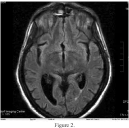
spine degenerative disc/joint disease, reduction of lordot-ic curve and moderate anterior head carriage. The neur-ologist ordered first a magnetic resonance image (MRI) of the brain (see Figure 1 and 2), which revealed mild ventricle enlargement due to ex vacuo dilatation second-ary to the diffuse, primarily parieto-occipital cortical atro-phy. In addition, a positron emission tomography (PET) study was performed, which indicated lack of radiotracer uptake in the parieto-occipital cortices bilaterally, more pronounced and with a larger volume of involvement on the right (see Figure 3). This study was performed to de-termine the areas of the brain that were not functioning properly. The clinical diagnosis of posterior cortical atro-phy (PCA) was accomplished after a thorough investi-gation, with corroborative findings from the neurologist consult. Communication between the neurologist and the chiropractic physician regarding exchange of records, treatment and outcomes occurred several times. The neur-ologist recommended daily dosage of Donepezil, a com-mon medication prescribed for Dementia-like symptoms, which is standard of care for PCA, and regular follow-up regarding the condition’s prognosis. The patient decided to adhere to the suggestion, and also pursue chiropractic
Figure 1.
Coronal plane; Brain magnetic resonance imaging (MRI) revealing enlarged ventricles
Figure 2.
Transverse plane; Brain magnetic resonance imaging (MRI) revealing enlarged ventricles
Figure 3.
Brain positron emission tomography (PET) scan with FGD radiotracer. Brain positron emission tomography
manipulative therapy and musculoskeletal rehabilitation hoping to enhance his neuromuscular function.
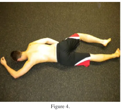
Our treatment consisted of manual medicine and re-habilitation procedures focused on the neuromuscular features of PCA, in a 42-week period initially, and then another 13-week period. Frequency and duration of treat-ments were variable throughout these periods. At times, visits were twice a week, others once every two weeks due to conflicting schedule. Treatment visits consisted of the use of vibratory stimuli therapy over the distal extrem-ities (to enhance vibratory and proprioceptive input to the CNS), with the use of a 128htz tuning fork pre and post high-velocity low-amplitude (HVLA) spinal manipula-tion. Proprioceptive retraining exercises standing on bal-ance training ball performing finger-nose exercise and catching various size objects was also incorporated. DNS therapy was performed in the supine and prone position, with 2 or 3 doctors asking the patient to perform specific motor pattern motions, average approximately 12 minutes (see Figure 4, 5 and 6). At-home therapeutic exercises for thoracic extension, and hamstrings/adductor stretch-hold exercises were encouraged.
Progress evaluation focused on functional improve-Figure 4.
Dynamic Neuromuscular Stabilization (DNS) procedure utilizing crawl position 1 with treatment points. This is
not the actual patient, rather only a model illustration about the procedure sequence.
Figure 5.
Dynamic Neuromuscular Stabilization (DNS) procedure utilizing crawl position 2 with treatment points. This is
not the actual patient, rather only a model illustration about the procedure sequence.
Figure 6.
Dynamic Neuromuscular Stabilization (DNS) procedure utilizing crawl position 3 with treatment points. This is
ments (FI) involving activities such as the ability to drive, dress, perform household chores, climb stairs, play golf, motor behavior, and overall global health status. The response to the DNS therapy, and proprioceptive exercises was used every visit to monitor progress. In a 10-month period, the Health Status Questionnaire (HSQ) demonstrated a 38% functional improvement. Within a 13-month period, HSQ and Back Bournemouth Question-naire (BBQ) demonstrated a 60% improvement, which is extremely significant regarding musculoskeletal pain and FI. These two outcome measurements are a short, self-re-ported questionnaire that measures objective improve-ments with different dimensions in patients with muscu-loskeletal pain, aiming pain and disability, but also takes the affective and cognitive aspects of pain into account. They are well known for their validity, reliability and re-sponsiveness. Likewise, functional impairments previous noted, such as inability to play golf, self-care (dressing), climbing stairs and motor control were enhanced. Better control of involuntary motion, right and left discrimina-tion, and refined motor skills for reaching objects were observed. No progression in memory loss, space orienta-tion, and speech disturbances was observed.
Significant functional improvement of neuromuscu-loskeletal control and pain was accomplished. Altered movement patterns between agonists and antagonists sec-ondary to dysfunctional sensory inputs were restored with improvement in the synergistic function between differ-ent muscle groups and fasciae. In this case, the integration of chiropractic care with DNS resulted in a number of remarkable musculoskeletal restorations, with improved mechanics and tolerance to activities of daily living. The patient was encouraged to continue with conservative care, in addition to daily routine of exercise, and active rehabilitative program.
Discussion
PCA is a progressive, debilitating, condition character-ized by difficulties in visuospatial tasks, writing, and
motor control.6 The loss of cognitive motor function
related to PCA, and the presence of prominently asym-metric limb apraxia, illustrates an uncommon feature of
this syndrome.7 Musculoskeletal examination in most
cases is unremarkable; however, severe progressive vis-ual and cognitive findings are common. According to Kas and colleagues8, the visual symptoms are perhaps more
likely to be detected than other deficits due to its greater impairment level, nevertheless, neuromuscular dysfunc-tions are equally as important to address. Because of poor awareness of PCA by physicians, patients usually receive incomplete care. Adjunctive rehabilitative treatment is considered fundamental for individuals with cognitive and motor disturbances. Special efforts must be centered on the neuromuscular features of PCA, and appropriate management with manual medicine, rehabilitation, exer-cises and cognitive therapy is recommended as an option for care, aiming to enhance quality of life, and improve neuromusculoskeletal functional.9 According to Kolar10, influenced by the work of Vojta, Dynamic Neuromuscu-lar Stabilization may be considered an adjunctive therapy in such cases to help enhance specific functions of the musculoskeletal system. Such an approach was chosen by the clinician as the most suitable therapy to facilitate passive movement patterns in this case, while enhancing primitive subcortical kinesiological patterns to stimulate appropriate neuromuscular function.
ipsilateral pain syndrome, generalized spinal stiffness and deconditioning syndrome.11,12,13
As evidenced by the present case, this clinical pres-entation of PCA was associated with musculoskeletal dysfunctions and asymmetric cortical modulation. The patient’s presentation with ipsilateral flexor angulation of upper limb, extensor angulation of lower limb, spinal stiffness, deconditioning, and imbalance motor patterns supports the application of manual medicine and rehabili-tation as adjunctive therapeutic procedures. SMT and DNS were utilized focusing on movement pattern analy-sis. According to Vacek14 if the central nervous system (CNS) is not functioning normally, the postural program will change and kinesiological analysis of postural activ-ity and re-actions can be used to evaluate the CNS. Also, mobilization and manipulation of spinal joints and fascia may influence global movement patterns allowing higher levels of cortical function, stimulating appropriate cortex output through balanced peripheral afferent stimuli to the PMRF.11 Other therapeutic procedures included inhibitory stretching protocols to the facilitated muscles as described in the upper and lower cross syndrome, as well as facili-tating exercises to increased activity in the inhibited mus-cles.13 Balance, vibration therapy over the extremities and proprioception exercises using solid ground and 2-way wobble boards were used with eyes open and closed, to stimulate appropriate cortical awareness of propriocep-tive and cerebral pathways.14,15
According to several studies11,13,16,17, a number of path-ways may explain the potential effects of manual medi-cine procedures on the neuraxis, and therefore theoretic-ally corroborate the positive neuromuscular improve-ments in this case. These include, but are not limited to, exciting spinoreticular pathways and dorsal column path-ways to the PMRF, modulation of vestibulosympathetic pathways and vestibulocerebellar activation of the nucle-us tractnucle-us solitarinucle-us, dorsal motor nuclenucle-us of vagnucle-us, and nucleus ambiguous. Yet, spinal manipulation and affer-ent peripheral stimulation may result in brain hemisphere influence via descending excitation of PMRF pathways, inhibitory control of IML cell column, which may alter central integration of the brain stem and hypothalamus via spinoreticular and spinothalamic afferent direct con-nections.11 14-19 These manual medicine procedures tend to facilitate global stability and enhance musculoskeletal functional patterns addressing a number of anatomical
structures that help maintain appropriate neuromuscular stimulation. Mechanoreceptors, proprioceptors, Golgi tendon organs, muscle spindles, and her sensory organ are all directly influenced by mobilization and directive manual therapy approach.20-23 Ultimately, extensive sen-sory input into the CNS contributes to the neuromuscular adaptation demanded by the cortex needs in its process of reorganization. Hence, manual medicine procedures play a fundamental role in adding sensory stimulation to per-ipheral receptors, known to influence motor response pat-terns, which directly affect posture, balance, locomotion, and musculoskeletal function.24-26
The manual medicine procedures used in this case im-proved the neuromuscular function and quality of life of this patient. Yet, no changes in memory, visual spatial or writing ability were seen. Furthermore, the results of this case report, and likewise cases 27, 28, where similar proced-ures were utilized, supports the value of such procedproced-ures in the co-management of neurological conditions asso-ciated with musculoskeletal dysfunctions. Chiropractic, DNS, and adjunctive therapies can help manage neuro-muscular compensations and altered movement patterns, secondary to improper spinal mechanics, and dysfunc-tional sensory inputs between muscle groups focusing in enhancing structural mobility, postural integrity and en-hance neuromuscular function.29,30 Our clinical approach for the management of musculoskeletal dysfunction, such as in this case, emphasizes the utilization of manu-al stimulation of specific zones of the body by properly mobilizing/manipulating joints to evoke predetermined efferent motor patterns by the central nervous system, facilitating sensory input to enhance cortex assimilation, and appropriate re-organization or adaptation.
Limitations
This report has the limitations of all case reports that rep-resent the experience of a single patient. Therefore, find-ings may not be generalizable to other patients.
Conclusion
con-sidered essential for successful results. Chiropractic, DNS and other procedures were utilized in this case. There was no progression in memory loss, speech or visuospatial orientation noted, though significant muscu-loskeletal improvements were achieved, as endorsed by an improvement in 60%, according to the HSQ, and BBQ outcome assessment tools, especially in quality of life, and musculoskeletal function. Our goal with this study is to increase multidisciplinary management awareness, and include chiropractic care in future research consid-ering its contributory use for functional enhancement in the co-management of neurological disorders with mus-culoskeletal dysfunctions.
Acknowledgments
We would like to thank Dana Lawrence, DC, MMedED, MA for his comments and review of this article.
References
1. Benson DF, Davis RJ, Snyder BD. Posterior cortical atrophy. Arch Neurol. 1988; 45:789-793.
2. Renner JA, Burns JM, Hou CE et al. Progressive posterior cortical atrophy dysfunction: a clinicopathologic series. Neurology. 2004; 63:1175-1180.
3. Crutch SJ, Lehmann M, Schott JM et al. Posterior cortical atrophy. Lancet Neurol. 2012; 11:170-78.
4. Migliaccio R, Agosta F, Rascovsky K et al. Clinical syndromes associated with posterior atrophy: early age at onset AD spectrum. Neurology. 2009; 73:1571-1578. 5. Mendez MF, Ghajarania M, Perryman KM. Posterior
cortical atrophy: clinical characteristics and difference compared to Alzheimer’s disease. Dement Geriatr Cogn Disord. 2002: 14:33-40.
6. McMonagle P, Deering F, Berliner Y et al. The cognitive profile of posterior cortical atrophy. Neurology. 2006; 66:331-338.
7. Tang-Wai DF, Josephs KA, Boeve BF et al. Visuospatial dysfunction as the presenting feature in corticobasal degeneration. Neurology. 2003; 61:1134-1135.
8. Kas A, de Souza LC, Samri D et al. Neural correlates of cognitive impairment in posterior cortical atrophy. Brain. 2011; 134:1464-78.
9. Roca M, Gleichgerrcht E, Torralva T et al. Cognitive rehabilitation in posterior cortical atrophy Neuropsychol Rehabil. 2010; 20:528-40.
10. Kolar P, Kobesova A. Three levels of motor control in the assessment and treatment of the motor system. J Bodyw Mov Ther. 2014; 18:23-33.
11. Beck RW. Fundamental concepts in functional
neurology. In: Functional Neurology for practitioners of
manual therapy. Churchill Livingtstone: Philadelphia, 2009: 1-20.
12. Savic I, Pauli S, Thorell JO et al. In vivo demonstration of altered benzodiazepine receptor density in patients with generalized-epilepsy. J Neurology, Neurosurgery and Psychiatry. 1994; 57:784-797.
13. Nyberg-Hansen R. Sites and mode of termination of reticulospinal fibers in the cat. An experimental study with silver impregnation methods. J Comparative Neurology. 1965; 124:74-100.
14. Vacek J. Sensory-motor approach to the stabilization system of the spine in patient with chronic back pain. International Musculoskeletal Medicine. 2012; 34:48-50. 15. Page P, Frank C, Lardner R. Assessment and Treatment
of Muscle Imbalance: The Janda Approach. 2009. Human Kinetics.
16. Rojas Vegas S, Abel T, Lindschulten R et al. Impact of exercise on neuroplasticity-related proteins in spinal cord injured humans. Neuroscience. 2008; 20:40-50.
17. Garcia-Larrea L, Maarrawi J, Peyron R et al. On the relation between sensory deafferentation, pain and thalamic activity in Wallenberg’s syndrome: a PET-scan study before and after motor cortex stimulation. Eur J Pain. 2006; 10:677-88.
18. Holt K, Beck RW, Sexton S. Reflex effects of a spinal adjustment on blood pressure. Proceeding of the Association of Chiropractic Colleges: Research agenda conference 2006. Washington, DC.
19. Carrick FR. Changes in brain function after manipulation of the cervical spine. J Manipulative Physiol Ther. 1994; 20:529-545.
20. Page SJ, Gater DR, Bach-Rita P. Reconsidering the motor recovery plateau in stroke rehabilitation. Arch Phys Med Rehab. 2004; 85:1377-1381.
21. Panjabi MM. A hypothesis of chronic back pain: ligament sub failure injuries lead to muscle control dysfunction. Eur Spine J. 2006; 15:668-676.
22. Karni A, Mayer G, Jezzard P et al. Functional MRI evidence for adult motor cortex plasticity during motor skill learning. Nature. 1995; 377:155-158.
23. Krakauer J. Motor learning: its relevance to stroke recovery and neurorehabilitation. Current Opinion in Neurology. 2006; 19:84-90.
24. Rome L. Neurovertebral influence upon the autonomic nervous system: some of the somato-autonomic evidence to date. Chiropr J Austr. 2009; 39:9-33.
25. Rome L. Neurovertebral influence upon the autonomic nervous system: some of the somato-autonomic evidence to date – part II: somatovisceral. Chiropr J Austr. 2010; 39:9-33.
26. Dishman DJ. Spinal reflex attenuation associated with spinal manipulation. Spine. 2000; 25:2519-2525. 27. Oppelt M, Juehring DD, Sorgenfry G et al. A case study
stabilization care to enhance function of a post cerebrovascular accident patient. J Bodyw Mov Ther. 2014; 18:17-22.
28. Juehring DD, Barber MR. A case study utilizing Vojta/ Dynamic Neuromuscular Stabilization therapy to control symptoms of a chronic migraine suffer. J Bodyw Mov Ther. 2011; 15:538-41.
29. Colloca CJ, Keller TS, Gunzburg R et al.
Neurophysiological response to intraoperative lumbosacral spinal manipulation. J Manipulative Physiol Ther. 2000; 23:447-457.