R E S E A R C H
Open Access
The subunits of the S-phase checkpoint complex
Mrc1/Tof1/Csm3: dynamics and interdependence
Sonya Dimitrova Uzunova
†, Alexander Stefanov Zarkov
†, Anna Marianova Ivanova, Stoyno Stefanov Stoynov
and Marina Nedelcheva Nedelcheva-Veleva
*Abstract
Background:The S-phase checkpoint aims to prevent cells from generation of extensive single-stranded DNA that predisposes to genome instability. TheS. cerevisiaecomplex Tof1/Csm3/Mrc1 acts to restrain the replicative MCM helicase when DNA synthesis is prohibited. Keeping the replication machinery intact allows restart of the replication fork when the block is relieved. Although the subunits of the Tof1/Csm3/Mrc1 complex are well studied, the impact of every single subunit on the triple complex formation and function needs to be established.
Findings:This work studies the cellular localization and the chromatin binding of GFP-tagged subunits when the complex is intact and when a subunit is missing. We demonstrate that the complex is formed in cell nucleus, not the cytoplasm, as Tof1, Csm3 and Mrc1 enter the nucleus independently from one another. Viain situchromatin binding assay we show that a Tof1-Csm3 dimer formation and chromatin binding is required to ensure the attachment of Mrc1 to chromatin. Our study indicates that the translocation into the nucleus is not the process to regulate the timing of chromatin association of Mrc1. We also studied the nuclear behavior of Mrc1 subunit in the process of adaptation to the presence hydroxyurea. Our results indicate that after prolonged HU incubation, cells bypass the S-phase checkpoint and proceed throughout the cell cycle. This process is accompanied by Mrc1 chromatin detachment and Rad53 dephosphorylation.
Conclusions:InS. cerevisiaethe subunits of the S-phase checkpoint complex Mrc1/Tof1/Csm3 independently enter the cell nucleus, where a Tof1-Csm3 dimer is formed to ensure the chromatin binding of Mrc1 and favor DNA replication and S-phase checkpoint fork arrest. In the process of adaptation to the presence of hydroxyurea Mrc1 is detached from chromatin and Rad53 checkpoint activity is diminished in order to allow S-phase checkpoint escape and completion of the cell cycle.
Keywords:Cell cycle, Protein localization, Adaptation, In situ chromatin binding assay, Mrc1, Tof1, Csm3
Background
The activation of the S-phase checkpoint aims to pre-serve genome stability when an impediment for strict DNA synthesis arises. It turns on a cascade of events and results in replicational block that provides time for the repair systems to eliminate the problem. Then the ordinary dynamics of the cell cycle is restored and DNA synthesis and segregation completed. A complex of three
proteins, named in S. cerevisiaeTof1/Csm3/Mrc1, plays
a critical role in that process. Those proteins are
con-served among organisms. Tof1’s familiar orthologs are
Swi1 in S. pombe and Timeless (Tim1) in human.
Csm3’s orthologs are Swi3 for fusion yeast and Tipin for
H. sapiensand Mrc1’s are respectively Mrc1 inS. pombe
and Claspin in higher eukaryotes [1-5]. Today they are categorized as S-phase checkpoint mediators [2]. Media-tors act as protein bridges (platforms) that bring to-gether sensor or effector kinases, functioning afterwards in the signal cascades. The three proteins are found to co-localize at normal and stalled replication forks [5,6]. They co-precipitate together in dynamic and stalled rep-lication forks [7], suggesting that the three proteins form a complex. Tof1, Csm1 and Mrc1 also co-precipitate with subunits of the MCM replicative helicase both from exponentially growing culture and arrested by hydro-xyurea (HU) one [7-9]. The synthetic lethality between * Correspondence:[email protected]
†Equal contributors
Institute of Molecular Biology“Roumen Tsanev”, Bulgarian Academy of Sciences, 21“Acad. George Bonchev”Str., 1113 Sofia, Bulgaria
deletions of tof1, csm3or mrc1 and mutations in
poly-merase α/primase complex, shows that the products of
these genes are interdependent in order to guarantee the correctness of the replication process [7]. These depen-dencies suggest that the Tof1/Csm3/Mrc1 complex aims to keep together the polymerase and helicase in order to prevent lethality of cells, when DNA synthesis is compromised.
It was shown that lack of Mrc1 leads to MCM-Cdc45 and Pol ε separation [9-11]. A function to stabilize the forks for Mrc1 and its orthologs was suggested [11,12].
Another S. cerevisiae protein – Ctf4 (Mcl1 in S. pombe
and And1 in X. laevis and H. sapiens) [13-17] was also
found to be involved in cell cycle progression and sister chromatide cohesion [18,19]. It physically interacts with
GINS and Pol α at replication progressing complex
(RPC) [20-25]. Recent data demonstrate that the
ortho-log of Ctf4 - And1 from Xenopus egg extract binds
Tipin, and their binding is necessary for the stable Polα association to chromatin under unchallenged conditions [26]. Indirect binding to Mrc1 was also revealed [27] and was demonstrated that this binding is sufficient for E3 ubiquitine ligase SCFDia2 association to the replica-tion complex.
Another important actor to regulate replication fork progression is the F-box protein Dia2. It is known that Dia2 interacts with many replication proteins, such as MCMs and GINS [13,27]. Recently, its function was as-sociated with its ability to form a complex with the modular ubiquitin ligase SCF (Skp1/cullin/F box) [28]. A physical interaction between Dia2 and Mrc1 and Ctf4
was demonstrated and was shown that SCFDia2
des-tabilizes Mrc1 and Ctf4 in a proteasome-dependent manner [29].
Other key actors in the process of S-phase checkpoint activation are the sensor kinase Mec1 (in a complex with Ddc2) and the effector kinase Rad53 [30-32]. Mrc1 is a substrate for Mec1 and is known to directly interact with Rad53 [33]. At first, in Rad53 independent manner, Mrc1 is Mec1 phosphorylated [34]. Thus, Mrc1 becomes competent to bind Rad53 and predisposes it to Mec1 phosphorylation [35]. The activated Rad53 additionally phosphorylates Mrc1. Although its fundamental function is to bring together Rad53 and Mec1 kinases in order to turn the S-phase checkpoint and to stabilize the replica-tion forks [35-37], mrc1 deletion mutation is not lethal. When knocked out, its function seems to be taken by the specific checkpoint mediator Rad9 [38]. Nevertheless, stalled forks restart much harder inmrc1Δcells when HU is removed from the media, suggesting a role for Mrc1 to promote stable fork-pausing complex formation and to guarantee recovery after fork arrest [39]. Tourriere and co-workers also demonstrate that in mrc1Δ cells, as well
as in tof1Δ mutants, the S-phase seems to be about
20 min longer, compared to the wild type (WT) yeast cells, most probably as a result of the approximately 40% slower progression of the replication fork. Claspin depletion also affects fork progression rates in human cell lines [40]. Al-though the rate of fork progression seems to be reduced intof1Δas well, it is less pronounced than that inmrc1Δ.
The absence of Tof1 seems to reflect much stronger on the pausing of the replication forks at the rDNA replica-tion fork barrier (RFB), protein-DNA barriers sites at the tRNA promoters and centromeres [39,41]. In contrast, in
tof1Δand inmrc1Δyeast cells, fork stalling is significantly increased at inverted repeat (IR) provoked hairpins than in WT cells [42]. The authors suggest that both Tof1 and Mrc1 counteract replication fork stalling at such DNA secondary structures.
All the above data indicate the key role of the Tof1/ Csm3/Mrc1 complex for normal replication fork move-ment and the establishmove-ment and regulation of the S-phase checkpoint. Our study examines the importance of every protein of Tof1/Csm3/Mrc1 complex for the nuclear localization and consequent chromatin binding of the other two. The specific role of Mrc1 in the process of adaptation to reduced nucleotide levels is also studied.
Results
Independent nuclear localization of the subunits of the Tof1/Csm3/Mrc1 complex
As our study aims to examine the interdependences of the
S. cerevisiaeS-phase checkpoint proteins Tof1, Csm3 and Mrc1 with regard to their nuclear localization, we first carried out sequence analysis of those proteins for pre-dictive Nuclear Localization Signals (NLS). The NLS is a sequence on the surface of a protein that is used to target the protein to enter the cell nucleus. We used the ‘PredictNLS’software [43] that is located at https://rostlab. org/owiki/index.php/PredictNLS. The analysis of the three examined proteins revealed that only Tof1 possesses hypothetical NLS (KKDKRKRRK), starting at the 1013th amino acid. According to the program, this NLS is com-mon for 28 proteins from various organisms, all of them located in nucleus. This prediction data suggest that Tof1 might be the leading protein to target the complex into the nucleus.
As Tof1 is the only one of the three that possesses canonical NLS, we checked whether it is the protein that is responsible for the nuclear localization of Csm3
and consequently Mrc1 inS. cerevisiae. To do that, we
used GFP-tagged proteins [Invitrogen™; [44]], shortly
named TOF1-GFP, CSM3-GFP and MRC1-GFP (See
Methods).
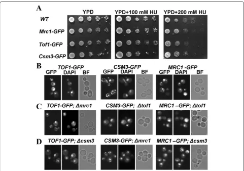
revealed viability comparable to the wild-type control (Figure 1A). The GFP-tagged strands also revealed ability to withstand chronic exposure to two different concen-trations of the S-phase checkpoint inducing agent HU, similar to that of the wild-type cells (Figure 1A). Then, as-suming that the three GFP-tagged strains function as their untagged versions, we used them to delete a gene coding a partner subunit of the S-phase checkpoint complex Tof1/Csm3/Mrc1 that is not GFP-tagged (See Table 1, Methods). As a result, a full set of deletion mutants of the complex’s subunits was achieved. We will refer to those
strains as:TOF1-GFP; csm3Δ,TOF1-GFP; mrc1Δ,
CSM3-GFP; tof1Δ, CSM3-GFP; mrc1Δ, MRC1-GFP; tof1Δ and
MRC1-GFP; csm3Δ. Asynchronous, exponentially growing cells from the constructed strains, as well as the initial GFP strains without deletions, were paraformaldehyde fixed and subjected to fluorescent microscopy analysis to detect the position of GFP-tagged proteins in the cell.
DAPI DNA staining was used for all of the probes to visualize the position of nucleus. Data were documented and analyzed for all of the examined strains (Figure 1B, C,
D). As was expected, the control TOF1-GFP,CSM3-GFP
and MRC1-GFP strains revealed co-localization of DAPI and GFP signals, indicative of nuclear localization of the respective subunit. Interestingly, all other strains - TOF1-GFP; csm3Δ, TOF1-GFP; mrc1Δ, CSM3-GFP; tof1 Δ,
CSM3-GFP; mrc1Δ, MRC1-GFP; tof1Δ and MRC1-GFP;
csm3Δ, also revealed co-localization of their GFP and
DAPI signals (Figure 1), suggestive of nuclear localization of the three subunits, regardless of the lack of their part-ners. Additionally, neither of the studied strains with dele-tions revealed cytoplasmic accumulation of a GFP-tagged protein. These results demonstrate that inS. cerevisiaethe three subunits of the Tof1/Csm3/Mrc1 S-phase check-point complex are independent with regard to their nu-clear translocation.
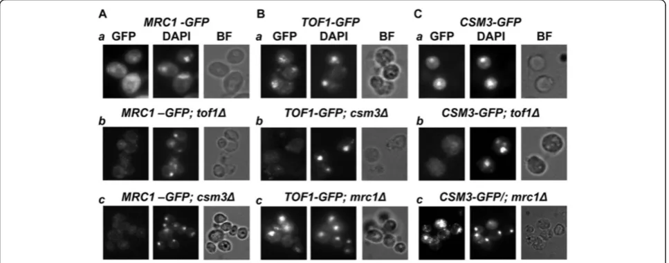
Interdependence of the subunits of the Tof1/Csm3/Mrc1 complex for their chromatin binding
Next we studied the interrelations of the three subunits for the chromatin assembly of the Tof1/Csm3/Mrc1 complex. In order to develop a whole cell study that can permit direct visualization of chromatin attached pro-teins, we used the same set of strains and carried out a “soft wash”by TritonX-100 detergent of partially
sphero-plasted S.cerevisieae cells (Methods). The most
im-portant step in that procedure was to determine the percentage of detergent to use. First we treated the cells with 100 mM HU for 3 hours to ensure that the three
studied proteins are chromatin bound. Then we tested various amounts of TritonX-100 on every GFP-strain without deletion. The maximum percentage of detergent that does not detach the protein from chromatin and re-veal nuclear GFP signal was used for further studies. For
MRC1-GFP, 0.5% w/v of detergent proved to be
appro-priate and for TOF1-GFP and CSM3-GFP – 3.0%. w/v
(Figure 2). As a positive control, a strain, carrying HTB2
protein (S. cerevisiae histone H2B) fused with mCherry
was also subjected to the same TritonX-100 washing procedure (Additional file 1: Figure S1). This method permitted us to perform simple multichannel fluorescent Table 1 List ofS. cerevisiaestrains used in this study
Strains (ORF name) Genotype Source
TOF1-GFP(YNL273W) MATa his3Δ1 leu2Δ0 met15Δ0 ura3Δ0 tof1-GFP::His3MX6 Invitrogen™
MRC1-GFP(YCL061C) MATahis3Δ1 leu2Δ0 met15Δ0 ura3Δ0 mrc1-GFP-HIS3MX6 Invitrogen™
CSM3-GFP(YMR048W) MATahis3Δ1 leu2Δ0 met15Δ0 ura3Δ0 csm3-GFP-HIS3MX6 Invitrogen™
RAD53-GFP (YPL153C) MATa his3Δ1 leu2Δ0 met15Δ0 ura3Δ0 rad53-GFP-HIS3MX6 Invitrogen™
TOF1-GFP; csm3Δ MATahis3Δ1 leu2Δ0 met15Δ0 ura3Δ0 tof1-GFP-HIS3MX6 csm3Δ::KanMX This study
TOF1-GFP; mrc1Δ MATahis3Δ1 leu2Δ0 met15Δ0 ura3Δ0 tof1-GFP-HIS3MX6 mrc1Δ::KanMX This study
CSM3-GFP; tof1Δ MATahis3Δ1 leu2Δ0 met15Δ0 ura3Δ0 csm3-GFP-His3MX6 tof1Δ::KanMX This study
CSM3-GFP; mrc1Δ MATahis3Δ1 leu2Δ0 met15Δ0 ura3Δ0 csm3-GFP-HIS3MX6 mrc1Δ::KanMX This study
MRC1-GFP; csm3Δ MATahis3Δ1 leu2Δ0 met15Δ0 ura3Δ0 mrc1-GFP-HIS3MX6 csm3Δ::KanMX This study
MRC1-GFP; tof1Δ MATahis3Δ1 leu2Δ0 met15Δ0 ura3Δ0 mrc1-GFP-HIS3MX6 tof1Δ::KanMX This study
HTB2-mCherry SLJ3517 (MATα, htb2::HTB2-mCherry-HYGMX) Sue L. Jaspersen
microscopy and direct visualization of the insoluble, chromatin bound GFP-tagged proteins. DAPI staining of the paraformaldehyde fixed cells was carried out. The match of GFP and DAPI signals was analyzed as an indi-cator of chromatin binding of the GFP-fused protein. As the treatment with TritonX-100 of partially sphero-plasted yeast cells leads to cell shape deformations, the compactness of the achieved DAPI signal was also repre-sentative of nuclear integrity.
The microscopy revealed that whentof1orcsm3genes
are deleted (in CSM3-GFP; tof1Δ or TOF1-GFP; csm3Δ
strains respectively), the reciprocal binding partner was not attached to chromatin (Figure 2), showing that Tof1 and Csm3 are interdependent for their chromatin bin-ding. In contrast, such a dependence of Tof1 and Csm3 on Mrc1 was not observed. After TritonX-100 wash of the soluble proteins and fluorescent microscopy of the
TOF1-GFP; mrc1Δ and CSM3-GFP; mrc1Δ strains, the examined GFP signals coincided with the corresponding DAPI signals (Figure 2). These results show indepen-dence of Tof1-Csm3 dimer chromatin binding on Mrc1. In contrast, Mrc1 required intact Tof1-Csm3 complex in order to associate to chromatin (Figure 2).
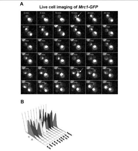
Mrc1 is positioned in the nucleus throughout the cell cycle
The three studied proteins co-precipitate together. Inte-restingly, Tof1 and Csm3 co-precipitate in
stoichiomet-ric amounts, but Mrc1 (as well as MCM’s complex
subunits) is in substoichiometric amounts [7], suggesting that Mrc1 is not constantly attached to Tof1-Csm3 dimer. As the function of Mrc1 is assumed to be re-stricted to S-phase, when it is DNA bound [33], we checked whether it is positioned in cell nucleus during that phase of the cell cycle only. To check this possibil-ity, we examined the nuclear localization of Mrc1 during the cell cycle.S. cerevisiaeexponentially growing MRC1-GFPcells were subjected to time-lapse live cell imaging. The obtained results indicated that Mrc1 fluorescent sig-nal is detected in the cell nucleus during the entire cell cycle (Figure 3A, B). These data show that translocation into the nucleus is not the key process to restrict the attachement of Mrc1 to Tof1-Csm3. Probably, some other mechanisms, such as protein-protein and/or pro-tein DNA interactions, as well as degradation of Mrc1 are the responsible mechanisms, which control its com-plex binding and S-phase functions.
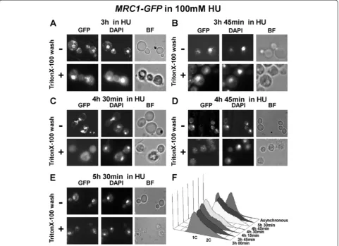
Mrc1 is removed from chromatin when the S-phase checkpoint is overcome in the presence of HU
Tof1, Csm3 and Mrc1 are all necessary for normal fork progression as well as stable checkpoint fork arrest [8,45], but just Mrc1 is sufficient to guarantee recovery after fork arrest [46]. It was shown that replication forks
reveal restart difficulties after HU block in mrc1Δ cells [39]. As Mrc1 is important for stable fork arrest, we de-cided to check for alterations in the nuclear behavior of Mrc1 in the process of adaptation. The adaptation is a process of overcoming the S-phase checkpoint and is noticed to take place when cells are subjected to per-sistent agent treatment or impossibility to repair specific DNA damage.
MRC1-GFPcells were treated with 100 mM HU. Sam-ples, starting from the third hour, from indicated time points, were taken and either fixed straight away or washed with TritonX-100 before fixing. The fluorescent microscopy results indicated the presence of Mrc1 at chromatin until 3 h 45 min (Figure 4A, B). Interestingly, the next samples, taken at 4 h 30 min and 4 h 45 min (Figure 4C, D), indicated that Mrc1 was still located in cell nucleus (although its amount seemed to be dimi-nished) but removed from chromatin. Then, after 5 h 30 min from HU addition (Figure 4E), Mrc1 protein seemed to reappear at chromatin. The flow cytometry analysis, carried out with cell probes from the same time points, indicated a small shift towards two contents of DNA (Figure 4F) after 4 h 30 min, suggesting that the S-phase checkpoint arrest had been by-passed and that the yeast cells had overcome the ribonucleotide reduc-tase inhibition.
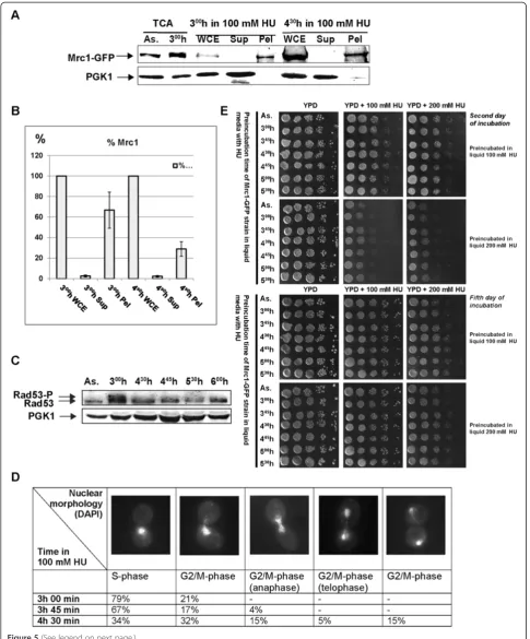
The observed Mrc1 chromatin diminishment was also detected by bulk chromatin fractioning assay (Figure 5A). The Western blot band analysis of that experiment re-vealed that the amount of chromatin bound Mrc1-GFP protein from 4 h 30 min time point, relative to the pro-tein value from the whole cell extract of the same time-point, indicated 42% diminishment, compared to the same correlation from the 3 h 00 min in 100 mM HU (Figure 5B).
To ensure that cells that proceed throughout the cell cycle in the presence of HU had overcome the S-phase checkpoint arrest, we analyzed the Rad53 kinase. We carried out Western blot analysis of total protein
ex-tracts from 100 mM HU treated yeast MRC1-GFPcells
(Figure 4G). The immunodetection indicated (Figure 5C) that as expected, at the third hour Rad53 appeared to be in hyperphosphorylated state that corresponds to S-phase checkpoint activation. The samples taken later on show that the amount of phosphorylated Rad53 seemed to significantly decrease. Both detachment of Mrc1 from chromatin and the decrease of phosphorylated Rad53 are indicative for S-phase checkpoint escape.
Confirmatory of that S-phase checkpoint escape are
the results we obtained from Mrc1-GFP yeast nuclear
of the counted cells revealed S-phase characteristic cel-lular and nuclear morphology. In contrast, at 4 h and 30 min time point, that percentage dropped to 34%. This shows that 45% of the cells had exited the S-phase, a value close to the calculated 42% decrease of chromatin bound Mrc1. The rest of the cells indicated G2/M-phase
related morphology, indicating that cells were no longer S-phase arrested and are trying to continue the cell cycle (Figure 5D). In addition, to visualize the behavior of studied proteins during S-phase checkpoint escape on a single cell level, we carried out time-lapse live cell
mi-croscopy of Mrc1-GFP and Rad53-GFP strains in the
presence of 100 mM HU (Additional file 1: Figure S2B and C and Additional file 2: Movie S2 and Additional file 3: Movie S4). The results from those experiments confirmed that cells continue their cell cycle progression in the presence of HU, but the duration of the cell cycle was much longer (compare Additional file 1: Figure S2A with S2B and Figure S2C with Figure 3). Movies of all time-lapse experiments are available in the (Additional file 2: Movie S2, Additional file 3: Movie S4, Additional file 4: Movie S1 and Additional file 5: Movie S3).
To check whether cells can steadily surmount the HU provoked nucleotide deficiency, a HU-viability test was
carried out. Samples from MRC1-GFP cells,
prelimi-narily arrested with HU (100 mM HU and 200 mM HU), were taken at indicated time points and grown on plates with or without HU (100 or 200 mM respectively). All samples indicated a visible cell growth (Figure 5E). Not surprisingly, a difference in growth was observed between cells, which after HU preincubation were plated
on YPD containing HU and those – on plates without
HU (best visualized after two days of incubation –
Figure 5E). The results show that all samples, even those, incubated on plates containing 200 mM HU, continued to grow, demonstrating that cells had surmounted the new conditions.
Discussion
Our study aimed to estimate the interdependencies of the three proteins with regard to compartmentalization of complex assembly. Recent data from other eukaryotes lead to the idea that Tof1/Csm3/Mrc1 complex is as-sembled in cell cytoplasm. Tanaka and co-workers
de-monstrated in S. pombe that the amount of Mrc1-GFP
nuclear signal is significantly reduced when Swi1 or Swi3 is deleted [48]. The authors suggest that the Swi1 and Swi3 are important for Mrc1 nuclear localization and consequent DNA binding. Their results presume that a dimer of Tim-Tipin is assembled in cytoplasm
and is responsible for the nuclear translocation of Mrc1. In HeLa cells, when Tim or Tipin were knocked-down, the respective binding partner was relocated to cyto-plasm. In addition, the amount of that partner was sig-nificantly reduced [49]. Such dependence for Claspins’s amount was not found for asynchronous growing cells. But when Tim or Tipin siRNA treated cells were sub-jected to HU, the amount of nuclear Claspin seemed to diminish. The authors suggest that Tim and Tipin facili-tate Claspin nuclear localization under replication stress. To check whether the complex is formed in the cyto-plasm inS. cerevisiae, we studied the cellular localization of GFP-tagged subunits of Tof1/Csm3/Mrc1, when the complex is intact and when a subunit is missing. As in higher eukaryotes [49], our analysis revealed a hypo-thetical NLS for Tof1, suggesting its responsibility for the nuclear translocation of the other subunits of the complex. However, in contrast to higher eukaryotes, in-dependence with regard to nuclear translocation of all three subunits was observed. Our findings demonstrate that in contrast toS. pombeand human cells, theS. cere-visiae Tof1/Csm3/Mrc1 S-phase checkpoint complex is most probably assembled in cell nucleus, as every sub-unit can enter it independently of the others. Deletion of each of the three genes did not lead to cytoplasmic accu-mulation of any partner subunit. The amounts of the undeleted, GFP-tagged proteins also seemed to be un-affected when observed under fluorescent microscope. As budding yeast is a preferred model organism for rep-lication studies, such a difference in proteins relation-ships and cell positioning control must be taken into account. As yeast cell nucleus is not disassembled during cell division, but undergoes nuclear division into two daughter nuclei, it might be speculated that those dif-ferences in the localization of complex assembly are a result of evolutionary adaptation and management of higher eukaryotes.
As Tof1, Csm3 and Mrc1 enter nucleus independently in S. cerevisiae, a question about the mechanism of
complex assembly arises. The stoichiometric interaction of Tof1 and Csm3 (in co-precipitation experiments on asynchronous cultures) indicates that by default they form a heterodimer (Nedelcheva et al. [7]). In contrast, the non- stoichiometric amount of Mrc1 suggests that it joins the complex occasionally. Is then the Tof1-Csm3 dimer formation required to ensure attachment to chro-matin and how is Mrc1 related to those relationships? In higher eukaryotes data are variable in accordance with
the model system applied in the study. TheXenopusegg
extract data show that Tipin and Claspin fail to bind to chromatin when Tim1 is depleted and that Tim1-Tipin is required for binding of Claspin but not vice versa [50]. In HeLa cells Tim and Tipin are interdependent for their chromatin binding [49], but insoluble Claspin is not af-fected by Tipin siRNA in asynchronous culture. Such dependence is noticed in HU treated cells only.
To visualize the chromatin association and depen-dencies of the subunits via a whole cell approach, we adapted the higher eukaryotes protocol for in situ chro-matin binding assay [36,37] toS. cerevisiae. This method permits performance of direct multichannel fluorescent microscopy to visualize the insoluble, chromatin bound GFP-tagged proteins. The results demonstrate the neces-sity of Tof1 for the chromatin binding of Csm3 and vice versa. Our results also show that Mrc1 is Tof1 and Csm3 dependent for its chromatin association, but in con-trast, Mrc1 is not required for the chromatin binding of Tof1 and Csm3. These data are in unison with ChIP-chip results of Bando and co-workers for the co-dependences of those proteins for association with replication forks [1].
It can be summarised that in S. cerevisiaeTof1-Csm3
initial dimer formation is required for chromatin asso-ciation. The nuclear import is not a regulatory step as every single subunit enters the nucleus independently. As a dimer Tof1-Csm3 is responsible for the chromatin binding of Mrc1.
A logical question to answer in that relation is whether the controlling mechanism for the restricted binding of
(See figure on previous page.)
Mrc1 to the Tof1 - Csm3 dimer (as co-precipitations in-dicated) is a result of cell cycle oscillations of its cellular localization. As Mrc1 known functions are restrained to S-phase of the cell cycle, when it is DNA bound [33], we checked whether it is positioned in cell nucleus during that phase of the cell cycle only. Our results indicated that there is nuclear Mrc1 during the entire cell cycle, confirming that translocation into the nucleus is not the leading process to regulate the association of Mrc1 to the S-phase checkpoint complex. A regulatory function of the timing of Mrc1’s triple S-phase complex associ-ation can be suggested, but further studies are required to elucidate the fine mechanisms of these interactions.
All three proteins Tof1, Csm3 and Mrc1 are involved in normal fork progression and stable checkpoint fork arrest [8,45], but just Mrc1 is assumed to be responsible for fork rehabilitation after fork arrest [39]. Stalled forks restart
much harder in mrc1Δ cells when HU is removed from
the media, suggesting a role for Mrc1 to promote stable fork-pausing complex formation and to guarantee reco-very after fork arrest. Some recent data connect Mrc1 with SCFDia2. This interaction was shown to be responsible for destabilization of Mrc1 in a proteasome-dependent man-ner during S-phase of the cell cycle in vivo [29]. It was also demonstrated that Dia2 contributes to Mrc1 degra-dation during S-phase checkpoint recovery [46]. As Mrc1 is important for stable fork arrest, we checked for alter-ations in the nuclear behavior of the protein in the process of overcoming the S-phase checkpoint, called adaptation. Generally, the adaptation is a process of loosening the S-phase checkpoint when cell meets impediments for coping with persistent agent or damage. The reason for such cell decision is not quite clear. It was suggested that when the cell is unable to cope with the problem, it allows restoration of the cell cycle to provide opportunities to re-pair the damage in a subsequent cell cycle, enhancing its chances for survival. The intimate mechanisms of execut-ing and controllexecut-ing this phenomenon are still vague. Some data point out the specific role of the amount of polo-like kinase CDC5 to suppress the hyperphosphorylation of Rad53 that leads to relieve of cell division arrest [51,52].
We studied the nuclear behavior of Mrc1 after a pro-longed period of incubation in the presence of HU. The fluorescent microscopy results in combination with flow cytometry data and bulk chromatin fractioning indicated that when theS. cerevisiaecell by-passes S-phase check-point arrest (after 4 h and 30 min) to proceed further into the cell cycle in the presence of the blocking agent, Mrc1 dissociates from chromatin. This finding empha-sizes the specific role of Mrc1 for keeping the stability of forks arrest. It shows that the physical presence of Mrc1 at replication pausing complex is required not only for stable fork arrest in response to S-phase checkpoint agent, but for the duration of that arrest as well.
In support of our findings, cell free studies onXenopus
egg extract in aphidicolin-induced DNA replication block show that after a prolonged interphase arrest, the extracts undergo adaptation and enter into mitosis with unfinished DNA replication. In this process Chk1 undergoes inactiva-tion and Claspin dissociates from chromatin [53].
One of the major functions of the S-phase checkpoint is to sufficiently enlarge the nucleotide pool in the cell
[54]. The key enzyme to regulate the levels of dNTPs–
ribonucleotide reductase (RNR) is regulated by the Mec1/Rad53/Dun1 kinases via two different mechanisms [55,56]. The first one aims the transcriptional induction of the RNR genes and the second results in phosphoryl-ation and removal of the RNR inhibitor Sml1 [57,58]. When HU is introduced into the media, it provokes the S-phase checkpoint activation that results in Sml1 deg-radation and free nucleotide pool enlargement. On the other hand, the HU itself is an inhibitor of RNR and ef-fects limitation of the amount of dNTPs. Therefore, the net effect of the two counteractive processes is measured and the predominant process takes control over cell fate, directing it either towards nucleotide synthesis or to-wards nucleotide synthesis inhibition. The dose of HU itself might be of major importance to target the pro-cess. In our experiments 100 mM HU was used. Our FACS analysis (Figures 3B and 4F) and the phospho-rylation of Rad53 at the third hour of HU treatment (Figure 5C) indicate that the amount of HU used pro-vokes S-phase checkpoint activation. At the same time, the live-cell imaging of Rad53-GFP and Mrc1-GFP cells (Additional file 1: Figure S2), as well as the viability test that we carried out (Figure 5E) undoubtedly indicate that yeast cells somehow succeed to survive and grow in 100 and even 200 mM HU for a long period of time. This shows that the decision for cell arrest was abolished and probably the nucleotide levels were sufficiently ad-equate to allow cell cycle restoration. Interestingly, the ordinary dynamics of the cell cycle is not absolutely restored. Alvino and co-workers [59] as well as our time-lapse live cell imaging experiments indicate that the dur-ation of the cell cycle in HU is much longer than the ordinary one. A probable explanation is that cells adapt to HU by raising the level of nucleotides to permit progres-sion of the cell cycle, but those levels remain limited and do not allow full restoration of the dynamics of the cell cycle. How the cell weighs the two opposite effects of the HU on the dNTPs pool and takes its decisions is still un-clear. Many other experimental data are required to estab-lish the mechanisms of this decision making. Our results indicate that the detachment of Mrc1 from chromatin and the diminishment of Rad53 phosphorylation give a proof of S-phase checkpoint bypass that follows that adaptation.
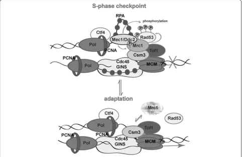
chromatin. One possible explanation for that detach-ment is the specific role of Mrc1 to prevent replicative helicase movement when the polymerase meets an obstacle for correct DNA synthesis. We hypothesize that
the pausing structure is possible when a “clutch” of
Mrc1 on MCM is present. This clutch is required for docking Mec1and Rad53 to ensure their checkpoint kinase activities, leading to fork arrest. Via some regula-tory mechanisms (perhaps by means of Polo-like kinase attachment and consequent phosphorylation) Mrc1 is dissociated from chromatin by detachment or degra-dation. The lack of Mrc1 leads to loss of Rad53/Mrc1 activity and loosens MCM helicase (Figure 6). As Mrc1 is required for normal replication fork progression, later on, when the cell division arrest has been relieved, the protein rebinds Tof1-Csm3 to ensure DNA synthesis.
Conclusions
Our findings demonstrate that in contrast to S. pombe
and human cells, the S. cerevisiae Tof1/Csm3/Mrc1
S-phase checkpoint complex is most probably assembled in cell nucleus, as every subunit can enter it indepen-dently and deletion of each of the three genes did not lead to cytoplasmic accumulation of any partner subunit.
Our data also indicates that inS. cerevisiaeTof1-Csm3 initial dimer formation is required for chromatin asso-ciation of Mrc1. This process is not controlled by the cell cycle as the protein is constantly positioned in the nucleus.
Our results indicate that in the process of adaptation to the presence of HU Mrc1 is detached from chromatin to relax Rad53 activity and thus to allow completion of the cell cycle. Our study emphasizes the specific role of Mrc1 for keeping the stability of fork arrest. It shows that the physical presence of Mrc1 at replication pausing complex is required not only for stable fork arrest in re-sponse to S-phase checkpoint agent, but also for the duration of that arrest.
Methods Strains and media
S. cerevisiae strains (Invitrogen™, Cat.# 95702) YNL273W (MATa his3Δ1 leu2Δ0 met15Δ0 ura3Δ0 TOF1-GFP-HIS3MX6), YCL061C (MATa his3Δ1 leu2Δ0 met15Δ0 ura3Δ0 MRC1-GFP-His3MX6), YMR048W (MATahis3Δ1 leu2Δ0 met15Δ0 ura3Δ0 CSM3-GFP-His3MX6) and YPL153C (MATahis3Δ1 leu2Δ0 met15Δ0 ura3Δ0 RAD53-GFP-HIS3MX6) are used [44]. We refer to those strains as
TOF1-GFP, MRC1-GFP, CSM3-GFPand RAD53-GFP, re-spectively. All are with BY 4741 background. We used YNL273W, YCL061C and YMR048W to delete a gene coding a partner subunit of the S-phase checkpoint complex Tof1/Csm3/Mrc1 that is not GFP-tagged. As a control for in situ chromatin binding assays SLJ3517 (MATα, htb2::HTB2-mCherry-HYGMX) or shortly - HTB2-mCherry, was used. All strains are listed in Table 1. Strains were cultivated in YPD medium (1% (w/v) yeast extract (Difco), 2% (w/v) Bacto peptone (Difco), 2% (w/v) dex-trose). Before fluorescent and confocal microscopy pro-cedures, yeast cells were grown in minimal medium (1.7 g/l YNB, 0.04 g/l CSM-His (Bio101, Inc.) and 2% (w/v) dextrose) to diminish the autofluorescence.
Construction of strains
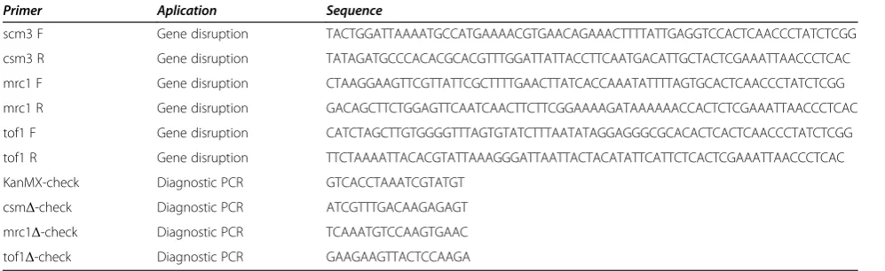
The plasmid pKS-KanMX6-1 (4571 bp) was used to PCR amplify the disruption cassettes for tof1, mrc1 andcsm3. For construction of the disruption cassettes, primers 1–6 were used (Table 2). These cassettes contained a selection marker KanMX for geneticine (G418) resistance. The dis-ruption cassettes carried 50 bp flanking sequences (intro-duced by the PCR primers) homologous to regions, which
surround the target genes. StrainsTOF1-GFP,MRC1-GFP
andCSM3-GFPwere transformed with the respective cas-sette [60]. The selection for integration was carried out on
YPD medium, containing 200 μmol/ml G418. The
inte-gration of the disruption cassette was also confirmed by diagnostic PCR [61]. The pairs of primers used in these re-actions were designed so that one of them is comple-mentary to the sequence from the KanMX gene, and the other–to the yeast genome region, neighboring the inte-grated cassette (Table 2).
GFP-fixation
A protocol published at“Koshland Web Site/Methods”was applied http://mcb.berkeley.edu/labs/koshland/Protocols/
MICROSCOPY/gfpfix.html. 250 μl of S. cerevisiae cells
were resuspended in 100 μl of paraformaldehyde/sucrose
(4 g paraformaldehyde, 3.4 g sucrose in 100 ml water). After 15 min of room temperature incubation, cells were washed and resuspended in appropriate quantity of KPO4/ sorbitol (2 M sorbitol, 1 M KPi, pH 7.5 (made of 183.4 ml 1 M K2HPO4 and 16.6 ml 1 M KH2PO4) and H2O in a 6:1:3 proportion).
Fluorescent microscopy
Glass slides were covered with 0.1% w/v Poly-L-Lysine
so-lution (SIGMA-ALDRICH, No. P 8920). 2.5 μg/ml of
DAPI (SIGMA-ALDRICH, No. 9542) was added to the fixed cells. They were incubated at 30°C, for 15 min in dark. Yeast cells were pelleted and washed in 1×PBS
(137 mM NaCl, 2.7 mM KCl, 4.3 mM Na2HPO4, 1.4 mM
KH2PO4). After resuspending in 1xPBS, they were ready
for DNA nuclear observation. 5 μl cell suspension was
pipetted onto the slide and after 10 sec deposition, cov-ered with coverslip. Observations are made by EC-Plan Neofluar 100×/1.3 Oil-immersion objective mounted on a Axiovert 200 M inverted fluorescent microscope, Carl Zeiss, using AxioCam MRm CCD camera, Carl Zeiss and filters: Filter set 01 (excitation: BP 365/12; beamsplitter: FT 395; emission: LP 397) and Filter set 38HE (excitation: BP 470/40; beamsplitter: FT 495; emission: BP 525/50). Images were acquired and processed by Carl Zeiss AxioVision Rel.4.7 and ImageJ software.
Time-lapse live cell imaging experiments
The glass-slide part of the“Glass bottom dishes”(Mattech, #s: P50G-1.5-14-) was covered with 0.1% w/v Poly-L-Lysine solution (SIGMA-ALDRICH, No. P 8920) in order to mount the yeast cells. To diminish the autofluorescence, the yeast cells were grown in minimal medium (1.7 g/l YNB, 0.04 g/l CSM-His (Bio101, Inc. and 2% (w/v) dex-trose) with 20 μg/ml extra adenine. All procedures were carried out at 25°C and according to the protocol, de-scribed by Silva and co-workers [62] (Cite). Observations were made by CFI Apo TIRF 100X Oil 1.49 NA objective mounted on Yokogawa CSU-X1 spinning disc confocal
Table 2 PCR primers used in this study
Primer Aplication Sequence
scm3 F Gene disruption TACTGGATTAAAATGCCATGAAAACGTGAACAGAAACTTTTATTGAGGTCCACTCAACCCTATCTCGG
csm3 R Gene disruption TATAGATGCCCACACGCACGTTTGGATTATTACCTTCAATGACATTGCTACTCGAAATTAACCCTCAC
mrc1 F Gene disruption CTAAGGAAGTTCGTTATTCGCTTTTGAACTTATCACCAAATATTTTAGTGCACTCAACCCTATCTCGG
mrc1 R Gene disruption GACAGCTTCTGGAGTTCAATCAACTTCTTCGGAAAAGATAAAAAACCACTCTCGAAATTAACCCTCAC
tof1 F Gene disruption CATCTAGCTTGTGGGGTTTAGTGTATCTTTAATATAGGAGGGCGCACACTCACTCAACCCTATCTCGG
tof1 R Gene disruption TTCTAAAATTACACGTATTAAAGGGATTAATTACTACATATTCATTCTCACTCGAAATTAACCCTCAC
KanMX-check Diagnostic PCR GTCACCTAAATCGTATGT
csmΔ-check Diagnostic PCR ATCGTTTGACAAGAGAGT
mrc1Δ-check Diagnostic PCR TCAAATGTCCAAGTGAAC
microscope (Andor Revolution XD system with Nikon TiE microscope stand and incubator for temperature and humidity control). Data were documented by iXon 897EMCCD camera with TiCAM. All time-lapse expe-riments were run using the following parameters: 11 Z-stacks, 0.5μm apart, acquired with 18% laser power on 488 nm and 200 ms exposure. Acquisition was made on every 5 min for 12–16 h. Maximum intensity projections of the stacks were prepared using ImageJ. Images and movies were processed by ImageJ software.
In situ chromatin binding assay
First the procedure was done onMrc1-GFP,Tof1-GFPand
Csm3-GFP strains. Cells were treated with 100 mM HU for 3 hours to ensure that the three GFP-fused proteins are chromatin bound. The cells were washed and
resus-pended in Spheroplasting buffer (0.1 М КРi, рН 7.5 –
described above; 1.2 М sorbitol; 0.5 mM MgCl2). 2% of
beta-mercapto-ethanol, diluted 1:10, was added and cells
were incubated for 7 min at 30°C. 4.0 μl of LongLife™
Zymolyase® (Geno Technology,Inc; Cat. # 786–036)
[1.5 U/μl] was added and incubation at 37°C for 13 min was carried out. After centrifugation, spheroplasts were resuspended in Spheroplasting buffer, containing protease inhibitors (Complete Mini EDTA-free tablete, Roche) and various amounts of TritonX-100 were tested for every strain. The maximum percentage of detergent that does not detach the protein from chromatin and reveal nuclear
GFP signal was used for further studies. ForMRC1-GFP,
0.5% w/v of detergent proved to be appropriate and for
TOF1-GFPandCSM3-GFP–3.0%. w/v. After incubation with TritonX-100 at 20°C for 7 min, cells were washed in Spheroplasting buffer containing protease inhibitors, spun down at 3000 rpm and resuspended in paraformaldehyde/ sucrose for GFP-fixation as described above, avoiding
vigorous shaking. To study the other S. cerevisiae GFP
strains with deletions, asynchronous cell cultures were used.
Spot assays
For 10-fold serial dilutions assays, yeast samples were prepared from exponentially growing cultures with con-centration 3.4 × 10⁶cells/ml. 5 μl of each dilution were then spotted onto YPD and YPD supplemented with 100 mM and 200 mM hydroxyurea (HU). Plates were in-cubated at 25°C for 3 days.
For HU viability test, exponentially growingS. cerevisiae
cells from MRC1-GFPstrain were arrested with 100 mM
HU or 200 mM HU in liquid YPD medium. Aliquots, con-taining 3.4 × 10⁶ cells/ml were taken at indicated time points. 10-fold serial dilutions were prepared and 5 μl of each dilution were spotted onto YPD and YPD, containing 100 or 200 mM HU, respectively. Plates were incubated at 25°C for 5 days.
Bulk chromatin fractionation
Whole cell, soluble and chromatin fractions were pre-pared as previously described [63], with modifications.
1 × 10⁹cells from logarithmic, 100 mM HU treated
cul-ture were harvested and 0.1% NaN3 was added. After
incubation for 5 min at 30°C cells were treated with 3 ml of prespheroplasting buffer [100 mM PIPES (pH 9.4), 10 mM DTT] for 10 min and then in 2 ml
spheroplasting buffer [50 mM KH2PO4/K2HPO4
(pH 7.5), 0.6 M Sorbitol, 10 mM DTT]. After LongLife™
Zymolyase® (Geno Technology,Inc; Cat. # 786–036)
di-gestion, the spheroplast pellets were washed with 1 ml of ice-cold wash buffer [100 mM KCl, 50 mM
HEPES-KOH (pH 7.5), 2.5 mM MgCl2, and 0.4 M Sorbitol],
pel-leted at 4000 rpm for 1 min at 4°C, and resuspended in an equal to the resultant pellet volume of extraction buf-fer [EB; 100 mM KCl, 50 mM HEPES-KOH (pH 7.5),
2.5 mM MgCl2, 50 mM NaF, 5 mM Na4P2O7, 0.1 mM
NaVO3], containing 1.5% Triton X-100, 1 mM PMSF
and protease inhibitors cocktail (cOmplete Mini EDTA-free Protease Inhibitor Cocktail Tablets, # 05892791001, Roche)]. The suspension was divided into two equal
parts–one for whole cell extract (WCE) and the second
for crude soluble (Sup) and chromatin (Pel) fractions. After 10 min incubation at 4°C, the lysates were passed through a thin syringe needle and spun at 300 × g in order to pellet and discard the aggregated and unlyzed cells. The Sup + Pel fraction lysate was underlayered with 50% volume of 30% sucrose and spun at 12 000 rpm for 10 min at 4°C. The supernatant (Sup) was kept for soluble fraction. Pellet was washed with 25% volume of EB containing 1.5% Triton X-100 (EBX), and spun again at 10 000 rpm for 5 min at 4°C. The crude chromatin pellet was dissolved in EBX. Finally, the volumes of WCE, Sup and Pel were equalized with EBX and 2× Laemmli’s buffer was added to each fraction. Samples were boiled for 3 min, and spun at 10 000 rpm for 1 min before loading to 6-15% gradient SDS PAGE gels.
Western blotting
Yeast total protein extracts were prepared according to Foiani and co-workers by means of TCA precipitation [64]. All solutions contained protease inhibitors cocktail (cOmplete Mini EDTA-free Protease Inhibitor Cocktail Tablets, # 05892791001, Roche) and the phosphatase
in-hibitors 0.1 mM Na3VO4and 1 mM NaF. The protein
aliquots were loaded on 6-15% gradient SDS-PAGE and run on 140 V. The samples were transferred onto Protran nitrocellulose membrane and immunodetected using goat polyclonal anti Rad53 antibody (Rad53 y-19 from Santa Cruz Biotechnology, Santa Cruz, CA). The results were visualized on Odyssey Infrared Imaging system (Li-Cor) by means of IRDye 680RD Donkey
Mrc1 protein, mouse monoclonal Anti-GFP Antibody (# 11 814 460 001, Roche) and IR Dye 800CW Goat Anti-Mouse Antobody (#926-32210, Li-Cor) were used. PGK1 was immunodetected by mouse monoclonal anti-PGK1 antibody [22C5D8] (ab113687, Abcam) and IR Dye 800CW Goat Anti-Mouse Antobody (#926-32210, Li-Cor).
Additional files
Additional file 1: Figure S1.Treating of HTB2-m Cherry S. cerevisiae strain with various amounts of detergent.Figure S2.Cells continue the cell cycle progression in the presence of HU, but the duration of the cell cycle is prolonged.
Additional file 2: Movie S2.Live cell imaging of Mrc1-GFP in 100 mM HU. Time-lapse live cell imaging ofMRC1-GFPyeast cells in the presence of 100 mM HU.
Additional file 3: Movie S4.Live cell imaging of Rad53-GFP in 100 mM HU. Time-lapse live cell imaging ofRad53-GFPyeast cells in the presence of 100 mM HU.
Additional file 4: Movie S1.Live cell imaging of Mrc1-GFP. Time-lapse live cell imaging ofMRC1-GFPyeast cell throughout the cell cycle. Additional file 5: Movie S3.Live cell imaging of Rad53-GFP. Time-lapse live cell imaging ofRad53-GFPyeast cell throughout the cell cycle.
Abbreviations
HU:Hydroxyurea; WT: Wild type; MCM: Minichromosome maintenance proteins; NLS: Nuclear Localization Signals; GFP: Green Fluorescent Protein; DAPI: 4′-6-diamidino-2-phenylindole; ChIP: Chromatin
immunoprecipitation; RNR: Ribonucleotide reductase; SDS-PAGE: Sodium Dodecyl Sulfate-Polyacrylamide Gel Electrophoresis.
Competing interests
The authors declare that they have no competing interests.
Authors’contributions
SDU carried out the viability tests, bulk chromatin fractionation, all western blotting experiments and some of the microscopy experiments. ASZ participated in the construction of yeast strains, some of the microscopy experiments and carried out the in situ chromatin binding assays. AMI constructed some of the yeast strains. SSS participated in the design of the study. MNN-V designed the experiments, drafted the manuscript and guided all of the experiments. All authors read and approved the final manuscript.
Authors’information
ASZ and AMI were Master degree diploma students, supervised by MNN-V, SDU is a PhD student, supervised by MNN-V, SSS is a collaborator of the group.
Acknowledgements
This work was supported by the National Science Fund of the Bulgarian Ministry of Education and ScienceМУ-Б-1507/05 andДОО2 291/18.12.2008 (MU01/0137) andДО3-198/10.04.2014 -“Science and Business”, financed by the Operational Programme“Human Resources Development”at the European Social Fund. We would like to thank Dr. Sue Jaspersen for kindly providing us the yeast strain SLJ3517 (HTB2-mCherry). We are also thankful to Dr. Anastas Gospodinov and Miroslav Velev for critical reading of the manuscript.
Received: 16 July 2014 Accepted: 17 October 2014 Published: 31 October 2014
References
1. Bando M, Katou Y, Komata M, Tanaka H, Itoh T, Sutani T, Shirahige K:Csm3, Tof1, and Mrc1 form a heterotrimeric mediator complex that associates with DNA replication forks.J Biol Chem2009,284:34355–34365.
2. Alcasabas AA, Osborn AJ, Bachant J, Hu F, Werler PJ, Bousset K, Furuya K, Diffley JF, Carr AM, Elledge SJ:Mrc1 transduces signals of DNA replication stress to activate Rad53.Nat Cell Biol2001,3:958–965.
3. Foss EJ:Tof1p regulates DNA damage responses during S phase in Saccharomyces cerevisiae.Genetics2001,157:567–577.
4. Mayer ML, Pot I, Chang M, Xu H, Aneliunas V, Kwok T, Newitt R, Aebersold R, Boone C, Brown GW, Hieter P:Identification of protein complexes required for efficient sister chromatid cohesion.Mol Biol Cell2004,15:1736–1745. 5. Noguchi E, Noguchi C, McDonald WH, Yates JR III, Russell P:Swi1 and Swi3
are components of a replication fork protection complex in fission yeast. Mol Cell Biol2004,24:8342–8355.
6. Calzada A, Hodgson B, Kanemaki M, Bueno A, Labib K:Molecular anatomy and regulation of a stable replisome at a paused eukaryotic DNA replication fork.Genes Dev2005,19:1905–1919.
7. Nedelcheva MN, Roguev A, Dolapchiev LB, Shevchenko A, Taskov HB, Francis Stewart A, Stoynov SS:Uncoupling of unwinding from DNA synthesis implies regulation of MCM Helicase by Tof1/Mrc1/Csm3 checkpoint complex.J Mol Biol2005,347:509–521.
8. Shimaoka Y, Tajima S, Fujimori F, Yamabayashi C, Moriyama H, Terada M, Takada T, Suzuki E, Bando M, Sugiyama Y, Narita I:Effects of IS-741, a synthetic anti-inflammatory agent, on bleomycin-induced lung injury in mice.Lung2009,187:331–339.
9. Komata M, Bando M, Araki H, Shirahige K:The direct binding of Mrc1, a checkpoint mediator, to Mcm6, a replication helicase, is essential for the replication checkpoint against methyl methanesulfonate-induced stress. Mol Cell Biol2009,29:5008–5019.
10. Pursell ZF, Isoz I, Lundstrom EB, Johansson E, Kunkel TA:Yeast DNA polymerase epsilon participates in leading-strand DNA replication. Science2007,317:127–130.
11. Lou H, Komata M, Katou Y, Guan Z, Reis CC, Budd M, Shirahige K, Campbell JL:Mrc1 and DNA polymerase epsilon function together in linking DNA replication and the S phase checkpoint.Mol Cell2008,32:106–117. 12. Bjergbaek L, Cobb JA, Tsai-Pflugfelder M, Gasser SM:Mechanistically distinct roles for Sgs1p in checkpoint activation and replication fork maintenance.Embo J2005,24:405–417.
13. Ho Y, Gruhler A, Heilbut A, Bader GD, Moore L, Adams SL, Millar A, Taylor P, Bennett K, Boutilier K, Yang L, Wolting C, Donaldson I, Schandorff S, Shewnarane J, Vo M, Taggart J, Goudreault M, Muskat B, Alfarano C, Dewar D, Lin Z, Michalickova K, Willems AR, Sassi H, Nielsen PA, Rasmussen KJ, Andersen JR, Johansen LE, Hansen LH,et al:Systematic identification of protein complexes in Saccharomyces cerevisiae by mass spectrometry. Nature2002,415:180–183.
14. Warren CD, Eckley DM, Lee MS, Hanna JS, Hughes A, Peyser B, Jie C, Irizarry R, Spencer FA:S-phase checkpoint genes safeguard high-fidelity sister chromatid cohesion.Mol Biol Cell2004,15:1724–1735.
15. Pan X, Ye P, Yuan DS, Wang X, Bader JS, Boeke JD:A DNA integrity network in the yeast Saccharomyces cerevisiae.Cell2006,124:1069–1081. 16. Collins SR, Miller KM, Maas NL, Roguev A, Fillingham J, Chu CS, Schuldiner
M, Gebbia M, Recht J, Shales M, Ding H, Xu H, Han J, Ingvarsdottir K, Cheng B, Andrews B, Boone C, Berger SL, Hieter P, Zhang Z, Brown GW, Ingles CJ, Emili A, Allis CD, Toczyski DP, Weissman JS, Greenblatt JF, Krogan NJ:
Functional dissection of protein complexes involved in yeast chromosome biology using a genetic interaction map.Nature2007,
446:806–810.
17. Bermudez VP, Farina A, Tappin I, Hurwitz J:Influence of the human cohesion establishment factor Ctf4/AND-1 on DNA replication.J Biol Chem2010,285:9493–9505.
18. Hanna JS, Kroll ES, Lundblad V, Spencer FA:Saccharomyces cerevisiae CTF18 and CTF4 are required for sister chromatid cohesion.Mol Cell Biol 2001,21:3144–3158.
19. Williams DR, McIntosh JR:Mcl1p is a polymerase alpha replication accessory factor important for S-phase DNA damage survival.Eukaryot Cell2005,4:166–177.
20. Gambus A, van Deursen F, Polychronopoulos D, Foltman M, Jones RC, Edmondson RD, Calzada A, Labib K:A key role for Ctf4 in coupling the MCM2-7 helicase to DNA polymerase alpha within the eukaryotic replisome.Embo J2009,28:2992–3004.
21. Zhou Y, Wang TS:A coordinated temporal interplay of nucleosome reorganization factor, sister chromatin cohesion factor, and DNA polymerase alpha facilitates DNA replication.Mol Cell Biol2004,
22. Williams DR, McIntosh JR:mcl1+, the Schizosaccharomyces pombe homologue of CTF4, is important for chromosome replication, cohesion, and segregation.Eukaryot Cell2002,1:758–773.
23. Tsutsui Y, Morishita T, Natsume T, Yamashita K, Iwasaki H, Yamao F, Shinagawa H:Genetic and physical interactions between Schizosaccharomyces pombe Mcl1 and Rad2, Dna2 and DNA polymerase alpha: evidence for a multifunctional role of Mcl1 in DNA replication and repair.Curr Genet2005,48:34–43.
24. Zhu W, Ukomadu C, Jha S, Senga T, Dhar SK, Wohlschlegel JA, Nutt LK, Kornbluth S, Dutta A:Mcm10 and And-1/CTF4 recruit DNA polymerase alpha to chromatin for initiation of DNA replication.Genes Dev2007,
21:2288–2299.
25. Tanaka H, Katou Y, Yagura M, Saitoh K, Itoh T, Araki H, Bando M, Shirahige K:
Ctf4 coordinates the progression of helicase and DNA polymerase alpha. Genes Cells2009,14:807–820.
26. Errico A, Cosentino C, Rivera T, Losada A, Schwob E, Hunt T, Costanzo V:
Tipin/Tim1/And1 protein complex promotes Pol alpha chromatin binding and sister chromatid cohesion.Embo J2009,28:3681–3692. 27. Morohashi H, Maculins T, Labib K:The amino-terminal TPR domain of Dia2
tethers SCF(Dia2) to the replisome progression complex.Curr Biol2009,
19:1943–1949.
28. Koepp DM, Kile AC, Swaminathan S, Rodriguez-Rivera V:The F-box protein Dia2 regulates DNA replication.Mol Biol Cell2006,17:1540–1548. 29. Mimura S, Komata M, Kishi T, Shirahige K, Kamura T:SCF(Dia2) regulates
DNA replication forks during S-phase in budding yeast.Embo J2009,
28:3693–3705.
30. Melo JA, Cohen J, Toczyski DP:Two checkpoint complexes are independently recruited to sites of DNA damage in vivo.Genes Dev2001,15:2809–2821. 31. Paulovich AG, Hartwell LH:A checkpoint regulates the rate of progression
through S phase in S. cerevisiae in response to DNA damage.Cell1995,
82:841–847.
32. Kiser GL, Weinert TA:Distinct roles of yeast MEC and RAD checkpoint genes in transcriptional induction after DNA damage and implications for function.Mol Biol Cell1996,7:703–718.
33. Osborn AJ, Elledge SJ:Mrc1 is a replication fork component whose phosphorylation in response to DNA replication stress activates Rad53. Genes Dev2003,17:1755–1767.
34. Tomonaga T, Nagao K, Kawasaki Y, Furuya K, Murakami A, Morishita J, Yuasa T, Sutani T, Kearsey SE, Uhlmann F, Nasmyth K, Yanagida M:
Characterization of fission yeast cohesin: essential anaphase proteolysis of Rad21 phosphorylated in the S phase.Genes Dev2000,14:2757–2770. 35. Grallert B, Kearsey SE, Lenhard M, Carlson CR, Nurse P, Boye E, Labib K:A
fission yeast general translation factor reveals links between protein synthesis and cell cycle controls.J Cell Sci2000,113(Pt 8):1447–1458. 36. Todorov IT, Attaran A, Kearsey SE:BM28, a human member of the
MCM2-3-5 family, is displaced from chromatin during DNA replication. J Cell Biol1995,129:1433–1445.
37. Kearsey SE, Montgomery S, Labib K, Lindner K:Chromatin binding of the fission yeast replication factor mcm4 occurs during anaphase and requires ORC and cdc18.Embo J2000,19:1681–1690.
38. Amano Y, Enomoto M, Bando M, Kawakami M, Sugiyama Y:Hypersensitity pneumonitis in a greenhouse rose grower.Nihon Kokyuki Gakkai Zasshi 2009,47:960–964.
39. Tourriere H, Versini G, Cordon-Preciado V, Alabert C, Pasero P:Mrc1 and Tof1 promote replication fork progression and recovery independently of Rad53.Mol Cell2005,19:699–706.
40. Petermann E, Helleday T, Caldecott KW:Claspin promotes normal replication fork rates in human cells.Mol Biol Cell2008,19:2373–2378. 41. Hodgson B, Calzada A, Labib K:Mrc1 and Tof1 regulate DNA replication
forks in different ways during normal S phase.Mol Biol Cell2007,
18:3894–3902.
42. Voineagu I, Narayanan V, Lobachev KS, Mirkin SM:Replication stalling at unstable inverted repeats: interplay between DNA hairpins and fork stabilizing proteins.Proc Natl Acad Sci U S A2008,105:9936–9941. 43. Cokol M, Nair R, Rost B:Finding nuclear localization signals.EMBO Rep
2000,1:411–415.
44. Huh WK, Falvo JV, Gerke LC, Carroll AS, Howson RW, Weissman JS, O’Shea EK:Global analysis of protein localization in budding yeast.Nature2003,
425:686–691.
45. Tonami Y, Murakami H, Shirahige K, Nakanishi M:A checkpoint control linking meiotic S phase and recombination initiation in fission yeast. Proc Natl Acad Sci U S A2005,102:5797–5801.
46. Fong CM, Arumugam A, Koepp DM:The Saccharomyces cerevisiae F-box protein Dia2 is a mediator of S-phase checkpoint recovery from DNA damage.Genetics2013,193:483–499.
47. Calvert ME, Lannigan J:Yeast cell cycle analysis: combining DNA staining with cell and nuclear morphology.Curr Protoc Cytom2010,Chapter 9.
Unit 9 32 31–16.
48. Shimmoto M, Matsumoto S, Odagiri Y, Noguchi E, Russell P, Masai H:
Interactions between Swi1-Swi3, Mrc1 and S phase kinase, Hsk1 may regulate cellular responses to stalled replication forks in fission yeast. Genes Cells2009,14:669–682.
49. Yoshizawa-Sugata N, Masai H:Human Tim/Timeless-interacting protein, Tipin, is required for efficient progression of S phase and DNA replication checkpoint.J Biol Chem2007,282:2729–2740.
50. Tanaka H, Kubota Y, Tsujimura T, Kumano M, Masai H, Takisawa H:Replisome progression complex links DNA replication to sister chromatid cohesion in Xenopus egg extracts.Genes Cells2009,14:949–963.
51. Vidanes GM, Sweeney FD, Galicia S, Cheung S, Doyle JP, Durocher D, Toczyski DP:CDC5 inhibits the hyperphosphorylation of the checkpoint kinase Rad53, leading to checkpoint adaptation.PLoS Biol2010,8:e1000286. 52. Pellicioli A, Lee SE, Lucca C, Foiani M, Haber JE:Regulation of
Saccharomyces Rad53 checkpoint kinase during adaptation from DNA damage-induced G2/M arrest.Mol Cell2001,7:293–300.
53. Yoo HY, Kumagai A, Shevchenko A, Dunphy WG:Adaptation of a DNA replication checkpoint response depends upon inactivation of Claspin by the Polo-like kinase.Cell2004,117:575–588.
54. Chabes A, Georgieva B, Domkin V, Zhao X, Rothstein R, Thelander L:
Survival of DNA damage in yeast directly depends on increased dNTP levels allowed by relaxed feedback inhibition of ribonucleotide reductase.Cell2003,112:391–401.
55. Zhao X, Rothstein R:The Dun1 checkpoint kinase phosphorylates and regulates the ribonucleotide reductase inhibitor Sml1.Proc Natl Acad Sci U S A2002,99:3746–3751.
56. Branzei D, Foiani M:The checkpoint response to replication stress. DNA Repair (Amst)2009,8:1038–1046.
57. Zhao X, Chabes A, Domkin V, Thelander L, Rothstein R:The ribonucleotide reductase inhibitor Sml1 is a new target of the Mec1/Rad53 kinase cascade during growth and in response to DNA damage.Embo J2001,
20:3544–3553.
58. Zhou Z, Elledge SJ:DUN1 encodes a protein kinase that controls the DNA damage response in yeast.Cell1993,75:1119–1127.
59. Alvino GM, Collingwood D, Murphy JM, Delrow J, Brewer BJ, Raghuraman MK:Replication in hydroxyurea: it’s a matter of time.Mol Cell Biol2007,
27:6396–6406.
60. Gietz RD, Jean AS, Woods RA, Schiestl RH:Improved method for high efficiency transformation of intact yeast cells.NAR1992,20:1425. 61. Akada R, Murakane T, Nishizawa Y:DNA extraction method for screening
yeast clones by PCR.Biotechniques2000,28:668–670. 672, 674. 62. Silva S, Gallina I, Eckert-Boulet N, Lisby M:Live cell microscopy of DNA
damage response in Saccharomyces cerevisiae.Methods Mol Biol2012,
920:433–443.
63. Liang C, Stillman B:Persistent initiation of DNA replication and chromatin-bound MCM proteins during the cell cycle in cdc6 mutants. Genes Dev1997,11:3375–3386.
64. Foiani M, Marini F, Gamba D, Lucchini G, Plevani P:The B subunit of the DNA polymerase alpha-primase complex in Saccharomyces cerevisiae executes an essential function at the initial stage of DNA replication. Mol Cell Biol1994,14:923–933.
doi:10.1186/1747-1028-9-4
Cite this article as:Uzunovaet al.:The subunits of the S-phase
checkpoint complex Mrc1/Tof1/Csm3: dynamics and interdependence.