Abstract
Central venous catheter placement is indicated in patients requiring long-term therapy. With repeated venous catheterisations, conventional venous access sites can be exhausted. This case illustrates the expanding role of radiology in managing difficult venous access. We present a case of translumbar, transhepatic, and transcollateral placement of central catheter in a woman with a difficult venous access problem who required lifelong parenteral nutrition secondary to short bowel syndrome. This case highlights the technical aspects of interventional radiology in vascular access management.
Keywords:central nervous system neoplasms, child, oncology, recurrence, rhabdoid tumour, teratoma
Submitted:4 Feb 2011
Accepted:29 May 2011
Introduction
Central venous catheter placement is indicated in patients requiring long-term parental nutrition or intravenous drug therapy. Long-term dependency on central catheters predisposes patients to complications such as central vein stenosis or thrombosis. Inevitably, access to common venous sites such as the femoral and neck veins becomes impossible. An interventional radiology unit that is properly equipped with imaging guidance knowledge and catheter skills can be a saviour in treating these patients.
Case Report
Here, we report a case of a 39-year-old woman with history of colon carcinoma complicated by multiple laparotomies as well as adhesion colic for the past 8 years. Secondary to multiple small bowel resections, she developed short bowel syndrome, requiring long-term total parental nutrition. She had implantable venous ports and percutaneous central cannulations inserted through the jugular, femoral, and subclavian veins. She then developed central vein stenosis and collateral formation of the upper body, resulting in minimal access to
conventional venous sites. We decided to insert a subcutaneous venous port into the inferior vena cava (IVC) using the translumbar approach for vascular access. However, the port only lasted for 5 months due to line-related sepsis. Computed tomography (CT) examination also revealed a stenotic infrarenal IVC post–translumbar catheter insertion. Our next option was to insert a central venous catheter via the transhepatic route. The transhepatic venous access remained functional over the next 4 months. Lastly, we successfully inserted a central cannula from the left basilic vein via a large collateral that reached the superior vena cava. However, the central cannula only lasted for another 3 months. In total, we managed to provide venous access using the percutaneous approach with image guidance and provided vascular access for at least a year before surgical intervention. These were the techniques used to provide central venous access for the patient.
Translumbar access
The procedure was performed under CT guidance using aseptic technique. CT fluoroscopy guidance was used to puncture the IVC. The patient was positioned prone with a 15- to 20-degree
Access in a Patient Requiring Translumbar,
Transhepatic, and Transcollateral Central
Catheter Insertion
Yazmin Yaacob1, Rozman Zakaria1, Zahiah MohaMMad1, Ahmad Razali Md ralib2, Ahmad Sobri Muda1
1 Department of Radiology, Universiti Kebangsaan Malaysia Medical Centre,
Jalan Yaacob Latif, Bandar Tun Razak, 56000 Cheras, Kuala Lumpur, Malaysia
2 Department of Radiology, Kulliyyah of Medicine, International Islamic
elevation on the right side. A temporary entry site was marked. A short scanning range, targeting the infrarenal IVC segment, was performed to detect a suitable entry site. The vital signs were monitored. After the location of the IVC puncture site was determined, local anaesthetic was infiltrated into the skin and the subcutaneous tissue. A small incision was made with a scalpel.
We used the AccuStick II introducer system (Boston Scientific, Natick, MA). A 21-gauge, 15-cm diagnostic needle with a stylet was used to puncture the IVC. The anterolateral margin of the vertebra was used as the directional landmark. The needle was advanced cephalad and medially at the level of L2 and L3 below the renal veins (Figure 1). A successful puncture of the IVC was confirmed by CT fluoroscopy and free aspiration of blood from the IVC. A nitinol 0.018-inch guide wire was manoeuvred into the needle under intermittent fluoroscopy guidance and advanced well into the IVC. The introducer needle was exchanged with a 6 Fr co-axial vascular sheath that was introduced over the 0.018-inch wire. A 0.035-inch guide wire, 150 cm in length, was advanced into cavo-atrial junction.
The port was implanted just above the anterior superior iliac spine, which was approximately 10 to 15 cm lateral to the entry site. Local anaesthetic was injected from the port site along the tunnelling pathway towards the entry site. The catheter was a 10 Fr single lumen (B Braun® subcutaneous port set) silicone rubber catheter, which was tunnelled subcutaneously and brought to the entry site.
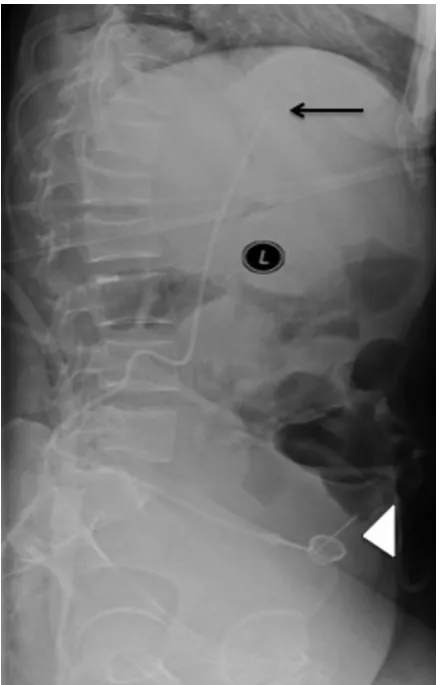
With the guide wire placed within the IVC, the tract was progressively dilated. A 10 Fr, 16 cm peel-away vascular sheath, which was part of the set, was inserted. For an ideal translumbar catheter insertion, a separate peel-away sheath with a recommended length between 18 and 20 cm is needed given the distance between the tissue and the entry site. However, this was not available at the time of the procedure. The peel-away sheath facilitated the insertion of the catheter into the IVC after catheter measurement was performed. The peel-away sheath was removed, and the final position of the catheter tip was confirmed with CT fluoroscopy. The proximal end of the catheter was then attached to the port and anchored to the adjacent muscle layer. The incision was closed with double-layer absorbable sutures. Similarly, the entry site was also sutured (Figure 2).
Figure 1: Computed tomography fluoroscopy showing the puncture needle (white arrow) approaching the infrarenal inferior vena cava (white arrowhead).
Figure 2: Lateral radiograph showing the
Transhepatic access
The transhepatic access was easier in comparison to the translumbar approach. The transhepatic route can be achieved with a 22G Chiba needle, as in percutaneous cholangiography under fluoroscopy. However, the aim is to reach the hepatic veins instead of the biliary tree. Another alternative, which we used in this patient, is a direct puncture with ultrasound guidance. After the hepatic vein was identified, a 0.018-inch wire was advanced to the IVC. In our patient, we used the AccuStick II (Boston Scientific) system to exchange the microwire to a 0.035-inch hydrophilic guide wire. The tip of the guide wire was placed at the distal superior vena cava. Similarly, subsequent dilatation and insertion of an appropriately sized peel-away sheath was advanced to the cavo-atrial junction. The external portion of the catheter was then tunnelled subcutaneously in the anteroinferior direction (Figure 3).
Transcollateral access
The patient had documented angiographic evidence of central vein stenosis from previous catheterisations of the jugular and subclavian veins (Figure 4). Our approach was via the left basilic vein; a 4 Fr vascular sheath was inserted, and a 4 Fr Cobra catheter was used to aid manoeuvring. Because multiple collaterals were observed, we chose the largest venous collateral and the most direct route to the superior vena cava. We achieved the catheter placement with multiple attempts and sheer persistence. The catheter was manoeuvred over the wire with the tip placed within the distal superior vena cava (Figure 5).
Discussion
Central venous access in a patient with central vein stenosis remains a challenge for both the referring physicians and the interventional radiologists. Non-conventional methods have been developed, as described above. Among all of the aforementioned techniques, translumbar catheter insertion using CT-guided fluoroscopy appears to be the best option, in our opinion. Kenney et al. (1) first described the technique for translumbar placement for central venous access in 1985. We used CT fluoroscopy for entry into the IVC. Conventionally, the IVC can be accessed with normal C-arm fluoroscopy either with or without a marker from a pigtail catheter or a guide wire placed in the IVC from the femoral venous
Figure 3: Frontal radiograph showing the transhepatic subcutaneous port placement of the patient.
Figure 4: Left upper limb venogram showing central vein stenosis with collateral formation.
the IVC using CT fluoroscopy. The patency of the IVC also can be assessed concurrently. During puncturing, the direction of the needle can be monitored without over-shooting to the adjacent structures, particularly the ureters and the aorta. Once the IVC was punctured, it was easy to place the distal tip of the guide wire at the junction of the IVC and the right atrium. However, CT-guided puncture is associated with higher radiation doses and, in certain circumstances, necessitates the use of an angiographic fluoroscopy machine that requires patient transfers. Newer versions of CT fluoroscopy have lower radiation doses with more protection devices for the operators. In this patient, we used a single-lumen chemoport for hyperalimentary nutrition. The translumbar venous access can also be successfully used and is suitable for a dual-lumen haemodialysis catheter (2,3). Additionally, it should be noted that although radiation is a concern, CT-guided insertion is safer than conventional fluoroscopy guidance (4). Resuscitation is difficult in these patients because they have limited venous access to begin with, should there be any fatal event.
Transhepatic catheterisation, which was first described by Po et al. (5) in 1994, is a well-known technique for central venous access. It was technically easier compared with the translumbar approach but presented a number of possible complications, such as bleeding, catheter dysfunction, and biliary-related complications. These complications had been known to be mainly associated with larger catheters intended for haemodialysis (6,7).
Transcollateral access is technically challenging because the route of a collateral is usually tortuous. Retrospectively, we feel that this approach should have been tried first to preserve the IVC. It appears to be safe and effective, and also provided our patient with a reliable venous access for three months (8–10).
Non-conventional image-guided percutaneous catheter placements are the exception and are requested only when traditional access sites are unavailable. They are good alternative routes for permanent venous access, and with proper technique and care, will be associated with minimal post procedural complications. Proper anatomical, radiological, and technical knowledge will influence the success of the procedures, which are best performed by a trained interventional radiology unit, as illustrated in the case.
Authors’ Contributions
Conception and design: ASM
Collection and assembly of the data, final approval of the article: YY
Drafting of the article: YY, RZ, ZM Critical revision or the article: ARMR, YY
Correspondence
Dr Yazmin Yaacob
MBChB (Otago), MMed Radiology (UKM) Department of Radiology
Universiti Kebangsaan Malaysia Medical Centre Jalan Yaacob Latif, Bandar Tun Razak
56000 Cheras
Kuala Lumpur, Malaysia Tel: +6012-201 0582
Email: [email protected]
References
1. Rajan DK, Croteau DL, Sturza SG, Harvill ML, Mehall CJ. Translumbar placement of inferior vena caval catheters: A solution for challenging hemodialysis.
Radiographics. 1998;18(5):1155–1167.
2. Denny DF Jr, Greenwood LH, Morse SS, Lee GK, Baquero J. Inferior vena cava: Translumbar catheterization for central venous access. Radiology.
1989;172(3 Pt 2):1013–1014.
3. Lund GB, Lieberman RP, Haire WD, Martin VA, Kessinger A, Armitage JO. Translumbar inferior vena cava catheters for long-term venous access.
Radiology. 1990;174(1):31–35.
4. Kariya S, Tanigawa N, Kojima H, Komemushi A, Shomura Y, Ha-Kawa SK, et al. Percutaneous translumbar inferior vena cava cannulation under
computed tomography guidance. Jpn J Radiol.
2009;27(4):176–179.
5. Po CL, Koolpe HA, Allen S, Alvez LD, Raja RM.
Transhepatic PermCath for hemodialysis. Am J
Kidney Dis. 1994;24(4):590–591.
6. Smith TP, Ryan JM, Reddan DN. Transhepatic
catheter access for hemodialysis. Radiology.
2004;232(1):246–251.
7. Yap DY, Tso WK, Chu FS, Chan TM, Lai KN, Tang SC. Transhepatic catheter placement of haemodialysis catheter: A solution for vascular access exhaustion.
Nephrology (Carlton). 2010;15(6):661–662
8. Forauer AR, Brenner B, Haddad LF, Bocchini TP. Placement of hemodialysis catheters through dilated external jugular and collateral veins in patients with internal jugular vein occlusions. Am J Roentgenol.
9. Teichgraber UK, Streitparth F, Gebauer B, Benter T. Placement of a port catheter through collateral veins in a patient with central venous occlusion. Cardiovasc Intervent Radiol. 2010;33(2):417–420.
10. Powell S, Chan TY, Bhat R, Lam K, Narlawar RS, Cullen N, et al. A retrospective comparative study of tunneled haemodialysis catheters inserted through occluded or collateral veins versus conventional methods.