Inter-Simple Sequence Repeat (ISSR) Markers:
Are We Doing It Right?
W.L. Ng1
* and S.G. Tan
2,3Inter-simple sequence repeats (ISSRs) are regions in the genome flanked by microsatellite sequences. PCR amplification of these regions using a single primer yields multiple amplification products that can be used as a dominant multilocus marker system for the study of genetic variation in various organisms. ISSR markers are easy to use, low-cost, and methodologically less demanding compared to other dominant markers, making it an ideal genetic marker for beginners and for organisms whose genetic information is lacking. Here, we comment upon some of the intricacies often overlooked in designing an ISSR experiment, clarify some misconceptions, and provide recommendations on using ISSR markers in genetic variation studies.
Key words: DNA marker; dominant marker; ISSR; RAM; genetic variation; DNA fingerprinting; ISSR troubleshooting
Overview
Soon after the discovery of the polymerase chain reaction (PCR) in 1983, new PCR-based DNA marker systems were continuously being developed. In the early 1990’s, the development of what would become today’s ‘inter-simple sequence repeat (ISSR)’ markers was independently reported by several research groups (e.g. Meyer et al. 1993, Gupta et al. 1994, Wu et al. 1994, Zietkiewicz et al. 1994). Today, ISSR markers are also popularly known as random amplified microsatellites (RAMs).
Microsatellites, simple sequence repeats (SSRs), or short tandem repeats (STRs) are regions in the genome that consist of short DNA motifs (usually 2-5 nucleotides long) repeated multiple times in a row, e.g. …ACACACACACAC… Subsequently, ISSRs are segments of DNA that are flanked at both ends by such microsatellite sequences. Using arbitrarily designed primers that contain repetitive sequences complementary to microsatellite regions in the genome (= ISSR primers), random DNA segments in the genome can be PCR-amplified (provided that a segment is within the amplifiable size range) and used as markers for genetic variation studies, hence the term ‘ISSR markers’. Figure 1 shows the basic concept behind the PCR amplification of ISSRs (= ISSR-PCR).
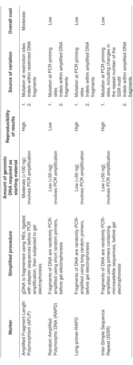
The ISSR marker belongs to a class of multilocus, mostly dominant genetic markers that also include the amplified fragment length polymorphism (AFLP), random amplified polymorphic DNA (RAPD) markers, and their derivatives (Table 1). Dominant markers do not allow clear distinction between homozygotes and heterozygotes. These markers, however, usually produce multiple DNA fragments (each of which is considered a locus) in a single reaction, allowing the generation of a large number of loci across the genome of any species without the need to first know the DNA sequences of the target regions. Apart from its usage as genetic markers, these dominant markers can also be used as initial steps for the development of co-dominant markers: RAPD for the development of
single-1Institute of Bioscience, Universiti Putra Malaysia, 43400 Serdang, Selangor, Malaysia 2Fellow of the Academy of Sciences, Malaysias
3Department of Cell and Molecular Biology, Faculty of Biotechnology and Biomolecular Sciences, Universiti Putra Malaysia,
43400 Serdang, Selangor, Malaysia
locus co-dominant ‘sequence characterised amplified region (SCAR)’ markers (e.g. Paran & Michelmore 1993), and ISSR for the development of single-locus co-dominant microsatellite markers (e.g. Fisher et al. 1996; Lian et al. 2001; Adibah et al. 2012).
For most genetic variation studies, a good genetic marker is defined by high genetic variability and the ability to generate multilocus data from the genome under study (Anne 2006). The generation of ISSR markers makes use of microsatellite sequences that are highly variable and ubiquitously distributed across the genome, at the same time achieving higher reproducibility compared to using RAPDs and costs less in terms of time and money compared to using AFLPs. All these make ISSR an ideal genetic marker for various studies, most notably on genetic variation/diversity (e.g. Wang et al. 2012; Shafiei-Astani et al. 2015), DNA fingerprinting (e.g. Shen et al. 2006), and phylogenetics (e.g. Iruela et al. 2002).
Over the years, there have been several reviews on the applications of ISSR markers (e.g. Godwin et al.
1997; Bornet & Branchard 2001; Reddy et al. 2002), mainly in plants genetics. However, few actually addressed the important considerations or potential problems that beginners ought to be aware of before embarking on an experiment using ISSR markers. In the following sections of this paper, we attempt to fill in that knowledge gap by clarifying several factors that are often overlooked, or misconceptions that many users have regarding the practical usage of ISSR markers in their experiments.
Gel electrophoresis
PCR amplification with primer containing
short repeated sequences gDNA (template DNA)
ISSR primer TACACACACACACAC
CACACACACACACAT
5’
5’
3’ 3’
Samples 1 2 3
ATGTGTGTGTGTGTG
GTGTGTGTGTGTGTA
Table 1. Comparison of different dominant DNA markers for genetic variation studies
Marker
Simplified procedure
Amount of genomic DNA required as starting material Reproducibility of results
Source of variation
Overall cost
Amplified Fragment Length Polymorphism (AFLP) gDNA is fragmented using REs, ligated with adapter sequences before PCR amplification, then subjected to gel electrophoresis
Moderate (>100 ng);
involves PCR amplification
High
1.
Mutation at restriction sites
2.
Indels within restricted DNA fragments
Moderate
Random Amplified Polymorphic DNA (RAPD) Fragments of DNA are randomly PCR- amplified using short random primers, before gel electrophoresis
Low (<50 ng);
involves PCR amplification
Low
1.
Mutation at PCR priming sites
2.
Indels within amplified DNA fragments
Low
Long primer-RAPD
Fragments of DNA are randomly PCR- amplified using long random primers, before gel electrophoresis
Low (<50 ng);
involves PCR amplification
High
1.
Mutation at PCR priming sites
2.
Indels within amplified DNA fragments
Low
Inter-Simple Sequence Repeat (ISSR)
Fragments of DNA are randomly PCR- amplified using primers containing microsatellite sequences, before gel electrophoresis
Low (<50 ng);
involves PCR amplification
High
1.
Mutation at PCR priming sites, including changes in the repeat number of the SSR motif
2.
Indels within amplified DNA fragments
Low
TeCHNiCAL CONSiDerATiONS
The basic procedure to conduct an ISSR genotyping experiment is simple:
1. PCR, using an ISSR primer, with genomic DNA (gDNA) as its template;
2. Use of agarose or polyacrylamide gel electrophoresis of PCR amplification products;
3. Scoring of ISSR bands; and
4. Data analysis.
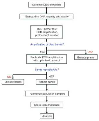
However, as with most other scientific experiments, the actual procedure will require additional steps for planning and evaluation before the final genotyping of samples. Figure 2 summarises the general procedure for the evaluation and usage of an ISSR primer for genotyping.
Genomic DNA extraction
Standardise DNA quantity and quality
ISSR primer test: PCR amplification,
protocol optimisation
Genotype population samples
Score recruited bands
Analysis Exclude bands
Exclude primer
Recruit bands Replicate PCR amplification
with optimised protocol YES
YES
NO
NO
Amplification of clear bands?
Bands reproducible?
For the purpose of this paper, we discuss in detail several important components of an ISSR experiment, as follows:
(a) gDNA as template for ISSR-PCR
Genomic DNA is commonly used as the template for ISSR-PCR, and is therefore an integral part for a successful ISSR experiment. Often overlooked in most experimental protocols, however, is the need to obtain high quality DNA as the starting material, and to standardise the quantity (amount) of template DNA used in each PCR reaction. DNA extracts, depending on the extraction method and type of sample, may contain traces of cell debris and components that potentially inhibit PCR reactions. As a result, fewer, if any, DNA fragments would be amplified compared to what we would expect when using purified DNA. Furthermore, using inconsistent amounts of DNA across PCR reactions would result in inconsistent concentrations of PCR amplification products, affecting band intensities across samples. In most cases, conventional DNA extraction methods would suffice to obtain good quality DNA. If not, further purification of the DNA extract using commercially available column-based DNA extraction/purification kits often helps. Then, the DNA concentrations are adjusted accordingly, to an approximate standard, before it is used in PCR reactions. Typically, 10–50 ng of good quality DNA is sufficient for each reaction.
(b) ISSR primer design
An ISSR primer is usually 16–25 base pairs (bp) in length, and comprises mainly, or solely, of repeated DNA motifs (2–4 bp each) meant to be complementary to microsatellite regions in the genome. Depending on the usage, there are 3 forms of ISSR primers: unanchored (primer consists only of a repeated motif, e.g.
5’–(AC)8–3’), 5’-anchored (primer consists of a repeated motif with one or several non-motif nucleotides at
the 5’-end, e.g. 5’–GA(AC)8–3’), and 3’-anchored (primer consists of a repeated motif with one or several
non-motif nucleotides at the 3’-end, e.g. 5’–(AC)8AG–3’). Reddy et al. (2002) discussed in detail the effects of
using these different primers for the generation of ISSR bands. Thus, for studies that aim to evaluate genetic variability, we recommend using either the 3’- or 5’-anchored ISSR primers. Unanchored ISSR primers may slip along the length of the complementary microsatellite region during PCR, producing inconsistent amplification in every cycle, and thus affecting the reproducibility of results.
Once the above points have been thoroughly considered, ISSR primers can be easily designed or customised to fit the needs of the experiment. Alternatively, previously reported primers can be used, with the ISSR primers designed at the University of British Columbia (primer names usually starting with ‘UBC’) being one of the more popular choices.
(c) PCR amplification with ISSR primers (ISSR-PCR)
Slightly different from the usual PCR reaction that involves a pair of different primers, ISSR-PCR involves only one primer in each reaction, e.g. single-primer PCR amplification. However, many do not realise that this single primer actually acts as both the ‘forward’ and ‘reverse’ primers which are essential for an amplification to take place (Figure 1).
ISSR-PCR is usually conducted with an annealing temperature (Ta) of 45–60°C, depending on the melting
temperature (Tm) of the ISSR primer (Reddy et al. 2002). While trying out new ISSR primers, it is important
to test several temperatures, usually Ta = 45°C, 50°C, 55°C, and 60°C for a standard PCR reaction profile,
to obtain an optimum Ta that amplifies clear and reproducible DNA bands. We have also used a touch-down
(d) Gel electrophoresis
ISSR-PCR amplification products are commonly electrophoresed through 1.5–2.0% weight/volume (w/v) agarose gel to achieve adequate separation of the DNA bands for easy scoring. Based on our experience, agarose gels made to higher concentrations (3.0% w/v or higher) may crack easily while solidifying. In fact, Bornet & Branchard (2001) found that 2.0% w/v agarose gels performed best among several concentrations (0.8, 1.0, 1.5, 2.0, and 3.0% w/v) in resolving ISSR bands. Alternatively, ISSR bands can be resolved using polyacrylamide gel electrophoresis (e.g. Godwin et al. 1997; Reddy et al. 2002).
(e) Scoring of bands
The standards or criteria of scoring DNA bands generated for most dominant DNA markers have so far been very much subjective, and band-scoring results may differ from person to person (Pompanon et al. 2005; Meudt & Clarke 2007). That being said, in order to minimise human and stochastic errors, we recommend observing several points when scoring ISSR bands on a gel: (1) Score only clearly distinctive bands. Smeared bands could be the result of unspecific binding of ISSR primers causing unintended amplification, or the overlapping of several bands with similar DNA fragment sizes, both of which would make scoring difficult and inconsistent. (2) Score only bands with strong intensities. Bands with weak intensities tend to have low reproducibility and thus are best avoided. (3) Set a standard band-scoring size range before scoring; usually in the range of 100–2000 bp. Electrophoresed through a 2.0% w/v agarose gel at 80–100 V, band sizes <100 bp are usually less sharp and could be the products of primer-dimer amplification; band sizes >2000 bp are difficult to amplify during PCR, and so have weak intensities and tend to have low reproducibility. As a rule-of-thumb when designing a PCR protocol, 1 minute of extension time is used for every 1000 bp of desired amplification product. Therefore, in theory, a 2-minute extension time will allow the amplification of bands up to a maximum size of approximately 2000 bp. Increasing the extension time during ISSR-PCR does not necessarily produce better data, since such a PCR reaction will not only be time-consuming, but shorter DNA fragments are known to be preferentially amplified during the PCR (Walsh et al. 1992). Finally, bands are recorded into the binary symbols, 1, for band presence, whereas, 0, for band absence (sometimes ‘+’ and ‘–’), for subsequent analyses.
(f) Estimation of basic parameters
When tabulating ISSR band-counts, one must understand some basic terminologies and descriptive statistics commonly reported in studies that use ISSR markers: (1) a ‘band’ scored in an ISSR experiment can also be termed as a ‘locus’. Regardless of whether a series of ISSR bands are generated from the same PCR reaction using the same ISSR primer, each ISSR band is separately considered as one locus, and hence one data point in any analysis; (2) “total number of bands” is the total number of different ISSR band sizes observed across all samples in a study; (3) “number of polymorphic bands” is the number of ISSR bands that show variation, i.e. the bands are present for some samples and absent for the others; and (4) “percentage of polymorphic bands” is represented by the formula:
% of polymorphic bands = No. of polymorphic bands × 100%
Total no.of bands
Therefore, for the example shown in Figure 3, the • total number of bands = 8
• number of polymorphic bands = 3
Samples
Sample Sample Sample Locus 1 1 1 1 2 0 0 0 0 3 1 1 1 1 1 1 1 1 1 1 1 1 1 1 1 1 1 1 L1 L2 L3 L4 L5 L6 L7 L8 2 3
Figure 3. Scoring ISSR bands
OTHer CONSiDerATiONS
(a) How many ISSR bands are needed?
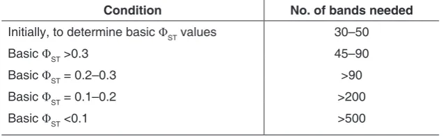
One common question that we often encounter from young researchers is, “How many ISSR bands should I have for my study?” This concern is not unfounded, as too small a number of bands (= too few data points) will negatively affect the resolution of an analysis and its ability to address a particular research objective. For genetic diversity estimation, Mariette et al. (2002) showed that at least 4–10 times more dominant loci should be used in order to attain a similar estimating power as with co-dominant markers (= microsatellite markers, in the case of the mentioned study). A recent review by Nelson and Anderson (2013) also concluded that for the analysis of population structure using AMOVA (Analysis of Molecular Variance) and STRUCTURE, the level of genetic differentiation among populations was the most critical factor in determining the number of loci to use. AMOVA was found to be accurate even with a small number of bands (30–50 bands), while more bands were usually required for the STRUCTURE analysis (Nelson & Anderson 2013). The recommended guideline is summarised in Table 2.
Table 2. Recommended guideline for the determination of the number of bands needed for STRUCTURE analysis using dominant markers. ΦST (or PHIST) values can be estimated using AMOVA
Condition No. of bands needed
Initially, to determine basic ΦST values 30–50
Basic ΦST >0.3 45–90
Basic ΦST = 0.2–0.3 >90
Basic ΦST = 0.1–0.2 >200
Basic ΦST <0.1 >500
(b) Reliability and comparability of results
with at least 5–10% of random samples can also be used to assess reproducibility (Bonin et al. 2004), whereas data points (DNA bands) that are not reproducible should be left out of the study. Finally, homology in dominant markers, although still an issue when it comes to accuracy, was shown to not be as bad as to render results from dominant markers unreliable (Simmons et al. 2007).
While results of ISSR genotyping may be reproducible within a study using the same equipment and protocol, we recommend caution when comparing band-scoring results across different studies. In our experience, even when two separate studies are on the same species using the same primers, the ISSR banding patterns may vary considerably (e.g. in Lo 2010 vs. Ng & Szmidt 2014). The reason for this is unknown, but it could be due to the effects of using different reagents (each manufacturer possibly have their own reagent concoction, including additives) and settings during PCR and/or the different scoring criteria adopted by different researchers. These inconsistencies make the reporting of experimental protocol very important in studies using ISSR markers. To demonstrate the robustness of studies involving ISSR markers, we have adapted recommendations by Crawford et al. (2012) and urge researchers to explicitly report the following in their manuscripts:
• Steps taken throughout the ISSR experiment
• Names and sequences of the ISSR primers used, as well as the PCR reaction protocol associated with each primer
• Standards/criteria used to ensure reliability of genotyping and scoring of bands, i.e. by selecting only loci that are clear, unambiguous, and reproducible, shown in replicated experiments
• Number and proportion of samples used in the replicated experiment
(c) Analysis of ISSR (dominant) data
The basis of most population genetic analyses is the assumption of Hardy-Weinberg equilibrium (HWE), including all its underlying assumptions – diploid organism, sexual reproduction, random mating, non-overlapping generations, no genetic drift, no migration, no mutation, no natural selection, and equal allele frequencies in both
sexes. In the dominant marker system, given 2 alleles M and m for a particular locus, if M is the amplifiable
‘dominant’ allele and m is the non-amplifiable ‘null’ allele, genotypes MM and Mm would both show bands,
hence the frequencies of each genotype cannot be exactly calculated. The frequencies of each allele would then have to be inferred from the frequency of the ‘null’ homozygotes (= the absent ‘bands’), assuming HWE. Such inference would be sensitive to the sampling strategy and life history of the organism under study, as any deviation from the assumptions of HWE will affect the precision of the estimation of allele frequencies, as well as any subsequent parameters derived using these estimates. For genetic diversity and genetic differentiation estimations, for instance, Krutovskii et al. (1999) and Mariette et al. (2002) found that while dominant data would be robust for investigating population structuring and genetic differentiation among populations, the same could not be said for genetic diversity estimates. We therefore urge users to be aware of the theoretical frameworks behind each analysis before using them for their ISSR data, and if possible, to use only programmes that have been designed to accommodate dominant data.
CONCLUSiON
Today, genetic markers are increasingly used to address various questions in ecology and agriculture. Although microsatellites are undoubtedly still the marker of choice for many genetic variation studies that require markers with high resolution, hypervariability, and co-dominance (Guichoux et al. 2011; Kalia et al. 2011), ISSR with its ease of application makes preliminary studies on genetic variation more accessible to beginners and less-funded projects. We envision that with an understanding of the advantages and limitations of ISSR markers, as well as a proper execution of experiments, robust and useful inferences can be made to provide justification for more sophisticated studies to be done in the future.
reFereNCeS
Adibah, AB, Liew, PL, Tan, SG, Faridah, QZ & Christianus, A 2012, ‘Development of single-locus DNA microsatellite markers using 5 anchored ISSR-PCR method for the mangrove horseshoe crab, Carcinoscorpius rotundicauda (Latreille, 1802) in Peninsular Malaysia’, Molecular Biology Reports, vol. 39, pp. 3815–3820.
Anne, C 2006, ‘Choosing the right molecular genetic markers for studying biodiversity: from molecular evolution to practical aspects’, Genetica, vol. 127, pp. 101–120.
Bonin, A, Bellemain, E, Bronken Eidesen, P, Pompanon, F, Brochmann, C & Taberlet, P 2004, ‘How to track and assess genotyping errors in population genetics studies’, Molecular Ecology, vol. 13, pp. 3261–3273.
Bornet, B & Branchard, M 2001, ‘Nonanchored inter simple sequence repeat (ISSR) markers: Reproducible and specific tools for genome fingerprinting’, Plant Molecular Biology Reporter, vol. 19, pp. 209–215.
Cavers, S, Degen, B, Caron, H, Lemes, MR, Margis, R, Salgueiro, F & Lowe, AJ 2005, ‘Optimal sampling strategy for estimation of spatial genetic structure in tree populations’, Heredity, vol. 95, pp. 281–289.
Crawford, LA, Koscinski, D & Keyghobadi, N 2012, ‘A call for more transparent reporting of error rates: the quality of AFLP data in ecological and evolutionary research’, Molecular Ecology, vol. 21, pp. 5911–5917.
Fisher, PJ, Gardner, RC & Richardson, TE 1996, ‘Single locus microsatellites isolated using 5’ anchored PCR’, Nucleic Acids Research, vol. 24, pp. 4369–4371.
Godwin, ID, Aitken, EAB & Smith, LW 1997, ‘Application of inter simple sequence repeat (ISSR) markers to plant genetics’, Electrophoresis, vol. 18, pp. 1524–1528.
Guichoux, E, Lagache, L, Wagner, S, Chaumeil, P, Leger, P, Lepais, O, Lepoittevin, C, Malausa, T, Revardel, E, Salin, F & Petit, RJ 2011, ‘Current trends in microsatellite genotyping’, Molecular Ecology Resources, vol. 11, pp. 591–611.
Gupta, M, Chyi, YS, Romero-Severson, J & Owen, JL 1994, ‘Amplification of DNA markers from evolutionarily diverse genomes using single primers of simple-sequence repeats’, Theoretical and Applied Genetics, vol. 89, pp. 998–1006.
Iruela, M, Rubio, J, Cubero, JI, Gil, J & Millan, T 2002, ‘Phylogenetic analysis in the genus Cicer and cultivated chickpea using RAPD and ISSR markers’, Theoretical and Applied Genetics, vol. 104, pp. 643–651.
Kalia, RK, Rai, MK, Kalia, S, Singh, R & Dhawan, AK 2011, ‘Microsatellite markers: An overview of the recent progress in plants’, Euphytica, vol. 177, pp. 309–334.
Kremer, A, Caron, H, Cavers, S, Colpaert, N, Gheysen, G, Gribel, R, Lemes, M, Lowe, AJ, Margis, R, Navarro, C & Salgueiro, F 2005, ‘Monitoring genetic diversity in tropical trees with multilocus dominant markers’, Heredity, vol. 95, pp. 274–280.
Krutovskii, KV, Erofeeva, SY, Aagaard, JE & Strauss, SH 1999, ‘Simulation of effects of dominance on estimates of population genetic diversity and differentiation’, The Journal of Heredity, vol. 90, pp. 499–502.
Lian, C, Zhou, Z & Hogetsu, T 2001, ‘A simple method for developing microsatellite markers using amplified fragments of inter-simple sequence repeat (ISSR)’, Journal of Plant Research, vol. 114, pp. 381–385.
Lo, EYY 2010, ‘Testing hybridization hypotheses and evaluating the evolutionary potential of hybrids in mangrove plant species’, Journal of Evolutionary Biology, vol. 23, pp. 2249–2261.
Mariette, S, Le Corre, V, Austerlitz, F & Kremer, A 2002, ‘Sampling within the genome for measuring within-population diversity: trade-offs between markers’, Molecular Ecology, vol. 11, pp. 1145–1156.
Meudt, HM & Clarke, AC 2007, ‘Almost forgotten or latest practice? AFLP applications, analyses and advances’, Trends in Plant Science, vol. 12, pp. 106–117.
Nelson, MF & Anderson, NO 2013, ‘How many marker loci are necessary? Analysis of dominant marker data sets using two popular population genetic algorithms’, Ecology and Evolution, vol. 3, pp. 3455–3470.
Ng, WL & Szmidt, AE 2014, ‘Introgressive hybridization in two Indo-West Pacific Rhizophora mangrove species, R. mucronata and R. stylosa’, Aquatic Botany, vol. 120, pp. 222–228.
Nybom, H 2004, ‘Comparison of different nuclear DNA markers for estimating intraspecific genetic diversity in plants’, Molecular Ecology, vol. 13, pp. 1143–1155.
Paran, I & Michelmore, RW 1993, ‘Development of reliable PCR-based markers linked to downy mildew resistance genes in lettuce’, Theoretical and Applied Genetics, vol. 85, pp. 985–993.
Pompanon, F, Bonin, A, Bellemain, E & Taberlet, P 2005, ‘Genotyping errors: Causes, consequences and solutions’, Nature Reviews Genetics, vol. 6, pp. 847–859.
Reddy, MP, Sarla, N & Siddiq, EA 2002, ‘Inter simple sequence repeat (ISSR) polymorphism and its application in plant breeding’, Euphytica, vol. 128, pp. 9–17.
Shafiei-Astani, B, Ong, AHK, Valdiani, A, Tan, SG, Yong, CSY, Ahmady, F, Alitheen, NB, Ng, WL & Kaur, T 2015, ‘Molecular genetic variation and structure of Southeast Asian crocodile (Tomistoma schlegelii): comparative potentials of SSRs versus ISSRs’. Gene, vol. 571, pp. 107–116.
Shen, J, Ding, X, Liu, D, Deng, G, He, J, Li, X, Tang, F & Chu, B 2006, ‘Intersimple sequence repeats (ISSR) molecular fingerprinting markers for authenticating populations of Dendrobium officinale Kimura et Migo’, Biological and Pharmaceutical Bulletin, vol. 29,
pp. 420–422.
Simmons, MP, Zhang, LB, Webb, CT & Muller, K 2007, ‘A penalty of using anonymous dominant markers (AFLPs, ISSRs, and RAPDs) for phylogenetic inference’, Molecular Phylogenetics and Evolution, vol. 42, pp. 528–542.
Walsh, PS, Erlich, HA & Higuchi, R 1992, ‘Preferential PCR amplification of alleles: Mechanisms and solutions’, PCR Methods and Applications, vol. 1, pp. 241–250.
Wang, X, Yang, R, Feng, S, Hou, X, Zhang, Y, Li, Y & Ren, Y 2012, ‘Genetic variation in Rheum palmatum and Rheum tanguticum (Polygonaceae), two medicinally and endemic species in China using ISSR markers’, PLOS One, vol. 7, e51667.
Wu, KS, Jones, R, Danneberger, L & Scolnik, PA 1994, ‘Detection of microsatellite polymorphisms without cloning’, Nucleic Acids Research, vol. 22, pp. 3257–3258.