Studying of the Expression of BAX and BCL-2 Genes in Men
with Sperm DNA Fragmentation Based on DFI and Comet
Assay
Hossein Taheri 1, Mohammad Salehi 2, 3*, Tarlan Eslami Arshaghi 4, Majid Mosahebi 4
1. Department of Anatomical Sciences and Biology of Reproduction, Faculty of Medicine, Shahid Beheshti University of Medical Sciences, Tehran, Iran. 2. Cellular and Molecular Biology Research Center, Shahid Beheshti University of Medical Sciences, Tehran, Iran.
3. Department of Biotechnology, Faculty of Advanced Technologies in Medicine, Shahid Beheshti University of Medical Sciences, Tehran, Iran. 4. Department of Transgenic, Stem Cell Technology Research Center, Tehran, Iran
* Corresponding Author: Mohammad Salehi, PhD
Address: Cellular and Molecular Biology Research Center, Shahid Beheshti University of Medical Sciences and Health Services, Tehran, Iran. Tel: +98 (21) 23872552 Fax: +98 (21) 22439956
E-mail: [email protected]
Introduction: The amount of expression of BAX and BCL-2 genes in infertile men’s sperm as well as its association with sperm parameters and DNA fragmentation index is an issue which has not been studied yet. In this research, it is assumed that up-regulation of BAX and down-regulation of BCL-2 are directly associated with sperm DNA fragmentation.
Methods: After obtaining semen samples from the patients by using gradient centrifugation method, the semen samples were centrifuged using gradient method in order to obtain pure sperm. Sperm is divided into three parts based on which flow cytometry, Real Time-PCR and Comet Assay techniques were conducted. After extracting RNA and producing cDNA, the amount of expression of BAX and BCL-2 genes was measured using Real Time-PCR. The amount of sperm DNA fragmentation was measured using flow cytometry and Comet Assay techniques. Based on the amount of DNA fragmentation index (DFI), samples were divided into the two groups of control (DFI<30) and DNA fragmentation (DFI≥30). Using WHO criteria, sperm parameters (morphology and motility) were evaluated.
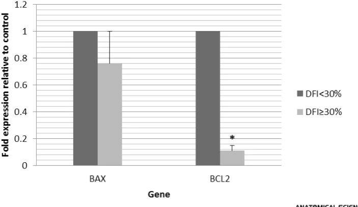
Results: This study showed that the amount of expression of BAX in the DNA fragmentation group was not significant compared to the control group but the expression of BCL-2 gene decreased significantly (P<0.05). Also, in many cases, there was a significant difference between the two groups in terms of parameters of sperm DNA fragmentation and morphological parameters (P<0.05).
Conclusion: This study showed that reduction of expression of BCL-2 increases sperm DNA damage and this result can be helpful for therapeutic purposes.
A B S T R A C T
Article info:
Received: 01 Jul. 2014 Accepted: 28 Oct. 2014
Key Words:
Sperm DNA damage, BAX, BCL-2
Mohammad Salehi is currently an academic member and embryologist of Shahid Beheshti University of Medical Sciences in Tehran, Iran. His main research interest is preimplantation development of human and animal embryo and somatic cell
1. Introduction
perm DNA integrity is essential for com-plete and accurate transfer of genetic ma -terial to the next generation. DNA frag-mentation is the first sign of programmed cell death [1]. Much evidence indicates abnormalities in the sperm produced by the man. These abnormalities, which affect fertility, oc -cur in the nucleus, cytoskeleton and organelles of the sperm [2].
It seems that there is a relationship between sperm fer -tility and apoptotic activity of sperm. Indirect evidence shows that in couples with normal sperm parameters, blastocyst formation rate is significantly higher as com-pared to couples with severe sperm parameters defi-ciency [3].
Male infertility is associated with poor sperm DNA in-tegrity, in a way that in infertile men, 25% of the sperm show denaturation and 28% show DNA fragmentation. This amount in fertile men includes 10% denaturation and 13% DNA fragmentation [4]. During spermato-genesis, through a mechanism called ‘screening germ cells’, Sertoli cells induce apoptosis in 50 to 60 percent of germ cells entering meiosis Ι. These germ cells, which have been marked by Fas apoptosis markers, must be phagocytosed by Sertoli cells; but this mechanism is not always done correctly and defective germ cells with dif -ferent percentages enter the DNA rearrangement pro-cess in spermiogenesis [5].
Apoptosis is a complex process that occurs through two main internal and external pathways. Either path -way is set at different levels. The internal path-way, which is intramitochondrial, contains key apoptosis fac -tors such as cytochrome C. The most important regula
-tory factors of the internal path of apoptotic and anti-apoptotic members of the BCL-2 family [6].
It has been reported that some of the members of BCL-2 protein family (Bcl-w, Mcl-7, Bcl-xl, Bcl-BCL-2) play a role in repressing apoptosis and some of them (Bad, Bak, Bcl-xs, Bax) in advancing apoptosis. These protein components are responsible for regulating apoptosis in many kinds of cells [7]. In normal cells, Bax is often found in cytosol but when the cell is affected by apop -totic stimuli, Bax is transferred to mitochondrial outer membrane [8].
The balance between these processes has a regula -tory effect at the transcriptional or post-transcriptional level and influences the apoptosis. For example, high Bax/Bcl ratio shows the pro-apoptotic tendency of the cell [9]. As the amount of Bax increases and Bcl-2 de-creases, the suitable pro-apoptotic environment is pre-pared for the cell [10]. The most important apoptosis regulators are Bcl-2 family members which exert their regulatory effect on apoptosis through regulation of mi -tochondrial changes prior to activation of caspases and nucleases. At the time of stress, pro-apoptotic proteins which are located inside cytosol are transferred to the mitochondrial level, where anti-apoptotic proteins also exist. The interaction between pro-apoptotic and anti-apoptotic proteins disrupts the normal function of the anti-apoptotic Bcl-2 proteins and can lead to the forma-tion of pores in the mitochondria and the release of cy -tochrome C and other pro-apoptotic molecules from the intermembrane space. By binding to Apaf-1, these ele-ments activate Caspase 9 and through targeting Caspase effectors eventually lead to cell death [11].
There are different tests to assess DNA damage. These tests assess DNA fragmentation based on the kind of damage and sensitivity of DNA. DNA fragmentation
S
Table 1. Primers used for analyzing the amount of expression of the studied genes.
GenBank Accession no. Product size (bp) Primer sequence (5′─3′ orientation) Genes name
NM_001291431.1 178 Forward: CAA ACT GGT GCT CAA GGC
Reverse: CAC AAA GAT GGT CAC GGT C Bax
NM_000657.2 148 Forward: GTA CTT AAA AAA TAC AAC ATC ACA G
Reverse: CTT GAT TCT GGT GTT TCC C BCL-2
XM-006715764.1 85 Forward: CTT CCT TCC TGG GCA TG
tests which are conducted in acid or base environments include a DNA denaturation phase in order to identify fragmentation or potential fragmentation of DNA [12]. Sperm Chromatin Structure Assay (SCSA) measures the percentage of DNA fragmentation, which is known as DFI. This technique is based on Acridine Orange (AO) fluorescence intensity or application of flow cy-tometry. Binding of Acridine Orange to normal DNA leads to emission of green light. In case of binding to fragmented DNA, the light will be red. The ratio of red light to the total of red and green lights indicates the percentage of DNA fragmentation [13].
During clinical application, various techniques report different percentages of DNA damage. Most of these
techniques do not record the percentage of DNA dam-age within a specific cell. An exception is Comet Assay which records the percentage of damage to each sperm. This technique shows some recognizable levels of dam -age in all sperms (even the fertile sperm population) [14].
Since so far no research has been conducted on the relationship between the expression of BAX and BCL-2 apoptotic genes and DFI, sperm parameters and Comet Assay, in this research, we study the expression of BAX and BCL-2 genes in patients with sperm DNA damage based on DFI and Comet Assay.
Figure 1. Graphs related to SCSA in the two studied groups; A: Histogram of control group DFI, B: Histogram of DNA damage group DFI.
2. Materials & Methods
This study was conducted after obtaining informed consent of the patients and its approval by the Ethics Committee of the Research Department of Shahid Be -heshti University of Medical Sciences.
Studied groups
After obtaining samples from patients visiting Taleqa-ni Hospital (20 patients), the samples were centrifuged with AllGrad (Life Global) using density gradient tech-nique (80%:40%). The washed samples were used to apply the techniques.
In order to study the degree of sperm DNA fragmen-tation or damage, Sperm Chromatin Structure Assay (SCSA) was used.
Method of conducting SCSA
First a volume of gradient semen containing 1.2 mil-lion sperm was mixed with TNE buffer (0.01M Tris-HCl, 0.15M NaCl, 1mM EDTA, pH 7.4). This suspen-sion was treated with acid solution (0.1% Triton X-100, 0.15mol/L NaCl, 0.08N HCl, pH 1.2) for 30 seconds.
Then it was dyed with 6mg/L Acridine Orange in phos-phate-citrate buffer with pH of 6.0 and was examined using flow cytometry device.
Based on the obtained curve and the value of DFI, pa-tients were divided into two groups (control group with DFI<30 and DNA damage with DFI≥30).
Quantitative analysis of expression of distinctive genes using Real-Time PCR
Quantitative real-time PCR analysis
Frequency of expression of BCL-2 and BAX genes was studied by using quantitative Real-time PCR with special primers. The details of primers have been pre -sented in Table 1. Reaction was conducted in the total volume of 13 microliter based on DNA Master SYBR Green I mix instruction using 1 microliter of each prim-er and 1 microlitprim-er of cDNA.
Cycling parameters include: 5 seconds in 95°C and 3 minutes in 95°C for denaturation, 15 seconds in 60°C and 10 seconds in 72°C for amplification and 40 elon-gation cycles. Amplification reactions were assessed by examining the melting curves to confirm the presence of Table 2. Data related to sperm DNA damage, assessed by Comet Assay.
DFI<30 DFI≥30
Parameter
58.64±0.85a 70.62±1.26a
Comet length
55.61±0.74 57.35±0.92
Comet height
2838.16±98.84b 3962.26±115.08b
Comet area
86942.23±4099.16c 111122.77±3332.95c
Comet intensity
30.27±0.54d 36.54±0.70d
Comet mean intensity
52.02±0.76e 45.21±0.71e
Head diameter
2632.89±90.05f 3064.39±91.46f
Head area
77318.38±3686.71 81255.40±2593.89
Head intensity
29.17±0.53g 33.61±0.65g
Head mean intensity
89.89±0.57h 74.98±0.62h
%DNA in head
6.61±0.37i 25.41±0.80i
Tail length
205.27±18.01j 897.86±42.21j
Tail area
9623.85±837.13k 29867.36±1226.52k
Tail intensity
77.91±28.59 101.15±10.41
Tail mean intensity
10.10±0.57l 25.01±0.62l
%DNA in tail
1.10±0.11m 8.55±0.40m
Tail moment
1.67±0.11n 7.21±0.28n
Olive moment
a single gene-specific peak. β–actin was considered as housekeeping gene for BAX and BCL-2 genes in order to normalize the data. Changes in the level of mRNA in each sample were normalized by the levels of house -keeping mRNA.
Comet assay
Conventional microscope slides were covered with 1.5% normal agarose (Sigma) dissolved in PBS (Ca2+
and Mg2+ free) maintained for at least one day at room
temperature to dry. 20 microliter of sperm sample with 80 microliter of 1% low melting agarose (Sigma), which had been dissolved in PBS, were mixed and placed on agarose slide. A coverslip was placed on the slide and it was refrigerated at 4°C for 20 minutes. Then, the cov-erslip was removed and another layer of low melting agarose was placed on the slide and was refrigerated at
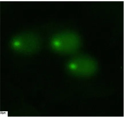
4°C for 20 minutes. After removing the coverslip, the slides were immersed in a Lysis buffer (2.5 M NaCl [Merck], 100mM EDTA [Merck], 10 mM Tris [Merck], 10% DMSO [Sigma], 1% Triton X-100 with PH=10) at 4°C for 80 minutes. Also, sufficient DIT was added. Then the slides were removed from the Lysis buffer and placed in horizontal gel tank filled with base buffer (300mM NaOH [Merck, Germany] 1 and mM EDTA and pH>13) for at 4°C for 20 minutes so that the DNA may be unwound. Electrophoresis was conducted at 4°C for 20 minutes using 20 V and 300 mA direct cur-rent. After electrophoresis, the slides were dyed with CYBER green. Using Nikon Eclipse 600 microscope equipped with 515-560nm stimulation filter, a 100w mercury lamp and an inhibitor filter, the slides were observed and photographed. The images were analyzed using Triteck Comet Score software and the follow -ing parameters were measured: Comet length, Comet
Table 4. The relationship between BAX expression and comet parameters.
P-Value BAX (P.C)
Parameter
0.003 0.864
Comet intensity
0.04
0.690 Head diameter
0.01
0.799 Head area
0.00
0.932 Head intensity
0.041
0.686 Tail mean intensity
Table 3. Comparison of the mean of sperm parameters in the sample with DNA damage and the normal sample. DFI<30 DFI≥30
Parameter
9.00±2.08a 2.33±0.49a
Morphology
10.66±1.76 7.66±0.61
Large head
10.66±1.76b 7±0.68b
Tappered head
4.00±0.00c 1.66±0.61c
Loose head
8.00±2.30 8.83±0.90
Bent
11.33±2.40 8.66±0.66
Cyto
10.66±2.40 8.50±1.08
Coiled
35.66±1.85d 56.00±2.35d
Amorph
25.33±12.97 28.66±9.92
Count
68.33±8.33e 14.00±1.52e
Motility
36.33±1.85f 10.33±1.5f
MotilityA
32.00±6.50g 13.66±1.56g
MotilityB
15.66±3.48h 47.00±4.85h
MotilityC
16.00±5.85 29.00±5.19
height, Comet area, Comet intensity, Comet mean in -tensity, head diameter, head area, head in-tensity, head mean intensity, %DNA in head, tail length, tail area, tail intensity, tail mean intensity, %DNA in tail, tail mo-ment, olive moment [15].
Sperm parameters
Based on WHO criteria, sperm parameters evaluated using light microscope.
3. Results
Sperm DNA damage was assessed using SCSA tech-nique (Figure 1) and the studied samples were divided into the two groups of DFI<30% and DFI≥30%. In order to study the amount of DNA damage more ac-curately, Comet Assay method was also used (Figure 3). The images obtained from Comet Assay were ana-lyzed by Triteck Comet Score software and seventeen parameters, which have been shown in Table 2, were studied. A summary of the results for the two studied groups has been presented in Table 2. Statistical analy -sis showed that there is a significant difference between the parameters of Comet, Comet Length, Comet Area, Comet Intensity, Comet Mean Intensity, Head Diam -eter, Head Area, Head Mean Intensity, %DNA in Head, Tail Length, Tail Area, Tail Intensity, Tail Mean Inten-sity, %DNA in Tail, Tail Moment, Olive Moment in the control group and DNA damage group (P<0.05).
Statistical study of sperm parameters have been pre -sented in Table 3. As it is seen, there is a significant difference between sperm morphology and motility in the control group and DNA damage group. This signifi-cant difference was also observed in Tappered Head, Loose Head, Amorph, and Motility A, B, C parameters
(P<0.05). In order to study the relationship between BAX and BCL-2 genes and Comet parameters, Pearson correlation was used.
There was a significant and direct difference in Pear-son correlation between BAX gene and Comet Inten-sity, Head Diameter, Head Area, Head Intensity and Tail Mean Intensity parameters (P<0.05). Also, there was a significant and direct difference in Pearson correla-tion between BCL-2 gene and Comet Mean Intensity, Head Mean Intensity and %DNA in Head parameters (P<0.05). Pearson correlation for DFI showed a signifi-cant and inverse difference with morphology and motil -ity (P<0.05).
4. Discussion
Numerous studies have shown that two members of Bcl-2 family, that is, BAX and BCL-2 play a crucial role in apoptosis. In the present research, it was observed that the amount of expression of BAX in the control and DNA damage groups does not show a significant dif-ference but the expression of BCL-2 has significantly decreased. The decrease in the expression of BCL-2 in-creases DFI and consequently, inin-creases the amount of sperm DNA fragmentation.
Also, studying sperm DNA damage by using Comet Assay technique suggests the existence of damage in all sperm samples. Except for 3 cases, other parameters significantly differ from one another (Table 2).
Comparison of the parameters of sperm DNA damage and BAX and BCL-2 genes showed that the expression of BCL-2 gene has a direct and significant relation-ship with Comet mean intensity, Head mean intensity and %DNA in head parameters. This shows that as the Table 5. The relationship between BCL-2 expression and Comet parameters.
P-Value BCL-2 (P.C)
Parameter
0.022 0.741
Comet mean intensity
0.012
0.784 Head mean intensity
0.005
0.837 %DNA in head
Table 6. The relationship between DFI and sperm morphology and mobility parameters.
P-Value DFI(P.C)
Parameter
0.009 -0.804
Morphology
0.048
-0.671
Mobility
amount of expression of BCL-2 decreases, the amount of Head mean intensity and %DNA in head decreases and is drawn to the tail, which indicates DNA fragmen-tation (Table 5).
Also, increase in the expression of BAX has a direct and significant relationship with Comet intensity, head diameter, head area, head intensity and tail mean inten -sity. This indicates that excessive expression of BAX has initiated apoptosis waterfall and sperm chromatin has been damaged. As the amount of BAX, and con-sequently DNA damage increases during electropho-resis, denatured DNA is drawn to anode and the more the damage, the more DNA is drawn to the tail. Based on the comparison of Comet parameters in the control and damaged groups, since the difference between the parameters related to tail is significant, it is evident that DNA has been damaged (Table 4). Findings of studies on bull sperm showed that the amount of expression of BAX was not statistically different between the fertile and infertile groups and there is no significant relation-ship between fertility and expression of BAX [16].
The amount of expression of Bcl-xl protein, which is a Bcl-2 homologue, is responsible for the survival of Sertoli cells, spermatogonia and the spermatocytes and Bax/Bcl-xl ratio affects the fate of these cells [17]. Se-lective expression of Bax and Bcl-2 proteins in germ cells strongly suggests that these proteins are involved in various phases of spermatogenesis, differentiation and maturation [7]. Bcl-2 family members regulate apoptosis. This molecule, which was first found in hu-man lymphocyte (B cell lymphoma/2), is found in cells with high division activity and normal tissues and as
proto-oncogene, induces immortality to the cell [18]. Deactivation of Bcl-2 and its homologue, Bcl-xl causes imbalance between the apoptotic and anti-apoptotic factors, which leads to cell death [19]. The amount of BAX in the semen of patients with varicocele increases significantly and is associated with up-regulation of apoptosis. In addition, this increase has a negative and significant relationship with concentration, motility and normal form of sperm, while significant decrease of BCL-2 has a positive and inverse relationship with them [20]. Study of expression of Bax and Bcl-2 in mice testis in the inflammatory phases showed that a few number of germ cells are in positive Bax and Fas phase but in the post-inflammatory phase, this positive phase disappears. In addition, in the post-inflammatory phase, Bcl-2 was observed as tiny spots on germ cells as well as on all the epithelial and interstitial cells of the seminiferous tubule [21].
Statistical analysis showed that sperm fragmentation index has a significant inverse relationship with sperm morphology and motility. Higher DFI indicates more damage to the structure of chromatin and higher dam -age directly affects sperm morphology and motility (Table 6).
Findings of Navaian et al. showed that the difference in the mean of sperm parameters between fertile men and infertile men with varicocele was significant. Also, the difference in the mean abnormal sperm morphol -ogy, the mean sperm number and the mean percentage of sperm motility between the two groups of fertile men and infertile men with varicocele was significant [22]. The results of the present research showed that the mean normal morphology in the control group and in the patients with DNA damage had a significant differ-ence and the mean sperm motility in normal men and in patients with DNA damage also showed a significant difference. Regarding the number of sperm, the differ -ence was not significant (Table 3).
The results indicate that the decrease in the expression of anti-apoptotic BCL-2 and increase in the expression of BAX targets sperm chromatin and leads to sperm DNA fragmentation. It is obvious that an increase in DFI affects sperm parameters such as morphology and motility.
Acknowledgement
ogy Research Center and Department of Anatomical Sciences and Biology of Reproduction, Faculty of Med-icine, Shahid Beheshti University of Medical Sciences that provided the context for conducting this research.
References
[1] Donnelly ET, O’Connell M, McClure N, Lewis SE. Differ-ences in nuclear DNA fragmentation and mitochondrial integrity of semen and prepared human spermatozoa. Human Reproduction. 2000; 15(7):1552-61.
[2] Sakkas D, Moffatt O, Manicardi GC, Mariethoz E, Tarozzi N, Bizzaro D. Nature of DNA damage in ejaculated hu-man spermatozoa and the possible involvement of apop-tosis. Biology of Reproduction. 2002; 66(4):1061-7. [3] Seli E, Gardner DK, Schoolcraft WB, Moffatt O, Sakkas D.
Extent of nuclear DNA damage in ejaculated spermatozoa impacts on blastocyst development after in vitro fertiliza-tion. Fertility and Sterility. 2004; 82(2):378-83.
[4] The Practice Committee of the American Society for Re-productive Medicine. The clinical utility of sperm DNA integrity testing. Fertility and Sterility. 2006; 86(5):S35-S7. [5] Sakkas D, Alvarez JG, Sperm DNA fragmentation:
mecha-nisms of origin, impact on reproductive outcome, and analysis. Fertility and Sterility. 2010; 93(4):1027-36. [6] Fridman JS, Lowe SW. Control of apoptosis by p53.
Onco-gene. 2003; 22(56):9030-40.
[7] Oldereid N, De Angelis P, Wiger R, Clausen O. Expression of Bcl-2 family proteins and spontaneous apoptosis in nor-mal human testis. Molecular Human Reproduction. 2001; 7(5):403-8.
[8] Czabotar PE, Westphal D, Dewson G, Ma S, Hockings C, Fairlie WD, et al. Bax crystal structures reveal how BH3 domains activate Bax and nucleate its oligomerization to induce apoptosis. Cell. 2013; 152(3):519-31.
[9] Li L, Wu W, Huang W, Hu G, Yuan W, Li W. NF-κB RNAi decreases the Bax/Bcl-2 ratio and inhibits TNF-α-induced apoptosis in human alveolar epithelial cells. Inflammation Research. 2013; 62(4):387-97.
[10] Wood WG, Igbavboa U, Muller WE, Eckert GP. Statins, Bcl-2, and apoptosis: cell death or cell protection? Molecu-lar Neurobiology. 2013; 48(2):308-14.
[11] Malik T. Male Infertility: Role of Cellular, Biochemical and Molecular Events. Theriogenology Insight-An Inter-national Journal of Reproduction in all Animals. 2014; 4(2):71-84.
[12] González-Marín C, Gosálvez J, Roy R. Types, causes, de-tection and repair of DNA fragmentation in animal and human sperm cells. International Journal of Molecular Sci-ences. 2012; 13(11):14026-52.
[13] Chohan KR, Griffin JT, Lafromboise M, Jonge CJ, Carrell DT. Comparison of chromatin assays for DNA
fragmen-tation evaluation in human sperm. Journal of Andrology. 2006; 27(1):53-9.
[14] Robinson L, Gallos ID, Conner SJ, Rajkhowa M, Miller D, Lewis S, et al. The effect of sperm DNA fragmentation on miscarriage rates: a systematic review and meta-analysis. Human Reproduction. 2012; 27(10):2908-17.
[15] Mohammad HN-E, Mohammad S, Shahnaz R, Maryam A, Shahla R, Fariba M, et al. Effect of sperm DNA damage and sperm protamine deficiency on fertilization and em-bryo development post-ICSI. Reproductive Biomedicine Online. 2005; 11(2):198-205.
[16] Dogan S, Mason MC, Govindaraju A, Belser L, Kaya A, Stokes J, et al. Interrelationships between apoptosis and fertility in bull sperm. Journal of Reproduction and Devel-opment. 2013; 59(1):18.
[17] Yan W, Samson M, Jégou B, Toppari J. Bcl-w forms com-plexes with Bax and Bak, and elevated ratios of Bax/Bcl-w and Bak/Bcl-w correspond to spermatogonial and sper-matocyte apoptosis in the testis. Molecular Endocrinol-ogy. 2000; 14(5):682-99.
[18] Thomas S, Quinn BA, Das SK, Dash R, Emdad L, Dasgup-ta S, et al. Targeting the Bcl-2 family for cancer therapy .Expert Opinion on Therapeutic Targets. 2013; 17(1):61-75. [19] Manchado E, Guillamot M, Malumbres M. Killing cells
by targeting mitosis. Cell Death & Differentiation. 2012; 19(3):369-77.
[20] Mostafa T, Rashed L, Nabil N, Amin R. Seminal BAX and BCL2 Gene and Protein Expressions in Infertile Men With Varicocele. Urology. 2014; 84(3):590-5.
[21] Kuerban M, Naito M, Hirai S, Terayama H, Qu N, Musha M, et al. Involvement of Fas/Fas‐L and Bax/Bcl‐2 Systems in Germ Cell Death Following Immunization With Synge-neic Testicular Germ Cells in Mice. Journal of Andrology. 2012; 33(5):824-31.