Introduction
Benfluron [5-(2-dimethylaminoethoxy)-7-oxo-7H-ben-zo[c]fluorene hydrochloride] is a potential antineoplastic agent (5,6) exhibiting interesting pharmacodynamic pro-perties in experiments carried out in vitro(in animal and human cells) and in vivo (2,9,10,11). Biotransformation of benfluron was also studied with the use of experimental in vivoand in vitromethods. A number of metabolites genera-ted during Phase I biotransformation have been detecgenera-ted (see Fig. 1) (4,13). A high-performance liquid chromato-graphic method was developed to determine benfluron and its metabolites in extracts from biological samples (7). However, previous studies on benfluron biotransformation revealed significant differences in recovery measurements, i.e. a difference in the total amount of benfluron given to the rat and the sum of the nine metabolites found in faeces and in urine over eleven days after oral administration of the parent compound. Oral administration of benfluron has been shown to lead to very poor absorption due to strong affinity of this compound to the rat stomach wall (1,14). The second possibility of how to explain the disproportion of recovery in the previous experiment is that benfluron
may form metabolites and their derivatives which are poor-ly extracted and/or are hardpoor-ly detectable because of their changed structure and polarity (e.g. polar metabolites or conjugates formed during Phase II biotransformation).
The aim of the present study was to find, isolate and identify further metabolites of Phase I and to evaluate, ma-inly, Phase II biotransformation of benfluron using the iso-lated perfused rat liver (IPRL). The pharmacokinetic profiles of benfluron and its metabolites during the perfu-sion were also estimated.
Material and Methods
ChemicalsSodium chloride, potassium chloride, calcium chloride, potassium dihydrogen phosphate, magnesium sulfate, sodi-um hydrogen carbonate, glucose, and polyvinylpyrolidone K 25 (all of analytical grade, Fluka) were used to prepare Krebs-Henseleit bicarbonate buffer (8) used in the isolated perfused liver.
Acetonitrile (HPLC grade, Merck), nonylamine (pu-rum, Fluka), chloroform, methanol, 2-propanol, phospho-ric acid (85%), aqueous ammonia (26%), triethylamine,
ORIGINAL ARTICLE
STUDY OF THE BIOTRANSFORMATION OF BENFLURON
USING THE ISOLATED PERFUSED RAT LIVER
Zbyněk Svoboda1, Milan Nobilis1, Jaroslav Květina1, Karel Lemr2
Institute of Experimental Biopharmaceutics, Joint Research Centre of PRO.MED.CS Praha a.s. and the Czech Academy of Sciences, Hradec Králové1; Palacky University, Olomouc: Department of Analytical Chemistry2
Summary: The isolated perfused rat liver method (IPRL) was used to find, isolate and identify further metabolites of Phase I and Phase II biotransformation of the potential cytostatic agent benfluron with special regard to the conjugation pro-cesses. Its pharmacokinetic profile during the perfusion was also estimated. The rat liver was isolated from the body and perfused in vitrousing a recirculating perfusion system. Benfluron was added to the reservoir as a bolus in doses of 200, 100, 30 mg/kg of body weigh and 1 mg/perfusate volume and also as a continual infusion in a dose of 0.1 mg/min in se-parate series of experiments. The following metabolites formed during Phase I biotransformation were found in the per-fusion liquid as well as in the bile: benfluron N-oxide, 9-hydroxy benfluron, demethylated 9-hydroxy benfluron, demethylated benfluron, and reduced benfluron. The major Phase II metabolite found in the bile samples was the glucu-ronide of 9-hydroxy benfluron. The pharmacokinetic profile of benfluron in IPRL indicated its main disposition and me-tabolic pathway, i.e. its rapid extraction from perfusate by the liver (t1/2α = 3.76 min), 9-hydroxylation followed up O-glucuronidation and excretion to the bile. It was revealed that 12 % of the total dose of the parent compound was exc-reted to the bile in the form of conjugates during the first hour of perfusion, 32 % during 1.5 hour, and 70 % during 2 hours after the administration of benfluron. The conjugates with glucuronic acid represented 96-98 % of all metabolites found in the bile.
Key words:Isolated perfused rat liver; Benfluron biotransformation; O-glucuronide conjugation
References
1. Abe K, Horiuchi M, Yoshimura K. Potentiation by DSP-4 of EEG slowing and memory impairment in basal forebrainlesioned rats. Eur J Pharmacol 1997;321:149-55.
2. Altman HL, Stone WS, rgen SO. Evidence for a possible functional interaction between serotonergic and cholinergic mechanisms in memory retrieval. Behav Neural Biol 1987;48:49-62.
3. Davis WM and Hatoum HL. Comparison of stimulants and hallucinogens on shuttle avoidance. Gen Pharmac 1987;18;123-8.
4. Delini-Stula A, Mogilnicka E, Hunn C, Dooley DJ. Novelty-oriented behavior in the rat after selective damage of locus coeruleus projection by DSP-4, a new no-radrenergic neurotoxin. Pharmacol Biochem Behav 1984;20:613-8.
5. Fritschy JM, Geffard M, Grzanna R. The response of noradrenergic axons to sys-temically administered DSP - 4 in the rat: an immunohistochemical study using antibodies to noradrenaline and dopamine—hydroxylase. J Chem Neuroanat 1990;3:309-21.
6. Gibson CJ. Inhibition of MAO B, but no MAO A, blocks DSP-4 toxicity on cent-ral NE neurons. European J Pharmacol 1987;141:135-8.
7. Grzanna R, Berger U, Fritschy JM, Geffard M. Acute action of DSP-4 on central norepinephrine axons: biochemical and immunohistochemical evidence for diffe-rential effects. J Histochem Cytochem 1989;37:1435-42.
8. Herink J, Koupilová M, Krs O, Bajgar J, Patočka J. Modelling of some neuro-pathological states of the central nervous system by aziridine derivatives. Cesk Fyziol 1992;41:7-10.
9. Jaim-Etcheverry G and Zieher LH. DSP-4: a novel compound with neurotoxic ef-fects on noradrenergic neurons of adult and developing rats. Brain Res 1980;188:513-23.
10. Koupilová M and Herink J. Effects of mescaline and its derivative N-(3,4,5-tri-methoxyphenylethyl)-aziridine on the spatial orientation of rats in a T-maze. Physiol Bohemoslov 1989;38:497-502.
11. Koupilová M, Herink J, Bajgar J. Effects of aziridine derivative N-(3,5-dimetho-xy-4-propoxyphenylethyl)-aziridine on learning and memory in laboratory rats. Homeostasis 1993;34:117-9.
12. Moran PM, Le Maitre MH, Philouze V, Reyman JM, Allain H, Leonard BE. Reversal of learning and memory impairment following lesion of the nucleus ba-salis magnocellularis (NMB) by concurrent noradrenergic depletion using DSP-4 in the rat. Brain Res 1992;595:327-33.
13. Morley MJ, Shah K, Bradshaw CM, Szabadi E. DSP - 4 and Herrnsteins equati-on: further evidence for a role of noradrenaline in the maintenance of operant behaviour by positive reinforcement. Psychopharmacology 1988;96:551-6. 14. Ohno M, Yoshimatsu A, Kobayashi M, Watanabe S. Noradrenergic DSP-4
lesi-ons aggravate impairment of working memory produced by hippocampal musca-rinic blockade in rats. Pharmacol Biochem Behav 1997;57:257-61.
15. Santucci AC, Haroutunian V, Davis KL. Pharmacological alleviation of combi-ned cholinenergic/noradrenergic lesion-induced memory deficits in rats. Clin Neuropharmacol 1991;14:1-8.
16. Sirvi J, Riekkinen JrP, Valjaka A, Jolkkonen J, Riekkinen PJ. The effects of no-radrenergic neurotoxin, DSP - 4, on the performance of young ang aged rats in spatial navigation task. Brain Res 1991;563:297-302.
17. Takasuna M, Iwasaki T. Active and passive avoidance learning in rats neonatally treated with intraventricular 6-hydroxydopamine. Behav Brain Res 1996;74:119-26.
Toledano-Gasca A. Hypothesis concerning the etiology of Alzheimers disease. Pharmacopsychiat. 1988;21:17-25.
18. Tuček S, Doležal V, Nedoma J. Cholinergic mechanisms in the brain. Activ Nerv Sup (Praha) 1986;28:42-3.
19. Zagrodska J, Wieczorek M, Romaniuk A. Social interactions in rats: behavioral and neurochemical alterations in DSP-4 - treated rats. Pharmacol Biochem Behav 1994;49:541-8.
20. al-Zahrani SS, al-Ruwaitea AS, Ho MY, Bradshaw CM, Szabadi E. Destruction of central noradrenergic neurones with DSP-4 impairs the acquisition of temporal discrimination but does not affect memory for duration in a delayed conditional discrimination task. Psychopharmacology (Berl) 1997;130:166-73.
21. Zieher LM and Jaim-Etcheverry G. Neurotoxicity of N-(2-chloroethyl)-N-ethyl-2-bromobenzylamine hydrochloride (DSP-4) on noradrenergic neurons is mimic-ked by its cyclic aziridinium derivative. Eur J Pharmacol 1980;65:249-52.
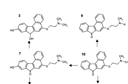
Fig. 1: Chemical structures of benfluron (compound No. 10) and its metabolites with their hypothetical pathways.
Fig. 2:Diagram of a recirculating liver perfusion apparatus:
1. thermometer; 2. constant head device; 3. electromanometer; 4. three-way stopcock; 5. liver platform with organ; 6. vial to collect bile; 7. flow meter;
8. thermostatically controlled reservoir; 9. thermostat;
10.filter;
11. peristaltic pump; 12.pneumoxide; 13.oxygenator; 14. manometer;
15.thermostatic control unit; 16.constant temperature cabinet.
1 16
15
14
13
12
11
10 2
3
4
5
6
7
8
9
and ethyl acetate (all of analytical grade, Lachema) were used for sample preparation, thin-layer chromatography (TLC) and high-performance liquid chromatography (HPLC).
Nonylamine buffer for the mobile phase and five ben-zo[c]fluorenes standard mixtures (compounds 4, 7, 8, 9 and 10, synthesis: M. Nobilis) were used for HPLC assay.
Laboratory animals
Male rats Wistar Han II (Rattus norvegiusvar. alba, con-ventional breeding facility of the Research Institute for Pharmacy and Biochemistry, Konárovice nad Labem, Czech Republic; 250-300 g) were used. They were fasted overnight and were allowed free access to water before the experiment. The experiments were approved by the local et-hics committee.
Liver Perfusion
The rat liver was perfused in vitrousing a modified sur-gical and perfusion technique described previously (8). Animals were anaesthetized with pentobarbital (60 mg/kg) before surgery. Freshly prepared and filtered albumin- and erythrocytes-free Krebs-Henseleit bicarbonate buffer (pH 7.4), supplemented with glucose (0.1 %), polyvinylpyrroli-done (3.5 %) as the plasma expander, and heparin (6.7 i.u./ml) and bubbled with humidified mixture of 95 % CO2 and 5 % O2, was delivered to the portal vein catheter. A flow rate of about 4 ml/g liver/min was maintained. The temperature of the perfusion cabinet and perfusion medi-um was thermostatically controlled at 37±0.5°C. Perfusion was conducted using the recirculating mode. Cannulation of the common bile duct permitted collection of bile, the flow rate of which was determined gravimetrically. An ini-tial stabilization period of 30 min was allowed before ad-ding benfluron to the perfusion medium.
The perfusate flow rate, bile production and organ ap-pearance were determined during the perfusion, organ we-igh and organ histopathology were determined after the perfusion to evaluate liver viability. The IPRL method was also previously established according to organ oxygen con-sumption using a Clarke-Type oxygen electrode (Lazar Research Institute, USA).
In the first series of experiments, IPRL preparations were perfused with different bolus doses of benfluron: 200, 100 and 30 mg/kg (n = 2 for each dose) and 1 mg/perfusa-te volume (n = 4). These experiments were performed to de-fine the metabolic profile of benfluron and to assess its various metabolites in the perfusate and bile. Perfusate and bile samples were collected at 15-min intervals after the ad-dition of benfluron.
Additional two IPRL experiments were conducted in which the livers were perfused with constant infusion rate of benfluron (0.1 mg/min) to produce a sufficient quantity of conjugates for their further identification. Bile samples were collected at 10-min intervals during 120 min of perfu-sion.
Sample preparation
The samples of perfusion liquid were alkalized with the same volume of 15 % aqueous ammonia to pH 9-10 and re-peatedly extracted (three times) with 10 ml of ethyl aceta-te. Ethyl acetate extracts were evaporated in vacuo (max. 40°C) to dryness. The residues were dissolved in a known volume (usually 1-2 ml) of the mobile phase, to be used in HPLC. The collected bile was only diluted with the mobile phase used in HPLC or in methanol for preparative TLC.
Chromatography
A Thermo Separation Products chromatograph setup was used. An HPLC column LiChroCART 125 x 4 mm with a precolumn LiChrospher 100 RP-18 (Merck) were used. The samples were assayed using a Spectra FOCUS high speed scanning UV detector. Detection was performed in dual wavelength mode (295 and 340 nm) or in high-spe-ed scanning mode (range 195-365 nm).
A preparative TLC was used for the isolation of the newly found metabolites and their conjugates in bile. For more details see the literature (12).
Liquid Chromatography-Mass Spectrometry
A Beckman System Gold setup (pump 125S, diode-ar-ray UV detector 168) and a Finnigan MAT setup (LCQ ion trap mass spectrometer coupled with a liquid chromato-graph by an electrospray interface) were used for metaboli-tes identification (LC/MS). It allowed to follow molecular masses of the compounds in the sample as well as to per-form fragmentation of selected ions.
Calculation
A Table Curve 2D software (SPSS Inc., version 4) was used to calculate pharmacokinetic data (t1/2α= half-time associated with the rapid elimination phase, t1/2β = half-time associated with the slow elimination phase) from mean perfusate and bile concentrations of benflurone and its metabolites. Bile excretion rate of metabolite was ex-pressed as mean ± standard deviation.
Results
It revealed, in a short retention time of HPLC analyses, the main metabolite of benfluron, identified by LC/MS as the product of conjugation of 9-hydroxy benfluron with glu-curonic acid formed during the Phase II biotransformation, i.e. O-glucuronide of 9-hydroxy benfluron.
Kinetic disposition of benfluron in the IPRL system.
A two-compartment model was used to depict benflu-ron elimination from an isolated perfused liver preparation. The rate of disappearance of benfluron from the perfusate of such a system is described by the following biexponenti-al equation:
c[benfl.]t= 2757e-0.18425.t+ 94e-0.01095.t
Evaluation of the pharmacokinetic parameters revealed the half-time of the rapid distribution phase of benfluron t1/2α= 3.76 min. The half-time of the elimination phase of benfluron was determined to be t1/2β = 63.30 min. The course of elimination of benfluron from the perfusion medium in dependence on time is shown in Fig. 3. Graphic representation of excretion of the principal me-tabolite (glucuronide 9-hydroxy benfluron) into bile in de-pendence on time is shown in Fig. 4. The half-life of achieving its steady-state phase of excretion was estimated to be 10.40 min.
Fig. 3. The elimination kinetics of benfluron from the per-fusate after administration of 1 mg of benfluron into the IPRL system.
Fig. 4. The kinetics of biliary excretion of the principal metabolite of benfluron (conjugate of 9-hydroxy benflu-ron) after administration of 1 mg of benfluron into the IPRL system.
Percentile share of the conjugates from the administe-red dose represented 70 % of the total amount of excreted benfluron and its metabolites into bile after 2 hours of per-fusion. For details see Tab. 1.
0 500 1000 1500 2000 2500 3000
25
0 50
Time (min)
Benf
lur
on (nmol)
75 100 125
0 25 50 75 100 125 150 175 200
25
0 50
Time (min)
Con
jugate of 9-HO benf
lur
on (nmol)
75 100 125
Tab. 1: Percentile representations of the conjugates from the administered dose in bile in vitro in dependence on time.
Discussion
The use of the IPRL method made it possible to find metabolites of Phase I and Phase II biotransformation of benfluron. The presented results clearly show that benflu-ron undergoes massive biotransformation in the liver com-partment, above all hydroxylation in position 9 and subsequent conjugation with glucuronic acid.
It was not possible to implement the original intention of finding minority metabolites by saturating biotransfor-mational pathways by using large doses of benfluron. The use of high benfluron concentrations in the IPRL system resulted in marked changes in the examined parameters of functional capacity of the isolated liver, i.e. a decrease in perfusate flow, a decrease in oxygen consumption, and fai-lure of biliary excretion. For this reason the doses were gra-dually decreased from 200 mg/kg to 1 mg/volume of perfusion medium. This dose did not result in the described changes and neither did continual infusion at a rate of 0.1 mg/min which was employed to produce sufficient amounts of metabolites intended for their identification using the LC/MS method. Interpretation of the changes in the functional capacity of the liver preparation after admi-nistration of large doses of benfluron can be based on a re-cent paper by Kopecký F. and Kopecká B. (3), which solves the physico-chemical properties of benfluron in aqueous medium in dependence on the ionic strength of the soluti-on. Benfluron hydrochloride is a substance relatively well soluble in aqueous media. However, it has been found that in the presence of potassium chloride (and other electroly-tes) in approximately osmotic concentrations (cKCl = 0.15 mol/l) benfluron produces ionic pairs and molecular asso-ciates without an outer electric charge, which results in mic-roprecipitation of benfluron in the solution. These multimers, as substances with a relatively large molecular mass, could be the cause of the above-mentioned loss of the functional capacity of IPRL, as under these conditions the solubility of benfluron in aqueous medium rapidly decrea-ses, which can result in an impairment of the microvascular system of the liver. Under these conditions the solubility of benfluron resembles that of the unprotonized benfluron base and it is therefore only cbenfluron = 1.1.10-4 mol/l.
Recalculation of ion concentrations of employed Krebs-Henseleit solution and the achieved concentrations of ben-fluron in perfusion medium (cbenfluron= 1.9.10-5mol/l) has revealed that the used concentration of benfluron in a dose of 1 mg/volume of perfusion medium lies just below the li-mit of the concentration region of the formation of the abo-ve-mentioned multimers.
An analysis of absorption spectra obtained by high-spe-ed UV scanning detection revealhigh-spe-ed the products of Phase I biotransformation, i.e. demethylated 9-hydroxy benfluron and twice demethylated 9-hydroxy-benfluron, including other previously described metabolites (see Results). In detailed analysis of elution zones in the region of short retention ti-mes, the spectra characteristic of benzo[c]fluorene structu-res were found. For this reason the HPLC method was modified, which resulted in prolongation of elution times of metabolites and made possible to separate the found struc-tures. Their isolation and identification using the LC/MS method have revealed that it is the product of conjugation of 9-hydroxy benfluron with glucuronic acid. In the regions of short retention times (HPLC), the method of mass spect-rometry has revealed other masses of molecules, which could correspond to other conjugates (but their full identifi-cation has not been completed yet).
Conjugates excreted into bile in the course of 2 hours of perfusion represent 70 % (Table 1) of the administered dose of the parent drug and 96-98 % of all metabolites found. It gives evidence for a large share of conjugation me-chanisms in biotransformation of benfluron by the liver and suggests possible enterohepatic kinetics of benfluron in
in vivoconditions. The construction of a probable scheme of benfluron biotransformation is shown in Fig. 1.
Acknowledgement
We thank prof. MUDr. Vladimír Herout, DrSc. for his-topathological examinations.
References
1. Francová V, Šmolík S, Schlehrová M et al. Derivatives of benzo[c]fluorene XVI. Absorption, distribution and elimination of 3H-benfluorene, 5-[2-(N,N-dimethy-lamino)ethoxy]-7-oxo-7H-benzo[c]fluorene hydrochloride in rats after oral and intravenous administration. Neoplasma 1985;32:529-36.
2. Jantová S, Horáková K. 9-Hydroxybenfluron Induced Inhibition of Proliferation and Metabolism in HeLa Cells. Cell Biochem Funct 1993;11:131-5.
3. Kopecký F, Kopecká B. Basicity, ionic associations and their effect on solubility of the antioneoplastic benflurone. Chem Papers 1997;51:99-106.
4. Koruna I, Ryska M, Poláková L et al. Metabolity benfluronu in vivo. Cesk Farm 1986;35:451-5.
5. Křepelka J, Roubík J, Holoubek J, Vančurová I. 5-, 6- and 7-substitution derivati-ves of 7-oxo-7H-benzo(c)fluorene. Coll Czechoslovak Chem Commun 1982;47:1258-66.
6. Křepelka J, Vančurová I, Holoubek J, Mělka M, Řežábek K. Derivatives of ben-zo[c]fluorene: II. Synthesis and biological effect of basic ethers of 7-oxo-7H -ben-zo[c]fluorene. Coll Czechoslov Chem Commun 1982;47:1856-66.
7. Kvasničková E, Nobilis M, Hais IM. Chromatographic characterization of in vit-ro metabolites of 5[2-(N,N-dimethylamino)ethoxy]-7-oxo-7H-benzo[c]fluorene. J Chromatogr 1984;295:201-9.
8. Květina J, Guaitani A. A Versatile Method for the in vitroPerfusion of Isolated Organs of Rats and Mice with Particular Reference to Liver. Pharmacology 1969;2:65-81.
Time of perfusion after Conjugates
benfluron administration [cumulative %
[h] from administered dose]
1 12±5
1.5 32±11
9. Mělka M, Křepelka J. Benfluron hydrochloride. Drugs Future 1987;12:745-8. 10. Miko M, Křepelka J, Mělka M. 9-Hydroxybenfluron: cytostatic effects and
inhi-bition of macromolecular biosynthesis in Ehrlich ascites and P388 murine leuke-mia cells. Anti-Cancer Drugs 1991;2:289-95.
11. Miko M, Křepelka J, Mělka M. Cytolytic activities of benfluron metabolites and loss of transplantability. Int J Tiss Reac 1989;XI:143-51.
12. Nobilis M, Anzenbacher P, Pastera P et al. Study of the biotransformation of a potential benzo[c]fluorene antineoplastic using high-performance liquid chro-matography with high-speed-scanning ultraviolet detection. J Chrochro-matography B 1996;681:143-51.
13. Nobilis M, Kvasničková E, Šroler A, Hais IM. Elimination of benflurone and its metabolites in the faeces and urine of rats. Drug Metab Drug Interact 1991;9:225-40.
14. Roubal Z, Poláková L, Grimová J et al. Deriváty benzo(c)fluorenu. XVII. Farmakokinetika Benfluronu u zvířat. Cesk Farm 1985;34:311-3.
Mgr. Zbyněk Svoboda, Institute of Experimental Biopharmaceutics, Joint Research Centre of PRO.MED.CS Praha a.s. and the Czech Academy of Sciences, Heyrovského 1207, 500 02 Hradec Králové 2, Czech Republic. e-mail: [email protected]
During the last Novem-ber we recalled the 70th an-niversary of opening the new public hospital in Hradec Králové, the institu-tion that subsequently has become a basis for the University Hospital of the new local Medical Faculty. Since that anniversary the-re has althe-ready elapsed more than half a year and today’s reminder may thus be taken for rather delayed. Nevertheless, the purport of this article is not only to com-memorate the very famous day when the long lasting desi-re of doctors and various men of light and learning of Hradec Králové came to its fulfilment. I would like to de-monstrate also all the efforts, which had preceded this hos-pital creation.
The care for human health was developing during the history only very slowly. Things that we nowadays consider for quite natural ones were neither dreamed of for many centuries. Today’s hospitals were preceded in the Middle Ages, as well as even for a long part of the New Ages, by so called sick houses. Their tasks
did not lie in the proper treat-ment yet; they rather represented mere shelters for old and sick pe-ople. The first such sick houses were established by Church and consisted mostly from very mo-dest institutions, as proved by the photograph of Jan Smit that do-cuments the by a miracle preser-ved small baroque sick house in Rabštejn upon Střela, erected most probably by Servits in the 18th century.
Also in Hradec Králové and its close vicinity such sick houses arose and disappeared again. The existence of the first one, estab-lished most probably as soon as in the 13th century by the com-mend (monasterial settlement) of German Sick House Brethren, German Knights of the Cross, is
documented in written from the year 1362. Another sick house was founded in the middle of the 14th century at Saint Anna’s Church. By the end of 14th century the ma-gistrate and aldermen together with the parish priest from Holy Ghost founded a big sick house with the St. Anthony Church on the riverside of Orlice. All these sick houses came to a lot of harm during the Hussite wars. That big one at Orlice river ceased to exist during the fortification works in Hradec Králové. Besides the above mentioned sick hou-ses for old and sick people also the special houhou-ses for lep-rous, and later on also for plague and cholera patients were established during the Middle Ages.
As late as by the end of the 18th and beginning od 19th centuries the character of hospitals in this country, as well as in all over Central Europe, started changing into the to-day’s form of existence. In Hradec Králové, that was in the year 1765 by the decision of Emperor Joseph the 2nd con-verted into the military fortified town, the situation was rat-her difficult. Vast suburbs had been taken down and the historical town centre on the hill above the rivers Labe and Orlice confluence were squeezed by the star-shaped walls. The whole city became a bizzare housing estate with conti-nuing existence of offices, trades, church and school insti-tutions, and, above all, housing the strong military garrison. It was quite natural then, that at first the military hospital
HISTORICAL ARTICLE
Dr. Vladimír Panoušek *1936
LOOKING BACK
(To our hospital jubilee)
Vladimír Panoušek
Baroque sick house in Rabštejn upon Střela. Picture taken by Jan Smit.