LABORATORY EXAMINATION
IN NERVE AGENT INTOXICATION
Jiří Bajgar
University of Defence, Faculty of Military Health Sciences, Hradec Králové, Czech Republic: Department of Toxicology; University of South Bohemia České Budějovice, Faculty of Social and Health Studies, Czech Republic: Department of Radiology and Toxicology
Summary: Diagnosis of nerve agent intoxication is based on anamnestic data, clinical signs and laboratory examination. For acute poisoning, cholinesterase activity in the blood (erythrocyte AChE, plasma/serum BuChE) is sensitive, simple and most frequent laboratory examination performed in biochemical laboratories. Specialized examinations to precise treatment (reactivation test) or to make retrospective diagnosis (fluoride induced reactivation etc.) can be conducted. Other sophisticated methods are available, too.
Keywords: Nerve agents; Metabolites; Diagnosis; Blood; Acetylcholinesterase; Erythrocytes; Butyrylcholinesterase; Plasma; Reactivation
REVIEW ARTICLE
Introduction
Chemical weapons (CW) belong to the weapons of mass destruction. Their development, production, use etc. are prohibited and controlled by the internatonal coonven-tion (Convencoonven-tion on the Prohibicoonven-tion of the Development, Production, Stockpiling and Use of Chemical Weapons and on their Destruction, CWC) (16). After ratification with sufficient number of the State Parties, CWC entered into force (29 April 1997) and at present, 188 State Parties are involved including Czech Republic. In the Czech Repub-lic, the State Institute for Nuclear Security (Státní ústav pro jadernou bezpečnost, SÚJB), was established as the exec-utive and control organ responsible for implementation of the Convention in the Czech Republic. Simultaneously, this organ licensed laboratories for ability to work/handle with highly toxic chemicals such as chemical warfare agents (CWA). Without the license it is impossible to work/manip-ulate with CWA (e.g. sarin, soman etc.) and this condition is limiting factor for analysis of these agents. Though the use of CWA is in general prohibited by the Convention, it is not excluded their misuse either in military or terroristic actions (57).
The group of nerve agents is the most important group of CWA. The stocks of these compounds (weaponized or stored) in the Albania, India, South Korea, United States and Russia (officially declared) represent about 69,429 tons (53); it is quite clear that the paper is focused to these agents. Laboratory diagnostics following poisoning with nerve agents is focused mostly to:
– determination of the mother compound or metabolites; examination is specific for such compound but it is
lim-ited to the acute stage of intoxication; usually it is not very sensitive, depending on the dose administered; – determination of changes caused by the agent (group of
agents) in question; it is mostly less specific indicating more group of agents. However, it is sensitive and de-tectable long time interval after the intoxication. Both determinations are realized mostly in the blood (erythrocytes/serum/plasma) but some other materials are not excluded (e.g. tissues such as the brain, muscle, saliva, cerebrospinal fluid etc.). It can be used for forensic purpos-es, too. The problem is determination/information on the normal values in biological sample. For further studies, it can be recommended the monography dealing with many aspects of CWA edited by Professor Gupta (25).
Nerve agents
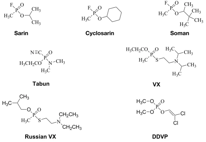
Nerve agents belong to the highly toxic agents; they are the most important group from CWA having high toxicity, rapid action at all routes of administration including inha-lation and percutaneous exposure. They can be divided into two main groups, G-agents and V-agents; between them, group of agents combining in their structures G an V group is observed, called GV (GP) agents. It can be pointed out that compounds of the same basic structure (organophosphates, OP) are used in industry, veterinary or human medicine and in agriculture, e.g.. Metathion, Malathion, Actellic, Dichlor-vos (DDVP), Paraoxon, Parathion, In-stop etc. (Fig. 1)
In the CWC, examples of nerve agents containing gen-eral structure are given (Schedule 1, CWC) (16):
O-Alkyl (≤C10, incl. cycloalkyl) N,N-dialkyl (Me, Et, n-Pro or i-Pro) phosphoramidocyanidates, e.g. tabun;
O-Alkyl (≤C10, incl. cycloalkyl) S-2-dialkyl (Me, Et, n-Pro or i-Pro)-aminoethyl alkyl (Me, Et, n-Pro or i-Pro) phosphonothiolates and corresponding alkylated or proto-nated salts, e.g. VX.
Thus, all these chemicals having above mentioned struc-ture are under control of the CWC.
Mechanism of their action, symptoms, principles of diagnosis and treatment for OP and nerve agents are very similar. Structures of some nerve agents and OP are shown in Fig. 2.
Pharmacodynamics of nerve agents
Irreversible acetylcholinesterase (AChE, EC 3.1.1.7) inhibition at cholinergic synapses is basic toxicodynamic
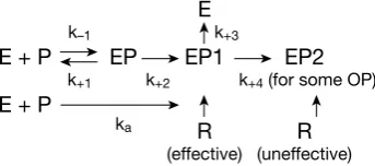
action of nerve agents. Increased level of the neuromediator acetylcholine causes symptomas observed in the course of poisoning. These symptoms are muscarinic, nicotinic and central as described in detail many times (3, 5, 14, 25, 36, 44, 58 etc.). However, before action at cholinergic synapses, nerve agent is penetrating into the blood, and, by this trans-port system is distributed to the target organs – peripheral and central nervous system. During penetration nerve agents react with the blood cholinesterases – erythrocyte AChE and plasma butyrylcholinesterase (BuChE, EC 3.1.1.8). The part of the agent bound to cholinesterases is excluded from toxic action. To the targets, only a part of the dose administered is penetrating and inhibiting AChE at cholinergic synapses. Schematic representation of interaction of AChE with nerve agent/organophosphate is shown in Fig. 3.
AChE (E) is reacting with nerve agent (P) to an in-termediate complex (EP) and then to phosphorylated (phosphonylated) complex EP1. The complex EP1 is reac-tivatable by cholinesterase reactivators (R). EP1 complex is able in some cases to be changed to non-reactivatable complex (EP2) – reactivators are not effective. This reaction (EP1 → EP2) is called aging of inhibited AChE and, typi-cally, it is observed for interaction of AChE with sarin and soman (Fig. 4). Relevant rate constants (k+1,−1,+2,+3,+4 and ka) for the reactions are indicated. The complex EP is not eas-ily detectable, EP1 and EP2 complexes can be determined. Their distinction is difficult. Non-cholinergic effects are not specific and they are observed in long term intervals (hours– days) following intoxication (3, 5, 32, 36, 44).
Spontaneous recovery of AChE activity (reactivation) is long lasting as well as the synthesis of AChE de novo (3, 5, 36, 44, 58, 59). Reaction of AChE and nerve agents is of Fig. 1: General structure of organophosphates: R1–2 are
hy-drogen, alkyl (including cyclic), aryl and others, alkoxy, alkylthio and amino groups. R3 is a dissociable group, e.g. halogens, cyano, alkylthio group, rest of inorganic or organic acid. More about the chemistry of organophosphates see e.g. Fest and Schmidt (23); modern trends in the development of nerve agents was described by Halámek and Kobliha (27)
Fig. 2: Structural formulae of some nerve agents and DDVP R2
R1 P R3 O
P O F
O H3C
CH3
CH3
PO F
O H3C
P O F
O H3C
CH3
H3C CH3
CH3
PO C
N H3CH2CO
H3C
CH3
N H3CH2COPO
S
H3C N
H3C CH 3
CH3
H3C
Sarin Cyclosarin Soman
P O O
S
H3C N
CH2CH3
CH2CH3
H3C
CH3
P O O
O O H3C
H3C Cl
Cl
Russian VX DDVP
system) reaches to a small fraction of the dose administered (10% and less). Detoxification of nerve agents and organo-phosphates was described in detail by Jokanovič (29). The binding of nerve agent to cholinesterases is important and advantageous for diagnostic purposes: blood cholinesterases are easily accessible for laboratory examination.
AChE a BuChE differences
AChE and BuChE are similar enzymes differing in their localisation, enzymatic properties, sequence of amino acids and physiological function (3, 5, 13, 17, 37, 45, 52, 61). AChE is observed at cholinergic synapses, erythrocytes and BuChE is contained mostly in plasma/serum and liver. AChE and BuChE have different sensitivity to substrates and inhibitors; inhibition by substrate is typical for AChE. The function of AChE is splitting neuromediator acetylcho-line at choacetylcho-linergic synapses; its function in erythrocytes is not yet known. The function of BuChE is not known, it can play a complementary role in cholinergic transmission or it is involved in non-specific detoxification reactions and neurological diseases (5, 17,18, 29).
A qualitative difference between the BuChE of suxam-ethonium sensitive individuals and that of other patients were described in detail by Whittaker (60). The biosynthe-sis of BuChE is controlled by two allelic genes, Eu
1 and Ea1. Individuals with the combination Eu
1Eu1 are homozygotes with normal BuChE activity; a combination of Ea
1Eu1 (hete-rozygotes) and Ea
1Ea1 (homozygotes) resulted in diminished BuChE activity. The presence of a silent gene (Es
1) was also proposed and a fourth gene controlling biosynthesis of Bu-ChE (fluoride resistant, Ef
1) was recognized; the hypothesis was in general established by family studies. When Bu-ChE is genetically changed to lower activity, all of drugs containing ester bond (succinylcholine, some local anaes-thetics) are not hydrolysed by BuChE. Thus, the dose of
Fig. 4: Demonstration of aging for sarin and soman. It is the change of reactivatable complex (EP1) to not reactivatable complex (EP2) and forming relevant alcohol
E
k–1 k+3
E + P EP EP1 EP2
k+1 k+2 k+4 (for some OP)
E + P
ka R R
(effective) (uneffective)
Fig. 3: Schematic representation of interaction between AChE (E) and nerve agent/organophosphate (P)
Sarin
O P
O O
H3C E H2O
HO P
O O
H3C E
OH
+ Aging +
H3C
H3C
H3C
H3C
Soman
O P
O O
H3C E
H2O
HO P
O O
H3C E
OH
+ Aging +
CH3
CH3
H3C
H3C
H3C
H3C
H3C
H3C
EP1 EP2 classic inhibition kinetics: reactions important for laboratory
diagnosis are shown in Fig. 3.
The effect of reactivators is limited by the rate of ag-ing process; it is chemical reaction when inhibited AChE is changed to be resistant to the effect of reactivators. The rate of this reaction is dependent on the time and the structure of inhibitor. The half-lives of aging for soman are reported to be in minutes; for sarin, the half-life is about 10 hours and AChE inhibited with VX is dealkylated within 24 and more hours (e.g. 5, 36). This fact is limiting factor for the treat-ment using reactivators. For OP insecticides, this reaction is not practically important (they are not dealkylated) (Fig. 4). Pharmacodynamics of nerve agent can be described by the simple scheme containing following steps – penetration (resorption) through biological barriers (dependent of the route of administration), transport and distribution by the transport system (blood) to sites of metabolic and toxic ef-fect (Fig. 5).
these drugs adminitered to the patient can be relatively high and some complications (e.g. succinyl choline apnoea) can be observed.
Diagnosis
Anamnestic data are very important for diagnosis of OP/nerve agent poisoning especially in connection with possible terroristic attack. Detection or identification of the agent is also very important, simultaneously with the clinical status of poisoned patiens and laboratory examination. De-tection of nerve agent is possible using special equipment. Clinical signs such as miosis and failure of accomodation, salivation, lacrimation and sweating, breath difficulties, nausea and vomiting and, later on, fasciculations and con-vulsions are important clinical signs.
The most important diagnostic test is determination of cholinesterase activity in the blood (erythrocyte AChE or plasma/serum BuChE) (5, 31, 56, 57). Specialized examinations mostly needs more sophisticated methods (11, 12).
From practical point of view, in nerve agent intoxication, erythrocyte AChE activity is determined. It correlates with symptoms of poisoning and with AChE activity/inhibition not only in the peripheral target organs (diaphragm), inhi-bition is very similar to that observed in the central nervous system (ponto medullar area of the brain). Therefore, AChE
activity in the red blood cells corresponds with pharmacody-namics. It is more valid for nerve agents; for OP insecticides, the correlation is not so closed (5, 55, 58).
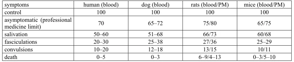
Table 1 shows changes in erythrocyte and brain (pon-tomedullar area) AChE activity following intoxication with nerve agents (sarin, soman, VX). When the blood activity is decreased to 70% of normal values (without any symptoms), it is indication for exclusion of the worker from contact with nerve agent. At this inhibition level, no symptoms are ob-served. When the activity is decreased to 40–70% of normal values, muscarinic symptoms are observed; further decrease to 15–30% correlates with fasciculations and convulsions, and, at the time of death, the activity determined is cca 5% and less. Moreover, the decrease of AChE activity in the erythrocytes copied the activity changes in the target or-gans. However, it is necessary to point out that activity of erythrocyte AChE and plasma BuChE varied individually (more for BuChE), and therefore, individual normal activity in workers with nerve agents is recommended.
From non-specific examinations, leukocytosis and an in-crease of aminotransferases are detected. For final diagnosis, reactivation test is very useful. The principle of the test is double determination of cholinesterase activity – the first one without reactivator, the second one with reactivator. The direct determination of the nerve agent (or metabolites) is possible but it is limited to short period after the exposure. Retrospective diagnosis based on specialized examination is also valuable (as it was demonstrated in sarin victims in Japan) but it requires sophisticated approach (usually GC with MS) (42).
Smear and detection
Smear and detection of contaminated material is possible but these methods are not suitable for clinical laboratory, it is purely chemical approach.
Free nerve agent
Detection of the free agent is possible in the blood/ plasma but the detection is timely limited to hours after
METABOLIZATION
NA/OP PENETRATION TRANSPORT, DISTR.
TOXIC EFFECT
smear blood urine, organs, faeces
(detection) fluids (amniotic, cerebrospinal, saliva etc.)
Fig. 5: Importants steps for toxicodynamics (material for laboratory diagnostics ante or post mortem)
Tab. 1: Symptoms of nerve agent intoxication and AChE activity changes in erythrocytes (man, dog) and in erythrocytes and brain (pontomedullar area, PM) in rats and mice. Table elaborated using data Bajgar et al. (6, 7); Bajgar (5); Capacio et al. (14); Jun et al. (30)
symptoms human (blood) dog (blood) rats (blood/PM) mice (blood/PM)
control 100 100 100 100
asymptomatic (professional
medicine limit) 70 65–72 75/80 65/75
salivation 50–60 51–68 66/73 60/68
fasciculations 20–30 25–38 27/36 25–29
convulsions 10–20 12–18 13/15 10/11
the exposure. Disadvantage is the necessity of calibration requiring original agent not known at the beginning of in-toxication. This situation is similar for metabolites.
Metabolites
In the case of sarin, soman and cyclosarin exposure, these agents are metabolized through their relevant acids – isopropylmethyl-, pinacolylmethyl- a cyclohexylmethyl-phosphonic acids, finally forming methylcyclohexylmethyl-phosphonic acid MPA):
O ║ H3C ─ P ─ OH
│ OH
In case of VX exposure, diisopropyl aminoethyl methyl sulphate or MPA can be detected. Metabolites mentioned (mostly MPA) can be detected in urine, too. The methods for detection are mostly combination of GC-MS, LC-MS or GC-FPD and other combinations (9, 20, 51, 54). In case of some OP (e.g. parathion, paraoxon) is possible to detect their metabolite in urine (p-nitrophenol) using simple spec-trophotometric method (4, 22).
AChE activity in erythrocytes
Determination of AChE activity can be performed using different methods based on various principles: in each case, substrate (either natural – acetylcholine or artificial – e.g. acetylthiocholine) is added to biological sample and un-hydrolysed substrate or reaction products are determined by different methods: spectrophotometric, pH changes indicators, colorimetric, polarographic, fluorimetric, radi-ochemical, enzymatic and others. The determination can be performed continually or discontinually, it is possible to increase the specificity using selective AChE or BuChE in-hibtors (review see e.g. 4, 5, 14, 42, 43).
The most common method is original Ellman’s method (21) and its modifications. It is based on hydrolysis of the thiocholine substrates (acetyl- and butyrylthiocholine or others). After enzymatic hydrolysis, the relevant acid and thiocholine are released and SH-group of thiocholine is detected using 5,5′-dithiobis-2-nitrobenzoic acid (DTNB) forming 5-mercapto-2-nitrobenzoate anion determined spectrophotometrically at 412 nm. Sometimes this method is used with specific inhibitors and there are many modifi-cations described in the literature. This method is in good correlation with other methods. It is sufficiently specific and sensitive and it is used for different purposes in many labo-ratories around the world. It is very useful in monitoring of workers exposed to nerve agents or OP; evaluation and 40 years experience with this determination including suspect intoxications was described by Jun et al. (30). The disadvan-tage of this method is that the method is not used routinely in clinical laboratories. Theoretically, it would be easily
per-formed in the clinical biochemical laboratory by the change of substrate (not butyrylthiocholine but acetylthiocholine) or by the use of specific BuChE inhibitors. It is possible to determine AChE activity in the whole blood (3, 26); it is represented by 90% of AChE and 10% of BuChE (human) and 29% of BuChE and 71% of AChE (rat) (3). Advantage of this method is sampling – the blood is obtained from finger tip (50 µl) and it is not separated to erythrocytes and plasma by centrifugation (4, 5). The determination of indi-vidual molecular forms of AChE or BuChE is also possible using separation techniques (electrophoresis, ultracentrifu-gation etc.) – high molecular forms are more sensitive to nerve agents than low molecular forms (3). It is used more frequently for OP insecticides.
The expression of activity is important for comparison of different results described by various laboratories. It is ex-pressed as mmoles of substrate hydrolyzed per time (minute) by defined volume (plasma/serum) or weight of the material (wet, dry, content of nitrogen etc.). Expression in Units is also possible. In the clinical laboratory, catals/l are used for the expression of enzymatic activity (kat/l) – it is 1 mol of substrate hydrolyzed per sec/liter or kg.
Reference values: normal AChE aktivity in erythrocytes or in the whole blood varies about 110.2 (±11.2) µkat/l and 133.3 (±14.5) µkat/l; optimal choice seems to be determina-tion of individual activity before possible exposure or before work with nerve agents. This examination in these cases is obligatory.
BuChE activity in serum/plasma
Determination of cholinesterase activity is based on sim-ilar principles as those described for AChE (reviews see e.g. 3, 4, 5, 14, 43). The most common method is mentioned Ell-man’s method (21) and its modifications. The determination of BuChE in the blood is the basic method for diagnosis and therapy monitoring for OP poisoning; however, it is neces-sary to be combined with clinical observation.
BuChE determination in the plasma or serum is routine examination in clinical biochemistry. It is used more fre-quently than that of AChE in the red blood cells. BuChE decrease indicates either a diminishment of the enzyme synthesis or a decrease in the number of production cells in the liver (4); some drugs also influnce the plasma BuChE activity. A special case of diminished BuChE activity is the hereditary affected presence of atypical variants of BuChE (59). BuChE activity can be affected by different drugs and diseases – except OP/nerve agents and carbamates there are hormones (e.g. peroral anticonception), some heavy met-als, carbon disulphide; BuChE can be changed in gravidity, antitumor therapy, in some psychiatric and neurological diseases, γ-irradiation, liver diseases, myocardial infarction (2–5, 18, 37).
determined to be 150.5 (±31.3) µkat/l; individual activity (at the first examination) is recommended. It is possible to sepa-rate individual isoenzymes of BuChE, however, there arises a question of separation medium (agar, polyacrylamide, etc.) and the method of quantitative evaluation of the activity of these isoenzymes (3, 4, 30, 41, 47, 58).
Reactivation test
The principle of the test is double determination of cholinesterase activity in pooled blood sample. The first determination is without adding of the reactivator and rep-resents normal activity; if it is decreased, it is indication of exposure to nerve agents. According to the degrese of inhibition, the symptoms and steps of the poisoning can be derived. The second determination in the presence of reac-tivator shows the response of inhibited cholinesterase to the oxime added; usually, it is higher than that without reactiva-tor. The increase indicates that the enzyme can be reactivated (it is EP1); if the enzyme is unreactivatable, it indicates that the enzyme is in unreactivatable step (EP2) and the activity of the second sample will be the same or lower than the first one. The level of reactivation could be helpful in differential diagnosis – what type of nerve agent was used: in case of soman, zero or very low reactivatability is observed. Modere increase indicates intoxication with sarin, and, high reacti-vatability suggests VX intoxication (Table 2).
It is possible to use different reactivators for second determination and – depending on the results – to choose the best reactivator for the treatment. Disadvantage of this method is non routine realization in clinical laboratory. This test was experimentally verified on dogs and rats in vivo and on human and dog blood in vitro (1–3, 5, 7).
Other markers
Generally, hydrolases and esterases and phosphatases (alkaline and acid) are sensitive to nerve agents but the sen-sitivity and specificity is not very high. Transaminases (AST, ALT) can be slightly elevated as well as leukocytes but there are also non specific changes. In acute intoxication, it is necessary to search blood oxygen (and CO2 saturation) and pH of the blood and the levels of acid metabolites (possible acidosis) (4, 5, 14, 36).
Stresogennic parameters
It is mostly corticosterone; its level is elevated 3–8 hours after the exposure. The concentrations of c-AMP and c-GMP are also elevated at these intervals (3, 44). Stress-ogenic effects as well as behavioral changes are observed also following exposure to low level of nerve agents (32).
Fluoride induced reactivation
The method of cholinesterase determination in the blood indicates the decrease of activity only (i.e. the presence of inhibitor). It does not to identify the mother compound, and, when the inhibition is too low (10–20%, i.e. the rest of ac-tivity is about 80–90%), it is in the limit of sensiac-tivity. It is not suitable for retrospective diagnosis. The method of fluoride-induced reactivation has not these disadvantages. The method was developed by TNO-Prins Maurits Labora-tory, Rijswijk, the Netherlands; it is based on reactivation of phosphylated cholinesterase and carboxylesterase (CaE, EC 3.1.1.1) by fluoride ions. Treatment of the inhibited en-zyme with fluoride ions can inverse the inhibition reaction yielding restored enzyme and a phosphofluoridate which is subsequently isolated and quantified by GC and phospho-rus-specific or mass spectrometric detection (46). Important fact is the presence of CaE or BuChE in the material exam-ined. Human (and monkey) plasma does not contain CaE but its BuChE concentration is relatively high [70–80 nM (10, 39)], much higher than the concentration of AChE in blood [ca. 3 nM (28)]. Plasma of laboratory animals, such as rats and guinea pigs, contains considerable concentrations of CaE in addition to the cholinesterases. The method allows partial identification of the OP whereas the lifetime of the phosphylated esterase (and consequently the retrospectivity of the method) is only limited by spontaneous reactivation, in vivo sequestration and aging. The rate of the latter pro-cess (aging) depends on the structure of the phosphyl moiety bound to the enzyme and on the type of esterase. Phosphyl-ated CaEs generally do not age.
Based on this method for retrospective detection of ex-posure to OP, exex-posure of victims of the Tokyo incident to a nerve agent, probably sarin, could be established from analysis of their blood samples (46). Later on, this method was used for the study of the effect of low inhalation dose of soman (6).
Alternatively, assays based on mass spectrometry have recently been developed which will also diagnose on the basis of the aged phosphonylated BuChE (24). The method was also modified and improved (19, 38).
Combination of different appoaches was able to establish definitive diagnosis of sarin use in Tokyo subway (40, 49). Thus, fluoride induced reactivation of nerve agent-inhibited AChE is a reliable and retrospective method to establish nerve agent-exposure. It is limited to compounds that regen-erated with fluoride ions.
Tab. 2: Results of the reactivation test for human, dog and rat erythrocyte AChE following inhibition by nerve agents in vitro (human) and in vivo (rats and dogs). Table elaborated using data of Bajgar (1–3, 5); Bajgar et al. (7)
Nerve
agent human Erythrocyte AChEdog rat
soman 00–10 15 (10–24) 10 (5–15)
sarin 25–65 63 (50–78) 55 (41–72)
Tab. 3: Comparison of different methods for diagnosis of nerve agent intoxication
method diagnostic validity for poisoning instrumen- tation time frequency of use difficult- ness demanding- ness
smear for exposure + – +++ minutes ++ + +
free + metab acute +++ hours ++ ++ ++
AChE ery/wh bl acute + minutes ++ + +
BuChE pl/se acute + minutes ++++ + +
react test acute + treatment + hour + ++ ++
other developed ++ minutes + + +
stress mark developed ++ hour + + ++
fluoride react retrospective(low doses) +++ hour ++ ++ +++
enzym dig retrospective +++ hours + +++ +++
adducts retrospective +++ hours + ++ ++
Enzymatic digestion
Fluoride induced reactivation is limited to compound able to react to adequate manner. This disadvantage is eliminated by the method of enzymatic digestion. This is a novel and general procedure for diagnosis of exposure to OP, which surpasses the limitations of the fluoride reactivation method (48). It is based on the rapid isolation of BuChE from the plasma by the affinity chromatography, digestion with pepsin followed by liquid chromatography with the mass spectrometric analysis of phosphylated nonapeptides resulting after the digestion of inhibited BuChE with pepsin (15). The method can be applied for the detection of expo-sures to various OP pesticides and nerve agents including soman. This approach is very valuable and represents a new field for the improvement of diagnosis with nerve agents and OP. A comprehensive review of the methods for retro-spective detection of exposure to toxic scheduled chemicals has been published by Noort et al. (43). Method allows to confirm retrospective exposure to different OP and nerve agents including soman. For review see also e.g. ref. 4, 12, 14, 42, 43, 56.
Adducts
New approach was described by Lockridge et al. (35). It is based on detection of OP/nerve agent adducts with tyrosine (from proteins in the blood), without binding to cholinesterases (8, 50). This method opens new possibilities of detection aspecially in case of OP having less inhibition efficacy to plasma BuChE (33).
Summarization of the methods shows Table 3.
Conclusions
From laboratory examinations used for diagnosis of nerve agennt poisoning, determination of AChE/BuChE ac-tivity in the blood remains as basic, sensitive and frequently
used method important for acute poisoning. Other methods are available for specification of therapeutic countermeas-ures, low doses exposure or retrospective diagnosis.
Acknowledgements
The study was supported by the grant of Ministry of Defence “A long term organization development plan 1011”.
References
1. Bajgar J. The influence of inhibitors and other factors on cholinesterases. Sbor Ved Pr LFUK (Hradec Kralove) 1991; 34: 3–75.
2. Bajgar J. Biological monitoring of exposure to nerve agents. Brit J Ind Med 1992; 49: 648–653.
3. Bajgar J. Organophosphates/nerve agent poisoning: mechanism of action, diag-nosis, prophylaxis and treatment. In: Makowsky GM, ed. Advances in Clinical Chemistry, vol. 38, Elsevier Academic Press, San Diego, CA, 2004: 151–216. 4. Bajgar J. Laboratory diagnosis of organophosphates/nerve agent poisoning. Klin
Biochem Metab 2005; 13: 40–47
5. Bajgar J. Complex view on poisoning with nerve agents and organophosphates. Acta Medica (Hradec Králové) 2005; 48: 3–21.
6. Bajgar J, Schans van der MJ, Fusek J, et al. (2003): Biochemical effects of low level exposure to soman vapour(Final report). Cooperative project of TNO Prins Maurits Laboratory and Purkynĕ Military Medical Academy, Czech Republic. Assignment number A00D448. 2003, 41 pages incl. 2 Annexes.
7. Bajgar J, Fusek J, Bartosova L, Jun D, Kuca K. Evaluation of reactivation test in anaesthetized dogs with experimental intoxication with nerve agents. J Appl Toxicol 2006; 26: 439–443.
8. Bao Y, Liu Q, Chen J, et al. Quantification of nerve agents adducts with albumin in rat plasma using liquid chromatorgaphy-isotope dilution tandem mass spectromtry. J Chromat A 2012; 1229: 164–171.
9. Barr JR, Driskell WJ, Aston LS, Martinez RA. Quantitation of metabolites of the nerve agents sarin, soman, cyclohexylsarin, VX and Russian VX in human urine-using isotope-dilution gas chromatography. J Analyt Toxicol 2004; 28: 372–378. 10. Bisschop de HC, De Meerleer WAP, Willems JL. Stereoselective phosphonylation of human serum proteins by soman. Biochem Pharmacol 1987; 36: 3587–3591. 11. Black RM. An overview of biological markers of exposure to chemical warfare
agents. J Analyt Toxicol 2008; 32: 2–9.
12. Black RM, Read RW. Biological markers of exposure to organophosphorus nerve agents. Arch Toxicol 2013; 87: 421–437.
13. Bosak A, Katalinic M, Kovarik Z. Cholinesterases: structure, role, and inhibition. (in Croatian). Arch Indust Hyg Toxicol 2011; 62: 175–190.
14. Capacio BR, Smith R, Gordon RK, et al. Medical diagnostics. In: Lenhart MK, Tuorinski, SD, eds. Textbook in Military Medicine . Office of the Surgeon General, Department of the Army, United States of America and US Army Med-ical Department Center and School, Fort Sam Houston, San Antonio, TX 2008: pp. 691–752.
on-line pepsin digestion-liquid chromatography-tandem mass spectrometry con-figuration for the rapid analysis of protein adducts of chemical warfare agents. J Chromat. B 2008; 870: 91–97.
16. CWC: Convention on the Prohibition of the Development, Production, Stockpiling and Use of Chemical Weapons and on their Destruction. OPCW 1994, 168 pages. OPCW, The Hague 1994.
17. Čolovič MB, Krstič DZ, Lazarevič-Pašu TD, Bondžič AM, Vasic VM. Acetylcho-linesterase inhibitors: pharmacology and toxicology. Curr Neuropharmacol 2013; 11: 315–335.
18. Darvesh S, Hopkins DA, Geula C. Neurobiology of butyrylcholinesterase. Nature Rev Neurosci 2003; 4: 131–138.
19. Degenhardt CEAM, Pleijsier K, van der Schans MJ, et al. Improvement of the fluoride reactivation method for the verification of nerve agent exposure. J Analyt Toxicol 2004; 28: 364–371.
20. Driskell WJ, Shih M, Needham LL, Barr DB. Quantitation of organophosphorus nerve agent metabolites in human urine using isotope dilution gas chromatogra-phy-tandem mass spectrometry. J Analyt Toxicol 2002; 29: 6–10.
21. Ellman GL, Courtney DK, Anders V, Featherstone RM. A new and rapid colori-metric determination of acetylcholinesterase activity. Biochem Pharmacol 1961; 7: 88–95.
22. Eyer F, Eyer P. Enzyme-based assay for quantification of paraoxon in blood of parathion poisoned patients. Hum Exp Toxicol 1998; 17: 645–651.
23. Fest C, Schmidt K-J. The chemistry of organophosphorus pesticides. Second Re-vised Edition., Berlin, Heidelberg, New York: Springer-Verlag 1982: 360. 24. Fidder A, Hulst AG, Noort D, de Ruiter R, van der Schans MJ, Benschop HP,
Langenberg JP. Retrospective detection of exposure to organophosphorus anti-cholinesterases: mass spectrometric analysis of phosphylated human buty-rylcholinesterase. Chem Res Toxicol 2002; 15: 582–590.
25. Gupta, R.C., Editor. Handbook of Toxicology of Chemical Warfare Agents, Am-sterdam, Boston, Heidelberg, London, New York, Oxford, Paris, San Diego, San Francisco, Singapore, Sydney, Tokyo, Elsevier/AP, 2009, 1147 pages. 26. Haigh JR, Lefkowitz IJ, Capacio BR, Doctor BP, Gordon RK. Advantages of the
WRAIR whole blood cholinesterase assay: comparative analysis to the micro-Ell-man, Test-mate ChETM, and Michel (ΔpH) assays. Chem-Biol Interact 2008; 175:
417–420.
27. Halámek E, Kobliha Z. Potenciální bojové chemické látky. Chem listy 2011; 105: 323–333.
28. Heath DF. Organophosphorus poisons. Anticholinesterases and related com-pounds. In: Alexander P, Bacq ZM, eds. Modern trends in physiological sciences. Pergamon Press, Oxford, London, New York, Paris, 1961: 241.
29. Jokanovic M. Current understanding of the mechanisms involved in metabolit detoxification of warfare nerve agents. Toxicol Lett 2009; 188: 1–10. 30. Jun D, Bajgar J, Kuca K, Kassa J. Monitoring of blood cholinesterase activity in
workers exposed to nerve agents. In: Gupta RC, ed. Handbook of Toxicology of Chemical Warfare Agents, Elsevier/AP, 2009: 877–886.
31. Knaack JS, Zhou Y, Abrey CW, et al. A high-throughput diagnostic method for measuring human exposure to organophosphorus nerve agents. Analyt Chem 2012; 84: 9470–9477.
32. Kassa J, Kuca K. Exposure to organophosphorus compounds – oximes, neu-roprotection and cognitive functions. In: The neurochemical consequences of organophosphate poisoning in the CNS. Weissman BA, Raveh L. eds. TRN, Kera-la, India 2010: 77–91.
33. Li B, Eyer P, Eddleston M, et al. Protein tyrosine adduct in humans self-poisoned by chlorpyrifos. Toxicol Appl Pharmacol 2013; 269: 215–225.
34. Lockridge O, Masson P. Pesticide and susceptible populations: people with bu-tyrylcholinesterase genetic variants may be at risk. Neurotoxicology 2000; 21: 113–126.
35. Lockridge O, Schopfer LM, Masson P. Biomarkers of exposure to organophospho-rus poisons: a new motif for covalent binding to tyrosine in proteins that have no active site serine. In: Gupta RC, ed. Handbook of Toxicology of Chemical Warfare Agents, Elsevier/AP, 2009: 847–858.
36. Marrs TC, Maynard RL, Sidell FS. Chemical warfare agents. Toxicology and treatment. J. Wiley and Sons, Chicester, New York, Brisbane, Toronto, Singapore 1996, 243 pages.
37. Massoulié J, Pezzementi L, Bon S, Krejci E, Vallette FM. Molecular and cellular biology of cholinesterases. Progr Neurobiol 1993; 41: 31–91.
38. Meer van der JA, Trap HC, Noort D, Schans van der MJ. Comprehensive gas chromatography with Time of Flight MS and large volume introduction for the
detection of fluoride-induced regenerated nerve agent in biological samples. J Chromat B 2010; 878: 1320–1325.
39. Myers DK. Cholinesterase. VII. Determination of the molar concentration of pseu-docholinesterase in serum. Biochem J 1952; 51: 303–311.
40. Nagao M, Takatori T, Matsuda Y, et al. Definitive evidence for the acute sarin poisoning diagnosis in the Tokyo subway. Toxicol Appl Pharmacol 1997; 144: 198–203.
41. Nagayama M, Akahori F, Chiwata H, et al. Effects of selected organophosphate insecticides on serum cholinesterase isoenzyme patterns in the rat. Vet Hum Tox-icol 1996; 38: 196–199.
42. Noort D, Benschop HP, de Jong LPA. Methods for retrospective detection of exposure to toxic scheduled chemicals: an overview. Voj zdrav Listy 2001; 70: 14–17.
43. Noort D, van der Schans MJ, Bikker FJ, Benschop HP. Diagnosis of exposure to chemical warfare agents: an essential tool to counteract chemical terrorism. In: Dishovsky C, Pivovarov A, eds. Counteraction to Chemical and Biologicalv Terrorism in East European Countries. Book Series: NATO Science for Peace and Security Series A -Chemistry and Biology, 2009: 195–201.
44. Patočka, J. et al. Vojenská toxikologie. Praha, Grada-Avicenum, 2004: 178 pages (in Czech).
45. Pohanka M. Cholinesterases, a target of pharmacology and toxicology. Biomed Papers 2011; 155: 219–230.
46. Polhuis M, Langenberg JP, Benschop HP. New method for retrospective detection of exposure to organophosphorus anticholinesterases: application to alleged sarin victims of Japanese terrorists. Toxicol Appl Pharmacol 1997; 146: 156–161. 47. Sakaguchi K, Nagayama M, Masaoka T, et al. Effects of fenthion, isoxathion,
dichlorvos and propaphos on the serum cholinesterase patterns of dogs. Vet Hum Toxicol 1997; 39: 1–5.
48. Schans van der MJ, Noort D, Fidder A, Degenhardt CEAM, et al. Retrospective de-tection of exposure to organophosphorus anticholinesterases: fluoride reactivation and mass spectrometric analysis of phosphylated human butyrylcholinesterase. The meeting of NATO TG 004 Task Group on Prophylaxis and Therapy of Chem-ical Agents. 4–7 November 2002, Oslo, Norway.
49. Schans van der MJ Laboratory analysis of chemical warfare agents and metabolites in biomedical samples. In: Gupta RC, ed. Handbook of Toxicology of Chemical Warfare Agents, Elsevier/AP, 2009: 827–835.
50. Schopler LM, Lockdridge O. Analytical approaches for monitoring exposure to organophosphorus and carbamate agents through analysis of protein adducts. Drug Test Anal 2012; 4: 246–261.
51. Shih ML, McMonagle JD, Dolzine TW, Gresham WC. Metabolite pharmacokinet-ics of soman, sarin and GF in rats and biological monitoring of exposure to toxic otrganophosphorus agents. J Appl Toxicol 1994; 14: 195–199.
52. Stefanidou M, Athanaselis S, Spiliopoulou H. Butyrylcholinesterase: biomarker for exposure to organophosphorus insecticides. Int Med J 2009; 39: 57–60. 53. Středa L, Patočka J. Zneschopňující chemické látky – ohrožení účelu a cíle
Úmlu-vy o zákazu chemických zbraní. Kontakt 2013, in press.
54. Swaim LL, Johnson RC, Zhou Y, Sandlin C, Barr JR. Quantification of organ-ophosphorus nerve agent metabolites using a reduced-volume, high-throughout sample processing format and liquid chromatography-tandem mass spectrometry. J Analyt Toxicol 2008; 32: 774–777.
55. Thiermann H, Szinicz L, Eyer P, Zilker T, Worek F. Correlation between red blood cell acetylcholinesterase activity and neuromuscular transmission in organophos-phate poisoning. Chem-Biol Interact 2005; 157–158: 345–347.
56. Thiermann H, Kehe K, Steinritz D, Mikler J, Hill I, Zilker T, Eyer P, Worek F. Red blood cell acetylcholinesterase and plasma butyrylcholinesteras status: important indicators for the treatment of patients poisoned by organophosphorus compounds. Arh Hig Rada Toksikol 2007; 58: 359–366.
57. Vale A, Marrs TC, Rice P. Chemical terrorism and nerve agents. Medicine 2012; 40: 77–79.
58. Voicu V, Bajgar J, Medvedovici A, Radulescu FS, Miron DS. Pharmacokinetics and pharmacodynamics of some oximes and associated therapeutic consequences: a critical review. J Appl Toxicol 2010; 30: 719–729.
59. Voicu V, Radulescu FS, Medvedovici A. Toxicological considerations of acetyl-cholinesterase reactivators. Exp Opin Drug Metab Toxicol 2013; 9: 31–50. 60. Whittaker M. Plasma cholinesterase variants and the anaesthesist. Anaesthesia
1980; 35: 174–197.
61. Wiesner J, Kriz Z, Jun D, Kuca, K. Acetylcholinesterase – the structural similarities and differences. J Enz Inhib Med Chem 2007; 22: 417–424.
Received: 08/07/2013 Accepted in revised form: 24/09/2013 Corresponding author: