R E S E A R C H
Open Access
Altered glial marker expression in autistic
post-mortem prefrontal cortex and cerebellum
Catherine Edmonson
1,2†, Mark N Ziats
1,3,4*†and Owen M Rennert
1Abstract
Background:The cellular mechanism(s) underlying autism spectrum disorders (ASDs) are not completely
understood, but ASDs are thought to ultimately result from disrupted synaptogenesis. However, studies have also shown that glial cell numbers and function are abnormal in post-mortem brain tissue from autistic patients. Direct assessment of glial cells in post-mortem human brain tissue is technically challenging, limiting glial research in human ASD studies. Therefore, we attempted to determine if glial cell-type specific markers may be altered in autistic brain tissue in a manner that is consistent with known cellular findings, such that they could serve as a proxy for glial cell numbers and/or activation patterns.
Methods:We assessed the relative expression of five glial-specific markers and two neuron-specific markers via qRT-PCR. We studied tissue samples from the prefrontal cortex (PFC) and cerebellum of nine post-mortem autistic brain samples and nine neurologically-normal controls. Relative fold-change in gene expression was determined using theΔΔCtmethod normalized to housekeeping geneβ-actin, with a two-tailed Student’st-testP<0.05 between groups considered as significant.
Results:Both astrocyte- and microglial-specific markers were significantly more highly expressed in autistic PFC as compared to matched controls, while in the cerebellum only astrocyte markers were elevated in autistic samples. In contrast, neuron-specific markers showed significantly lower expression in both the PFC and cerebellum of autistic patients as compared to controls.
Conclusions:These results are in line with previous findings showing increased glial cell numbers and
up-regulation of glial cell gene expression in autistic post-mortem brain tissue, particularly in the PFC, as well as decreased number of neurons in both the PFC and cerebellum of autistic patients. The concordance of these results with cell-level studies in post-mortem autistic brain tissue suggests that expression of glial cell-type specific markers may serve as a useful alternative to traditional cellular characterization methods, especially when appropriately-preserved post-mortem tissue is lacking. Additionally, these results demonstrate abnormal glial-specific gene expression in autistic brains, supporting previous studies that have observed altered glial cell numbers or activation patterns in ASDs. Future work should directly assess the correlation between cell-type specific marker levels and cell number and activation patterns.
Keywords:Astrocyte, Autistic disorder, Gene expression, Glia, Interneuron, Microglia, Neuron
* Correspondence:[email protected]
†Equal contributors
1Laboratory of Clinical and Developmental Genomics, National Institute of
Child Health and Human Development, National Institutes of Health, 49 Convent Drive, Building 49, Room 2C078, Bethesda, MD 20814, USA
3
University of Cambridge, Robinson College, Grange Rd, Cambridgeshire CB3 9AN, UK
Full list of author information is available at the end of the article
Background
Autism spectrum disorders (ASDs) are neurodevelop-mental syndromes defined by impairments in language, verbal and non-verbal communication, and restrictive/ repetitive patterns of behavior [1]. ASD symptoms mani-fest within the first two years of life, with their severity and presentation varying considerably between individ-uals, thus yielding the‘spectrum’classification. ASDs are estimated to affect 1 in 88 children in the United States, and the prevalence of ASDs is at least four times more common in males than females [2].
The etiology of autism is complex and the neurobio-logical mechanism(s) that result in the clinical pheno-type remain to be fully understood. However, there is strong evidence that the autism phenotype ultimately results from aberrant synaptic wiring in the developing brain [3]. In particular, studies have shown that long-distance communication between disparate neocortical areas may be disrupted in ASDs, causing delays in infor-mation processing within the brain that manifest as the communication, language, and social development problems seen in children with autism [4]. Additionally, parallel research has shown that neuronal micro-circuitry within brain areas may also be disrupted in ASDs, and that this may result in local processing defi-cits within brain regions related to higher functioning, such as the prefrontal cortex (PFC) [5]. Underlying these circuit disruptions is a large body of evidence that has demonstrated decreased numbers of neurons (and their various subtypes) throughout the autistic brain by early childhood in post-mortem studies [6].
In addition to the body of evidence implicating aberrant local and long-distance synaptic dysfunction in ASDs, many studies have demonstrated microglial and astrocyte dysfunction in ASD brains. For instance, post-mortem pathological studies of autistic brain using immunocytochemistry (IHC) and/or stereology have identified microglial activation patterns [7-9], and have demonstrated increased microglial cell density in mul-tiple brain regions [8,10]. Glial activation refers to the well characterized cascade of events that occurs upon reaction of glia to stimuli, resulting in their mobilization, release of cytokines, and ability to phagocytize, among other processes [11]; glial activation is associated with specific gene expression patterns that are distinct from
‘resting’glial gene expression [12]. Furthermore, positron emission tomography (PET) using a microglial-specific radiotracer also demonstrated microglial activation in multiple brain regions of autistic cases [13]. Additionally, studies in a Rett syndrome mouse model, a single-gene deletion disorder with autism as a component, have also demonstrated cellular microglial abnormalities [14], and a remarkable study demonstrated that autistic-like phe-notypes can be partially reversed by replacing mutant
Mecp2 (−/−) microglia with their respective wild-type cells [15].
Increased numbers of astrocytes, with altered cell size and branching patterns, have also been demonstrated in post-mortem autistic brains [16]. Additionally, astrocyte-specific cell marker proteins are increased in multiple autistic brain regions [17,18]. Similar to microglial stud-ies in ASD mouse models, astrocytes have been shown to be abnormal in a number of single-gene ASD models, including Rett [19,20], Fragile X [21], and Tuberous Sclerosis [22]. In parallel to the aforementioned microglial study, it was also shown that replacing mutant astrocytes in Mecp2 (−/−) mice can correct some aspects of the phenotype [23].
However, it is not clear how these separate lines of evidence—one demonstrating immune/glial dysfunction in ASDs and the other implicating synaptic abnormal-ities—may converge into a common mechanism in the autistic brain that ultimately results in the shared clinical phenotype. Because separate studies have shown that microglia and astrocytes play critical roles in sculpting developing synapses during normal neurodevelopment [24,25], it is reasonable to hypothesize that inherent defects or aberrant numbers of microglia and astrocytes in the developing autistic brain may be causative of the synaptic abnormalities by affecting the proper wiring of developing neuronal connections. However, because appropriately-preserved post-mortem autistic brain tis-sue is lacking [26], cellular-level studies assessing glial numbers and activation in human autistic brains have been limited. Moreover, quantification of cell numbers in postmortem tissue by stereology is technically chal-lenging, further limiting the ability of researchers to assess the few appropriately-preserved tissue samples that are available. Finally, no studies have concurrently specifically assessed for microglia, astrocytes, and neurons in the same set of autistic brain samples. As a conse-quence, a comprehensive understanding of the relation-ship between glial and neuron cells in autistic brains is needed.
Therefore, the purpose of this study was two-fold. First, we sought to determine if microglia, astrocyte, and neuron-specific markers were altered in post-mortem autistic brain tissue, in order to further investigate the role of glia in ASDs. Then, we determined if glial and neuronal cell-type specific marker expression patterns are consistent with known cellular-level findings, be-cause gene expression studies of post-mortem human brain are often easier to perform than cell-level studies, and therefore this approach may serve as a valuable
brain tissue from patients with autism and healthy con-trols. Our results provide further evidence for a role of glia in autism pathology, and suggest that assessment of glial cell-type specific markers may serve as a proxy for relative cellular numbers or activation patterns.
Methods
Post-mortem brain samples
Post-mortem brain tissue was obtained from the National Institute of Child Health and Human Development Brain and Tissue Bank, MD, USA. This source obtained consent to use brain tissue for research from each patient or their guardian prior to his/her death, and their protocol was approved by their Institutional Review Board. No patient-specific identifiable information was obtained. Because multiple brain regions have been implicated in ASDs, we performed our analysis in two separate areas that have been consistently demonstrated as abnormal in autism— the PFC and the cerebellum. We obtained post-mortem PFC brain tissue from five individuals with autism and from five healthy controls (Table 1). We obtained post-mortem cerebellum brain tissue from four individuals with autism and four healthy controls. The majority of sample pairs (PFC and cerebellum) were derived from the same donor brain. All cases were Caucasian males, and case controls were matched by age as closely as possible.
RNA isolation and quality control
RNA isolation and quality control analysis were performed as previously described [27]. Briefly, total RNA was extracted using TRIZOL Reagent (Life Technologies, Carlsbad, CA, USA) according to the manufacturer’s protocol. Quantification of RNA was performed using a NanoDrop ND-1000, and RNA integrity was assessed using an Agilent Bioanalyzer 2100 (Table 2).
Reverse transcriptase reaction
Total RNA (1μg) was used in a 20μL reverse transcript-ase reaction to synthesize cDNA with SuperScript3 Reverse Transcriptase (Life Technologies) according to the manufacturer’s protocol. Briefly, 1 μg of total RNA was added to an aqueous solution containing 250 ng/μL of random hexamer and 10 mM deoxyribonucleotide triphosphate. The RNA was denatured for 5 minutes at 65°C and then snap cooled on ice for 2 minutes. After which 0.1 M DTT, 5× First-Strand Buffer (250 mM Tris–HCl, 375 mM KCl, 15 mM MgCl2), RNaseOUT Recombinant Ribonuclease Inhibitor (40 Units/μL), and SuperScript3 Reverse Transcriptase (200 Units/μL) were added into each sample mixture. The reaction was carried out under the following conditions: 25°C for 5 minutes, 50°C for 60 minutes, and 70°C for 15 minutes.
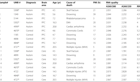
Table 1 Clinical characteristics and RNA quality of autistic and control brain samples
Sample# UMB # Diagnosis Brain
area
Age (yr) Cause of death
PMI (h) RNA quality
A260/280 A260/230 RIN
1 5308* Autism PFC 4.5 Skull fracture 21 2.051 2.266 4.9
2 1349 Autism PFC 5.6 Drowning 39 2.044 2.232 4.3
3 5144 Autism PFC 7.2 Rhabdomyosarcoma 3 2.058 2.271 5.4
4 5302* Autism PFC 16.3 DKA 20 2.031 2.238 4
5 4999* Autism PFC 20.8 Cardiac arrhythmia 14 2.039 2.232 6
6 4670* Control PFC 4.6 Commotio Cordis 17 2.048 2.276 5.2
7 1185 Control PFC 4.7 Drowning 17 2.026 2.243 4.7
8 4898 Control PFC 7.7 Drowning 12 2.056 2.183 5.9
9 4848* Control PFC 16.7 Drowning 15 2.044 2.185 6.7
10 4727* Control PFC 20.5 Multiple injuries (MVA) 5 2.066 2.209 6.5
11 5308* Autism Cere 4.5 Skull fracture 21 2.087 1.781 7.3
12 4899 Autism Cere 14.3 Drowning 9 2.077 2.314 9.3
13 5302* Autism Cere 16.3 DKA 20 2.083 1.646 2.2
14 4999* Autism Cere 20.8 Cardiac arrhythmia 14 2.081 2.114 9.2
15 4670* Control Cere 4.6 Commotio Cordis 17 2.088 2.161 6.1
16 4722 Control Cere 14.5 Multiple injuries (ATV) 16 2.073 2.828 6.5
17 4848* Control Cere 16.7 Drowning 15 2.087 2.327 6.8
18 4727* Control Cere 20.5 Multiple Injuries (MVA) 5 2.067 2.307 7.2
The cDNA produced from the reaction was diluted to 0.25× with nuclease free water.
Real time quantitative PCR
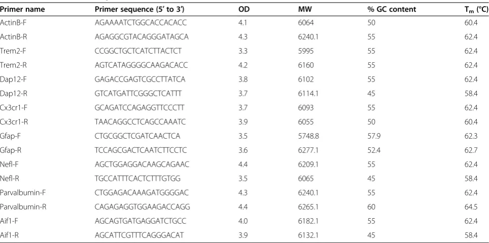
SYBR Green Expression Assay System (Applied Biosys-tems, Foster City, CA, USA) was used to measure relative, normalized, mRNA expression levels. We assessed four separate microglial-specific cell surface genes: Triggering receptor expressed on myeloid cells 2 (TREM2), DAP12, CX3C chemokine receptor 1 (CX3CR1), and allograft inflammatory factor 1 (AIF1) [28]. Two cell type spe-cific intermediate filaments, glial fibrillary acidic pro-tein (GFAP), which is astrocyte-specific [29], and the pan-neuronal cell marker NEFL [30], were used to assess for astrocytes and neurons, respectively. Add-itionally, we assessed for GABAergic interneurons specifically with parvalbumin (PVL) [31]. The intermedi-ate filament housekeeping gene beta-actin (ACTB) was used as a control. Forward and reverse primer sequences were generated using Primer3 software and synthesized by Eurofins MWG Operon (Huntsville, AL, USA) (Table 2).
Quantitative reverse transcriptase polymerase reaction (qRT-PCR) was performed using an ABI Prism 7900 Se-quence Detection System (Life Technologies) with a 96-well format. Each qRT-PCR reaction contained 6.5μL water, 12.5μL SYBR Green master mix (Applied Biosys-tems), 1μL forward primer (10μM), 1 μL reverse primer (10 μM), and 4 μL of cDNA (0.25×). Data was collected using the SDS2.3 Program (Applied Biosystems) under the following run parameters: 48°C for 30 minutes, 95°C for
10 minutes, 40 cycles of 95°C for 15 seconds, 60°C for 1 minute, and a final dissociation stage.
Data analysis
The target genes and the endogenous controls were measured with technical triplicates in each qRT-PCR reaction, and all genes were assessed in three separate, independent qRT-PCR runs. The cycle threshold num-ber (Ct) was calculated using RQ Manager 1.2 Software (Applied Biosystems). Relative expression of each target gene was normalized to ACTBusing theΔΔCtmethod. AllPvalues reported are based on a two-tailed Student’s t-test. Only results with a P value less than 0.05 were considered significant.
Results
The average post-mortem interval (PMI) was not sig-nificantly different between autistic and control tissue samples (Table 1; ASD = 17.9 h, ctrl = 13.2 h, P= 0.16). This remained true after sub-stratifying by brain region (ASD PFC = 19.4 h, ctrl PFC = 13.2 h,P= 0.28; and ASD cerebellum = 16.0 h, ctrl cerebellum = 13.25 h, P= 0.48). RNA isolated from post-mortem brain tissue was gener-ally of high quality, and the RNA Integrity Number (RIN) was not significantly different between autism and controls (ASD = 5.84, ctrl = 6.18,P= 0.67). The RIN was also not significantly different after sub-stratifying by brain region (ASD PFC = 4.92, ctrl PFC = 5.80, P= 0.13; and ASD cerebellum = 7.00, Ctrl cerebellum = 6.65, P= 0.85).
Table 2 Primers used for qRT-PCR
Primer name Primer sequence (5′to 3′) OD MW % GC content Tm(°C)
ActinB-F AGAAAATCTGGCACCACACC 4.1 6064 50 60.4
ActinB-R AGAGGCGTACAGGGATAGCA 4.3 6240.1 55 62.4
Trem2-F CCGGCTGCTCATCTTACTCT 3.3 5995 55 62.4
Trem2-R AGTCATAGGGGCAAGACACC 4.2 6160 55 62.4
Dap12-F GAGACCGAGTCGCCTTATCA 3.8 6102 55 62.4
Dap12-R GTCATGATTCGGGCTCATTT 3.7 6114.1 45 58.4
Cx3cr1-F GCAGATCCAGAGGTTCCCTT 3.7 6093 55 62.4
Cx3cr1-R TAACAGGCCTCAGCCAAATC 3.9 6055 50 60.4
Gfap-F CTGCGGCTCGATCAACTCA 3.5 5748.8 57.9 62.3
Gfap-R TCCAGCGACTCAATCTTCCTC 3.6 6277.1 52.4 62.7
Nefl-F AGCTGGAGGACAAGCAGAAC 4.4 6209.1 55 62.4
Nefl-R TGCCATTTCACTCTTTGTGG 3.5 6065 45 58.4
Parvalbumin-F CTGGAGACAAAGATGGGGAC 4.3 6240.1 55 62.4
Parvalbumin-R CAGAGAGGTGGAAGACCAGG 4.4 6265.1 60 64.5
Aif1-F AGCAGTGATGAGGATCTGCC 4.0 6182.1 55 62.4
Aif1-R AGCATTCGTTTCAGGGACAT 3.9 6132.1 45 58.4
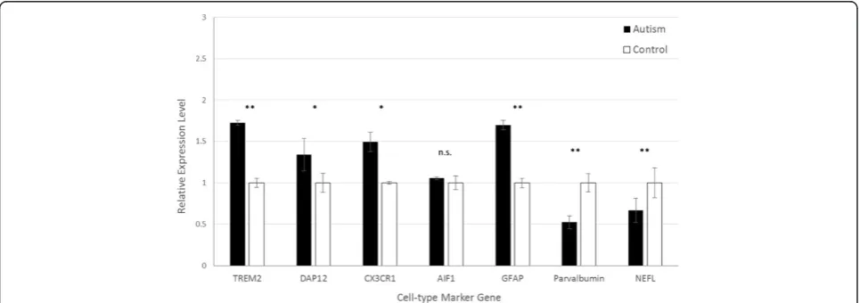
In the PFC, quantification of microglial markers dem-onstrated significantly increased expression in autistic samples ofTREM2, DAP12, and CX3CR1, but notAIF1 (Figure 1). The expression of TREM2was highest of all microglial markers, approximately 1.75-fold higher in autism brain tissue than controls (P= 0.0016). The levels of CX3CR1 and DAP12 were 1.50-fold (P= 0.0092) and 1.34-fold (P= 0.0086) higher in autistic samples relative to controls, respectively. Similarly, the expression of astrocyte markerGFAPwas significantly higher in autis-tic brains (1.70-fold, P= 0.0049). Conversely, however, both the pan-neuronal marker NEFL, and the GABAer-gic interneuron-specific marker PVA, were significantly lower in autistic samples compared to controls (0.68-fold,P= 0.0034; and 0.52-fold,P= 0.0020, respectively).
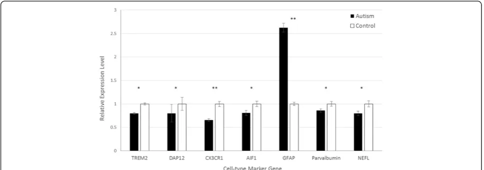
In post-mortem cerebellum, the expression of astro-cyte markerGFAPwas also significantly higher in autism samples than in healthy controls (2.63-fold, P= 0.0022; Figure 2). In contrast, the expression of microglial markersTREM2,DAP12,CX3CR1, and AIF1were lower in autism tissue than in control tissue, with fold changes of 0.780 (P= 0.0056), 0.797 (P= 0.0083), 0.659 (P= 0.0029), and 0.808 (P= 0.0052), respectively. Expression of neuronal markersPVAandNEFLwere also lower in autism samples than in control samples (0.862-fold, P= 0.033; and 0.798-fold,P= 0.013, respectively), as was found in the PFC.
Discussion
While there have been multiple studies assessing RNA expression levels in autistic tissue, here we report on the expression of microglial, astrocyte, and neuron-specific cell markers concurrently in two regions of autistic brains. Our results show that glial-specific markers demonstrate altered expression in autistic brains. More-over, the expression pattern of these cell-type specific markers parallels previous findings of glial cell number
and activation patterns in autistic brain assessed via cell-level techniques (discussed below). Therefore, this approach may be a useful alternative for assessing activation and/or cell numbers in post-mortem brain tissue studies of ASD patients.
Microglia cell-marker research is still a relatively new area, and thus the markers used to quantify microglial cell number and activation are still debated. To address this issue, we used four different markers that are putatively microglial-specific. Our results demonstrate that in the PFC, there is increased expression of all microglial markers assessed, although AIF1 did not reach statistical significance. However, previous reports have shown that AIF1 expression in the brain is low [32], potentially contributing to this result. The finding of increased microglial cell markers in autistic PFC is in agreement with a number of studies that have found in-creased numbers and activation of microglia in autistic brains. For instance, Morgan et al. showed increased microglial density in dorsolateral PFC grey matter of ASD brains via IHC and stereology [8], and they also demonstrated that microglia are more closely associated with neurons in autistic dorsolateral PFC than in con-trols [9]. Similarly, Tetreault et al. also demonstrated increased microglial density in the frontoinsular and visual cortex of autistic brains as compared to controls [10]. Additionally, a number of studies have specifically identified microglial activation in autistic frontal cortex, through both PET radiotracer imaging [13] and IHC/ cytokine profiling approaches [7]. Our results provide further support to the growing body of evidence demon-strating increased microglial numbers and activation in autistic PFC, and our cell marker gene expression results are largely in concordance with these cell level studies.
In contrast, our cerebellar results show significantly lower expression of all four microglial cell specific
markers in autistic brains. While other studies have identified microglial activation in the cerebellum of autistic tissue [33], no study has attempted to specifically quantify microglial cells in the cerebellum using the markers assessed here, and therefore histopathologic studies in the cerebellum are needed to confirm these findings. One report described increased microglial cell activation in the cerebellum, assessed via HLA-DR stain-ing in the white matter and granular cell layer of the cerebellum [7], and another showed increased microglial activation throughout the brain (although most promin-ently in the cerebellum) using anin vivoPET metabolic radiotracer [13]. However, HLA-DR expression in human microglial cells has been shown to be highly variable between individuals, and its expression actually decreases upon cytokine stimulation [34]. Moreover, as we previously discussed [35], the cerebellum is anatom-ically and physiologanatom-ically unique; thus metabolic and pathological findings in the cerebellum must be inter-preted with caution. Furthermore, our tissue samples from cerebellum contained all three layers of the cere-bellar cortex, as opposed to the molecular layer only. Until direct histologic assessment of these microglial markers are performed in post-mortem autistic cerebel-lum, our results must be interpreted cautiously. How-ever, our results do suggest that there are significant differences in microglial cell markers between ASD PFC and cerebellum, perhaps reflecting differences in micro-glial activation and/or cell numbers between these areas in ASD brain.
In both the PFC and the cerebellum, there was signifi-cantly increased expression of the astrocyte-specific marker GFAP in autistic brains. This trend was most prominent in the cerebellum, where GFAP expression was over two-fold higher in ASD brains than in healthy controls. Our findings parallel those of previous studies,
which have shown increased expression of GFAP protein in the cerebellum and cortex of patients of autism through IHC staining, western blotting, and mRNA ex-pression [7,17,36,37]. While studies have not been done to quantify astrocyte numbers in the autistic cerebellum, our results and those of previous studies provide evi-dence for astroglial reaction in autism.
Interestingly, we also found significantly decreased expression of the pan-neuronal marker NEFL in both the PFC and the cerebellum of autistic brains. This result is also supported by previous studies, which have shown decreasedNEFL mRNA expression in the anter-ior cingulate gyrus, motor cortex, and thalamus of autis-tic brains [38]. However, cell-level studies in autisautis-tic brain have produced conflicting results about neuron numbers. While a large body of evidence has suggested there is a loss of neurons in many areas of autistic brains, as recently reviewed in [39], other studies have shown that young autistic brains may have 70% more neurons in the PFC [40]. Importantly, though, is the age of the patient at time of death, as longitudinal studies have suggested that early brain overgrowth in ASDs quickly reverses to a phenotype of neuronal loss [41]. Consequently, the older age of patients in this study may bias our findings towards the neuronal loss spectrum of the disease.
Similarly, we found significant decreases in the GABAer-gic interneuron-specific markerPVAin both the PFC and cerebellum of autistic samples. Despite many studies demonstrating decreased GABAergic components across different areas of the autistic brain, as reviewed in [42], the one pathological analysis of parvalbumin-positive interneu-rons in ASD did not identify differences in the autistic cerebellum [43]. However, this study only assessed the molecular layer of the cerebellar cortex, whereas our tissue samples contained all three layers. Additionally, while
parvalbumin interneurons have been shown to be unchanged in the autistic posterior cingulate cortex and fusiform gyrus [44], and increased in the autistic hippo-campus [45], they have not been directly assessed in the autistic PFC.
In addition to potentially serving as markers of glial cell numbers and/or activation, many of the genes assessed have specific putative biological relevance to ASDs themselves. For instance, in brain, Dap12 (also known asTYROBP) encodes a microglial-specific trans-membrane signaling polypeptide [46]. The encoded protein acts as an activating signal transduction element with known roles in brain myelination and inflammation [47]. Its receptor,TREM2, encodes a membrane protein that functions in modulating the brain’s immune re-sponse via production of constitutive cytokines, and is critical for activating microglial phagocytosis [48]. Rare mutations within these two genes have been associated with Nasu-Hakola disease [49]. Nasu-Hakola disease is characterized neurologically by new onset psychiatric and cognitive symptoms in the fourth decade of life, evolving to memory loss and cognitive decline resem-bling Alzheimer’s disease [50]. Interestingly, single nu-cleotide polymorphisms in this receptor pathway were also recently linked to a significantly increased risk of Alzheimer’s disease, and are thought to relate to the inability of microglia to properly remove neurodegenera-tive debris such as beta-amyloid [51]. It is intriguing to speculate that defects in this same pathway in neurode-velopmental disorders such as ASDs may also result in defects in microglial phagocytosis, but in the develop-mental context it is the inability to prune overabundant synapses, as opposed to neurodegenerative debris, that results in the behavioral phenotype.
Similarly, CX3CR1, also known as the fractalkine receptor, encodes a protein receptor on the surface of microglia that binds to the chemokine CX3CL1 (also called fractalkine); fractalkine functions to induce micro-glial migration and adhesion during phagocytosis [52]. This pathway was recently demonstrated to play a critical role in the developing brain of mice allowing for migration of microglia to their synaptic targets, where phagocytosis and synaptic refinement occur. CX3CR1 knockout mice had more synapses on cortical neurons than wild-type mice, and displayed subtle neurological deficits [25].
AIF1, also known as IBA1, encodes a protein that is consistently up-regulated in expression during microglial activation, and therefore has been used to discriminate between resting and activated microglia [53]. The AIF1 gene is located within the highly variable locus contain-ing parts of the major histocompatibility complex, which itself has been linked to ASDs and dysfunctional micro-glia phagocytosis [54].
Glial fibrillary acidic protein (GFAP) is an intermediate filament protein that is expressed by astrocytes, and as discussed above, has been previously shown to be up-regulated in autistic brains as was also shown here. Interestingly, decreased expression of GFAP has been reported in other neurodevelopmental disorders, such as Schizophrenia and bipolar disorder [55]. GFAP, like all intermediated filament proteins, is important in maintaining the cellular cytoskeleton in astrocytes. The cytoskeleton plays a number of key functional roles in addition to maintaining cell shape, including inter-cellular communication, mitosis, and cellular mi-gration during phagocytosis. Mutations in GFAP are responsible for Alexander’s disease, a rare disorder characterized by severe developmental delay, increased head size, and seizures [56].
Overall, our findings demonstrate that the autistic brain by mid-childhood has molecular changes consist-ent with increased astrocyte expression and decreased neuronal and interneuron expression in both the PFC and cerebellum, with PFC-specific increased microglial marker expression. Furthermore, the specific glial molecules found to be abnormal in autistic brains are intimately involved in glial mobilization and phagocyt-osis pathways, they have previously been shown to be critical for normal neurodevelopment, and are known to be causative of other rare neurodevelopmental phenotypes. These findings support the notion that a complex interplay between glial dysfunction and neurogenesis may underlie the clinical manifestations of ASDs.
expertise, suggest this may be a valuable and simple alternative‘screening’approach.
Conclusions
In summary, assessment of glial numbers and activation in autism post-mortem brain research is hampered by the scarcity of appropriately-preserved tissue, and the tech-nical challenge of traditional stereotactic methods. We show that glial and neuron cell-type specific markers have mRNA expression patterns that parallel known cellular aberrations in ASDs. Our results provide further evidence that glial cells may play a role in the pathogenesis of ASDs, and suggest that assessing for glial cell-type specific marker expression may represent a viable approach to relatively quantify glial cell patterns in ASD post-mortem research.
Abbreviations
AIF1:Allograft inflammatory factor 1; ASD: Autism spectrum disorder; CX3CR1: CX3C chemokine receptor 1; IHC: Immunocytochemistry;
PET: Positron emission tomography; PFC: Prefrontal cortex; PMI: Post-mortem interval; qRT-PCR: Quantitative real-time polymerase chain reaction; RIN: RNA integrity number;TREM2: Triggering receptor expressed on myeloid cells 2.
Competing interests
The authors declare that they have no competing interests.
Authors’contributions
CE, MNZ, and OMR conceived of and designed the analysis. CE performed the experiments. CE, MNZ, and OMR analyzed results and wrote the manuscript. All authors read and approved the final manuscript.
Acknowledgements
The Intramural Research Program at the National Institute of Child Health and Human Development supported this work. MNZ was also supported by Baylor College of Medicine MSTP and the NIH-University of Cambridge Bio-medical Scholars Program. CE was additionally supported by the University of Florida College of Medicine. The funders had no role in study design, data collection and analysis, decision to publish, or preparation of the manuscript.
Author details
1Laboratory of Clinical and Developmental Genomics, National Institute of
Child Health and Human Development, National Institutes of Health, 49 Convent Drive, Building 49, Room 2C078, Bethesda, MD 20814, USA.
2
University of Florida College of Medicine, 1600 SW Archer Rd, Gainesville, FL 32603, USA.3University of Cambridge, Robinson College, Grange Rd,
Cambridgeshire CB3 9AN, UK.4Baylor College of Medicine MSTP, One Baylor Plaza, Houston, TX 77030, USA.
Received: 14 September 2013 Accepted: 23 December 2013 Published: 10 January 2014
References
1. American Psychiatric Association:Diagnostic and Statistical Manual of Mental Disorders.5th edition. Arlington: American Psychiatric Publishing; 2013. 2. Centers for Disease Control and Prevention:Prevalence of autism
spectrum disorders—autism and developmental disabilities monitoring network, 14 sites, United States, 2008.MMWR2012,3:1–24.
3. Zoghbi HY:Postnatal neurodevelopmental disorders: meeting at the synapse?Science2003,5646:826–830.
4. Just MA, Cherkassky VL, Keller TA, Kana RK, Minshew NJ:Functional and anatomical cortical underconnectivity in autism: evidence from an FMRI study of an executive function task and corpus callosum morphometry. Cereb Cortex2007,17(4):951–961.
5. Rubenstein JL, Merzenich MM:Model of autism: increased ratio of excitation/inhibition in key neural systems.Genes Brain Behav2003,2 (5):255–267.
6. Courchesne E, Pierce K, Schumann CM, Redcay E, Buckwalter JA, Kennedy DP, Morgan J:Mapping early brain development in autism.Neuron2007, 56(2):399–413.
7. Vargas DL, Nascimbene C, Krishnan C, Zimmerman AW, Pardo CA: Neuroglial activation and neuroinflammation in the brain of patients with autism.Ann Neurol2005,57(1):67–81.
8. Morgan JT, Chana G, Pardo CA, Achim C, Semendeferi K, Buckwalter J, Courchesne E, Everall IP:Microglial activation and increased microglial density observed in the dorsolateral prefrontal cortex in autism. Biol Psychiatry2010,68(4):368–376.
9. Morgan JT, Chana G, Abramson I, Semendeferi K, Courchesne E, Everall IP: Abnormal microglial-neuronal spatial organization in the dorsolateral prefrontal cortex in autism.Brain Res2012,1456:72–81.
10. Tetreault NA, Hakeem AY, Jiang S, Williams BA, Allman E, Wold BJ, Allman JM:Microglia in the cerebral cortex in autism.J Autism Dev Disord2012, 42(12):2569–2584.
11. Watkins LR, Milligan ED, Maier SF:Glial activation: a driving force for pathological pain.Trends Neurosci2001,24(8):450–455.
12. Hickman SE, Kingery ND, Ohsumi TK, Borowsky ML, Wang LC, Means TK, El Khoury J:The microglial sensome revealed by direct RNA sequencing. Nat Neurosci2013,16(12):1896–1905.
13. Suzuki K, Sugihara G, Ouchi Y, Nakamura K, Futatsubashi M, Takebayashi K, Yoshihara Y, Omata K, Matsumoto K, Tsuchiya KJ, Iwata Y, Tsujii M, Sugiyama T, Mori N:Microglial activation in young adults with autism spectrum disorder.JAMA Psychiatry2013,70(1):49–58.
14. Maezawa I, Jin LW:Rett syndrome microglia damage dendrites and synapses by the elevated release of glutamate.J Neurosci2010, 30(15):5346–5356.
15. Derecki NC, Cronk JC, Lu Z, Xu E, Abbott SB, Guyenet PG, Kipnis J:Wild type microglia arrest pathology in a mouse model of Rett syndrome. Nature2012,484(7392):105–109.
16. Cao F, Yin A, Wen G, Sheikh AM, Tauqeer Z, Malik M, Nagori A, Schirripa M, Schirripa F, Merz G, Brown WT, Li X:Alteration of astrocytes and Wnt/β-catenin signaling in the frontal cortex of autistic subjects. J Neuroinflammation2012,9(1):223.
17. Laurence JA, Fatemi SH:Glial fibrillary acidic protein is elevated in the superior, frontal, parietal and cerebellar cortices of patients with autism. Cerebellum2005,4:206–210.
18. Fatemi SH, Folsom TD, Reutiman TJ, Lee S:Expression of astrocytic markers aquaporin 4 and connexin 43 is altered in brains of subjects with autism.Synapse2008,62(7):501–507.
19. Maezawa I, Swanberg S, Harvey D, LaSalle JM, Jin LW:Rett syndrome astrocytes are abnormal and spread MeCP2 deficiency through gap junctions.J Neurosci2009,29(16):5051–5061.
20. Yasui DH, Xu H, Dunaway KW, Lasalle JM, Jin LW, Maezawa I:MeCP2 modulates gene expression pathways in astrocytes.Mol Autism2013, 4(1):3.
21. Yuskaitis CJ, Beurel E, Jope RS:Evidence of reactive astrocytes but not peripheral immune system activation in a mouse model of Fragile X syndrome.Biochim Biophys Acta2010, 1802(11):1006–1012.
22. Uhlmann EJ, Apicelli AJ, Baldwin RL, Burke SP, Bajenaru ML, Onda H, Kwiatkowski D, Gutmann DH:Heterozygosity for the tuberous sclerosis complex (TSC) gene products results in increased astrocyte numbers and decreased p27-Kip1 expression in TSC2+/−cells.Oncogene2002,21 (25):4050–4059.
23. Lioy DT, Garg SK, Monaghan CE, Raber J, Foust KD, Kaspar BK, Hirrlinger PG, Kirchhoff F, Bissonnette JM, Ballas N, Mandel G:A role for glia in the progression of Rett’s syndrome.Nature2011,475(7357):497–500. 24. Eroglu C, Barres BA:Regulation of synaptic connectivity by glia.Nature
2010,468:223–231.
25. Paolicelli RC, Bolasco G, Pagani F, Maggi L, Scianni M, Panzanelli P, Giustetto M, Ferreira TA, Guiducci E, Dumas L, Ragozzino D, Gross CT:Synaptic pruning by microglia is necessary for normal brain development.Science 2011,6048:1456–1458.
26. Abbott A:Tissue-bank shortage: brain child.Nature2011,478(7370):442–443. 27. Ziats MN, Rennert OM:Aberrant expression of long noncoding RNAs in
28. Ransohoff R, Perry V:Microglial physiology: unique stimuli, stimuli, specialized responses.Annu Rev Immunol2009,27:119–145. 29. Baba H, Nakahira K, Morita N, Tanaka F, Akita H, Ikenaka K:GFAP gene
expression during development of astrocyte.Dev Neurosci1997, 19(1):49–57.
30. Lépinoux-Chambaud C, Eyer J:Review on intermediate filaments of the nervous system and their pathological alterations.Histochem Cell Biol 2013,140(1):13–22.
31. Conde F, Lund JS, Jacobowitz DM, Baimbridge KG, Lewis DA:Local circuit neurons immunoreactive for calretinin, calbindin D-28 k or parvalbumin in monkey prefrontal cortex: distribution and morphology.J Comp Neurol 1994,341(1):95–116.
32. Imai Y, Ibata I, Ito D, Ohsawa K, Kohsaka S:A novel gene Iba1 in the major histocompatibility complex class III region encoding an EF hand protein expressed in a monocytic lineage.Biochem Biophys Res Commun1996, 224:855–862.
33. Casanova MF:The neuropathology of autism.Brain Pathol2007, 17(4):422–433.
34. Smith AM, Gibbons HM, Oldfield RL, Bergin PM, Mee EW, Curtis MA, Faull RL, Dragunow M:M-CSF increases proliferation and phagocytosis while modulating receptor and transcription factor expression in adult microglia.J Neuroinflammation2013,10:85.
35. Ziats MN, Rennert OM:The cerebellum in autism: pathogenic or an anatomical beacon?Cerebellum2013,12(5):776–777.
36. Bailey A, Luthert P, Dean A, Harding B, Janota I, Montgomery M, Rutter M, Lantos P:A clinicopathological study of autism.Brain1998,121 (5):889–905.
37. Purcell AE, Jeon OH, Zimmerman AW, Blue ME, Pevsner J:Postmortem brain abnormalities of the glutamate neurotransmitter system in autism. Neurology2001,57(9):1618–1628.
38. Anitha A, Nakamura K, Thanseem I, Yamada K, Iwayama Y, Toyota T, Matsuzaki H, Miyachi T, Yamada S, Tsujii M, Tsuchiya KJ, Matsumoto K, Iwata Y, Suzuki K, Ichikawa H, Sugiyama T, Yoshikawa T, Mori N:Brain region-specific altered expression and association of mitochondria-related genes in autism.Mol Autism2012,3(1):12.
39. Kern JK, Geier DA, Sykes LK, Geier MR:Evidence of neurodegeneration in autism spectrum disorder.Transl Neurodegener2013,2(1):17.
40. Courchesne E, Mouton PR, Calhoun ME, Semendeferi K, Ahrens-Barbeau C, Hallet MJ, Barnes CC, Pierce K:Neuron number and size in prefrontal cortex of children with autism.JAMA2011,306(18):2001–2010. 41. Courchesne E, Campbell K, Solso S:Brain growth across the life span in
autism: age-specific changes in anatomical pathology.Brain Res2011, 1380:138–145.
42. Fatemi SH, Blatt GJ:Alterations in GABAergic biomarkers in the autism brain: research findings and clinical implications.Anat Rec (Hoboken) 2011,294(10):1646–1652.
43. Whitney ER, Kemper TL, Rosene DL, Bauman ML, Blatt GJ:Density of cerebellar basket and stellate cells in autism: evidence for a late developmental loss of Purkinje cells.J Neurosci Res2009, 87(10):2245–2254.
44. Oblak AL, Rosene DL, Kemper TL, Bauman ML, Blatt GJ:Altered posterior cingulate cortical cyctoarchitecture, but normal density of neurons and interneurons in the posterior cingulate cortex and fusiform gyrus in autism.Autism Res2011,4(3):200–211.
45. Lawrence YA, Kemper TL, Bauman ML, Blatt GJ:Parvalbumin-, calbindin-, and calretinin-immunoreactive hippocampal interneuron density in autism.Acta Neurol Scand2010,121(2):99–108.
46. Paradowska-Gorycka A, Jurkowska M:Structure, expression pattern and biological activity of molecular complex TREM-2/DAP12.Hum Immunol 2013,74(6):730–737.
47. Kaifu T, Nakahara J, Inui M, Mishima K, Momiyama T, Kaji M, Sugahara A, Koito H, Ujike-Asai A, Nakamura A, Kanazawa K, Tan-Takeuchi K, Iwasaki K, Yokoyama WM, Kudo A, Fujiwara M, Asou H, Takai T:Osteopetrosis and thalamic hypomyelinosis with synaptic degeneration in DAP12-deficient mice.J Clin Invest2003,111(3):323–332.
48. Takahashi K, Rochford CD, Neumann H:Clearance of apoptotic neurons without inflammation by microglial triggering receptor expressed on myeloid cells-2.J Exp Med2005,201(4):647–657.
49. Kaneko M, Sano K, Nakayama J, Amano N:Nasu-Hakola disease: the first case reported by Nasu and review.Neuropathology2010,30(5):463–470.
50. Bianchin MM, Martin KC, de Souza AC, de Oliveira MA, Rieder CR: Nasu-Hakola disease and primary microglial dysfunction.Nat Rev Neurol 2010,6(9):2 p following 523.
51. Jonsson T, Stefansson H, Steinberg S, Jonsdottir I, Jonsson PV, Snaedal J, Bjornsson S, Huttenlocher J, Levey AI, Lah JJ, Rujescu D, Hampel H, Giegling I, Andreassen OA, Engedal K, Ulstein I, Djurovic S, Ibrahim-Verbaas C, Hof-man A, Ikram MA, van Duijn CM, Thorsteinsdottir U, Kong A, Stefansson K: Variant of TREM2 associated with the risk of Alzheimer’s disease.N Engl J Med2013,368(2):107–116.
52. Wolf Y, Yona S, Kim KW, Jung S:Microglia, seen from the CX3CR1 angle. Front Cell Neurosci2013,18:7–26.
53. Schwab JM, Frei E, Klusman I, Schnell L, Schwab ME, Schluesener HJ:AIF-1 expression defines a proliferating and alert microglial/macrophage phenotype following spinal cord injury in rats.J Neuroimmunol2001, 119(2):214–222.
54. Needleman LA, McAllister AK:The major histocompatibility complex and autism spectrum disorder.Dev Neurobiol2012,72(10):1288–1301. 55. Johnston-Wilson NL, Sims CD, Hofmann JP, Anderson L, Shore AD, Torrey
EF, Yolken RH:Disease-specific alterations in frontal cortex brain proteins in schizophrenia, bipolar disorder, and major depressive disorder. The Stanley Neuropathology Consortium.Mol Psychiatry2000,5(2):142–149. 56. Li R, Johnson AB, Salomons G, Goldman JE, Naidu S, Quinlan R, Cree B,
Ruyle SZ, Banwell B, D’Hooghe M, Siebert JR, Rolf CM, Cox H, Reddy A, Gutiérrez-Solana LG, Collins A, Weller RO, Messing A, van der Knaap MS, Brenner M:Glial fibrillary acidic protein mutations in infantile, juvenile, and adult forms of Alexander disease.Ann Neurol2005,57(3):310–326.
doi:10.1186/2040-2392-5-3
Cite this article as:Edmonsonet al.:Altered glial marker expression in autistic post-mortem prefrontal cortex and cerebellum.Molecular Autism
20145:3.
Submit your next manuscript to BioMed Central and take full advantage of:
• Convenient online submission
• Thorough peer review
• No space constraints or color figure charges
• Immediate publication on acceptance
• Inclusion in PubMed, CAS, Scopus and Google Scholar
• Research which is freely available for redistribution