BioMed Central
Cardiovascular Ultrasound
Open Access
Review
Tissue Doppler and strain imaging: anything left in the echo-lab?
Rodolfo Citro*
1, Eduardo Bossone
2, Bettina Kuersten
2, Giovanni Gregorio
1and Alessandro Salustri
3Address: 1Department of UTIC-Cardiology, "San Luca" Hospital, Vallo della Lucania (SA), Italy, 2Department of Cardiology, "Santa Maria
dell'Olmo" Hospital, Cava dei Tirreni e Costa d'Amalfi (SA), Italy and 3Department of Cardiology, Policlinico Luigi Di Liegro, Roma, Italy
Email: Rodolfo Citro* - [email protected]; Eduardo Bossone - [email protected]; Bettina Kuersten - [email protected]; Giovanni Gregorio - [email protected]; Alessandro Salustri - [email protected]
* Corresponding author
Abstract
Medline research indicates that an increasing number of manuscripts have been published in the last decade claiming, the feasibility and the potential clinical role of tissue Doppler and strain/strain rate imaging. However, despite this amount of scientific evidence, these technologies are still confined to dedicated, high-tech, research-oriented echocardiography laboratories. In this review we have critically evaluated these techniques, analysing their physical principles, the technical problems related to their current clinical application, and the future perspectives. Finally, this review explores the reasons why these technologies are still defined "new technologies" and the impact of their implementation on the current clinical activity of an echocardiography laboratory.
Background
In the past decade, numerous studies have been published addressing the feasibility and potential clinical applicabil-ity of tissue Doppler imaging (TDI) and its derived param-eters strain and strain rate (SR) [1-7] (Figure 1). The data reported in these studies strongly support that the differ-ent methods are attractive in regards to their underlying theory as well as for the information revealed about car-diac function, that can be obtained by analyzing the motion of the diverse structural components of the heart (myocardial walls, valvular rings); at the same time all these methods strive to eliminate everything subjective in the assessment of an echocardiogram. Thus, a large volume of scientific evidence has been produced in favor of clinical use of these methods and various parameters have been proposed with the ambitious goal to contribute towards additional diagnostic value with respect to various pathol-ogies. [8]. Despite this promising research, the parallel clinical diffusion of these methods never took place. To
this day these methods are used only in a few centers and mainly in conjunction with research protocols; why have these published data not led to wider application in prac-tice? Is this skepticism justified? Is the information obtained by TDI or SR 'redundant'? Do solid fields of clin-ical applications supported by these data already exist today or will they exist in the near future? In order to answer these questions, this review will first assess the var-ious aspects of this technology from the physical princi-ples to the technological implementation and then compile the information.
Physical principles
TDI, through proper modifications of the hardware and software of the ultrasound platform (elimination of the
high-pass filter and adjustment of gain) allows the analysis of velocity signals having high amplitude and low fre-quency originating from tissue, which are usually not detected in traditional Doppler examination. In its
vari-Published: 30 October 2008
Cardiovascular Ultrasound 2008, 6:54 doi:10.1186/1476-7120-6-54
Received: 11 September 2008 Accepted: 30 October 2008
This article is available from: http://www.cardiovascularultrasound.com/content/6/1/54
© 2008 Citro et al; licensee BioMed Central Ltd.
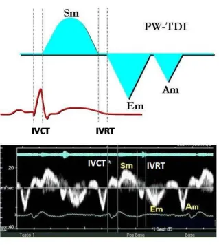
ous modalities (pulsed Doppler, 2D color mode and color M-mode), TDI renders possible on-line or off-line, the non invasive calculation of the time intervals and velocities of myocardial contraction and relaxation during the various phases of the cardiac cycle by using post-processing appli-cation software. (Figure 2). One of the primary reasons for the skepticism towards TDI lies in its very physical princi-ples. Doppler echocardiography seems to be the ideal method for the examination of intracardiac and vascular flow that obey the laws of fluid dynamics and for the cal-culation of pressure gradients comparable to those obtained by invasive methods like cardiac catheterization. [8]. However, the motion of the myocardial walls is differ-ent, occurring in more directions and planes, and is influ-enced by the motion of the other organs and structures of the thoracic cage, following complex mechanical phe-nomena often not completely understood and for which there exists no reference method [10]. This problem is par-ticularly evident when quantitative analysis of the myo-cardial function using TDI is applied to the study of segmental contraction abnormalities typical of ischemic cardiopathy.
In fact, if on one hand TDI has the merit of having brought back to mind very old concepts of physiopathol-ogy of the ischemic heart muscle, focusing the attention back on regional contraction phenomena, such as the delayed and reduced systolic shortening and the appear-ance of late systolic contraction, on the other hand, one must keep in mind that the measurement of regional myocardial velocities is not independent from the overall motion of the heart and suffers from tethering induced by collateral segments [3]. These limitations could poten-tially be overcome by strain imaging, which introduced the concept of myocardial deformation as a sensitive index of contractile capacity intrinsic to the myocardium [11] (Figure 3). However, strain is derived from the myo-cardial velocity gradient measured with TDI, from which the relative limitations in terms of angle-dependency are derived. Furthermore, the signal is strongly subjected to tedious noise problems (especially in the apical regions) which change the profiles of the spectral curve, making interpretation difficult, hardly reproducible and particu-larly arduous to reach interpretational consensus on, especially in the SR modality [4-6].
Number of articles per year, reported on PubMed, entering the search filter "tissue Doppler" or "myocardial Doppler" in title and/or abstract
Figure 1
Cardiovascular Ultrasound 2008, 6:54 http://www.cardiovascularultrasound.com/content/6/1/54
Two dimensional strain (2D strain), the most recent technique, has lately been proposed for obtaining veloci-ties and deformation of the myocardial walls as an alter-native to Doppler sampling. This method is based on the estimation of vectorial velocities instead of the analysis of the long component along the lines of the image. The algorithm identifies the vectorial velocities by 'tracking' the data obtained by the analysis of radiofrequency and black/white signals [12]. For every pixel of the image an angle-independent velocity is being estimated by selecting a pattern around the pixel examined, which is followed in the various frames, comprising the time period under examination. For those characteristics 2D strain could overcome the present limits (angle dependency and
sam-pling in the apical segments) of the strain obtained by using the Doppler data (Figure 4).
Reference methods
Thanks to its high temporal resolution, TDI and the meth-ods derived from it analyze phenomena that happen in such short time periods, to elude the resolution of the human eye and could not be considered during a tradi-tional echocardiographic evaluation [3,6]. These phe-nomena inherent to myocardial contractility have been addressed with methods commonly used in experimental studies, like sonomicrometric techniques, which are not reproducible in a clinical setting and for this reason can-not be defined as 'reference' methods. Consequently, the Example of a normal PW-TDI spectral curve: note the immediate representation of the mechanical events in the various phases of the cardiac cycle
Figure 2
real problem consists in the lack of a definition of myocar-dial contractility and of a 'gold standard' that identifies it. The strain imaging technique should be used to fill this very knowledge gap.
Technological problems
Up to now, studies conducted "in vivo" have been under-taken on a few selected patients admitted at highly spe-cialized centers, using specific software not always widely accessible. Furthermore, companies producing ultra-sound machines have not developed their products in comparable manners for reasons related to commerciali-zation or research. As a result, at the present time, not all machines are able to produce the same parameters with the same modality. Each company was kept busy by devel-oping its own software, then offering it prematurely and neglecting to consider whether it would turn into a com-mercial gadget or a truly useful tool. In addition, only recently attempts were made to standardize the nomen-clature and the parameters. Presently, no agreement has been reached on the modalities of temporal analysis, on how to identify the various waves of TDI or strain curves,
or on how to perform the measurements (positioning of the sample volume, size and form of the region of interest for the acquisition of the raw data). All of these issues are in part responsible for the difficulty of this method to enter "the real world".
Contemporary applications
a) One very interesting application of TDI is the diagnosis of acute myocardial ischemia, an event that induces a change of the subendocardial fibers with prevalent longi-tudinal contraction, determining a reduction of peak systolic velocity, of peak strain and systolic SR (which cor-relate with the reduction of regional myocardial blood flow) and of the protodiastolic velocity with Em/Am inversion only after a few seconds following occlusion of the left anterior descending coronary artery [13-15]. In numerous experimental studies TDI has been shown to be able to reproduce noninvasively the typical modifications of contractile function of acute ischemia, that have already been documented in experimental studies using sonomicrometry: delayed contractile shortening followed by a late asynchronous contraction that falls in the period New generation ultrasound platforms also allow to obtain simultaneous curves of velocity, strain rate and myocardial strain from a selected region of interest, for example the middle segment of the interventricular septum (note the white circle)
Figure 3
Cardiovascular Ultrasound 2008, 6:54 http://www.cardiovascularultrasound.com/content/6/1/54
of isovolumetric relaxation. This last phenomenon of ' tar-dokinesis' also defined as systolic shortening, post-systolic thickening, or post-post-systolic motion depending on the different technologies used to represent it, has been associated with the presence of ischemia and myocardial viability [16] (Figure 5). It also seems to have a potential role in the recognition of the genesis of ischemia in patients with left bundle branch block [17]. In addition to the observation of post-systolic motion, the pre-ejection peak velocity helps to identify myocardial ischemia [18]. TDI was proven to be feasible during physical and phar-macological stress echocardiography; however, the single multicenter study that has used TDI during dobutamine stress echocardiography, the MYDISE study, has yielded only partly encouraging results showing an advantage limited to the analysis of only a fraction of the myocardial
walls and predominantly in the basal segments [19,20]. Likewise, strain and SR have been proven to be feasible during stress echocardiography, and the correct interpre-tation of the possible variations of the peak systolic and of the post-systolic shortening induced by dobutamine at low and high doses can represent a guide in the recogni-tion of the different pathophysiological models of myo-cardial viability (stunned or hibernating myocardium), as well as after myocardial infarction of the different patho-logical substrates (transmural versus non-transmural necrosis) [21-23].
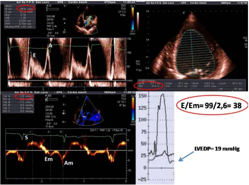
b) The role and value of TDI is most clearly established in the evaluation of diastolic function. In this context in fact the E/Em ratio correlates with pulmonary capillary wedge pressure and has been proven to be comparable to the Representation of 2D-strain (speckle tracking) of the left ventricle in the apical long axis view in a patient with systolic heart failure
Figure 4
plasma BNP values in the detection of elevated left ven-tricular filling pressures not only in critical patients admit-ted to intensive care units, but also in hospitalized patients independently of the value of the ejection frac-tion [24-27] (Figure 6). Parameters derived from the anal-ysis of tissue velocities of the mitral ring seem to have predictive value independently and additionally to mor-tality after 2 years, which confers them a fundamental prognostic impact which is absolutely not negligible [26,28]. However, one must keep in mind that the preload independency of Em partially decreases when ejec-tion fracejec-tion is normal, which obviously limits its use in these conditions, and that the values of E/Em between 8 and 15 lay in a gray zone in which it is necessary to utilize the Valsalva maneuver and to take into account the com-bined data of transmitral and pulmonary venous flow to ultimately be able to estimate the filling pressures [26]. The problem is that a considerable number of patients fall into this range and consequently, in these, the diagnostic contribution of TDI is diminished. This drawback is cer-tainly compensated for in two categories of patients: those with atrial fibrillation and those after heart transplant sur-gery, for which, each for different reasons, the value of
standard Doppler echocardiographic parameters for eval-uation of diastolic function is reduced [29]. On these grounds TDI has actually entered the clinical arena for the assessment of diastolic function [26]. Beyond global diastolic function, the analysis of regional diastolic func-tion with SR shows evidence of a reducfunc-tion of the proto-diastolic peak at rest that predicts with a specificity of 93% the presence of significant coronary heart disease at coro-nary angiography [30].
c) One of the new aspects introduced by TDI is the possi-bility to examine myocardial function along the heart's longitudinal axis. The ventricular myocardium comprises of longitudinal fibers (longitudinal contraction), which make up most of its subepicardial and subendocardial layers and the papillary muscles, and of circular fibers (radial contraction) which make up most its middle layer [31]. This spatial configuration results in the myocardium being anatomically and functionally heterogeneous. The longitudinal systolic function changes earlier in respect to radial systolic function, in ischemia as well as in myocar-dial hypertrophy [32,33]. Numerous clinical studies dem-onstrated the possibility to easily document signs of Example of a strain curve in a patient with anterior myocardial infarction
Figure 5
Cardiovascular Ultrasound 2008, 6:54 http://www.cardiovascularultrasound.com/content/6/1/54
global systolic longitudinal dysfunction by analyzing the systolic velocity of the mitral ring. A peak systolic velocity Sm > 5,4 cm predicts a left ventricular ejection fraction of 50% with a sensitivity of 88% and a specificity of 97% [34]. Peak and time to peak systolic velocity measured in the posterior wall correlate with LVEF and with the inva-sively measured dP/dt, in normals as well as in patients with heart disease [35]. A reduced Sm has been reported to
correlate with LVEF in patients with dilated cardiomyopa-thy [36]. It is important to emphasize the following aspects:
- Systolic velocity can be reduced in those parts of the mitral ring corresponding to walls with regional wall motion abnormalities. In those cases it is preferable to
cal-culate a medium of the values obtained in at least 4 regions of the mitral ring;
- The indices of longitudinal left ventricular dysfunction do not correlate linearly with the conventional indices like LVEF, because they depend on other factors such as ventricular volumes, wall thickness and cardiac rhythm;
- The analysis of the mitral annulus is affected by the ana-tomical and functional status of the left atrium as well as by the annulus itself (calcification, valvular prosthesis etc...);
- Even in the presence of a normal LVEF the longitudinal systolic function decreases with age.
Example of non-invasive estimation of left ventricular filling pressures in a patient with severe systolic left ventricular dysfunc-tion (LVEF 31%; upper right panel)
Figure 6
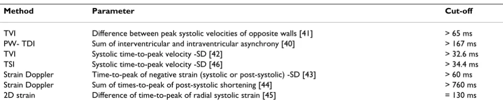
d) One field of application, which in itself would guaran-tee the survival of TDI is the study of ventricular contrac-tile dyssynchrony. The development of ultrasound platforms capable of imaging at high frame rates leads to temporal resolution high enough as to allow for a metic-ulous analysis of the different phases of the cardiac cycle. The measurement of TDI-derived time intervals has been more easily accepted, firstly because they were related to parameters cardiologists are already confident with, sec-ondly because they were validated in hemodynamic labo-ratories and thus have been confronted to a "gold standard" [37]. The application of these methods has brought upon the definition and the calculation of delays in global (interventricular) and regional (intraventricular) mechanical contraction [38]. All this has had the merit of drawing attention to the impact of dyssynchrony on car-diac performance and has shed light on the presence of significant ventricular asynchronicity even in the absence of left bundle branch block in patients with normal or near normal duration of QRS, as well as in patients with heart failure and without severely reduced LVEF [38]. The deciding driving force was a fortunate historical coinci-dence: the concurrent interest of the scientific community in cardiac resynchronization therapy in patients with severe heart failure despite optimal medical therapy and enlargement of the QRS complex in the electrocardiogram [39]. Evidence emerged in literature emphasizing the pos-sible superiority of TDI in respect to ECG and traditional echocardiography in the identification of a significant number of cases of mechanical ventricular dyssynchrony which is fundamental to identify those candidate patients for implantation of a biventricular pace-maker who will be "responders" and benefit from this procedure [38,40-48]. Further, it is possible to map the delays, knowing which of the walls are being activated with greater delay and among those more capable of recovery, and thus pro-viding important information to the electrophysiologist to plan the modality and the site of stimulation [38]. Even in this field, where it performed with great success, TDI remained a prisoner of excessive differences in methodol-ogies in use and modalities of measurement of ventricular delays. Some authors use spectral pulsed Doppler [40],
others color TDI [41,42] others strain Doppler [43,44], and others again 2D strain [45]; measuring the delays from the beginning of the QRS respectively to the begin-ning or the peak of the S wave; some have taken into account the absolute values, others calculated mean val-ues, the standard deviation; emphasis was laid by some on interventricular dyssynchrony, by others on intraven-tricular dyssnchrony, and still by others on the sum of both combined [38,40-49] (table 1). Although two indi-ces, those proposed by Bax et al. [41] and by Yu et al. [42,46], seem to have found broad consensus, in reality even these have failed when tested in the PROSPECT trial, a multicenter study, which gave disappointing results in the prediction of responders to resynchronization therapy [47]. The reasons for these dissatisfying results may lie in a lack of standardization in regards to the modalities of examination and of analysis. Measuring mechanical dys-synchrony is an opportunity which these new echocardio-graphic methods should not fail to take advantage of, because of their unique property to represent the non invasive "interpretation key" to myocardial function. The necessity for adequate training aimed at unifying the interpretation of TDI and strain curves to reduce interob-server variability is evident [47-49].
Future applications
a) It is undeniable that the potential development of TDI indices correlated to right ventricular function is being closely followed. This is because of the intrinsic limita-tions of standard echocardiography which does not allow for an examination of a heart chamber with such complex anatomy as that of the right ventricle. The problem becomes more evident in some patient categories, such as obese patients, those with chronic bronchopneumopathy or those patients in intensive care, for which for various reasons the acoustic window is suboptimal. In these cases it would be useful to have on hand "objective" parameters relatively independent of image quality. PW tissue Dop-pler seems to have this potential more than other meth-ods; in fact a direct relationship between isovolumetric relaxation time and systolic pulmonic arterial pressure has been observed. However, in this case broader studies are
Table 1: Indices of ventricular dyssynchrony obtained by tissue Doppler and strain imaging
Method Parameter Cut-off
TVI Difference between peak systolic velocities of opposite walls [41] > 65 ms PW- TDI Sum of interventricular and intraventricular asynchrony [40] > 167 ms
TVI Systolic time-to-peak velocity -SD [42] > 32.6 ms
TSI Systolic time-to-peak velocity -SD [46] > 34.4 ms
Strain Doppler Time-to-peak of negative strain (systolic or post-systolic) -SD [43] > 60 ms Strain Doppler Sum of times-to-peak of post-systolic shortening [44] > 760 ms 2D strain Difference of time-to-peak of radial systolic strain [45] = 130 ms
Cardiovascular Ultrasound 2008, 6:54 http://www.cardiovascularultrasound.com/content/6/1/54
needed to verify which indices can be used and in which patient categories [50-54]. In experimental studies, myo-cardial acceleration during isovolumetric contraction has been shown to be a load-independent parameter that cor-relates with telesystolic elasticity of the right ventricle, and if this is verified in clinical practice, it could become a use-ful and interesting parameter to measure right ventricular systolic function [55].
b) TDI and strain Doppler can catch preclinical signs of prevalently diastolic myocardial dyfunction in various cardiac pathologies, like hypertensive cardiopathy [56], diabetic cardiomyopathy [57], and secondary cardiopa-thies caused by neurological diseases like Friedreich's ataxy [6]. The distinction between pathological and phys-iological myocardial hypertrophy is of considerable clini-cal relevance, as well as the early identification of patients genetically predisposed to hypertrophic cardiomyopathy before the appearance of the very hypertrophy itself [58]. The use of TDI permits to reveal signs of early systolic dys-function, present when conventional indices of systolic function are still normal, as in diverse conditions like amyloidosis [50], thalassemia major [60], Fabry cardio-myopathy [61], Chagas disease [62] and other rare pathol-ogies. Besides, in Fabry's disease an improvement of peak systolic strain and SR after enzymatic substitution therapy has been documented [63].
c) Additionally, TDI and strain methods have been applied in the study of atrial mechanical function. Strain indices have been shown to be capable of identifying those patients with higher probability to maintain sinus rhythm after electrical cardioversion of atrial fibrillation [64]. These data seem promising because they can be applied to a wide population of patients with cardiopa-thies, a population with atrial fibrillation, taking in con-sideration recurring relapses (administration of
antiarrythimic drugs and duration of therapy, anticoagu-lants).
What will the future echocardiography laboratory look like?
For the purpose of establishing the measuring parameters and the best modalities of acquisition, it will be necessary on the one hand to plan prospective trials directed at test-ing the feasibility and reproducibility of specific protocols in defined clinical settings and on the other hand to con-front these echocardiographic parameters with other methods (like nuclear magnetic resonance) capable of analyzing the same phenomena with different physical principles and modalities [14]. This kind of effort will be fundamental to increase the type and number of clinical conditions in which these methods can pass from poten-tially useful to certainly useful (Tab. 2), accomplishing the leap in quality from being an "industrial gadget" to being a validated diagnostic "tool". In the meantime it would be desirable for cardiologists to be open minded towards possible clinical applications of the "new technologies" of echocardiography. Echocardiography gained its renown by becoming the "battle horse" of the clinical cardiologist thanks to its widespread availability (favored by portable echo devices) and most of all by the immediateness of the morphological and functional information, which ena-bles the experienced echocardiographer to make diagnos-tic judgments and prognosdiagnos-tic predictions within minutes. To apply all this with optimal efficacy it will be necessary to structure the echocardiography laboratory in two dis-tinctive procedural phases: A first phase dedicated to the mere acquisition of the images (possibly performed by echocardiography technicians or sonographers), and a second phase which does not require the patient's pres-ence during which the images are analyzed to obtain all the necessary information, utilizing specialized software. Tedious off-line analysis, however, could lead cardiolo-gists staying long periods of time in the "digital" echo lab
Table 2: Clinical applications of tissue Doppler and strain
Parameters Information Clinical condition TDI Strain SR
Basal systolic peak Global longitudinal systolic function Aspecific - -
+/-Systolic time-to-peak Mechanical dyssynchronicity Cardiomyopathy + +/-
-Systolic peak Right ventricular systolic function Heart failure - +/-
+/-Systolic peak and/or protodiastolic peak Regional systolic function at rest Ischemic Cardiopathy - +/-
+/-Systolic peak and/or protodiastolic peak Regional systolic function during stress Ischemic Cardiopathy - +/-
+/-Systolic peak Preclinical cardiac involvement Infiltrative Cardiomyopathy - -
+/-Systolic peak Monitoring of therapeutic effect Infiltrative Cardiomyopathy - +/-
+/-Protodiastolic peak Ventricular filling pressures Aspecific ++ -
+/-Protodiastolic peak Prognostic indicator Arterial Hypertension + -
-Protodiastolic peak Distinction between constriction and restriction Heart failure + -
+/-Protodiastolic peak Identification of pathological hypertrophy Ventricular Hypertrophy + -
+/-Atrial systolic peak Prediction of response to cardioversion Atrial Fibrillation - +
with a corresponding drastic extension of time dedicated to each exam and the inevitable impairment of the pro-ductivity of medical doctors and their laboratories. It is therefore necessary, for the companies to focus their efforts on developing software capable of improving the definition of spectral curves linked to semi-automated analysis systems so as to render the acquisition and inter-pretation of the data rapid and reliable. A faster applica-tion time of the method would render it more "user friendly" promoting didactics and its definite implemen-tation. Finally, as the Americas are no longer known as the "new world", the time will come when tissue Doppler and strain technologies will rid themselves of the limiting label "new technologies" to become familiar "partners" in our everyday cardiological diagnostic activity.
Competing interests
The authors declare that they have no competing interests.
Authors' contributions
RC wrote the paper, EB drafted the manuscript, BK trans-lated into English, GG participated in designe of the arti-cle, AS wrote and reviewed the article and participated in its designe. All authors read and approved the final man-uscript.
Acknowledgements
The authors give special thanks to Dr. Paolo Trambaiolo for his invaluable suggestions and for his critical ideas.
References
1. Isaaz K, Thompson A, Ethevenot G, Cloez JL, Brembilla B, Pernot C:
Doppler echocardiographic measurement of low velocity motion of the left ventricular posterior wall. Am J Cardiol 1989,
64:66-75.
2. Sutherland GR, Stewart MJ, Groundstroem KW, Moran CM, Fleming A, Guell-Peris FJ, Riemersma RA, Fenn LN, Fox KA, McDicken WN:
Color Doppler myocardial imaging: a new technique for the assessment of myocardial function. J Am Soc Echocardiogr 1994,
7:441-58.
3. Hatle L, Sutherland GR: Regional myocardial function – a new approach. Eur Heart J 2000, 21:1337-57.
4. Heimdal A, Stoylen A, Torp H, Skjaerpe T: Real-time strain rate imaging of the left ventricle by ultrasound. J Am Soc Echocardi-ogr 1998, 11:1013-19.
5. Edvardsen T, Gerber BL, Garot J, Bluemke DA, Lima JA, Smiseth OA:
Quantitative assessment of intrinsic regional myocardial deformation by Doppler strain rate echocardiography in humans: validation against three-dimensional tagged mag-netic resonance imaging. Circulation 2002, 106:50-56.
6. Sutherland GR, Di Salvo G, Claus P, D'hooge J, Bijnens B: Strain and strain rate imaging: a new clinical approach to quantifying regional myocardial function. J Am Soc Echocardiogr 2004,
17:788-802.
7. Yip G, Abraham T, Belohlavek M, Khandheria BK: Clinical applica-tions of strain rate imaging. J Am Soc Echocardiogr 2003,
16:1334-42.
8. Abraham TP, Dimaano VL, Liang HY: Role of Tissue Doppler and strain echocardiography in current clinical practice. Circula-tion 2007, 116:2597-2609.
9. Hatle L, Angelsen B: Physical principles and clinical applica-tions. In Doppler ultrasound in cardiology II edition. Lea & Febiger, Phil-adelphia; 1985.
10. Thomas G: Tissue Doppler echocardiography – A case of right tool, wrong use. Cardiovasc Ultrasound 2004, 2:12.
11. Abraham TP, Nishimura RA: Myocardial Strain: Can we finally Measure Contractility? J Am Coll Cardiol 2001, 37:731-34. 12. Becker M, Bilke E, Kühl H, Katoh M, Kramann R, Franke A, Bücker A,
Hanrath P, Hoffmann R: Analysis of myocardial deformation based on pixel tracking in two dimensionalechocardio-graphic images enables quantitative assessment of regional left ventricular function. Heart 2006, 92:1102-08.
13. Gorcsan J III, Strum DP, Mandarino WA, Gulati VK, Pinsky MR:
Quantitative assessment of alterations in regional left ven-tricular contractility. With color coded tissue Doppler echocardiography. Circulation 1997, 95:2423-33.
14. Derumeaux G, Ovize M, Loufoua J, André-Fouet X, Minaire Y, Cribier A, Letac B: Doppler tissue imaging quantitates regional wall motion during ischemia and reperfusion. Circulation 1998,
97:1970-77.
15. Garcia-Fernandez MA, Azevedo J, Moreno M, Bermejo J, Pérez-Cas-tellano N, Puerta P, Desco M, Antoranz C, Serrano JA, García E, Del-cán JL: Regional diastolic function in ischemic heart disease using pulsed wave Doppler tissue imaging. Eur Heart J 1999,
20:496-505.
16. Citro R, Galderisi M: Myocardial post-systolic motion in ischemic and not ischemic myocardium: the clinical value of tissue Doppler. Echocardiography 2005, 22:525-32.
17. Citro R, Galderisi M, Guarini P, Cicala S, Mattioli D, Bianco A, de Divitiis O, Gregorio G: Left bundle branch block with and with-out coronary artery disease: which value for a tissue Dop-pler-derived post-systolic motion? Ital Heart J 2003, 4:706-712. 18. Edvardsen T, Urheim S, Skulstad H, Steine K, Ihlen H, Smiseth OA:
Quantification of left ventricular systolic function by tissue Doppler echocardiography. Added value of measuring pre-and postejection velocities in ischemic myocardium. Circula-tion 2002, 105:2071-77.
19. Fraser AG, Payne N, Madler CF, Janerot-Sjøberg B, Lind B, Grocott-Mason RM, Ionescu AA, Florescu N, Wilkenshoff U, Lancellotti P, Wütte M, Brodin LA, MYDISE Investigators: Feasibility and repro-ducibility of off line tissue Doppler measurement of regional myocardial function during dobutamine stress echocardiog-raphy. Eur J Echocardiogr 2003, 4:43-53.
20. Citro R, Salustri A, Trambaiolo P, Gregorio G: Il Doppler tessutale nella valutazione della funzione miocardica durante ecocar-diografia da stress. Ital Heart J Suppl 2002, 3:161-9.
21. Edvardsen T, Skulstad H, Aakhus S, Urheim S, Ihlen H: Regional myocardial systolic function during acute myocardial ischemia assessed by strain Doppler echocardiography. J Am Coll Cardiol 2001, 37:726-30.
22. Jamal F, Strotmann J, Weidemann F, Kukulski T, D'hooge J, Bijnens B, Werf F Van de, De Scheerder I, Sutherland GR: Noninvasive quan-tification of the contractile reserve of stunned myocardium by ultrasonic strain rate and strain. Circulation 2001,
104:1059-65.
23. Weidemann F, Dommke C, Bijnens B, Claus P, D'hooge J, Mertens P, Verbeken E, Maes A, Werf F Van de, De Scheerder I, Sutherland GR:
Definig the transmurality of a chronic myocardial infarction by ultrasonic strain-rate imaging. Implications for identifying intramural viability. An experimental study. Circulation 2003,
107:883-88.
24. Nagueh SF, Mikati I, Kopelen HA, Middleton KJ, Quinones MA, Zoghbi WA: Doppler estimation of left ventricular filling pres-sure in sinus tachycardia. A new application of tissue doppler imaging. Circulation 1998, 98:1644-50.
25. Ommen SR, Nishimura RA, Appleton CP, Miller FA, Oh JK, Redfield MM, Tajik AJ: Clinical utility of Doppler echocardiography and tissue Doppler imaging in the estimation of left ventricular filling pressures: A comparative simultaneous Doppler-cath-eterization study. Circulation 2000, 102:1788-94.
26. Paulus WJ, Tschöpe C, Sanderson JE, Rusconi C, Flachskampf FA, Rademakers FE, Marino P, Smiseth OA, De Keulenaer G, Leite-Moreira AF, Borbély A, Edes I, Handoko ML, Heymans S, Pezzali N, Pieske B, Dickstein K, Fraser AG, Brutsaert DL: How to diagnose diastolic heart failure: a consensus statement on the diagno-sis of heart failure with normal left ventricular ejection frac-tion by the Heart Failure and Echocardiography Associations of the European Society of Cardiology. Eur Heart J 2007, 28:2539-50.
ven-Cardiovascular Ultrasound 2008, 6:54 http://www.cardiovascularultrasound.com/content/6/1/54
tricular filling pressures: a comparison of tissue Doppler echocardiography and B-type natriuretic peptide in patients with pulmonary artery catheters. Circulation 2004, 109:2432-39. 28. Wang M, Yip GW, Wang AY, Zhang Y, Ho PY, Tse MK, Lam PK, Sand-erson JE: Peak early diastolic mitral annulus velocity by tissue Doppler imaging adds independent and incremental prog-nostic value. J Am Coll Cardiol 2003, 41:820-26.
29. Sundereswaran L, Nagueh SF, Vardan S, Middleton KJ, Zoghbi WA, Quiñones MA, Torre-Amione G: Estimation of left and right ven-tricular filling pressures after heart transplantation by tissue Doppler imaging. Am J Cardiol 1998, 82:352-57.
30. Liang H-Y, Cauduro S, Pellikka P, Wang J, Urheim S, Yang EH, Rihal C, Belohlavek M, Khandheria B, Miller FA, Abraham TP: Usefulness of two-dimensional speckle strain for evaluation of left ven-tricular diastolic deformation in patients with coronary artery disease. Am J Cardiol 2006, 98:1581-86.
31. Jones CJH, Raposo L, Gibson DG: Functional importance of the long-axis dynamics of the human left ventricle. Br Heart J 1990,
63:215-20.
32. Vinereanu D, Florescu N, Sculthorpe N, Tweddel AC, Stephens MR, Fraser AG: Differentiation between pathologic and physio-logic lef ventricular hypertrophy by tissue Doppler assess-ment of long axis function in patients with hypertrophic cardiomyopathy or systemic hypertension and in athletes.
Am J Cardiol 2001, 88:53-58.
33. Bolognesi R, Tsialtas D, Barilli Al, Manca C, Zeppellini R, Javernaro A, Cucchini F: Detection of the early abnormalities of left ven-tricular function by hemodynamic, echo tissue Doppler imaging, and mitral Doppler flow techniques in patients with coronary artery disease and normal ejection fraction. J Am Soc Echocardiogr 2001, 14:764-72.
34. Gulati VK, Katz WE, Follansbee WP, Gorcsan J 3rd: Mitral annular descent velocity by tissue Doppler echocardiography as an index of global left ventricular function. Am J Cardiol 1996,
77:979-84.
35. Yamada H, Oki T, Tabata T, et al.: Assessment of left ventricular systolic wall motion velocity with pulsed tissue Doppler imaging: comparison with peak dP/dt of the left ventricular pressure curve. J Am Soc Echocardiogr 1998, 11:442-49.
36. Fukuda K, Oki T, Tabata T, Iuchi A, Ito S: Regional left ventricular wall motion abnormalities in myocardial infarction and mitral annular descent velocities studied with pulsed tissue Doppler imaging. J Am Soc Echocardiogr 1998, 11:841-48. 37. Zamorano J, Wallbridge DR, Ge J, Drozd J, Nesser J, Erbel R: Non
invasive assessment of cardiac physiology by tissue doppler echocardiography. A comparison with invasive haemody-namics. Eur Heart J 1997, 18:330-39.
38. Gorcsan J 3rd, Abraham T, Agler DA, Bax JJ, Derumeaux G, Grimm RA, Martin R, Steinberg JS, Sutton MS, Yu CM, American Society of Echocardiography Dyssynchrony Writing Group: Echocardiogra-phy for cardiac resynchronization therapy: recommenda-tions for performance and reporting – a report from the American Society of Echocardiography Dyssynchrony Writ-ing Group endorsed by the Heart Rhythm Society. J Am Soc Echocardiogr 2008, 21:191-213.
39. Abraham WT, Fisher WG, Smith AL, Delurgio DB, Leon AR, Loh E, Kocovic DZ, Packer M, Clavell AL, Hayes DL, Ellestad M, Trupp RJ, Underwood J, Pickering F, Truex C, McAtee P, Messenger J, MIRA-CLE Study Group. Multicenter InSync Randomized Clinical Evaluation:
Cardiac resynchronization in chronic heart failure. N Engl J Med 2002, 346:1845-53.
40. Penicka M, Bartunek J, De Bruyne B, Vanderheyden M, Goethals M, De Zutter M, Brugada P, Geelen P: Improvement of left ventricu-lar function after cardiac resynchronization therapy is pre-dicted by tissue Doppler imaging echocardiography.
Circulation 2004, 109:978-83.
41. Bax JJ, Bleeker GB, Marwick TH, Molhoek SG, Boersma E, Steendijk P, Wall EE van der, Schalij MJ: Left ventricular dyssynchrony pre-dicts response and prognosis after cardiac resynchronization therapy. J Am Coll Cardiol 2004, 44:1834-40.
42. Yu CM, Fung WH, Lin H, Zhang Q, Sanderson JE, Lau CP: Predictors of left ventricular reverse remodeling after cardiac resyn-chronization therapy for heart failure secondary to idio-pathic dilated or ischemic cardiomyopathy. Am J Cardiol 2003,
91:684-88.
43. Mele D, Pasanisi G, Capasso F, De Simone A, Morales MA, Poggio D, Capucci A, Tabacchi G, Sallusti L, Ferrari R: Left intraventricular myocardial deformation dyssynchrony identifies responders to cardiac resynchronization therapy in patients with heart failure. Eur Heart J 2006, 27:1070-78.
44. Porciani MC, Lilli A, Macioce R, Cappelli F, Demarchi G, Pappone A, Ricciardi G, Padeletti L: Utility of a new left ventricular asyn-chrony index as a predictor of reverse remodelling after car-diac resynchronization therapy. Eur Heart J 2006, 27:1818-23. 45. Suffoletto MS, Dohi K, Cannesson M, Saba S, Gorcsan J 3rd: Novel
speckle-tracking radial strain from routine black-and-white echocardiographic images to quantify dyssynchrony and pre-dict response to cardiac resynchronization therapy. Circula-tion 2006, 113:960-68.
46. Yu CM, Zhang Q, Fung JW, Chan HC, Chan YS, Yip GW, Kong SL, Lin H, Zhang Y, Sanderson JE: A novel tool to assess systolic asynchrony and identify responders of cardiac resynchroni-zation therapy by tissue synchroniresynchroni-zation imaging. J Am Coll Cardiol 2005, 45:677-84.
47. Chung ES, Leon AR, Tavazzi L, Sun JP, Nihoyannopoulos P, Merlino J, Abraham WT, Ghio S, Leclercq C, Bax JJ, Yu CM, Gorcsan J 3rd, St John Sutton M, De Sutter J, Murillo J: Results of the Predictors of Response to CRT (PROSPECT) trial. Circulation 2008,
117:2608-16.
48. Anderson LJ, Miyazaki C, Sutherland GR, Oh JK: Patient selection and echocardiographic assessment of dyssynchrony in car-diac resynchronization therapy. Circulation 2008, 117:2009-23. 49. Galderisi M, Cattaneo F, Mondillo S: Doppler echocardiography
and myocardial dyssynchrony: a practical update of old and new ultrasound technologies. Cardiovasc Ultrasound 5:28. 2007 Sep 6
50. Meluzin J, Spinarova L, Bakala J, Toman J, Krejcí J, Hude P, Kára T, Soucek M: Pulsed Doppler tissue imaging of the velocity of tri-cuspid annular systolic motion; a new, rapid, and non-inva-sive method of evaluating right ventricular systolic function.
Eur Heart J 2001, 22:340-48.
51. Kukulski T, Hubbert L, Arnold M, Wranne B, Hatle L, Sutherland GR:
Normal regional right ventricular function and its change with age: a Doppler myocardial imaging study. J Am Soc Echocardiogr 2000, 13:194-204.
52. Alam M, Wardell J, Andersson E, Samad BA, Nordlander R: Right ventricular function in patients with first inferior myocardial infarction: assessment by tricuspid annular motion and tri-cuspid annular velocity. Am Heart J 2000, 139:710-15.
53. Caso P, Galderisi M, Cicala S, Cioppa C, D'Andrea A, Lagioia G, Lic-cardo B, Martiniello AR, Mininni N: Association between myocar-dial right ventricular relaxation time and pulmonary arterial pressure in chronic obstructive lung disease: analysis by pulsed Doppler tissue imaging. J Am Soc Echocardiogr 2001,
14:970-77.
54. Abbas A, Lester S, Moreno FC, Srivathsan K, Fortuin D, Appleton C:
Noninvasive assessment of right atrial pressure using Dop-pler tissue imaging. J Am Soc Echocardiogr 2004, 17:1155-60. 55. Vogel M, Schmidt MR, Kristiansen SB, Cheung M, White PA, Sorensen
K, Redington AN: Validation of myocardial acceleration during isovolumic contraction as a novel noninvasive index of right ventricularcontractility: comparison with ventricular pres-sure-volume relations inan animal model. Circulation 2002,
105:1693-99.
56. Poulsen SH, Andersen NH, Ivarsen PI, Mogensen CE, Egeblad H:
Doppler tissue imaging reveals systolic dysfunction in patients with hypertension and apparent "isolated" diastolic dysfunction. J Am Soc Echocardiogr 2003, 16:724-31.
57. Boyer JK, Thanigaraj S, Schechtman KB, Perez JE: Prevalence of ventricular diastolic dysfunction in asymptomatic, normo-tensive patients with diabetes mellitus. Am J Cardiol 2004,
93:870-75.
58. Nagueh SF, Bachinski LL, Meyer D, Hill R, Zoghbi WA, Tam JW, Quiñones MA, Roberts R, Marian AJ: Tissue Doppler imaging consistently detects myocardial abnormalities in patients with hypertrophic cardiomyopathy and provides a novel means for an early diagnosis before and independently of hypertrophy. Circulation 2001, 104:128-30.
Publish with BioMed Central and every scientist can read your work free of charge
"BioMed Central will be the most significant development for disseminating the results of biomedical researc h in our lifetime."
Sir Paul Nurse, Cancer Research UK
Your research papers will be:
available free of charge to the entire biomedical community
peer reviewed and published immediately upon acceptance
cited in PubMed and archived on PubMed Central
yours — you keep the copyright
Submit your manuscript here:
http://www.biomedcentral.com/info/publishing_adv.asp
BioMedcentral
physiological form pathological pressure-overload left ven-tricular hypertrophy in rats. Circulation 2002, 105:1602-08. 60. Koyama J, Ray-Sequin PA, Falk RH: Longitudinal myocardial
func-tion assessed by tissue velocity, strain and strain rate tissue Doppler echocardiography in patients with AL (primary) cardiac amyloidosis. Circulation 2003, 107:2446-52.
61. Vogel M, Anderson LJ, Holden S, Deanfield JE, Pennell DJ, Walker JM:
Tissue Doppler Echocardiography in patient with Tha-lassemia detects early myocardial dysfunction related to myocardial iron overload. Eur Heart J 2003, 24:113-19. 62. Pieroni M, Chimenti C, Ricci R, Sale P, Russo MA, Frustaci A: Early
detection of Fabry caridomyopathy by tissue Doppler Imag-ing. Circulation 2003, 107:1978-84.
63. Barros MV, Ribeiro Al, Machado FS, Rocha MO: Doppler Tissue imaging to assess systolic function in Chagas'disease. Arq Bras Cardiol 2003, 80:31-40.
64. Weidemann F, Breunig F, Beer M, Sandstede J, Turschner O, Voelker W, Ertl G, Knoll A, Wanner C, Strotmann JM: Improvement of cardiac function during enzyme replacement therapy in patients with Fabry disease: a prospective strain rate imag-ing study. Circulation 2003, 108:1299-1301.
65. Di Salvo G, Caso P, Lo Piccolo R, Fusco A, Martiniello AR, Russo MG, D'Onofrio A, Severino S, Calabró P, Pacileo G, Mininni N, Calabró R: