Pharyngeal Airway Dimensions and Head Posture in Obstructive
Sleep Apnea Patients with and without Morphological Deviations
in the Upper Cervical Spine
Liselotte Sonnesen
1, Arne Petersson
2, Søren Berg
3, Palle Svanholt
11Section of Orthodontics, Department of Odontology, Faculty of Health and Medical Sciences, University of Copenhagen, Denmark.
2Department of Oral and Maxillofacial Radiology, Malmö University, Malmö, Sweden. 3Clinic for Infant and Adult Sleep Medicine, Lovisenberg Diakonale Sykehus, Oslo, Norway.
Corresponding Author: Liselotte Sonnesen
Section of Orthodontics, Department of Odontology
Faculty of Health and Medical Sciences, University of Copenhagen 20 Nørre Alle, DK-2200, Copenhagen
Denmark
Phone: 0045 35 32 66 70 E-mail: [email protected]
ABSTRACT
Objectives: The aim of the study was to analyse differences in pharyngeal airway dimensions and head posture between obstructive sleep apnea patients with and without morphological deviations in the upper cervical spine and to analyse associations between pharyngeal airway dimensions and head posture in the total sample.
Material and Methods: The sample comprised 53 obstructive sleep apnea (OSA) patients of which 32.1% had upper spine morphological deviations. Accordingly two groups were defined: 17 OSA patients with morphological deviations in the upper spine and 36 without upper spine deviations. Pharyngeal airway dimensions in terms of distances, cross-sectional areas and volume and upper spine morphological deviations were evaluated on cone-beam computed tomography. Head posture was evaluated on two-dimensional generated lateral cephalograms. Differences were analysed and adjusted for age and gender by multiple linear regression analysis.
Results: OSA patients with upper spine morphological deviations had a significantly more backward and curved neck posture (OPT/HOR, P < 0.01; OPT/CVT, P < 0.05) compared to OSA patients without spine deviations. No significant differences were found in airway dimensions between patients with and without upper spine deviations. In the total group significant associations were found between head posture and pharyngeal airway distances and cross-sectional area at the nasal floor, epiglottis and hyoid bone level (P < 0.05, P < 0.01, P < 0.001). No significant association was found between head posture and airway volume.
Conclusions: The results may contribute to differentiate obstructive sleep apnea patients and thereby may prove valuable in diagnosis and treatment planning of obstructive sleep apnea patients.
Keywords: nasopharynx; obstructive sleep apnea; oropharynx; standing position; vertebral column.
Accepted for publication: 26 September 2017 To cite this article:
Sonnesen L, Petersson A, Berg S, Svanholt P.
Pharyngeal Airway Dimensions and Head Posture in Obstructive Sleep Apnea Patients with and without Morphological Deviations in the Upper Cervical Spine
J Oral Maxillofac Res 2017;8(3):e4
INTRODUCTION
Obstructive sleep apnea (OSA) is by far the most common sleep-related breathing disorder, affecting 2 - 4% of the adult population, particularly males aged 60 years and older where the prevalence is 30 - 60% [1,2]. OSA is defined as cessation of airflow
with persistent respiratory effort, due to repeated anatomical obstruction or partial collapse of the oropharyngeal region, involving the soft palate, dorsum of the tongue and the posterior pharyngeal wall [1,3]. OSA is multifactorial with age, gender and body mass index (BMI) as predisposing factors [1,3]. Authors agree that there are craniofacial morphological and postural characteristics in OSA patients such as reduced posterior airway space, abnormally long soft palate, low position of the hyoid bone and an extended head posture [4,5].
The upper airway is normally maintained by the bony and cartilaginous structures surrounding the naso- and oropharynx. Located posterior to the pharynx, the cervical spine is composed of seven vertebrae that provide full range of motion to the neck through articulations with the occiput and between the vertebrae. Some studies have been performed on the relationship between the cervical spine and OSA. Pathological conditions as osteoarthritis of the cervical spine [6-8], cervical osteophytes [9-11] and osteochondromas [12,13] have been reported in relation to OSA patients. Recently, the morphology of the upper cervical spine in patients with OSA has been described on two-dimensional lateral cephalograms [14,15] and three-dimensional cone-beam computed tomography (CBCT) [16]. The prevalence of morphological deviations in the upper spine range between 32 and 46% and the
deviations occurred significantly more often in OSA
patients than in subjects with neutral occlusion and normal craniofacial morphology [14-16].In addition, associations between OSA patients with upper spine morphological deviations and mandibular advancement device (MAD) treatment outcome have been reported [17].
Studies have also shown an association between upper cervical spine morphology and head posture evaluated on lateral cephalograms in healthy subjects or subjects with malocclusion [18-21]. Furthermore, associations between head posture and pharyngeal airway dimensions have been documented on lateral cephalograms in adults [5,22-24]. According to a recent systematic review, there may be a lack of evidence on the effect of head posture on the upper airway dimensions in three-dimensional imaging due
to lack of standardized method of the head and tongue position during image acquisition [25]. Furthermore, the relationship between morphological deviations in the upper spine and pharyngeal airway dimensions in OSA patients has not previously been reported.
Due to the cervical spine’s and head posture’s close associations with the pharyngeal airway it seems clinically relevant to focus on the relationship between the upper cervical spine and sleep apnea on three-dimensional imaging of OSA patients in a standardized head position.
The aims of the present study were to analyse: 1) differences in pharyngeal airway dimensions between OSA patients with and without upper spine morphological deviations, 2) differences in head posture between OSA patients with and without upper spine morphological deviations and 3) associations between pharyngeal airway dimensions and head posture in the total sample of CBCTs taken in a standardized head position.
MATERIAL AND METHODS Subjects
The first 53 patients with apnoea-hypopnoea
index (AHI) > 5 were enrolled as a part of a longitudinal sleep apnea study at the Orthodontics Section, Department of Odontology, University of Copenhagen, Denmark, in the period from January 2012 to May 2013 form part of the present study. The patients were diagnosed with OSA by sleep studies, using overnight polygraphy (Embletta Portable Diagnostic System, Iceland). Definition of AHI: apnea as cessation of airflow more than 10 seconds, hypopnea as reduction in airflow of at least 30%
with an associated desaturation of 4%. The sample was divided according to a visual assessment of morphological deviations in the upper cervical spine on three-dimensional images using the NewTom NNT software (QR srl, Verona, Italy) and analysed according to Sandham [26] as fusion of the vertebra
units and posterior arch deficiencies. The findings
were categorized as morphological deviations of the upper cervical spine, yes/no and have previous been described in details [16]. A total of 32.1% of the OSA patients had morphological deviations in the upper spine. Accordingly two groups were
defined: 17 OSA patients, 7 women and 10 men aged
In OSA patients with morphological deviations in the upper spine AHI ranged between 9 and 35 (mean 20.41 [SD 8.49]) and in OSA patients without morphological deviations in the upper spine AHI ranges between 7 and 57 (mean 20.17 [SD 11.76]). No statistical gender or AHI differences were found between the groups, but the patients with morphological deviations in the upper spine were older than the patients without spine deviations (P < 0.05). All participants had read and signed the informed consent form. The study had been approved by the Ethical Committee for Copenhagen, Denmark (Ref. No. H-3-2011-086) and the Danish Data Protection Agency (J. No. 2012-54-0041).
Methods
The CBCTs were taken in a NewTom VGI unit (QR srl, Verona, Italy) of standing OSA patients with the teeth in occlusion and in the standardized head posture in the self-balance position as described by Siersbaek-Nielsen and Solow [27]. The settings were as followed: standard scan mode with an imaging volume of 15 x 15 cm, scan time 18 seconds and axial thickness of 0.3 mm. The pharyngeal airway distances and cross-sectional areas were analysed using the ITK-Snap software [28]and the pharyngeal airway volumes were analysed by Romexis software (Planmeca Company, Helsinki, Finland). Two-dimensional images were generated with the Romexis software in the mid sagittal plane and the head posture was analysed using View Box 3®
Figure 1. Pharyngeal reference levels illustrated in the mid sagittal
view. NF = nasal floor, through the most posterior and inferior point
of the cavum nasi; V = velum, the shortest antero-posterior distance behind velum palate; RL = radix linguae, the narrowest point behind radix linguae; EP = epiglottis, the level through the top of epiglottis; HY = hyoid bone, the level where the vocal chord is visible.
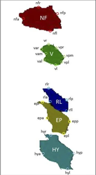
Figure 2. Pharyngeal reference points. NF = nasal floor
level: most anterior (nfa) and posterior (nfp) point in the
sagittal plane, the most right (nfr) and left (nfl) point.
V = velum level: the most anterior (vam) and posterior (vmp) point in the sagittal plane, the most anterior (var) and posterior point (vpr) in the right part, the most anterior (val) and posterior point (vpl) in the left part. RL = radix linguae level: most anterior (rla) and posterior point (rlp) in the sagittal plane, the most right (rlr) and left (rll) point. EP = epiglottis level: most anterior (epa) and posterior point (epp) in the sagittal plane, the most right (epr) and left (epl) point. HY = hyoid bone level: most anterior (hya) and posterior (hyp) point in the sagittal plane, the most right (hyr) and left (hyl) point.
(Demetrios Halaznetis, Athens, Greece).
Pharyngeal airway dimensions
The sagittal and transversal linear distances
were measured at five horizontal levels: the nasal floor (NF), velum (V), radix linguae (RL),
epiglottis (EP) and hyoid bone (HY) (Figure 1,
Table 1) according to the levels defined in the
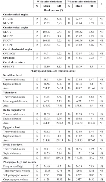
Table 1. Postural variables and pharyngeal dimensions in obstructive sleep apnea subjects with and without upper spine deviations With spine deviations Without spine deviations
P
N Mean SD N Mean SD
Head posture (°) Craniovertical angles
FH/VER 15 95.31 5.36 31 92.97 4.91 NS
NL/VER 15 93.02 6,93 30 89.64 8.59 NS
Craniocervical angles
NL/CVT 15 108.17 9.43 30 106.52 9.52 NS
NL/OPT 15 92.15 9.8 30 95.67 9.19 NS
FH/CVT 16 103.69 7.22 31 104.96 8.06 NS
FH/OPT 15 94.42 8.91 31 99.02 8.06 NS
Cervicohorizontal angles
CVT/HOR 16 74.71 6.22 36 73.07 7.92 NS
OPT/HOR 16 90.45 7.42 36 83.85 7.22 b,c
Cervical curvature
OPT/CVT 17 15.89 8.12 36 10.79 4.3 a
Pharyngeal dimensions (mm/mm2/mm3)
Nasal floor level
Transversal distance 17 28.31 4.39 36 27.84 5.47 NS Sagittal distance 17 21.56 4.06 36 20.33 3.4 NS
Area 17 533.33 154.53 36 469.2 121.68 NS
Velum level
Transversal distance 17 23.15 6.86 36 24.28 6.82 NS Mean sagittal distance 17 6.21 2.15 36 6.72 2.32 NS
Area 17 136.93 77.86 36 155.81 95 NS
Radix lingual level
Transversal distance 17 31.59 14.16 36 31.28 6.53 NS
Sagittal distance 17 10.75 3.96 36 10.92 4 NS
Area 17 264.49 152.63 36 257.62 103.47 NS
Epiglottis level
Transversal distance 17 30.62 6 36 33.03 5.44 NS Sagittal distance 17 13.13 4.7 36 13.07 3.83 NS
Area 17 314.46 158.13 36 311.15 117.65 NS
Hyoid bone level
Transversal distance 17 38.01 3.75 36 38.95 4.19 NS
Sagittal distance 17 16.73 3.84 36 17 3.56 NS
Area 17 418.5 119.32 36 440.38 114.2 NS
Pharyngeal high and volume
Pharynx total high 17 54.49 6.7 36 56.23 7.83 NS Total pharyngeal volume 17 12928 6274 36 12666 6381 NS Velopharyngeal volume 17 6799 3589 36 6729 5085 NS Oropharyngeal volume 17 6129 3952 36 5937 3436 NS
aP < 0.05; bP < 0.01; cgender effect (P < 0.05). Multiple linear regression analysis.
N = number of patients; SD = standard deviation; NS = not significant; FH/VER = the angle between Frankfort horizontal and true vertical
The cross-sectional areas were assessed by marking the edge of the pharynx aperture and the area was then
calculated by the software within the defined points
and margins (Figure 2, Table 1).
The pharyngeal volume was assessed as described by Anandarajah et al. [30]. The volumes were defined
according to Sears et al. [29] as the total pharyngeal volume between NF and EP, the oropharyngeal volume between V and EP and the nasopharyngeal volume as the difference between the total volume and the oropharyngeal volume (Figure 3, Table 1). The airway volumes were then automatically calculated by
the software within the defined margins (Table 1).
Head posture
Nine variables representing the craniovertical, craniocervical and cervicohorizontal postural relationships and the curvature of the upper cervical
spine was defined according to Solow and Tallgren
[31].As the cranial base and thereby the nasion-sella line (NSL) was not visible on some of the CBCTs the Frankfort horizontal line was used instead in all OSA patients. The reference points and lines are illustrated in Figure 4 and the mean values are shown in Table 1.
Reliability
The reliability of the variables describing the pharyngeal airway dimensions and head posture were assessed by re-measuring 25 lateral CBCTs selected at random from the previously evaluated CBCTs. The CBCTs were marked and measured again, and paired
t-test found no significant differences between the
two sets of recordings. The method errors calculated
by Dahlberg’s formula [32] for the head posture ranged from 0.04 to 2.5 degrees and the Houston
reliability coefficient [33] from 0.7 to 1. The method errors for the distances of the pharyngeal airway ranged from 0.08 to 3.35 mm and the Houston
reliability coefficient from 0.73 to 1. The method
errors for the cross-sectional areas of the pharyngeal airway ranged from 4.6 to 52.4 mm2 and the Houston reliability coefficient from 0.79 to 1. The method
errors for the volumes of the pharyngeal airway ranged from 549.1 to 1270.8 mm3 and the Houston reliability coefficient from 0.92 to 0.99.
Statistical analysis
The normality of distributions regarding the pharyngeal airway dimensions and the head posture was assessed by parameters of skewness and kurtosis and by Shapiro-Wilks W-test. Cervical lordosis (OPT/ CVT), velum area and the pharyngeal airway volume were not normally distributed. Once the variables were transformed logarithmically they became normally distributed.
Differences in means of pharyngeal airway dimensions and head posture between the two groups were assessed by unpaired t-test and adjusted for age and gender by multiple linear regression analyses. Associations between head posture and airway dimensions adjusted for age and gender were assessed by multiple linear regression analysis. Parametric data were expressed as mean and standard deviation (M [SD]).
The results from the tests were considered significant
at P-values below 0.05. The statistical analyses were performed using SPSS 20.00 (Inc., Chicago, Illinois, USA).
Figure 3. Illustration of pharyngeal volumes in the mid sagittal view. A = total pharyngeal volume between nasal floor plane and epiglottis
plane; B = oropharyngeal volume between velum plane and epiglottis plane.
Table 2. Significant associations between pharyngeal airway dimensions and head posture adjusted for age and gender in the total group
(n = 53)
Level Nasal floor level Epiglottis level Hyoid bone level
Variable Transversal distance distanceSagittal Area Transversal distance distanceSagittal Transversal distance distanceSagittal Area Craniovertical angles (°)
FH/VER 0.36a 0.39a 0.33a - - - -
-NL/VER - - -
-Craniocervical angles (°)
NL/CVT - - - 0.31a 0.42b 0.45b
NL/OPT - - - 0.42b 0.31a
FH/CVT - - - 0.32a 0.32a 0.4b 0.49c 0.5c
FH/OPT - - - 0.3a 0.3a 0.32a 0.5c 0.46b
Cervicohorizontal angles (°)
CVT/HOR - - - -0.43b -0.46b -0.57c
OPT/HOR - - - -0.53b,d -0.51b,d
aP < 0.05; bP < 0.01; cP < 0.001. dAge effect (P < 0.05). Multiple linear regression analysis.
FH/VER = the angle between Frankfort horizontal and true vertical line; NL/VER = the angle between nasal line and true vertical line; NL/CVT = the angle between nasal line and cervical vertebra tangent line; NL/OPT = the angle between nasal line and odontoid process tangent line; FH/CVT = the angle between Frankfort horizontal and cervical vertebra tangent line; FH/OPT = the angle between Frankfort horizontal and odontoid process tangent line; CVT/HOR = the angle between cervical vertebra tangent and true horizontal line; OPT/HOR = the angle between odontoid process tangent and true horizontal line; OPT/CVT = the angle between odontoid process tangent and cervical vertebra tangent line.
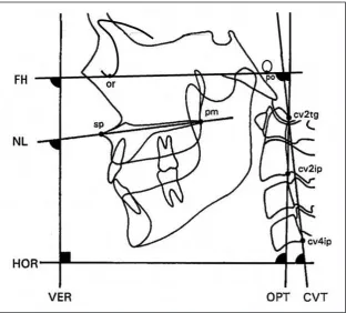
Figure 4. Reference points and lines describing the head posture on two-dimensional generated lateral cephalogram [31].
FH = Frankfurter horizontal through or and po; or = orbitale, the most inferior point of foramen orbitale; po = porion, the most inferior point of apertura acusticus.
NL = nasal line through sp and pm; sp = spinal point, the apex of in the anterior nasal spine; pm = pterygomaxillare, the intersection between
the nasal floor and the posterior contour of the maxilla.
OPT = odontoid process tangent through cv2ip and cv2tg; cv2ip = the most posteroinferior point on the corpus of C2; cv2tg = the tangent point on the dorsal contour of the odontoid process of C2 to a line from cv2ip.
RESULTS
Differences between the groups
The cervicohorizontal angle (OPT/HOR) and OPT/
CVT was significantly larger in OSA patients with
morphological deviations in the upper spine compared to OSA patients without upper spine deviations (P < 0.01, P < 0.05, respectively; Table 1), meaning a more backward and curved neck posture in OSA patients with upper spine morphological deviations compared to OSA patients without spine deviations.
No significant differences were found in airway
dimensions between patients with and without upper spine morphological deviations (Table 1).
Associations
When associations between head posture and pharyngeal airway distances and cross-sectional areas were adjusted for age and gender a clear pattern was found between head posture and the pharyngeal airway distances and cross-sectional area at the HY level (Table 2). Thus, the craniocervical posture (FH/
OPT, FH/CVT, NL/OPT, NL/CVT) was significantly
positively associated with airway distances and cross-sectional area at the HY level (P < 0.05, P < 0.01, P < 0.001; Table 2) and the cervicohorizontal posture
(OPT/HOR, CVT/HOR) was significantly negatively
associated with airway distances and cross-sectional area at the HY level (P < 0.01, P < 0.001; Table 2) meaning larger airway distances and cross-sectional area at the HY level with an extended and forward head posture. Furthermore, the craniocervical posture
(FH/OPT, FH/CVT) was significantly positively
associated with the pharyngeal airway distances at the EP (P < 0.05; Table 2), and the craniovertical posture
(FH/VER) was significantly positively associated with
the pharyngeal airway distances and cross-sectional
area at the NF level (P < 0.05; Table 2). No significant
associations were found between head posture and pharyngeal airway volume (Table 2).
DISCUSSION
The aim of the present study was to analyse differences in pharyngeal airway dimensions and head posture between OSA patients with and without morphological deviations in the upper cervical spine and to study associations between pharyngeal airway dimensions and head posture on three-dimensional CBCT taken in a standardized head position. To our knowledge differences in pharyngeal airway
dimensions and head posture between OSA patients with and without morphological deviations in the upper spine have not previously been reported in the literature. Furthermore it is still uncertain how the head posture affects the upper airway dimensions analysed on three-dimensional CBCT taken in a standardized head position [25].
Differences between groups
In the present study, OSA patients with morphological
deviations in the upper spine had a significantly more
backward and curved neck posture compared to OSA
patients without spine deviations. The findings were in
agreement with previous studies where an association was found between morphological deviations in the upper spine and posture of the head and neck in healthy adults [18] and in children with class II malocclusion [19].
A direct relationship between morphological deviations in the upper spine and pharyngeal airway
dimensions could not be verified in the present study.
Previous studies have shown that morphological
deviations in the upper spine occurred significantly
more often in OSA patients compared to healthy controls [14,15] and that the craniofacial profile
of OSA patients with block fusions in the upper cervical spine and fusion of two vertebrae differed
significantly from the craniofacial profile of other
OSA patients [15]. Therefore the morphological deviations in the upper spine contribute to phenotypic differentiations between types of OSA and thereby to the diagnosis and subdivision of patients with OSA [14,15]. Furthermore, OSA patients with morphological deviations in the upper spine show a worse response to MAD treatment compared to OSA patients without morphological deviations in the upper spine [17]. The findings indicate that the aetiology
in OSA patients with morphological deviations in the upper spine is characterised by other factors or combinations of different factors than in OSA patients without upper spine morphological deviations [14,15,17].
Specific cervical spine pathologies, such as
osteophytes, osteoarthritis and osteochondromas are conditions associated with OSA that may be due to a direct relationship to the pharyngeal dimensions resulting in a decrease in the pharyngeal airway dimensions [6-13]. Often these pathological conditions are parts of general syndromes that cause OSA [34] and thereby make it difficult to
and morphological deviations in the upper cervical spine found in previous studies [14-16] could not be explained by a direct relationship to the pharyngeal
airway dimensions. No significant differences in the
pharyngeal airway dimensions between patients with and without upper spine morphological deviations were found in the present study. Results from previous study [17] indicate that OSA patients with upper spine morphological deviations may respond poorly to MAD treatment. The results from the present study
showed no statistically significant differences in
upper airway dimensions between OSA patients with and without upper spine morphological deviations. Therefore it may be suggested the OSA patients with upper spine morphological deviations should be treated with constant positive airway pressure and OSA patients without upper spine morphological deviations should be treated with MAD. This recommendation should be investigated with future research.
Associations
When the CBCT was taken in the standardized head posture a clear pattern was found between head posture and the pharyngeal airway distances and cross-sectional area at the HY level: an extended and forward head posture was associated with larger airway distances and cross-sectional area at
the HY level. These findings were in agreement
with a previous study using two-dimensional lateral cephalograms of OSA patients taken in the standardized head position which showed that an extended and forward head posture was associated with the anterior-posterior airway distances especially in the lower part of the pharyngeal airway [5]. Since the previous study used a two-dimensional method the pharyngeal airway volume was not assessed. Another previous study using three-dimensional magnetic resonance imaging of children with sleep-disordered breathing, found an association between various head positions and the oropharyngeal volume [35]. Interestingly, the present study of CBCTs in the standardized head position did not show an association between head posture and pharyngeal airway volumes, only pharyngeal airway distances and cross-sectional area. This is new and important
knowledge as the previous described compensatory physiological postural mechanism that serves to maintain airway adequacy in OSA patients [5] may be due to a stretch of the pharyngeal airway at the HY level and not due to an increased airway volume. Pharyngeal airway patency depends on the
relative influence of pharyngeal airway dimensions,
pharyngeal airway compliance and the negative intraluminal pressure during inspiration [36,37]. Therefore, the stretch of the pharyngeal airway with an extended and forward head posture found in the present study may decrease the risk of airway collapsibility due to changes in pharyngeal airway dimensions and interluminal pressure during inspiration locally at the HY level and not by an increased airway volume as previously suggested [5,35].
CONCLUSIONS
Head posture was significantly different between
patients with and without upper spine morphological
deviations. No significant differences in airway
dimensions between patients with and without upper spine morphological deviations were found. Head posture was associated with pharyngeal airway distances and cross-sectional areas but not with pharyngeal airway volume. These results may contribute to help differentiate obstructive sleep apnea patients and thereby may prove valuable in future diagnosis and treatment planning of obstructive sleep apnea patients.
ACKNOWLEDGMENTS AND DISCLOSURE STATEMENTS
The authors report no conflicts of interest related to
this study. The authors would like to thank Eva Marie Reinwald (Department of Odontology, University of Copenhagen) for manuscript preparation. The study is funded by strategic funding from the Department of Odontology, Faculty of Health and Medical Sciences, University of Copenhagen, Denmark (Strategimidler OI 2011) and the Danish Dental Association Foundation (28-05-2014, KOF).
REFERENCES
1. Prisant LM, Dillard TA, Blanchard AR. Obstructive sleep apnea syndrome. J Clin Hypertens (Greenwich) 2006 Oct; 8(10): 746-50. [doi: 10.1111/j.1524-6175.2006.888139.x]
3. Guilleminault C, Tilkian A, Dement WC. The sleep apnea syndromes. Annu Rev Med. 1976;27:465-84. [doi: 10.1146/annurev.me.27.020176.002341]
4. Neelapu BC, Kharbanda OP, Sardana HK, Balachandran R, Sardana V, Kapoor P, Gupta A, Vasamsetti S. Craniofacial and upper airway morphology in adult obstructive sleep apnea patients: A systematic review and meta-analysis of cephalometric studies. Sleep Med Rev. 2017 Feb;31:79-90. [Medline: 27039222] [doi: 10.1016/j.smrv.2016.01.007] 5. Solow B, Skov S, Ovesen J, Norup PW, Wildschiødtz G. Airway dimensions and head posture in obstructive sleep
apnoea. Eur J Orthod. 1996 Dec;18(6):571-9. [Medline: 9009421] [doi: 10.1093/ejo/18.6.571]
6. Shoda N, Seichi A, Takeshita K, Chikuda H, Ono T, Oka H, Kawaguchi H, Nakamura K. Sleep apnea in rheumatoid arthritis patients with occipitocervical lesions: the prevalence and associated radiographic features. Eur Spine J. 2009 Jun;18(6):905-10. [Medline: 19365641] [PMC free article: 2899654] [doi: 10.1007/s00586-009-0975-z]
7. Nguyen HV, Ludwig SC, Silber J, Gelb DE, Anderson PA, Frank L, Vaccaro AR. Rheumatoid arthritis of the cervical spine. Spine J. 2004 May-Jun;4(3):329-34. [Medline: 15125859] [doi: 10.1016/j.spinee.2003.10.006]
8. Drossaers-Bakker KW, Hamburger HL, Bongartz EB, Dijkmans BA, Van Soesbergen RM. Sleep apnoea caused by rheumatoid arthritis. Br J Rheumatol. 1998 Aug;37(8):889-94. [Medline: 9734681] [doi: 10.1093/rheumatology/37.8.889] 9. Eyigor H, Selcuk OT, Osma U, Koca R, Yilmaz MD. Cervical osteophytes: a rare cause of obstructive sleep apnea.
J Craniofac Surg. 2012 Sep;23(5):e444-6. [Medline: 22976699] [doi: 10.1097/SCS.0b013e318260e9ea]
10. Ando E, Ogawa T, Shigeta Y, Hirai S, Ikawa T, Ishikawa C, Nejima J. A case of obstructive sleep apnoea with anterior cervical osteophytes. J Oral Rehabil. 2009 Oct;36(10):776-80. [Medline: 19702680] [doi: 10.1111/j.1365-2842.2009.01984.x] 11. Fuerderer S, Eysel-Gosepath K, Schröder U, Delank KS, Eysel P. Retro-pharyngeal obstruction in association
with osteophytes of the cervical spine. J Bone Joint Surg Br. 2004 Aug;86(6):837-40. [Medline: 15330024] [doi: 10.1302/0301-620X.86B6.14933]
12. Wang V, Chou D. Anterior C1-2 osteochondroma presenting with dysphagia and sleep apnea. J Clin Neurosci. 2009 Apr;16(4):581-2. [Medline: 19195892] [doi: 10.1016/j.jocn.2008.05.024]
13. Yoshida T, Matsuda H, Horiuchi C, Taguchi T, Nagao J, Aota Y, Honda A, Tsukuda M. A case of osteochondroma of the atlas causing obstructive sleep apnea syndrome. Acta Otolaryngol. 2006 Apr;126(4):445-8. [Medline: 16608803] [doi: 10.1080/00016480500416793]
14. Sonnesen L, Petri N, Kjaer I, Svanholt P. Cervical column morphology in adult patients with obstructive sleep apnoea. Eur J Orthod. 2008 Oct;30(5):521-6. [Medline: 18647950] [doi: 10.1093/ejo/cjn028]
15. Svanholt P, Petri N, Wildschiødtz G, Sonnesen L, Kjaer I. Associations between craniofacial morphology, head posture, and cervical vertebral body fusions in men with sleep apnea. Am J Orthod Dentofacial Orthop. 2009 Jun;135(6):702.e1-9; discussion 702-3. [Medline: 19524827] [doi: 10.1016/j.ajodo.2009.02.011]
16. Sonnesen L, Jensen KE, Petersson AR, Petri N, Berg S, Svanholt P. Cervical vertebral column morphology in patients with obstructive sleep apnoea assessed using lateral cephalograms and cone beam CT. A comparative study. Dentomaxillofac Radiol. 2013;42(6):20130060. [Medline: 23503808] [PMC free article: 3667528] [doi: 10.1259/dmfr.20130060]
17. Svanholt P, Petri N, Wildschiødtz G, Sonnesen L. Influence of craniofacial and upper spine morphology on mandibular advancement device treatment outcome in patients with obstructive sleep apnoea: a pilot study. Eur J Orthod. 2015 Aug;37(4):391-7. doi: 10.1093/ejo/cju064. Epub 2014 Oct 28. [Medline: 25351571] [doi: 10.1093/ejo/cju064]
18. Sonnesen L, Pedersen CE, Kjaer I. Cervical column morphology related to head posture, cranial base angle, and condylar malformation. Eur J Orthod. 2007 Aug;29(4):398-403. [Medline: 17607022] [doi: 10.1093/ejo/cjm010]
19. Arntsen T, Sonnesen L. Cervical vertebral column morphology related to craniofacial morphology and head posture in preorthodontic children with Class II malocclusion and horizontal maxillary overjet. Am J Orthod Dentofacial Orthop. 2011 Jul;140(1):e1-7. [Medline: 21724066] [doi: 10.1016/j.ajodo.2010.10.021]
20. Kylämarkula S, Huggare J. Head posture and the morphology of the first cervical vertebra. Eur J Orthod. 1985 Aug;7(3):151-6. [Medline: 3862587] [doi: 10.1093/ejo/7.3.151]
21. Huggare J. Association between morphology of the first cervical vertebra, head posture, and craniofacial structures. Eur J Orthod. 1991 Dec;13(6):435-40. [Medline: 1817068] [doi: 10.1093/ejo/13.6.435]
22. Anegawa E, Tsuyama H, Kusukawa J. Lateral cephalometric analysis of the pharyngeal airway space affected by head posture. Int J Oral Maxillofac Surg. 2008 Sep;37(9):805-9. [Medline: 18468864] [doi: 10.1016/j.ijom.2008.03.006] 23. Muto T, Takeda S, Kanazawa M, Yamazaki A, Fujiwara Y, Mizoguchi I. The effect of head posture on the pharyngeal airway
space (PAS). Int J Oral Maxillofac Surg. 2002 Dec;31(6):579-83. [Medline: 12521311] [doi: 10.1054/ijom.2002.0279] 24. Muto T, Yamazaki A, Takeda S, Kawakami J, Tsuji Y, Shibata T, Mizoguchi I. Relationship between the pharyngeal airway
space and craniofacial morphology, taking into account head posture. Int J Oral Maxillofac Surg. 2006 Feb;35(2):132-6. [Medline: 15961280] [doi: 10.1016/j.ijom.2005.04.022]
25. Gurani SF, Di Carlo G, Cattaneo PM, Thorn JJ, Pinholt EM. Effect of Head and Tongue Posture on the Pharyngeal Airway Dimensions and Morphology in Three-Dimensional Imaging: a Systematic Review. J Oral Maxillofac Res. 2016 Mar 31;7(1):e1. [Medline: 27099695] [PMC free article: 4837605] [doi: 10.5037/jomr.2016.7101]
26. Sandham A. Cervical vertebral anomalies in cleft lip and palate. Cleft Palate J. 1986 Jul;23(3):206-14. [Medline: 3524906] 27. Siersbaek-Nielsen S, Solow B. Intra- and interexaminer variability in head posture recorded by dental auxiliaries.
28. Yushkevich PA, Piven J, Hazlett HC, Smith RG, Ho S, Gee JC, Gerig G. User-guided 3D active contour segmentation of anatomical structures: significantly improved efficiency and reliability. Neuroimage. 2006 Jul 1;31(3):1116-28. [Medline: 16545965] [doi: 10.1016/j.neuroimage.2006.01.015]
29. Sears CR, Miller AJ, Chang MK, Huang JC, Lee JS. Comparison of pharyngeal airway changes on plain radiography and cone-beam computed tomography after orthognathic surgery. J Oral Maxillofac Surg. 2011 Nov;69(11):e385-94. [Medline: 21778015] [doi: 10.1016/j.joms.2011.03.015]
30. Anandarajah S, Abdalla Y, Dudhia R, Sonnesen L. Proposal of new upper airway margins in children assessed by CBCT. Dentomaxillofac Radiol. 2015;44(7):20140438. [Medline: 25806863] [doi: 10.1259/dmfr.20140438]
31. Solow B, Tallgren A. Natural head position in standing subjects. Acta Odontol Scand. 1971 Nov;29(5):591-607. [Medline: 5290983] [doi: 10.3109/00016357109026337]
32. Dahlberg G. Statistical Methods for Medical and Biological Students. Br Med J. 1940;2(4158):358-359. [PMC free article: 2179091]
33. Houston WJ. The analysis of errors in orthodontic measurements. Am J Orthod. 1983 May;83(5):382-90. [Medline: 6573846] [doi: 10.1016/0002-9416(83)90322-6]
34. Khan A, Than KD, Chen KS, Wang AC, La Marca F, Park P. Sleep apnea and cervical spine pathology. Eur Spine J. 2014 Mar;23(3):641-7. [Medline: 24121751] [PMC free article: 3940806] [doi: 10.1007/s00586-013-3046-4]
35. Pirilä-Parkkinen K, Pirttiniemi P, Pääkkö E, Tolonen U, Nieminen P, Löppönen H. Pharyngeal airway in children with sleep-disordered breathing in relation to head posture. Sleep Breath. 2012 Sep;16(3):737-46. [Medline: 21826530] [doi: 10.1007/s11325-011-0569-y]
36. Hudgel DW. Mechanisms of obstructive sleep apnea. Chest. 1992 Feb;101(2):541-9. [Medline: 1735286] [doi: 10.1378/chest.101.2.541]
37. Mohsenin V. Effects of gender on upper airway collapsibility and severity of obstructive sleep apnea. Sleep Med. 2003 Nov;4(6):523-9. [Medline: 14607346] [doi: 10.1016/S1389-9457(03)00168-0]
To cite this article:
Sonnesen L, Petersson A, Berg S, Svanholt P.
Pharyngeal Airway Dimensions and Head Posture in Obstructive Sleep Apnea Patients with and without Morphological Deviations in the Upper Cervical Spine
J Oral Maxillofac Res 2017;8(3):e4
URL: http://www.ejomr.org/JOMR/archives/2017/3/e4/v8n3e4.pdf doi: 10.5037/jomr.2017.8304
Copyright © Sonnesen L, Petersson A, Berg S, Svanholt P. Published in the JOURNAL OF ORAL & MAXILLOFACIAL RESEARCH (http://www.ejomr.org), 30 September 2017.