OPEN A
CCE
SS
JZ
AR R
esearch article
OPEN A
CCE
SS
Impacts of UVB provision on serum vitamin D3, pigmentation, growth
rates and total body mineral content in Mallorcan midwife toad larvae
(
Alytes muletensis
)
Catherine Whatley1,2, Benjamin Tapley1, Yu-Mei Ruby Chang3, Jade Newton-Yowens4, David Mckendry4, Christopher Michaels1 1Zoological Society of London, Regent’s Park, London Zoo, London, NW1 4QR, United Kingdom.
2The University of Manchester, Oxford Road, Manchester, M13 9PL, United Kingdom 3Royal Veterinary College, Royal College Street, London, NW1 0TU, United Kingdom 4Manchester Metropolitan University, Oxford Road, Manchester, M15 6BH, United Kingdom
Correspondence: Dr Christopher Michaels, [email protected]
Keywords: Amphibians, Calcium
Metabolism, UVB Radiation, Tadpole,
Vitamin D3
Article history: Received: 24 Nov 2018 Accepted: 01 Oct 2019 Published online: 31 Jan 2020
Abstract
Our understanding of captive husbandry for amphibians is rapidly improving as empirical knowledge in this area grows. Early evidence indicates that UVB radiation is an important aspect of captive husbandry for at least some species, and may be critical in combatting nutritional metabolic bone disease and other environmentally linked diseases. However, the limited evidence in this field is restricted to post-metamorphic anurans, and impacts of UVB provision on larvae are even less well known. We measured the effects of ecologically appropriate levels of UVB exposure on growth rates, pigmentation acquisition, serum vitamin levels in the blood plasma, and whole-body mineral content in the Mallorcan midwife toad (Alytes muletensis). There were no significant effects of UVB exposure on any parameters measured. We were therefore unable to provide clear evidence that UVB irradiation can be used to synthesise vitamin D3 in the skin. Our data suggest that when A. muletensis larvae have
access to a diet containing vitamin D3 and under husbandry conditions currently recognised as best
practice, UVB irradiation is not required to maintain this species successfully. These results do not indicate that UVB provision is unimportant for amphibian larvae in general, however. Further research is needed to elucidate how tadpoles interact with UVB radiation in nature and to examine how UVB radiation is provided in captivity, and to test for effects using a wider variety of species from a range of different habitats.
Introduction
As amphibians are currently the most threatened group of vertebrates (IUCN 2018), captive breeding programmes (CBPs) are considered necessary to ensure the survival of some amphibian populations (Wren et al. 2015). Amphibians have complex and poorly understood captive husbandry requirements, however, and several CBPs have faced many obstacles, including failing to keep animals in good health, and failing to breed them (Gagliardo et al. 2008; King et al. 2011; Tapley et al. 2015a). An important aspect of amphibian husbandry is calcium provision (for a review of the importance of this see Antwis and Browne 2009). Amphibians extract
calcium from their diet and environment, and produce stores
in bones and endolymphic sacks (Pilkington and Simkiss 1966), in circulating blood plasma (Stiffler 1993), and under the skin (Bentley 1984; Cheek et al. 1993).
Calcium content of prey items may be increased through dusting (Michaels et al. 2014) or gut loading (Finke 2003). However, provision of calcium alone is often insufficient for
maintaining normal calcium levels. Vitamin D3 acts to regulate
lighting in captive anurans. Diseases associated with poor calcium metabolism, including metabolic bone disease (MBD), may compromise the ability of institutions to successfully maintain amphibians in captivity, including those central to the survival of threatened species (Tapley et al. 2015b). Increasing knowledge of the specific requirements of a species is therefore crucial to the success of CBPs.
In nature, amphibians are often exposed to UVB radiation, either incidentally as they move through their environment (Kats et al. 2012; DeMarchi et al. 2018) or through active basking behaviour (Michaels and Preziosi 2013). Taxa that do not spend the day in lightless environments will be exposed to some UVB radiation on a daily basis. Some authors have found deleterious effects of UVB exposure in amphibians (Blaustein et al. 1994, Grant and Licht 1995; Blaustein et al.1998; Pandelova et al. 2006), but these are largely the result of either exposure above normal levels in nature, without a paired thermal gradient and/or no opportunity to escape exposure. Several studies in captive amphibians in the context of captive husbandry have shown beneficial effects of UVB radiation (Verschooren et al. 2011; Michaels et al. 2015; Tapley et al. 2015b) and UVB is frequently provided for captive adult amphibians without deleterious effects (C. Michaels; B Tapley pers. obs.). Aquatic amphibian larvae may be exposed to UVB radiation in nature as well as adults, but there is currently no husbandry focused work investigating the UVB requirements of this lifestage.
In this study, tadpoles of the Mallorcan midwife toad (Alytes
muletensis) were exposed to levels of UVB radiation appropriate
for its natural ecology, to measure its effects on growth and development, serum vitamin levels, and whole-body mineral content, in order to evaluate its relevance to optimising captive husbandry.
Materials and methods
Ethics statement: Methods were approved by the ZSL ethics committee (Project number ZDZ27) and deemed appropriate to carry out in the UK without a Home Office license under the Animals (Scientific Procedures) Act 1984.
Experimental animals
Alytes muletensis was selected for this study, as it is a common
non-model organism used in laboratories for amphibian disease
studies (Kraaijeveld-Smit et al. 2005; Garner et al. 2009) and represents a species with a long captive husbandry history (Tonge and Bloxham 1989).
All tadpoles were spawned from captive bred A. muletensis held
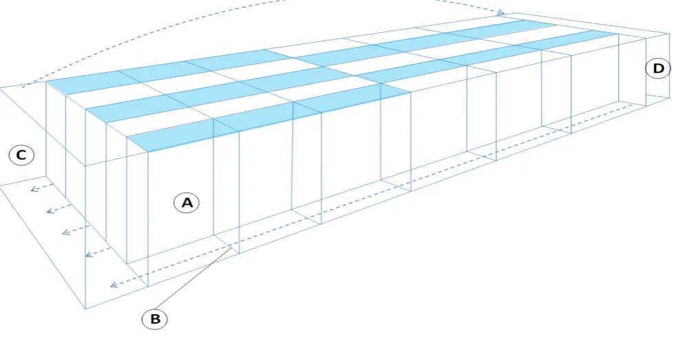
at ZSL London Zoo. Thirty tadpoles from six different clutches were used, and these tadpoles were collected from the water dishes of several enclosures immediately after deposition by brooding males. Each clutch was split equally between treatment groups. The tadpoles were photographed (see methods below), and then transferred immediately into individual compartments of the experimental tank (see Figure 1).
barrier at the surface and with a polythene barrier under water.
Husbandry
Powder free nitrile gloves were always used when servicing the animals. Animals were maintained in a mixture of reverse osmosis water and tap water in a 9:1 ratio respectively. Water was dosed appropriately with tap water conditioner (API/Tetra). Water parameters were maintained at an alkalinity between 30 and 60 mg/L, and at a pH of 7.3. Temperatures were kept at 17-21°C for the duration of the study following EAZA best practice guidelines (Wells et al. 2015). Water quality was maintained using biological and mechanical filtration (Eheim Lav filter media,
two Boyu sponge filters, and growth of Egeria densa –filtration
was not present in the actual experimental compartments; see below) and routine 30% daily water changes as well as detritus siphoning. Filter media was cleaned in tank water as necessary. Ammonia and nitrite were generally maintained at <0.05 mg/L and nitrate at <100 mg/L. Water parameters were monitored weekly using an aquarium test kit (Salifert Nitrate test kit; Nitrate), and a spectrophotometer (Interface photometer 7500, Palintest). Tadpoles were fed on a homogenised diet comprising of 10 parts fish flake, eight parts trout pellet, eight parts grass pellets, three parts cuttlefish bone, two parts dried tubifex, two parts dried river shrimp, and three parts spirulina in one batch (parts measured by weight). Brands varied between batches according to availability, but all tadpoles were fed on same diet at any given time. Food was offered as a sinking pellet to avoid surface interruption of UVB radiation decreasing the amount of UVB reaching the tadpoles. Tadpoles were also able to feed on surface algae within the tank at all times. We did not control for food intake, as in a practical husbandry setting, food is provided ad libitum and is not a limiting factor for growth.
Experimental array
The experimental array comprised of a glass aquarium split into 30 compartments that each housed a single tadpole (Figure 1). A small gap beneath each divide allowed water flow throughout the system whilst also preventing tadpoles from moving between compartments. Two larger compartments, one at each end of the tank, held the filter media and water was transferred through the tank using a small pump (Eheim pump compact+ 3000) at one end, with silicone tubing transporting water to the other end compartment through a spray bar. The tank was illuminated with five 12% T5 Arcadia UVB emitting lamps across each row of compartments, with half of each lamp alternately covered with a UVB filter (CLS200 XSR Clear Ultra Violet Filter) to block UVB radiation whilst still allowing passage of other wavelengths (Michaels et al. 2015). This resulted in 15 compartments being exposed to UVB (UVB+) with the other 15 not (UVB-). Irradiation
period was set at four hours on, 20 hours off as A. muletensis
from the room on a 12:12 photoperiod from a non UVB emitting array. Lamps above the experimental array were held in place using tape to prevent movement, and the UVB emitted by these lamps was measured every month from a distance equal to that of halfway through the water column, and lamps were replaced four months into the seven month experiment due to decreased UVB emissions. Each individual compartment was equipped with
a refugium comprising an eight cm piece of food grade PVC piping
(one-inch diameter; GF Harvel) to allow the tadpole to shelter from either UVB radiation (in UVB+ compartments), or from any perceived danger.
UVB+ compartments were irradiated with a maximum UVi of 2-3.7 (1.9-3.6 with the polythene barrier). Using a deeper chamber to allow for the height of the solarmeter, UVB+ chambers had a minimum UVi of c. 1 on the substrate at the bottom of the compartment. A zone of UVi 0 was available inside the PVC tube hide. These UVi are broadly concordant with a UVi of c. 2.4
collected underwater (at a range of depths) at an A. muletensis
breeding site at Coco de Bovas, Mallorca in July 2017 (J. Bosch pers. comm.). UVB- compartments received a UVI of 0, which was achieved through the placement of lamps, reflectors and glass panels and was confirmed using the Solarmeter 6.5. T5 fluorescent tubes produce markedly less UVB radiation towards the ends of the tubes; the array and light selection was designed to avoid any compartments being under the lower-UVB parts of the lamps; this was confirmed using the Solarmeter 6.5.
Data collection
Tadpoles were photographed next to a scale at the beginning of the experiment and weekly thereafter. Each tadpole was removed
from its enclosure using a net and photographed in a moist petri dish against the same scale, before being returned to its
compartment. The Gosner stage of development (Gosner 1960) of the tadpole was recorded weekly. Photographs were analysed using ImageJ (1.48v, NIH). Tail length (from tail tip to vent), head length (snout to vent length), and total tadpole length (sum of tail length and head length (McDiarmig and Altig 1999) was measured using this software. The tail:body ratio of the tadpoles was also calculated. Each tadpole was given a colour score from one to five with one being the lightest colour, and five being the darkest. This was to determine if UVB had any effect on colouration (Blaustein and Belden 2003). Non-experimental larvae were used as standards of each colour category and experimental larvae were matched to the closest standard by eye.
Once the tadpoles had reached Gosner stage 39 (their maximum length before shrinking during metamorphosis), or at seven months post introduction to the experimental array (whichever came sooner), they were euthanased using a buffered methocaine trisulphate (MS222) solution. A blood sample was taken from the heart of each tadpole immediately after euthanasia. This was done by making an incision in the ventral skin of the tadpole to expose the heart, removing any excess interstitial fluid that may dilute the blood sample using a cotton bud, puncturing the heart using a
scalpel, and extracting the blood that filled the body cavity using a heparinised capillary tube (Vitrex Medical BRIS microhematocrit tubes). Blood drops were then dropped onto test blotting strips obtained from City Assays (City Hospital, Birmingham, Sandwell and West Birmingham Hospital NHS trust; www.vitamindtest.org. uk). These were returned to this laboratory to measure 25-hydroxy
vitamin D3 by tandem mass spectrometry after derivatisation and liquid-liquid extraction, using in house calibrators and quality control material (Jaffe et al., 2019). The bodies of these tadpoles were then stored in Eppendorf tubes (50 ml capacity) at -20°C and sent for calcium and phosphorus analysis using inductively coupled plasma atomic emission spectroscopy (ICP-AES) (Michaels et al. 2014) at Manchester Metropolitan University.
Mineral analysis was carried out at Manchester Metropolitan University. Frozen tadpoles were dried out at 60°C in a GenLab incubator for 72 hours in a 50 ml glass vial, then washed with ultrapure water. A dry weight of all tadpoles was taken. The tadpoles were then transferred to digestion vessels and two ml Hydrogen peroxide (>30% w/v) was added to break down the organic matter. After 30 minutes, 8 ml of nitric acid (>68% PrimerPlus- trace analysis grade) was added and left to digest for two hours. The vessels (plus six blanks) were then transferred to the microwave (CEM Mars Xpress 5) and run for five minutes at 90°C, then 10 minutes at 170°C. This was then repeated to ensure digestion was complete. The volume of each sample was then made up to 50ml with distilled water, and 13 ml of each sample was added to 15 ml centrifuge tubes (Fisherbrand) for the ICP-AES analysis (Thermo scientific iCAP6300 Duo). Five repeats were run for each sample, and an average was taken to be used for analysis. Levels of calcium and phosphorus were measured.
Statistical Analysis
All statistics carried out used a significance threshold of P=0.05. From 10 randomly chosen images, the total length of each tadpole was measured three times using ImageJ (1.48v, NIH). Between each measurement, the scale was recalibrated, and repeatability (r) for each measurement was calculated using the following
formula:
r=S_(2A )/(S_2+ S_2A) where S_2A is the among-groups variance and S_2 is the within-groups variance (Lessels and Boag, 1987; Verschooren et al. 2011; Michaels et al. 2015).
Growth data and colouration
Data were plotted as histograms and no deviation from normal distribution was noted. Plotting residuals against the data showed no evidence of heterogeneity of variance, therefore parametric statistics were deemed appropriate for analysis. A longitudinal linear mixed effects model was used to assess the effects of UV treatment on the rate of tadpole growth over time. Fixed effects included UV treatment, and linear and quadratic polynomials of age and the interactions of UV treatment and two age polynomials to control for repeated measures. In addition to the random clutch effect, intercept, linear and quadratic polynomials of age for the tadpole were also modelled as random effects using unstructured covariance type. Analysis was carried out using Statistics software R (3.4.0) and the package <lme4>. A generalized estimating equation using a cumulative logit link function was used to assess the effect of UV treatment, age, and their interactions on the tadpole colouration. The order in which tadpoles was added to the experimental array was accounted for. First-degree autoregressive working correlation structure was used to account for the repeated measures from the same tadpole. This analysis was done using SPSS (Version 23).
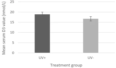
Vitamin D3 levels in blood serum
Serum was successfully recovered and tested for 25 tadpoles, 13 from UVB+ and 12 from UVB-. A Mann Whitney U test (two tailed)
was carried out on serum 25(OH)D3 levels for both treatment
groups after anomalous results were omitted. Two test runs, each representing the mean average of four tests are conducted per sample. Three individual results were omitted as they were several times higher than the other repeats within their test runs. Therefore, data were available from all 25 animals from which samples were recovered. This analysis was done using Graphpad Prism (7.03).
ICP-AES mineral content analysis
Calibration curves were calculated for each mineral. All calibration curves had a correlation of 0.999. A Mann Whitney U test (two tailed) was carried out on both calcium and phosphorus content of all tadpoles from both treatment groups. This analysis was done using Graphpad Prism (7.03).
Colouration score F1 = 0.385 0.755
Figure 2. Serum D3 levels: Average levels of vitamin D3 taken from tadpole
rainforests (La Marca et al. 2010) and B. orientalis inhabiting a wide range of water bodies within lowland temperate and subtropical forests (Kuzmin et al. 2004), as well as their phylogeny. However, both species actively bask in sunlight or artificial light sources given the opportunity. As these two studies (the only two experimental studies using controls so far focusing on amphibian UVB provision from a husbandry perspective) have demonstrated that UVB provision is arguably beneficial in both these species of basking anuran from otherwise differing ecologies, it is reasonable to assume that at least anurans that exhibit basking behaviour are likely to have a physiological requirement for UVB radiation. These studies only focus on UVB provision post-metamorphosis, and so the present study is the first to focus on provision of quasi-natural levels of UVB in larval amphibians from the perspective of captive husbandry.
We detected 25(OH)D3 in the serum of tadpoles in this study.
This suggests that this species may use vitamin D3 in active
absorption of calcium from the gut, and that it does not rely solely on passive absorption. This underpins the importance of
providing at least a diet containing vitamin D3. Wild A. muletensis
larvae are deposited in large, warm, shallow water bodies from May to September, where they develop and metamorphose in the autumn or following spring (Pinya and Pérez-Mellado 2014). Developing larvae are exposed to UVB and are active during the day (Spence 2002) when ambient UVB levels are higher. Although
A. muletensis tadpoles appear to avoid the most intense sun
exposure by hiding beneath the overhangs of rocky pools (Schley et al. 1998), even in these microhabitats tadpoles are exposed to a level of UVB similar to those used in this study (J. Bosch pers. comm.; see Methods). It is therefore surprising that we found no
effect of UVB exposure on blood serum 25(OH)D3 levels, growth
rates, or colouration of the tadpoles, as it would be reasonable to expect an organism to utilise a free source of energy such as UVB radiation from the sun (Michaels and Preziosi 2013). It is important
to note that comparisons with serum 25(OH) D3 levels in wild
individuals would inform the establishment of a normal baseline. Without these data it is unclear if tadpoles in this study were exhibiting normal, equally high, or equally low concentrations of
serum vitamin D3 (although no deleterious effects were noticed
either way). However, no clinical signs of hypovitaminosis D3 were
noted.
Results
Repeatability analysis
All measures taken were found to be highly repeatable with the lowest r value being 0.96.
Growth data and colouration
As the effects of the linear polynomial of tadpole age were significant, this was used to control for repeated measures and so the quadratic polynomial was not used. We report P values for the UVB treatment*age interaction effect, which account for repeated measures. The order in which tadpoles were added to the experimental array had no significant effect on any results (P>0.05 for all measures). There was no effect of UV treatment on any growth parameters or coloration score measured in this study (Table 1).
Vitamin levels in blood serum
There was no significant difference in serum vitamin D3 results
between treatment groups (U12=59, P=0.3134) (Figure 2).
ICP-AES mineral content analysis
There was no significant difference in calcium content (U14=85, P=0.5860,) or phosphorus content (U14=76, P=0.3389) (Figure 3). The average calcium to phosphorus ratio for the UVB+ treatment was 1.20:1, and the average calcium to phosphorus ratio for the UVB- group was 1.26:1 respectively.
Discussion
Michaels et al. (2015) found that captive post-metamorphic
fire-bellied toads (Bombina orientalis) that were exposed to
ecologically and physiologically relevant levels of UVB had significantly, although only slightly, more robust skeletal development, a significantly increased rate of adult colour acquisition, and a significantly increased level of serum vitamin
D3 than animals that were not exposed. Verschooren et al. (2011)
also showed that UVB exposure in recently metamorphosed
treefrogs (Trachycephalus resinifictrix) increased growth and
development of skeletal structures. These species differ greatly in
their ecology, with T. resinifictrix inhabiting the canopy of tropical
therefore possible that the dietary vitamin D3 present in the food,
which could not be eliminated for ethical and legislative reasons,
provided sufficient vitamin D3 to saturate serum levels of 25(OH)
D3. Figure 2 shows a non-significant increase in 25(OH)D3 levels,
which may be indicative of this dynamic. We were not able to
include treatments with no UVB irradiation or dietary vitamin D3 in
order to address this issue, as this would have fallen into the remit of the Animals (Scientific Procedures) Act 1986 in UK legislation, which would require licenses and facilities beyond the scope of this study.
Photobiosynthesis of vitamin D3 in the skin requires both UVB
irradiation and appropriate temperatures to enable biochemical reactions (see Michaels et al. 2015). Although the temperatures
used in this study fall within the c. 17-24oC range recorded in a
study of free-living tadpoles of this species (Schley et al. 1998), and within the range determined as preferred temperature and optimum for growth (Kadel and Hemmer 1984; Martens 1984) larvae in this study did not have access to a temperature gradient and therefore may not have become sufficiently warm to utilise
UVB radiation in the synthesis of vitamin D3. The duration of UVB
irradiation per day was relatively short. Tadpoles of this species often occur in rocky gorges, where direct insolation may be limited to a short period. However, light scatter from gorge sides may extend UVB irradiation in nature beyond the period of direct insolation (Caldwell et al 1980). Free living tadpoles modulate their use of deeper and shallower water and rock overhangs throughout the day, and aggregate under rock shelves in deeper water during periods of most intense sunlight (Schley et al. 1998). This means that the parameters used in the experimental array may have mimicked larval exposure to UVB and temperature in the field reasonably well, with a short period of UVB irradiance and relatively constant temperatures, but further studies looking at UVB exposure in wild tadpoles over time are required to inform future work. The inclusion of thermal as well as UVB gradients in further experiments would also be useful.
The experimental array consisted of a shared water body between all 30 tadpoles. This allowed maintenance of stable water quality, which would not have been possible if tadpoles were housed separately. Consistently good water quality is impossible to maintain in small, isolated unfiltered containers, which require frequent 100% water changes and results in fluctuation of parameters including nitrogenous waste, bacterial load and pH. Poor and fluctuating water quality causes stress, which influences growth and development rates in amphibians in a complex fashion (e.g. Denver 2009). As stocking density has been shown to influence growth rates (Dash and Hota 1980; Wilbur 1997), the order in which the tadpoles were added to the array was statistically controlled for as a proxy for stocking density, in addition to the systematic paired addition of animals under both treatments.
There are few available data regarding serum vitamin D3
levels in anurans (Michaels et al. 2015), and the dry blood spot method of quantifying this has not been validated for amphibians (or any other non-human animal), and in the only non-human
2019). However, for small animals, where analysis of liquid serum is not possible due to minimum sample volume requirements, the dry blood spot approach is the only viable method available,
Bearing these caveats in mind, Michaels et al. (2015) found a range of serum levels ranging from 73.59 –88.68 nM/L across UV- and UV+ treatment groups. However, the raw data collected in our study ranged from 3.8 – 52.5 nM/L across both treatment groups. This could result from differing physiological requirements, different relative impacts of captivity on each species, methodological error or a host of other reasons. Methodological validation and the acquisition of a larger data set from a variety of species is needed in order to better understand the implications of these data.
Whole body mineral analysis, including calcium and phosphorus,
of adult Cuban tree frogs (Osteopilus septentrionalis) was carried
out by Allen et al. (1993). Frogs were fed on either a high or a low calcium diet (no UVB exposure provided), and results showed much higher calcium:phosphorus ratios (approximately 2:1 in females and approximately 2.5:1 in males for both treatment groups) than were found in this study (approximately 1.2:1 in the UV+ group and 1.26:1 in the UV- group). This is probably because metamorphosed frogs with ossified skeletons will contain more mineralised calcium within these structures, compared to the tadpoles in this study which lack mineralised skeletons. However, figures from this study meet the recommended requirement of a 1:1 or 2:1 ratio of calcium to phosphorus in the diet respectively (Finke 2002), indicating that no signs of ill health would be expected
as a result of this, and that the levels of 25(OH)D3 that we detected
are likely to be sufficient for normal calcium homeostasis. Increased pigmentation with melanin may be used by amphibians as protection against harmful doses of UVB radiation (Brenner and Hearing 2008). However, we found no significant difference in pigmentation score between treatments, and neither did we observe any negative effects of UVB exposure (as found in other studies; Worrest and Kimmeldorf 1976; Grant and Licht 1995; Blaustein et al. 1998, 2003, 2005). Three tadpoles from the UVB negative group died of natural causes unrelated to UVB provision (confirmed by post mortem examination). No animals from the UVB+ group died during this study. This is unsurprising, as tadpoles were provided with an ecologically appropriate level of UVB with both a gradient, and refugia to allow exposure to be avoided if chosen. Whilst the tadpoles used the refugia provided, there seemed to be no increased usage amongst UVB positive animals, and many times animals from both treatment groups were observed swimming at the top of the water column where UVB exposure would have been greatest. The numbers of tadpole deaths were too small for statistical analysis.
Overall, our results indicate that the integration of ecologically appropriate UVB radiation into existing husbandry protocols for
larval A. muletensis had no effect on measured parameters while
anuran tadpoles. Further work exploring the impacts of diet, thermal gradients, and UVB dosage and exposure length may be
required to fully understand UVB requirements in A. muletensis
larvae. Moreover, UVB requirements may vary between taxa depending on their evolutionary and ecological context (Michaels et al. 2014).
This study focuses on UVB provision for calcium homeostasis, but irradiation may have other important effects. For example, UVB exposure has also been shown to kill bacteria found on human skin (Jekler et al. 1992; Fluhr and Gloor 1997; Yoshimura-Mishima et al. 1999). Should the same principles apply to amphibians, then UVB radiation may have effects on host-pathogen interactions, both by directly inhibiting bacterial growth on the skin, and through interactions with bacterial floras that may mediate infection. UVB provision could therefore potentially be used to help reduce the incidence of disease in CBPs, and its provision may still be useful for rearing tadpoles in capacities unrelated to calcium metabolism. This study provides a basis upon which further research methods into tadpole UV provision studies can be refined and improved to ultimately determine the role of UV provision for anuran larvae in captivity.
Acknowledgements
The authors would like to thank all members of staff involved in the care of tadpoles at ZSL London Zoo. We would also like to thank Jaimie Bosch for in situ UVi data collection, and an anonymous reviewer for their comments.
References
Alcover J.A., Mayol J., Jaume D., Alomar G., Pomar G., Jurado J. (1984)
Biologia i ecologia de les poblacions relictes de Baleaphryne muletensis
a la muntanya mallorquina. Història Biològica del Ferreret 129–151. Allen M.E., Oftedal O.T., Ullrey D.E. (1993) Effect of dietary calcium
concentration on mineral composition of fox geckos (Hemidactylus
garnoti) and Cuban tree frogs (Osteopilus septentrionalis). Journal of Zoo and Wildlife Medicine 24: 118–128.
Antwis R.E., Browne R.K. (2009) Ultraviolet radiation and vitamin D3 in
amphibian health, behaviour, diet and conservation. Comparative Biochemistry and Physiology Part A: Molecular & Integrative
Physiology 154: 184–190.
Baldwin G.F., Bentley P.J. (1980) Calcium metabolism in bullfrog tadpoles (Rana catesbeiana). Journal of Experimental Biology 88: 357–366. Bentley P.J. (1984) Calcium metabolism in the Amphibia. Comparative
Biochemistry and Physiology Part B: Physiology 79: 1-5.
Bianchi C.P., Shanes A.M. (1959) Calcium influx in skeletal muscle at rest, during activity, and during potassium contracture. The Journal of General Physiology 42: 803–815.
Blaustein A.R., Belden L.K. (2003) Amphibian defenses against ultraviolet-B radiation. Evolution & Development 5: 89–97.
Blaustein A.R., Hoffman P.D., Hokit D.G., Kiesecker J.M., Walls S.C., Hays J.B. (1994) UV repair and resistance to solar UV-B in amphibian eggs: a link to population declines?. Proceedings of the National Academy of
Sciences 91: 1791–1795.
Blaustein A.R., Kiesecker J.M., Chivers D.P., Hokit D.G., Marco A., Belden L.K., Hatch A. (1998) Effects of ultraviolet radiation on amphibians: field experiments. American Zoologist 38: 799–812.
Blaustein A.R., Romansic J.M., Scheessele E.A. (2005) Ambient levels of ultraviolet-B radiation cause mortality in juvenile western toads, Bufo boreas. The American Midland Naturalist 154: 375–382.
Bloxam Q.M., Tonge S.J. (1995) Amphibians: suitable candidates for breeding-release programmes. Biodiversity & Conservation 4: 636– 644.
Brenner M., Hearing V.J. (2008) The protective role of melanin against UV damage in human skin. Photochemistry and Photobiology 84: 539– 549.
Buley K.R., Garcia G. (1997) The recovery programme for the Mallorcan midwife toad Alytes muletensis: an update. Dodo 33: 80–90. Caldwell M.M., Robberecht R., Billings W.D. (1980) A steep latitudinal
gradient of solar ultraviolet-B radiation in the arctic-alpine life zone.
Ecology 61: 600-611.
Cheek T., Flik G., Hazon N. (1993). Calcium regulation and signalling. In
Joint COB-SEB Symposium on Calcium Regulation and Cell Signalling.
Canterbury, UK: Company of Biologists.
Dash M.C., Hota A.K. (1980) Density effects on the survival, growth rate,
and metamorphosis of Rana tigrina tadpoles. Ecology 61: 1025–1028. DeMarchi J.A., Britton A., O’Donnell K., Saporito R.A. (2018) Behavioural preference for low levels of UV-B radiation in two neotropical frog species from Costa Rica. Journal of Tropical Ecology 34: 336–340. Denver R.J. (2009) Stress hormones mediate environment-genotype
interactions during amphibian development. General and Comparative
Endocrinology 164: 20-31.
Finke M.D. (2002) Complete nutrient composition of commercially raised invertebrates used as food for insectivores. Zoo Biology 21: 269–285. Finke M.D. (2003) Gut loading to enhance the nutrient content of insects as
food for reptiles: a mathematical approach. Zoo Biology 22: 147–162. Fioletov V.E., McArthur L.J.B., Mathews T.W., Marrett L. (2009) On the
relationship between erythemal and vitamin D action spectrum weighted ultraviolet radiation. Journal of Photochemistry and Photobiology B: Biology 95: 9–16
Fluhr J.W., Gloor M. (1997) The antimicrobial effect of narrow-band UVB (313 nm) and UVA1 (345–400nm) radiation in vitro. Photodermatology,
Photoimmunology & Photomedicine 13:197–201.
Gagliardo R., Crump P., Griffith E., Mendelson J., Ross H., Zippel K. (2008) The principles of rapid response for amphibian conservation, using the programmes in Panama as an example. International Zoo Yearbook 42:
125–135.
Garner T.W.J., Garcia G., Carroll B., Fisher M.C. (2009) Using itraconazole
to clear Batrachochytrium dendrobatidis infection, and subsequent depigmentation of Alytes muletensis tadpoles. Diseases of Aquatic
Organisms 83: 257–260.
Gosner K.L. (1960) A simplified table for staging anuran embryos and larvae with notes on identification. Herpetologica 16: 183–190. Grant K.P., Licht L.E. (1995) Effects of ultraviolet radiation on life-history
stages of anurans from Ontario, Canada. Canadian Journal of Zoology
73: 2292–2301.
Heaney R.P., Armas L.A., Shary J.R., Bell N.H., Binkley N., Hollis B.W. (2008). 25-Hydroxylation of vitamin D3: relation to circulating vitamin D3 under various input conditions. The American Journal of Clinical
Nutrition 87: 1738-1742.
Holick M.F. (1981) The cutaneous photosynthesis of previtamin D3: a
unique photoendocrine system. Journal of Investigative Dermatology
77: 51–58.
IUCN (2018) The IUCN Red List of Threatened Species. Version 2018-1.
<http://www.iucnredlist.org> Downloaded on 17th August 2018. Jaffe J.E., Ferguson A., Michaels C.J. (2019) The utility of dried blood spots
for the assessment of avian vitamin D3 status compared with plasma analysis. Journal of Zoo and Aquarium Research 7: 138-143.
Jekler J., Bergbrant I.M., Faergemann J., Larkö O. (1992). The in vivo effect of UVB radiation on skin bacteria in patients with atopic dermatitis.
Acta Dermato-venereologica 72: 33–36.
Kadel K., Hemmer, H. (1984). Temperature dependence of larval development in the Mallorcan midwife toad, Baleaphryne muletensis.
In: Hemmer, H. and Alcover, J.A. (eds.) Historia Biologica del Ferreret.
Palma de Mallorca, Mallorca: Editorial Moll,169–174.
Kats L.B., Bucciarelli G.M., Schlais D.E., Blaustein A.R., Han B.A. (2012) Ultraviolet radiation influences perch selection by a Neotropical poison-dart frog. PloS one 7: 51364.
King J.D., Muhlbauer M.C., James A. (2011) Radiographic diagnosis of metabolic bone disease in captive bred mountain chicken frogs (Leptodactylus fallax). Zoo Biology 30: 254–259.
Kraaijeveld-Smit F.J.L., Beebee T.J.C., Griffiths R.A., Moore R.D., Schley L. (2005) Low gene flow but high genetic diversity in the threatened Mallorcan midwife toad (Alytes muletensis). Molecular Ecology 14:
3307–3315.
Kuzmin S., Pipeng L., Matsui M., Ishchenko V., Maslova I. (2004)
Bombina orientalis. The IUCN Red List of Threatened Species 2004: e.T54449A11146991. http://dx.doi.org/10.2305/IUCN.UK.2004.RLTS. T54449A11146991.en. Downloaded on 13 August 2017.
La Marca E., Azevedo-Ramos C., Reynolds R., Coloma L.A., Santiago R. (2010) Trachycephalus resinifictrix. The IUCN Red List of Threatened Species 2010: e.T55823A11373135. http://dx.doi.org/10.2305/IUCN. UK.2010-2.RLTS.T55823A11373135.en. Downloaded on 13 August 2017.
Michaels C.J., Antwis R.E., Preziosi R.F. (2015) Impacts of UVB provision and dietary calcium content on serum vitamin D3, growth rates,
skeletal structure and coloration in captive oriental fire-bellied toads (Bombina orientalis). Journal of Animal Physiology and Animal
Nutrition 99: 391–403.
Michaels C.J., Gini B.F., Preziosi R.F. (2014) The importance of natural history and species-specific approaches in amphibian ex-situ conservation. The Herpetological Journal 24: 135-145.
Moore R.D., Griffiths R.A., Roman A. (2004) Distribution of the Mallorcan midwife toad (Alytes muletensis) in relation to landscape topography and introduced predators. Biological Conservation 116: 327–332. Norman A.W. (1998) Sunlight, season, skin pigmentation, vitamin D, and
25-hydroxyvitamin D: integral components of the vitamin D endocrine system. The American Journal of Clinical Nutrition 67: 1108–1110. Pandelova I., Hewitt S.R., Rollins-Smith L.A., Hays J.B. (2006) UVB
Dose-toxicity Thresholds and Steady-state DNA-photoproduct Levels During Chronic Irradiation of Inbred Xenopus laevis Tadpoles. Photochemistry and Photobiology 82: 1080–1087.
Pilkington J.B., Simkiss K. (1966) The mobilization of the calcium carbonate deposits in the endolymphatic sacs of metamorphosing frogs. Journal of Experimental Biology 45: 329–341.
Pinya S., Pérez-Mellado V. (2014) Clutch size in wild populations of Alytes muletensis. Acta Herpetologica 9: 115–117.
Schley L., Griffiths R.A., Roman A. (1998) Activity patterns and microhabitat selection of Mallorcan midwife toad (Alytes muletensis) tadpoles in
natural torrent pools. Amphibia-Reptilia 19: 143–151.
muletensis in captivity. International Zoo Yearbook 28: 45-53. Verschooren E., Brown R.K., Vercammen F., Pereboom J. (2011) Ultraviolet
B radiation (UV-B) and the growth and skeletal development of the Amazonian milk frog (Trachycephalus resinifictrix) from
metamorphosis. Journal of Physiology and Pathophysiology 2: 34–42. Wilbur H.M. (1977) Density-dependent aspects of growth and
metamorphosis in Bufo americanus. Ecology 58: 196–200.
Wells E., Garcia-Alonso D., Rosa G.M., Garcia G., Tapley B. (2015)
Amphibian Taxon Advisory Group Best Practice Guidelines for Midwife
toads (Alytes sp.). https://www.eaza.net/assets/Uploads/CCC/2015-Midwife-toads-EAZA-Best-Practice-Guidelines-Approved.doc.pdf. Downloaded 14 October 2018.
Worrest R.C., Kimeldorf D.J. (1976) Distortions in amphibian development induced by ultraviolet-B enhancement (290–315 nm) of a simulated solar spectrum. Photochemistry and Photobiology 24: 377–382. Wren S., A. Angulo H. Meredith J. Kielgast Dos Santos M., Bishop P. (eds)
(2015) Amphibian Conservation Action Plan. April 2015. IUCN SSC Amphibian Specialist Group: http://www.amphibians.org/acap/ . Downloaded on 13 August 2017.