Open Access
Research article
Identification and validation of suitable endogenous reference genes
for gene expression studies in human peripheral blood
Boryana S Stamova*
1, Michelle Apperson
1, Wynn L Walker
1, Yingfang Tian
1,
Huichun Xu
1, Peter Adamczy
1, Xinhua Zhan
1, Da-Zhi Liu, Bradley P Ander
1,
Isaac H Liao
1, Jeffrey P Gregg
2, Renee J Turner
1, Glen Jickling
1, Lisa Lit
1and
Frank R Sharp
1Address: 1Department of Neurology and M.I.N.D. Institute, University of California at Davis Medical Center, Sacramento, CA 95817, USA and 2Department of Pathology, and M.I.N.D. Institute, University of California at Davis Medical Center, Sacramento, CA 95817, USA
Email: Boryana S Stamova* - [email protected]; Michelle Apperson - [email protected]; Wynn L Walker - [email protected]; Yingfang Tian - [email protected]; Huichun Xu - [email protected];
Peter Adamczy - [email protected]; Xinhua Zhan - [email protected]; Da-Zhi Liu - [email protected]; Bradley P Ander - [email protected]; Isaac H Liao - [email protected]; Jeffrey P Gregg - [email protected];
Renee J Turner - [email protected]; Glen Jickling - [email protected]; Lisa Lit - [email protected]; Frank R Sharp - [email protected]
* Corresponding author
Abstract
Background: Gene expression studies require appropriate normalization methods. One such method uses stably expressed reference genes. Since suitable reference genes appear to be unique for each tissue, we have identified an optimal set of the most stably expressed genes in human blood that can be used for normalization.
Methods: Whole-genome Affymetrix Human 2.0 Plus arrays were examined from 526 samples of males and females ages 2 to 78, including control subjects and patients with Tourette syndrome, stroke, migraine, muscular dystrophy, and autism. The top 100 most stably expressed genes with a broad range of expression levels were identified. To validate the best candidate genes, we performed quantitative RT-PCR on a subset of 10 genes (TRAP1, DECR1, FPGS, FARP1, MAPRE2, PEX16, GINS2, CRY2, CSNK1G2 and A4GALT), 4 commonly employed reference genes (GAPDH, ACTB, B2M and HMBS) and PPIB, previously reported to be stably expressed in blood. Expression stability and ranking analysis were performed using GeNorm and NormFinder algorithms.
Results: Reference genes were ranked based on their expression stability and the minimum number of genes needed for nomalization as calculated using GeNorm showed that the fewest, most stably expressed genes needed for acurate normalization in RNA expression studies of human whole blood is a combination of TRAP1, FPGS, DECR1 and PPIB. We confirmed the ranking of the best candidate control genes by using an alternative algorithm (NormFinder).
Conclusion: The reference genes identified in this study are stably expressed in whole blood of humans of both genders with multiple disease conditions and ages 2 to 78. Importantly, they also have different functions within cells and thus should be expressed independently of each other. These genes should be useful as normalization genes for microarray and RT-PCR whole blood studies of human physiology, metabolism and disease.
Published: 5 August 2009
BMC Medical Genomics 2009, 2:49 doi:10.1186/1755-8794-2-49
Received: 12 January 2009 Accepted: 5 August 2009
This article is available from: http://www.biomedcentral.com/1755-8794/2/49
© 2009 Stamova et al; licensee BioMed Central Ltd.
Background
Gene expression analysis is widely used to study various biological processes. Different transcript quantification methodologies exist, all of which rely on utilization of proper normalization techniques. Such analysis requires several variables need to be accounted for, including amount and quality of RNA, enzymatic efficiencies, and differences between tissues or cells in overall transcrip-tional activity. The current, most universally utilized nor-malization method for PCR and Northern blotting relies on the use of constitutively expressed endogenous refer-ence genes, often termed ''house-keeping genes.'' It has been shown, however, that the expression of such refer-ence genes can vary significantly between different tissues or conditions, thus making it impossible to use the same reference genes for various tissues [1-5]. Moreover, the identification of appropriate control genes presents a cir-cular problem, because in order to find suitable genes for normalization, prior knowledge of their stable gene expression is needed, which also relies on properly nor-malized data.
The conventional use of a single gene for normalization can introduce a significant error in the quantification of transcript levels [6]. It is recommended that an optimal set of reference genes be identified for each individual exper-imental setting or tissue. In this study, we used whole-genome human microarrays and surveyed the expression of over 39,500 genes in 526 whole-blood samples from control subjects and patients with Tourette syndrome, stroke, migraine, muscular dystrophy, and autism to iden-tify the best candidate control genes. The 10 best candi-date control genes, as well as commonly used house-keeping controls and PPIB, were validated with independ-ent samples of whole-blood RNA from both patiindepend-ents with multiple sclerosis (MS) and age- and gender-matched controls by qRT-PCR. GeNorm was used to identify an optimal set of the most stably expressed genes for normal-ization in whole-blood expression studies [6].
Methods
Patients and samples
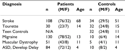
We used a large cohort of available human Affymetrx array data that we have generated over the last several years from previous and on-going studies (Table 1). Many, but not all, of the subjects included in our analyses have previously been reported in the following studies: stroke [7-10], Tourette syndrome [11-13], controls [14-18], migraine [19], muscular dystrophy [20,21] and autism [22,23].
RNA Isolation and Quality Control
Whole blood was collected and total RNA was isolated using PAXgene tubes and kits (Qiagen, Valencia, CA, USA). RNA samples were examined for concentration and
purity using a Nanodrop ND-1000 spectrophotometer, and integrity was checked using the Agilent 2100 Bioana-lyzer. High quality RNA with a RIN number above 8.0 was used for the qRT-PCR experiment. The RIN number takes into consideration not only the conventional ratio of 28 S to 18 S ribosomal RNA, but also additional critical regions of the entire RNA electrophoregram.
Affymetrix experiments
Affymetrix Human 2.0 Plus arrays (Affymetrix, Santa Clara, CA, USA), surveying over 54,000 probe-sets, were used in this study. The 54,000 probe-sets represent over 39,500 potential human genes (Affymetrix Manual). The standard Affymetrix protocol was followed for the sample labeling, hybridization, and image scanning [23].
qRT-PCR cDNA synthesis
The cDNA synthesis and real-time PCR was performed at the Lucy Whittier Molecular and Diagnostic Core Facility (University of California, Davis, U.S.A.). The following cDNA synthesis protocol was validated through the Lucy Whittier Molecular and Diagnostic Core Facility using a variety of sample types and species. 10 l (750 ng) of tem-plate RNA per sample, 1 l gDNA WipeOut Buffer, 1 l RNase-free water was incubated at 42°C for two minutes and briefly centrifuged. A 1 L aliquot was TaqMan® ana-lyzed with human GAPDH to confirm all gDNA had been digested. Then 8 l from the reverse transcription reaction mix (0.5 l Quantitect Reverse Transcriptase, 2 l 5× Quantitect RT buffer, 0.5 l RT Primer Mix, 0.5 l 20 pmol
Random Primers, 4.5 l RNase free water) was added to each well, incubated at 42°C for 40 minutes, inactivated at 95°C for 3 minutes and briefly centrifuged. Finally, 100 l of water was added to each well and mixed thoroughly.
Selection of TaqMan Assays
TaqMan assays were selected based on several criteria: 1) only pre-developed and pre-validated gene expression assays were selected from Applied Biosystems (ABI, Foster City, CA, U.S.A.), for which the company guarantees
Table 1: Characteristics of subjects who had gene expression assessed in whole blood on Affymetrix Human microarrarys.
Diagnosis Patients Controls
n (M/F) Age n (M/F) Age
amplification efficiency over 96%. 2) Only _m1 assays (spanning exon -exon boundaries) were selected to ensure no genomic DNA will be amplified. 3) If available, the assays were preferentially selected to amplify the same tar-get sequence where the Affymetrix probe-set was located. 4) If a gene selected for qRT-PCR validation was repre-sented by multiple probe-sets on the Affymetrix array, the standard deviation (SD) of the expression of all probe-sets was inspected and if some were found to have a high standard deviation, their position in the reference sequence was avoided.
Real-Time TaqMan® PCR
Pre-validated gene expression assays were purchased from Applied Biosystems. See Table 1 for the Assay and Gene IDs. An aliquot of the cDNA from a subset of samples was TaqMan® analyzed with human GAPDH prior to profiling. TaqMan® analysis was done in duplicate reactions using the low density array format as described by Osman et al [24]. This was validated and processed through the Lucy Whittier Molecular and Diagnostic Core Facility (1). The samples were placed in a 384 well plate and amplified in an automated fluorometer (ABI PRISM 7900 HTA FAST, ABI). ABI's standard amplification conditions were used: 2 min at 50°C, 10 minutes at 95°C, 40 cycles of 15 sec-onds at 95°C and 60 secsec-onds at 60°C. Fluorescent signals were obtained during the annealing temperature and Ct values exported with a threshold of 0.1 and a baseline of 3–10.
Data Analysis
Affymetrix Chips
All 526 arrays were summarized using the GC-RMA algo-rithm [25]. They were normalized within GeneSpring 7.3 software (Agilent Technologies, Palo Alto, CA) using a three-step normalization procedure, including data trans-formation, per chip and per gene normalization (Gene-Spring Manual).
qRT-PCR GeNorm Analysis
All samples for which the standard deviation (SD) of the technical duplicates was 1.41 (or higher than 2-fold change) were considered missing data points and were not included in the analysis. The GeNorm tool was used to calculate the internal control genes stability measures (M) [6]. In short, input data was generated using the com-parative Ct-method [26], where relative expression levels were calculated by setting the lowest Ct value of a particu-lar gene to 1, and the relative expression levels for the same gene in the rest of the samples were calculated. Then for every control gene, the pair-wise variation with all other control genes is estimated in GeNorm as the SD of the log-transformed expression ratios. The internal con-trol gene stability measure M is defined as the average pair-wise variation of a particular control gene with all
other control genes. Stepwise exclusion of the gene with the highest M value is performed. Because a pair of control genes is needed to calculate M, the two most stably expressed genes cannot be ranked any further. The opti-mal number of control genes for noropti-malization is deter-mined by calculating the pair-wise variation Vn/Vn+1 between each set of two sequential normalization factors [6]. Consistent with the Vandesompele recommenda-tions, the minimum number of genes for which the V was smaller than 0.15, was used to define the optimal set of control genes for normalization. A normalization factor is calculated based on the geometric mean of the optimal set of control genes.
qRT-PCR NormFinder Analysis
The NormFinder Excel plug-in [27] was used as an alter-native algorithm to the GeNorm algorithm for ranking the expression stability of the control genes. It ranks the can-didate control genes by estimating their expression stabil-ity while also taking into account the experimental design (control and disease groups). The input qRT-PCR results were the linearized quantities (2-Ct). The main differ-ences between the GeNorm algorithm [6] and the model-based approach in NormFinder [27] is that the latter takes into account the inter- and intra-group variation, as well as the estimation of variances.
Analysis of Variance (ANOVA)
Partek Genomics Suite (St. Louis, MI, USA) was used to perform an ANOVA using the Diagnosis as fixed and the Plate as a random factor ( + Diagnosis + Plate + (Diagno-sis × Plate) + ). The input data were the normalized data to the geometric average of the optimal set of control genes (TRAP1, DECR1, PPIB, and HMBS) using the com-parative Ct method.
Ct Method
The Ct method [28] was used to calculate the normal-ized calibrated data. TRAP1, identified by GeNorm as one of the two most stably expressed genes was used as a nor-malizer. The average of the healthy controls was used as the calibrator. In short, the Ct value of the normalizer was subtracted from the Ct value of each sample (Ct). Then the value of the calibrator was subtracted (Ct) and the linearized values were calculated (2-(Ct)). A prerequisite for using this method of analysis is that the normalizer and the gene of interest have very similar amplification efficiency. The ABI assays used in this study have been pre-validated by the company to have over 96% amplification efficiency.
Results
Patients and samples
con-trols) to age 68 (stroke study patients) with a range of 2 to 78 years of age (Table 1). There was a predominance of males (387) to females (139). Control subjects had no known medical, surgical, or psychiatric illnesses. Study subjects had a variety of medical illnesses with varying pathophysiologies, including neurodevelopmental and neurological disorders (autism, mental retardation, Tourette syndrome), stroke (ischemic brain injury), mus-cular dystrophy (genetic muscle disease), migraine head-aches, as well as a presumed autoimmune disease, multiple sclerosis (Table 1).
Selection of Reference Genes from Whole-Genome Affymetrix Arrays
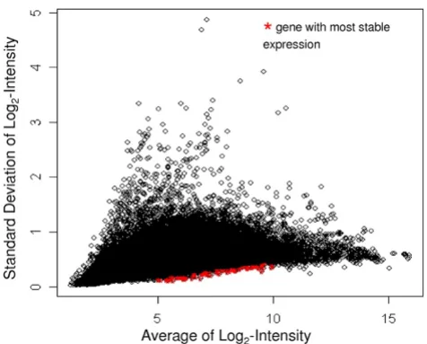
Our goal was to identify stably expressed genes with a broad range of expression levels from our Affymetrix data on the 526 samples (Table 1). We chose the 20 most sta-bly expressed genes from each of five intensity intervals (n = 100 genes): log2-transformed intensity values between 5–6, 6–7, 7–8, 8–9 and 9–10 (Figure 1). For a list of the most stable genes over all of the expression levels, see Additional file 1. Based on our selection criteria, these 100 most stably expressed genes covered a wide range of raw expression values from 32 to 1024. Functional categoriza-tion using the Database for Annotacategoriza-tion and Visualizacategoriza-tion, and Integrated Discovery (DAVID, http:// david.abcc.ncifcrf.gov/) showed that 50% of the anno-tated genes are involved in primary metabolic processes.
For the list of the 100 most stably expressed genes on the 526 microarrays see Additional file 1.
Selection of Candidate Control Genes for Validation by qRT-PCR
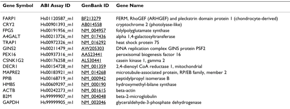
The set of 100 stably expressed genes on microarrays was filtered down to probe-sets with available gene annota-tion, as well as to probe-sets detecting only a single known gene (Affymetrix annotation *_at). Based on these criteria, 23 probe-sets were identified (data not shown). From them, 10 candidate genes were selected for validation with, on average, two genes per intensity interval. These 10 candidate genes included TRAP1, DECR1, FPGS, FARP1, MAPRE2, PEX16, GINS2, CRY2, CSNK1G2 and A4GALT. In the validation, we also included four com-monly employed reference genes, ACTB, GAPDH, B2M and HMBS. In addition, PPIB was included because it was previously reported to be stably expressed in blood [3]. Each gene and ABI assay information is presented in Table 2. The average expression values from the Affymetrix array data of the genes selected for validation by qRT-PCR are shown in Figure 2. The range of the SDs of the the log2 -transformed intensity values on the microarrays was between 0.10 and 0.42 for the 10 candidate reference genes on the microarrays (solid bars, Figure 2). The SDs for the remainder genes (open bars, B2M, ACTB, GAPDH, PPIB and HMBS) ranged between 0.47 and 0.72 on the microarrays (Figure 2). Based on the Affymetrix array data alone, FARP1 was the most stably expressed. A general
The Average log2- intensity (x-axis) of 39,500 genes from a total of 526 Affymetrix Human 2.0 Plus microarrays is plotted against the standard deviation of the log2- intensity (y-axis) of those genes
Figure 1
The Average log2- intensity (x-axis) of 39,500 genes from a total of 526 Affymetrix Human 2.0 Plus micro-arrays is plotted against the standard deviation of the log2- intensity (y-axis) of those genes. Probe-sets high-lighted in red are the 100 probe-sets with the lowest stand-ard deviation within an interval of the log2-transformed
intensity values between 5 and 10.
Average expression calculated from the 526 Affymetrix Human 2.0 Plus microarrays for the candidate control genes selected for qRT-PCR validation
Figure 2
trend of increasing SD with increasing intensity was noted for the candidate reference genes (Figure 2).
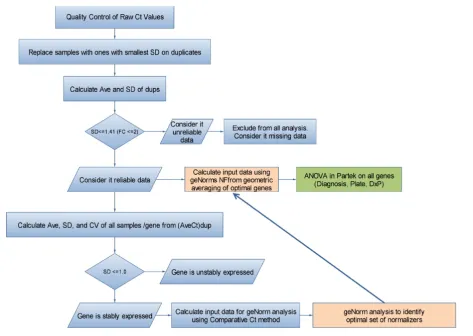
qRT-PCR Analysis and Expression of Candidate Control Genes
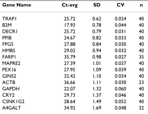
The analysis flowchart presented in Figure 3 describes the steps used to analyze the qRT-PCR data. The subset of 15 candidate reference genes (Table 2), validated on qRT-PCR, exhibited a broad range of expression levels from 17.93 to 36.66 cycle threshold (Ct) values (Table 3, Figure 4). Samples for which the SD on the technical replicates was higher than 1.4 (a fold-change higher than 2) were considered unreliable data and were not included in the downstream analysis, thus reducing the sample size for some assays to lower than 40 (Table 3). Genes that had SD 1.0 on the average raw Ct values of all samples were con-sidered to be stably expressed. Based on that criteria, TRAP1, B2M, DECR1, PPIB, FPGS, HMBS and FARP1 (Table 3, Figure 4, below the dotted line) showed stable expression over all 40 samples. In contrast, MAPRE2, PEX16, GINS2, ACTB, GAPDH, CRY2, CSNK1G2 and A4GALT did not show stable expression over the samples (Table 3, Figure 4 above the dotted line). The stably expressed genes covered a wide range of expression levels, from a high level of expression of 17.9 Ct for B2M, to low levels of 35.71 Ct for FARP1 (Table 3, Figure 4).
Expression Stability and Ranking Analysis
To identify the most suitable set of genes for normaliza-tion in whole-blood, the average expression stability (M) for those reference genes which showed SD of raw Ct-val-ues 1.0 on all samples was calculated using the GeNorm tool (see Methods). Those genes included TRAP1, FPGS, B2M, DECR1, PPIB and HMBS (Figure 4). All of these genes also had a SD of the Ct-values for the technical duplicates of 1.41 and a SD of the raw Ct values of all
samples 1.0. The FARP1 gene was omitted from the analysis of the expression stability because GeNorm does not allow missing values. However, re-analysis where FARP1 was included with the rest of the five stably expressed genes (with n = 35) did not change the final results (data not shown).
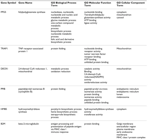
An important pre-requisite for estimating the Expression Stability Measure is that the genes are not coordinately regulated. The Gene Ontology terms of the six genes, ana-lyzed in GeNorm, are presented in Table 4. These genes are involved in different biological process, molecular functions and/or are located in different cellular compart-ments.
The GeNorm algorithm calculates the M value of a gene based on the average pair-wise variation between all genes in the analysis. Figure 5A plots the average expression sta-bility of the six reference genes. This curve was generated by a step-wise exclusion of the least stable reference gene. It shows that B2M was the least stable, while TRAP1 and FPGS were the two most stably expressed genes in whole blood of patients with MS and gender and age matched controls.
Determination of the optimal number of reference genes for normalization of the dataset uses a metric called V. It is the pair-wise variation of two sequential normalization factors. For consistency with the recommendations of Vandesompele et al [6], the least number of genes for which V<0.15 should be selected as the optimal set of genes for normalization. In the study, the four most stably expressed genes, TRAP1, FPGS, DECR1 and PPIB, repre-sent the optimal set of genes for normalization (Figure 5B). We estimated the gene expression stability and rank-ing of the control genes usrank-ing an alternative algorithm
Table 2: The panel of candidate reference genes chosen for validation by qRT-PCR.
Gene Symbol ABI Assay ID GenBank ID Gene Name
FARP1 Hs01120587_m1 BF213279 FERM, RhoGEF (ARHGEF) and pleckstrin domain protein 1 (chondrocyte-derived) CRY2 Hs00901393_m1 AB014558 cryptochrome 2 (photolyase-like)
FPGS Hs00191956_m1 NM_004957 folylpolyglutamate synthase A4GALT Hs00213726_m1 NM_017436 alpha 1,4-galactosyltransferase TRAP1 Hs00972326_m1 NM_016292 heat shock protein 75
GINS2 Hs00211479_m1 AW205303 DNA replication complex GINS protein PSF2 PEX16 Hs00937316_m1 AA523441 peroxisomal biogenesis factor 16
CSNK1G2 Hs00176258_m1 AL530441 casein kinase 1, gamma 2
DECR1 Hs00154728_m1 NM_001359 2,4-dienoyl CoA reductase 1, mitochondrial
MAPRE2 Hs00183921_m1 NM_014268 microtubule-associated protein, RP/EB family, member 2 PPIB Hs00168719_m1 NM_000942 peptidylpropyl isomerase B
HMBS Hs00609297_m1 NM_000190 hydroxymethyl-bilane synthase ACTB Hs00242273_m1 NM_001615 beta-actin
B2M Hs99999907_m1 NM_004048 beta-2-microglobulin
Flow chart of the data analysis performed for the qRT-PCR studies Figure 3
Flow chart of the data analysis performed for the qRT-PCR studies.
Expression (average raw Ct, x axis) is plotted versus Standard Deviation of average raw Ct (y axis) for all of the candidate con-trol genes
Figure 4
(NormFinder). The relative ranking of the genes was essentially the same as the one obtained using GeNorm. The most stable gene was TRAP1 (stability value 0.066), followed by FPGS (0.067), DECR1 (0.078), PPIB (0.079), HMBS (0.095) and B2M (0.108). The main differences between the GeNorm algorithm (reference) and the model-based variance estimation approach in NormFinder [27] is that the latter takes into account the inter- and intra-group variation and uses direct estimation of variances as opposed to pair-wise variation.
Analysis of Variance (ANOVA)
To identify genes that showed differential expression between the MS subjects and the healthy controls, an ANOVA was performed on the qRT-PCR data. Diagnosis and each PCR-plate were included as factors. CSNK1G2 showed significant differences in expression between MS and control samples (Table 5, p < 0.05). However, if mul-tiple-comparison correction is applied to this p-value, it is not statistically significant. Expression of the remaining genes did not differ significantly between the matched pairs (Table 5). No significant p-values (p 0.05) for plate, nor for the Diagnosis-versus-plate interaction were found, suggesting no significant technical bias in the study (data not shown). The fold-change of expression between the MS and the healthy controls for the stably expressed genes (Figure 4, below the dotted line) was clos-est to 1.0 for FPGS (Table 5). ACTB and A4GALT fold-changes could not be estimated in Partek due to multiple missing values. The greatest deviation from the 1.0 ratio
for all the genes was observed for CSNK1G2. We also used the Ct method [28], as described in the Methods sec-tion, to calculate fold-change. B2M showed the smallest deviation from the 1.0 ratio for the most stably expressed genes (Table 5). TRAP1, identified by GeNorm as one of the most stably expressed genes, was used as the Normal-izer; therefore, its ratio is not relevant. The highest devia-tion from the 1.0 ratio for all the genes was observed for CSNK1G2, as expected.
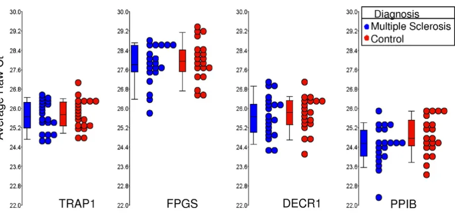
Data on the four genes, which constitute the optimal set of normalizers (TRAP1, FPGS, DECR1 and PPIB) on the MS and healthy control samples are shown separately as box-and-whisker plots (Figure 6). Data for both the Ct and Comparative Ct methods for MS versus control patients are shown in Table 5, with similar results being obtained with both methods.
Discussion
Gene expression techniques such as qRT-PCR, microar-rays and Northern blotting require accurate normaliza-tion methods in order to obtain reliable, quantitative data. A common approach is to use an endogenous refer-ence gene. The purpose of the referrefer-ence gene(s) is to remove differences not attributable to real biological var-iation. However, numerous reports indicate that differ-ences in the expression levels of commonly used endogenous reference genes vary considerably between different tissues and between different experimental con-ditions. Thus, there are no universal reference genes for all tissues or experimental conditions [2,3,29-35].
To address this problem, the best reference genes must be determined for each individual tissue or experimental set-ting. Moreover, a combination of several endogenous control genes produces more reliable normalization than any single control gene [6]. We therefore examined the expression of over 39,000 genes in 526 samples of whole blood from men, women and children that included healthy controls and individuals with a variety of different diseases. The 100 most stably expressed probe-sets were identified based upon having the least variance across all of these samples over a broad range of expression values. These 100 genes could also be useful as endogenous inter-nal controls for microarray studies – an approach not commonly used at present for microarray studies. Fifty percent of the annotated genes were involved in primary metabolic processes. Thus these genes appear to be good ''housekeeping genes'' for human blood because of their stable expression across various ages, genders, and dis-eases.
qRT-PCR validation was then performed on 10 candidate reference genes, four commonly used reference genes (ACTB, GAPDH, B2M, and HMBS) and PPIB, which is
Table 3: qRT-PCR on multiple sclerosis patients and normal controls
Gene Name Ct-avg SD CV n
reported to be stably expressed in blood [3]. The two fre-quently used control reference genes in nervous tissue, ACTB and GAPDH, were not stably expressed under our experimental conditions and are therefore not suitable reference genes for human whole blood. They have also been shown to vary in a variety of experimental settings and tissues [2,33,36-39], including in whole blood [3]. B2M and PPIB showed stable expression in our study. They were not identified as stably expressed in our Affymetrix data because their expression was higher than our upper limit for selection. PPIB was also reported to be
stably expressed in whole blood of patients with local and systemic inflammatory syndromes and healthy controls [3]. HMBS had a high SD based on our Affymetrix arrays on the 526 samples (SD of log-2-transformed values = 0.72). For comparison, the most stably expressed genes in the same intensity interval had a standard deviation (SD) of the log-2-transformed values around 0.2. HMBS, how-ever, showed stable expression when using qRT-PCR in the MS versus healthy controls experiment. Closer inspec-tion of the expression stability on the 526 samples from the Affymetrix chips showed that the HMBS SD of the
log-Table 4: Gene Ontology (GO) Annotation of genes analyzed in GeNorm
Gene Symbol Gene Name GO Biological Process
Term
GO Molecular Function Term
GO Cellular Component Term
FPGS folylpolyglutamate synthase nucleobase, nucleoside, nucleotide and nucleic acid metabolic process glycine metabolic process one-carbon compound metabolic
process
biosynthetic process nucleoside metabolic process
folic acid and derivative biosynthetic process
nucleotide binding Tetrahydrofolylpoly-glutamate-synthase activity ATP binding
ligase activity
cytoplasm mitochondrion cytosol
TRAP1 TNF receptor-associated protein 1
protein folding nucleotide binding receptor activity tumor necrosis factor receptor binding ATP binding
unfolded protein binding
Mitochondrion
DECR1 2,4-dienoyl CoA reductase 1, mitochondrial
metabolic process oxidation reduction
catalytic activity Binding 2,4-dienoyl-CoA reductase(NADPH) activity
oxidoreductase activity
mitochondrion
PPIB peptidylprolyl isomerase B (cyclophilin B)
protein folding peptidyl-prolyl cis-trans isomerase activity protein binding isomerase activity peptide binding unfolded protein binding
endoplasmic reticulum endoplasmic reticulum lumen
melanosome
HMBS hydroxymethyl-bilane synthase
porphyrin biosynthetic process heme biosynthetic process tetrapyrrole biosynthetic process
hydroxymethylbilane synthase activity
transferase activity
cytoplasm
B2M beta-2-microglobulin antigen processing and presentation of peptide antigen via MHC class I
immune response
protein binding Golgi membrane extracellular region plasma membrane early endosome membrane
2-transformed values for most of the studies was around 0.5, but it was 0.94 for the stroke study. This result sug-gests that there were study-specific differences for stroke versus the other diseases, leading to its unstable expres-sion. Our conclusion that HMBS was unstably expressed across our 526 samples is consistent with a previous study reporting that HMBS was unstably expressed in leukocytes [6]. Among the 10 candidate reference genes, TRAP1,
DECR1, FPGS, and FARP1 showed the most stable expres-sion.
In most research studies, the target genes or genes regu-lated within the study are expressed at different levels. In such situations, it is preferable if the comparable endog-enous or "housekeeping genes" are expressed at compara-ble levels as the target gene. This is essential if the microarray, RT-PCR or Northern blotting platform is non-Identification of the most stably expressed reference genes using GeNorm analysis
Figure 5
Table 5: Diagnosis effect on the candidate control genes.
Ct Method Comparative Ct Method Normalized to TRAP1 Normalized to Optimal Set of 4 Genes* Gene Fold Change (MS/H) Fold Change (MS/H) p-value(Diagnosis) FPGS 1.08 1.00 0.964
ACTb 1.08 ND 0.788
GINS2 1.21 1.11 0.691 HMBS 1.12 1.06 0.581 B2M -1.04 -1.10 0.575 FARP1 1.34 1.16 0.559 A4GALT -1.01 ND 0.416 TRAP1 ** -1.08 0.342 GAPDH -1.45 -1.61 0.198 DECR1 -1.09 -1.11 0.161 PPIB 1.32 1.22 0.155 PEX16 1.32 1.23 0.073 MAPRE2 1.42 1.30 0.072 CRY2 1.58 1.45 0.065 CSNK1G2 1.76 1.63 0.026
Expression of the candidate reference genes in whole blood of multiple sclerosis (MS) subjects is compared to age and gender matched control subjects using the Ct Method and the Comparative Ct Method. Genes are arranged in the order of decreasing p-value for diagnosis. *ANOVA: + Diagnosis + Plate + (Diagnosis × Plate) +
**TRAP1 fold change not calculated since that gene was used as a normalizer.
RNA expression values are shown for the four most stably expressed genes (TRAP1, FPGS, DECR1 and PPIB) for multiple sclerosis (MS) and controls subjects
Figure 6
RNA expression values are shown for the four most stably expressed genes (TRAP1, FPGS, DECR1 and PPIB) for multiple sclerosis (MS) and controls subjects. Circles represent individual qRT-PCR Ct average raw values for MS samples (blue, n = 20) and age and gender matched controls (red, n = 20). Boxes represent the 25th and 75th quartile and lines
linear at very low or at very high levels. Thus, having a set of endogenous reference genes that are stably expressed at different levels makes it possible to help select those whose expression is comparable to the target genes.
An optimal set of reference genes was identified based on the expression stability measure M as utilized in the GeNorm algorithm. It is based on the concept that the ratio between two ideal reference genes should always be constant regardless of the experimental conditions used. An important assumption here is that the genes in the analysis are not coordinately regulated and have inde-pendent functions. The six genes analyzed in GeNorm, seem to be involved in different biological processes, molecular functions and/or are located in different cellu-lar compartments (Table 4). TRAP1 and PPIB are involved in the same biological process however their cellular com-partmentalization is different. TRAP1 is a mitochondrial molecular chaperone also known as heat shock protein 75 (Hsp75). PPIB, encoding the cyclophillin B protein, is a cytoplasmic peptide isomerase. The broad spectrum of functions of the stably expressed genes makes it less likely that they are coordinately regulated.
An advantage of the approach taken in this study is that we performed a whole-genome survey of a large number of subjects of different ages, genders and diseases to iden-tify the most stably expressed genes in whole blood. Drawbacks to this approach include the fact that it is an array-based discovery method that is less likely to reliably detect very low expressing genes. In addition, the subjects chosen do reflect some bias related to the diseases studied, and reflect other biases in the subject selection – such as predominantly hospitalied and male patients.
It should be emphasized that the ultimate usefulness of the proposed endogenous reference genes for whole blood has not been demonstrated in this study. To do this, future studies must show that after correcting technical and individual variability according to the control genes, the discriminatory power of the diagnostic genes should be substantially improved.
It is important to point out the utility of the reference genes identified here for use in clinical human testing. Should the reference genes identified in this study provide sufficiently accurate normalization, then genes of interest could be normalized to these reference genes in healthy individuals and in individuals with a given disease or physiological condition. Once reference expression levels and standard deviations are derived in a large group of healthy individuals, deviations could be identified in tar-get individuals using these endogenous genes for normal-ization without the need for repeated samples from normal, healthy individuals. For non-clinical research
studies, such normalization to the same endogenous ref-erence genes would allow for comparison across studies.
Conclusion
We took advantage of the large amount of expression data on 33,000 genes from 526 individuals to select genes that had stable expression in whole blood. This gave us an added advantage to assess not only the gene expression of commonly used housekeeping genes but to perform a whole genome survey for stably expressed genes over mul-tiple ages, disease conditions and different expression lev-els. Ranking based on the expression stability from qRT-PCR data of the candidate control genes and estimation of the minimum number of genes needed for nomalization showed that the fewest, most stably expressed genes needed for acurate normalization in RNA expression stud-ies of human whole blood is a combination of TRAP1, FPGS, DECR1 and PPIB. These genes should be useful as normalization genes for a wide-range of whole blood expression studies in humans.
Competing interests
The authors declare that they have no competing interests.
Authors' contributions
BSS, MA, WLW and FRS designed the study, analyzed the results and wrote the manucript. MA provided the sam-ples from the MS patients for the RT-PCR and array stud-ies. YT, HX, PA, XA, DL, BPA, IHL, JPG, RJT, GCJ and LL either helped collect samples, helped analyze the data from the microarray studies and/or read and proof read the manuscript. JPG supervised the processing of a number of the arrays used in these studies. All authors read and approved the final manuscript.
Additional material
Acknowledgements
These studies were supported by grants from: the NIH/NINDS (NS056302); an American Stroke Association Bugher Foundation Center for Stroke Prevention Research; Cure Autism Now/Autism Speaks; the UC Discovery Program and Pediatric Biosciences; the Tourette Syndrome Association; and a gift from the Ron and Darrin Mittelstaedt family sup-ported Foundation.
Additional file 1
100 Most Stable Genes and 10 Affy Candidate Reference Genes. Infor-mation on sheet 2 represents the 100 most stable genes including Affyne-trix probe-set IDs, reference public IDs, gene symbols, and expression levels. The data on sheet 2 represents the 10 Affymetrix candidate refer-ence genes with probe-sets, ABI assay, and expression levels information.
Click here for file
References
1. Huggett J, Dheda K, Bustin S, Zumla A: Real-time RT-PCR nor-malisation; strategies and considerations. Genes Immun 2005,
6(4):279-284.
2. Lee PD, Sladek R, Greenwood CM, Hudson TJ: Control genes and variability: absence of ubiquitous reference transcripts in diverse mammalian expression studies. Genome Res 2002,
12(2):292-297.
3. Pachot A, Blond JL, Mougin B, Miossec P: Peptidylpropyl isomer-ase B (PPIB): a suitable reference gene for mRNA quantifi-cation in peripheral whole blood. J Biotechnol 2004, 114(1– 2):121-124.
4. Schmittgen TD, Zakrajsek BA: Effect of experimental treatment on housekeeping gene expression: validation by real-time, quantitative RT-PCR. J Biochem Biophys Methods 2000, 46(1– 2):69-81.
5. Thellin O, Zorzi W, Lakaye B, De Borman B, Coumans B, Hennen G, Grisar T, Igout A, Heinen E: Housekeeping genes as internal standards: use and limits. J Biotechnol 1999, 75(2–3):291-295. 6. Vandesompele J, De Preter K, Pattyn F, Poppe B, Van Roy N, De
Paepe A, Speleman F: Accurate normalization of real-time quantitative RT-PCR data by geometric averaging of multi-ple internal control genes. Genome Biol 2002,
3(7):RESEARCH0034.
7. Tang Y, Xu H, Du X, Lit L, Walker W, Lu A, Ran R, Gregg JP, Reilly M, Pancioli A, et al.: Gene expression in blood changes rapidly in neutrophils and monocytes after ischemic stroke in humans: a microarray study. J Cereb Blood Flow Metab 2006,
26(8):1089-1102.
8. Xu H, Tang Y, Liu DZ, Ran R, Ander BP, Apperson M, Liu XS, Khoury JC, Gregg JP, Pancioli A, et al.: Gene expression in peripheral blood differs after cardioembolic compared with large-vessel atherosclerotic stroke: biomarkers for the etiology of ischemic stroke. J Cereb Blood Flow Metab 2008, 28(7):1320-1328. 9. Zhan X, Kim C, Sharp FR: Very brief focal ischemia simulating transient ischemic attacks (TIAs) can injure brain and induce Hsp70 protein. Brain Res 2008, 1234:183-197.
10. Sharp FR, Xu H, Lit L, Walker W, Apperson M, Gilbert DL, Glauser TA, Wong B, Hershey A, Liu DZ, et al.: The future of genomic profiling of neurological diseases using blood. Arch Neurol
2006, 63(11):1529-1536.
11. Du X, Tang Y, Xu H, Lit L, Walker W, Ashwood P, Gregg JP, Sharp FR: Genomic profiles for human peripheral blood T cells, B cells, natural killer cells, monocytes, and polymorphonuclear cells: comparisons to ischemic stroke, migraine, and Tourette syndrome. Genomics 2006, 87(6):693-703.
12. Lit L, Enstrom A, Sharp FR, Gilbert DL: Age-related gene expres-sion in Tourette syndrome. J Psychiatr Res 2009, 43(3):319-330. 13. Lit L, Gilbert DL, Walker W, Sharp FR: A subgroup of Tourette's patients overexpress specific natural killer cell genes in blood: a preliminary report. Am J Med Genet B Neuropsychiatr Genet 2007, 144B(7):958-963.
14. Tang Y, Schapiro MB, Franz DN, Patterson BJ, Hickey FJ, Schorry EK, Hopkin RJ, Wylie M, Narayan T, Glauser TA, et al.: Blood expres-sion profiles for tuberous sclerosis complex 2, neurofibroma-tosis type 1, and Down's syndrome. Ann Neurol 2004,
56(6):808-814.
15. Tang Y, Lu A, Ran R, Aronow BJ, Schorry EK, Hopkin RJ, Gilbert DL, Glauser TA, Hershey AD, Richtand NW, et al.: Human blood genomics: distinct profiles for gender, age and neurofi-bromatosis type 1. Brain Res Mol Brain Res 2004, 132(2):155-167. 16. Tang Y, Glauser TA, Gilbert DL, Hershey AD, Privitera MD, Ficker DM, Szaflarski JP, Sharp FR: Valproic acid blood genomic expres-sion patterns in children with epilepsy – a pilot study. Acta Neurol Scand 2004, 109(3):159-168.
17. Tang Y, Gilbert DL, Glauser TA, Hershey AD, Sharp FR: Blood gene expression profiling of neurologic diseases: a pilot microar-ray study. Arch Neurol 2005, 62(2):210-215.
18. Tang Y, Lu A, Aronow BJ, Sharp FR: Blood genomic responses dif-fer after stroke, seizures, hypoglycemia, and hypoxia: blood genomic fingerprints of disease. Ann Neurol 2001,
50(6):699-707.
19. Hershey AD, Tang Y, Powers SW, Kabbouche MA, Gilbert DL, Glauser TA, Sharp FR: Genomic abnormalities in patients with migraine and chronic migraine: preliminary blood gene
expression suggests platelet abnormalities. Headache 2004,
44(10):994-1004.
20. Wong B, Gilbert DL, Walker WL, Liao IH, Lit L, Stamova B, Jickling G, Apperson M, Sharp FR: Gene expression in blood of subjects with Duchenne muscular dystrophy. Neurogenetics 2008, 9:9. 21. Walker WL, Liao IH, Gilbert DL, Wong B, Pollard KS, McCulloch CE,
Lit L, Sharp FR: Empirical Bayes accomodation of batch-effects in microarray data using identical replicate reference sam-ples: application to RNA expression profiling of blood from Duchenne muscular dystrophy patients. BMC Genomics 2008,
9(494):494.
22. Enstrom AM, Lit L, Onore CE, Gregg JP, Hansen RL, Pessah IN, Hertz-Picciotto I, Water JA Van de, Sharp FR, Ashwood P: Altered gene expression and function of peripheral blood natural killer cells in children with autism. Brain Behav Immun 2009,
23(1):124-133.
23. Gregg JP, Lit L, Baron CA, Hertz-Picciotto I, Walker W, Davis RA, Croen LA, Ozonoff S, Hansen R, Pessah IN, et al.: Gene expression changes in children with autism. Genomics 2008, 91(1):22-29. 24. Osman F, Leutenegger C, Golino D, Rowhani A: Comparison of
low-density arrays, RT-PCR and real-time TaqMan RT-PCR in detection of grapevine viruses. J Virol Methods 2008,
149(2):292-299.
25. Wu Z, Irizarry RA: Preprocessing of oligonucleotide array data. Nat Biotechnol 2004, 22(6):656-658.
26. Schmittgen TD, Livak KJ: Analyzing real-time PCR data by the comparative C(T) method. Nat Protoc 2008, 3(6):1101-1108. 27. Andersen CL, Jensen JL, Orntoft TF: Normalization of real-time
quantitative reverse transcription-PCR data: a model-based variance estimation approach to identify genes suited for normalization, applied to bladder and colon cancer data sets. Cancer Res 2004, 64(15):5245-5250.
28. Livak KJ, Schmittgen TD: Analysis of relative gene expression data using real-time quantitative PCR and the 2(-Delta Delta C(T)) Method. Methods 2001, 25(4):402-408.
29. Biederman J, Yee J, Cortes P: Validation of internal control genes for gene expression analysis in diabetic glomerulosclerosis.
Kidney Int 2004, 66(6):2308-2314.
30. Etschmann B, Wilcken B, Stoevesand K, Schulenburg A von der, Sterner-Kock A: Selection of reference genes for quantitative real-time PCR analysis in canine mammary tumors using the GeNorm algorithm. Vet Pathol 2006, 43(6):934-942.
31. Hoerndli FJ, Toigo M, Schild A, Gotz J, Day PJ: Reference genes identified in SH-SY5Y cells using custom-made gene arrays with validation by quantitative polymerase chain reaction.
Anal Biochem 2004, 335(1):30-41.
32. Jung M, Ramankulov A, Roigas J, Johannsen M, Ringsdorf M, Kris-tiansen G, Jung K: In search of suitable reference genes for gene expression studies of human renal cell carcinoma by real-time PCR. BMC Mol Biol 2007, 8(47):47.
33. Ohl F, Jung M, Radonic A, Sachs M, Loening SA, Jung K: Identifica-tion and validaIdentifica-tion of suitable endogenous reference genes for gene expression studies of human bladder cancer. J Urol
2006, 175(5):1915-1920.
34. Radonic A, Thulke S, Bae HG, Muller MA, Siegert W, Nitsche A: Ref-erence gene selection for quantitative real-time PCR analy-sis in virus infected cells: SARS corona virus, Yellow fever virus, Human Herpesvirus-6, Camelpox virus and Cytomeg-alovirus infections. Virol J 2005, 2(7):7.
35. Silver N, Best S, Jiang J, Thein SL: Selection of housekeeping genes for gene expression studies in human reticulocytes using real-time PCR. BMC Mol Biol 2006, 7(33):33.
36. Hamalainen HK, Tubman JC, Vikman S, Kyrola T, Ylikoski E, War-rington JA, Lahesmaa R: Identification and validation of endog-enous reference genes for expression profiling of T helper cell differentiation by quantitative real-time RT-PCR. Anal Biochem 2001, 299(1):63-70.
37. Roge R, Thorsen J, Torring C, Ozbay A, Moller BK, Carstens J: Com-monly used reference genes are actively regulated in in vitro stimulated lymphocytes. Scand J Immunol 2007, 65(2):202-209. 38. Ross DT, Scherf U, Eisen MB, Perou CM, Rees C, Spellman P, Iyer V,
Jeffrey SS, Rijn M Van de, Waltham M, et al.: Systematic variation in gene expression patterns in human cancer cell lines. Nat Genet 2000, 24(3):227-235.
Publish with BioMed Central and every scientist can read your work free of charge "BioMed Central will be the most significant development for disseminating the results of biomedical researc h in our lifetime."
Sir Paul Nurse, Cancer Research UK
Your research papers will be:
available free of charge to the entire biomedical community
peer reviewed and published immediately upon acceptance
cited in PubMed and archived on PubMed Central
yours — you keep the copyright
Submit your manuscript here:
http://www.biomedcentral.com/info/publishing_adv.asp
BioMedcentral
535 housekeeping/maintenance genes. Physiol Genomics 2000,
2(3):143-147.
Pre-publication history
The pre-publication history for this paper can be accessed here: