R E V I E W
Open Access
Tools to reverse-engineer multicellular
systems: case studies using the fruit fly
Qinfeng Wu, Nilay Kumar, Vijay Velagala and Jeremiah J. Zartman
*Abstract
Reverse-engineering how complex multicellular systems develop and function is a grand challenge for systems bioengineers. This challenge has motivated the creation of a suite of bioengineering tools to develop increasingly quantitative descriptions of multicellular systems. Here, we survey a selection of these tools including microfluidic devices, imaging and computer vision techniques. We provide a selected overview of the emerging cross-talk between engineering methods and quantitative investigations within developmental biology. In particular, the review highlights selected recent examples from theDrosophilasystem, an excellent platform for understanding the interplay between genetics and biophysics. In sum, the integrative approaches that combine multiple advances in these fields are increasingly necessary to enable a deeper understanding of how to analyze both natural and synthetic multicellular systems.
Keywords:Microfluidics, Image processing, Machine learning, Deep learning, Imaging
Background
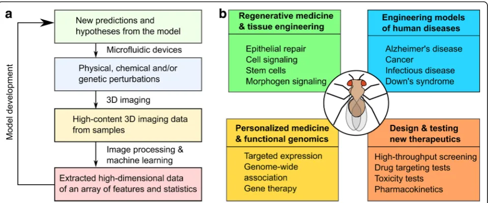
Answers to many human health challenges require an inte-grated systems-level understanding of the body [1]. Bio-complexity, the emergence of properties that are more than the sum of individual constituents, leads to profound impli-cations on how to solve problems in regenerative medicine, cancer therapy, and personalized medicine [2]. This com-plexity spans multiple spatial scales from molecules, such as proteins and DNA, to cells, tissues, organs and organ systems. It requires a systems-level analysis to understand this complexity [3]. The general paradigm of systems re-search adopts an iterative approach, which usually involves transitioning from experiments to model formulation then to revision of original hypotheses (Fig.1a) [4].
Genetic model systems, such as the worm—C. elegans, the zebrafish or the fruit fly—Drosophila melanoga-ster, serve as proof-of-principle platforms for developing tools to analyze multicellular systems or to test new tech-niques in forward-engineering living systems [5]. In par-ticular, Drosophila enables genetic studies of how genes are regulated to control morphogenesis [6–8] and physi-ology [9]. It is an excellent system for studies that are at the crossroad of biophysics, information processing, and
molecular and developmental biology. The fruit fly system provides many advantages, including cheap and easy hus-bandry, rapid life cycle, and many available genetic tools [5, 10–16]. These advantages contribute to the status of Drosophila as a premier model for reverse-engineering multicellular systems. Of note, several fundamental signal-ing pathways were first discovered inDrosophila, including Hedgehog [17], Notch [18] and Wingless pathways [19]. Therefore,Drosophila has been extremely crucial in biol-ogy and bioengineering researches in many areas and will surely continue to play a critical role in years to come [20]. Beyond fundamental research, Drosophila has been used to study many health challenges, including cancer [21–28], neurodegenerative disorders [29–31], infectious diseases [32], cardiac disease [33], aging and metabolic diseases [34], wound healing and organ regeneration [20, 35–38] (Fig. 1b).Drosophiladisease models can acceler-ate the racceler-ate of therapeutic drug testing and discovery due to the availability of genetic tools and a genome that lacks redundancy [11, 39–41]. Thus, Drosophila has a proven track record for understanding the biocomplexity of multicellular systems.
Here, we review a selected set of engineering tools and methodologies that are broadly applicable to reverse-engineer organ development. As a case in point, we focus on selected examples centered on the
© The Author(s). 2019Open AccessThis article is distributed under the terms of the Creative Commons Attribution 4.0 International License (http://creativecommons.org/licenses/by/4.0/), which permits unrestricted use, distribution, and reproduction in any medium, provided you give appropriate credit to the original author(s) and the source, provide a link to the Creative Commons license, and indicate if changes were made. The Creative Commons Public Domain Dedication waiver (http://creativecommons.org/publicdomain/zero/1.0/) applies to the data made available in this article, unless otherwise stated.
* Correspondence:[email protected]
quantitative analysis of Drosophila (Fig. 1). This review highlights selected engineering advances that have led to the development of tools in the field of high-throughput and high-content screening: microfluidic devices, im-aging technologies, and imim-aging analysis algorithms. Many novel and elegant engineering designs, such as various microfluidic devices and imaging modalities, have more precise manipulations and extract deeper in-sights from genetic systems, with a large breadth applied to the zebrafish, the fruit fly and the worm [42–45]. Rapid advances in machine learning and deep learn-ing have greatly increased researchers’ ability to ex-tract and analyze biological data. These tools are enabling increasingly quantitative characterization of fruit flies and other multicellular systems. Finally, the availability of many computational modeling tools (see, for example, reviews such as [46, 47]) has facili-tated and accelerated the iterative cycle of hypothesis testing and revision (Fig. 1a). The review concludes with a perspective on current trends and future po-tential directions for reverse-engineering of multicel-lular systems.
Microfluidic devices enable controlled imaging and perturbations of fruit fly development
Microfluidic devices refer to systems that use chan-nels with dimensions of tens to hundreds of
micrometers to manipulate a small amount of fluids [48]. A big challenge in studying the fruit fly is how to accurately apply perturbations and manipulate its organs due to their small size. Microfluidic devices are an increasingly important technique for address-ing this challenge. In the followaddress-ing section, we dis-cuss how microfluidic devices were applied in representative individual studies and how they have contributed to the improvement of current experi-mental approaches.
Sample preparation and immobilization
Immobilization is a critical step to achieve high reso-lution imaging and precise manipulation for moving samples, such as Drosophila larvae. For example, to study the larval nervous system, researchers require the larva to be immobilized to image neuronal physiological activities. However, immobilization of larvae is difficult because of its digging and burrowing motion. Trad-itional immobilization techniques, such as tape or glue, still allow minor larval movement and reduce larval via-bility [49,50]. Therefore, several strategies have been de-veloped to immobilize samples. For example, Mondal et al. used a deformable membrane controlled by a water column to mechanically restrain larvae. The device al-lows them to image vesicle trafficking in the neurons of Fig. 1Workflow for reverse-engineering multicellular systems and the broad applicability ofDrosophilaas an integrative test case.aA
Drosophila, C. elegans, and zebrafish at high resolution [51, 52]. Another chip designed by the same group im-mobilizes larvae by clamping the mouth region to re-duce digging movement. There is an additional design that pneumatically immobilizes larvae and allows for auto-mated larva loading, immobilization and unloading. Both methods achieved significant immobilization and resulted in high-resolution imaging of neural responses [53, 54]. Mechanical restraint achieves easy immobilization but leads to reduced viability and innate response to mechan-ical perturbation [53,54].
Anesthesia is an alternative to mechanical immobilization. Heemskerk et al. developed an immobilization chamber that uses desflurane for anesthesia [55]. A newer design uses both CO2and
com-pression to immobilize larvae [56]. The chip also incor-porates inputs for food feeding that allow for long-term (> 10 h) immobilization and imaging. Researchers were
able to observe regenerative axonal growth up to 11 h of injury of the larva, demonstrating that CO2 did not
affect the physiology of the larva in this study. An im-proved design uses coolant, instead of CO2, for
anesthesia and immobilization (Fig. 2a). This technique enabled the imaging of in vivo mitochondria movement in axons with high resolution without affecting the larva physiology [57].
Orienting a multicellular sample during loading is a frequently encountered problem. To overcome this, Ardeshiri et al. employed a rotatable glass that can suck onto the head of the larva to rotate the larva [49, 58]. Another creative solution allows samples to be prepared on the cover glass first before the sili-cone slab is placed on top to form the channels of the device [59]. This design allows more flexible prep-arations, better orientations and wider accommoda-tion of a variety of samples.
Microinjection
Delivery of genetic constructs into fly embryos requires precise microinjection. For perturbation studies, drugs/ toxins must also be accurately introduced into fragile embryos. Due to the requirement of precise placement and the small volume of injection, microinjectors have become tools of choice. Several microfluidic devices have been created to miniaturize this technique and to surpass the reliability of manual injection. First, Delubac et al. designed a microfluidic system for automatic em-bryo loading, detection and injection [60]. The device re-trieves and places the embryos in contact with the injector/needle. The injection begins when the system detects the embryo in front of the injector. This fully-au-tomated process enables high-throughput screening of embryos and/or creation of transgenic Drosophila lines. However, there is no control as to how deep the injector can go. Later, Ghaemi et al. incorporated a long-taper needle and a micro-positioner to control the depth of in-jection (Fig. 2c) [61]. This system enables deep (up to 250μm), highly-precise injections (a resolution of 5μm) and low injection volumes (as low as 30 ± 10 pL) with minimum damage because of the tapered needle. The precise (position and volume) injection of toxins (NaN3)
into specific locations of theDrosophilaembryo enables a detailed spatiotemporal study of how toxins affect em-bryo development [61].
Sorting, positioning and orienting of samples
One of the advantages of using Drosophila embryos is the high-throughput data collection enabled by the number of embryos that can be obtained at low cost. However, sorting, positioning and orienting of many em-bryos or other post-embryonic organs is a technical hur-dle that needs to be addressed. Furlong et al. adopted the concept of fluorescence-activated cell sorting (FACS) and designed a device for sorting embryos expressing a fluorescent protein marker [62]. The device uses a ro-botic valve to separate the embryos into fluorescent and non-fluorescent samples. In 2004, Chen et al. presented a pressure-controlled microfluidic sorter for Drosophila embryos that directs the flow direction of embryos into different outlets [63]. The computer simulation and flow experiment with dye demonstrated the functionality of the device. Chen et al. improved the design to allow for high-speed sorting, enabled by a deflecting jet to change the movement of the object [64].
Bernstein et al. presented an early attempt to pos-ition and orient Drosophila embryos in batch for high-throughput microinjection. They designed a micro-assembly of protruded hydrophobic surfaces to achieve large-scale positioning and orienting of the em-bryos [65]. Embryos are flowed through the device and are immobilized when in contact with the hydrophobic
surface. The designed achieved 95% immobilization rate and 40% alignment rate. They also presented a conceptual design of the high-throughput microinjection system that would work with the orientation array, still yet to be real-ized as a physical working model [66].
Lu and collaborators developed a series of array-based microfluidic devices for positioning and orienting Drosophila embryos. A first microfluidic array was designed to utilize passive hydrodynamics to trap, pos-ition and vertically orient Drosophila embryos (Fig. 2d) [67, 68]. The vertical orientation of the embryo allows the observation of dorsal-ventral patterning of proteins of interest. The device provided high-throughput dorso-ventral patterning data. Subsequently, the researchers modified the device to horizontally orient the embryo [69]. The Lu lab further improved the design to increase the loading efficiency to > 90% [70]. The new iteration also allows for anoxia perturbation of the embryos and potentially other forms of perturbation.
Multi-modal perturbations to organ systems
Spatiotemporal control over a range of perturbations (e.g. mechanical, chemical and electrical) on multicellu-lar samples often requires multi-modal microfluidic de-vice designs. Lucchetta et al. designed pioneering microfluidic devices to investigate how temperature reg-ulates embryogenesis [71, 72]. The device generates a temperature step between the two compartments of a Drosophilaembryo. This spatiotemporal perturbation of temperature created a way to understand the complex biochemical networks governing Drosophila embryogen-esis [73]. Researchers have adopted this design and used it for other perturbations. For example, a similar design exerts spatiotemporal control of oxygen gradient on liv-ing embryos [74]. To accommodate various Drosophila samples and apply different kinds of chemical stimuli, Giesen et al. came up with a device that can immobilize a range ofDrosophilaorgans and apply chemical stimu-lations [75]. The authors demonstrated the use of the device to perturb and image brain, leg and proboscis. They successfully measured calcium-based neuron re-sponses to chemical stimuli at single-cell resolution using this device.
mechanisms of Ca2+ signaling in wing discs, a model organ for investigating signal transduction during organ growth. The device allows accurate mechanical stimula-tion of the wing disc, and it can be modified to accom-modate other organoid-size systems and/or adding additional perturbations, such as electric stimulation [78].
Trends for microfluidic devices for multicellular systems Microfluidic devices enable high-throughput analysis and perturbation with high spatiotemporal resolution. Recent efforts have combined functionalities that were traditionally achieved by multiple microfluidic devices into one design. For example, Shorr et al. invented a de-vice that incorporates various automated operations of Drosophila embryo, including high-throughput auto-matic alignment, immobilization, compression, real-time imaging, and recovery of hundreds of live embryos [79]. These new devices have achieved multiplexing of various modalities, and allow for acceleration of research in de-velopmental biology and multicellular systems [80].
The possibilities brought up by microfluidic devices are numerous and the development of new manufactur-ing technologies is helpmanufactur-ing the democratization of microfluidic devices as well. Computer-aided design (CAD) and simulation have greatly increased the accuracy and functionality of newly-designed devices [63,64,79]. 3D printing is enabling the customizable pro-duction of microfluidic chips [81,82], as the resolution of those printers has improved significantly. 3D printers have brought down the cost of manufacturing and enabled the easy transfer of designs [80]. Other quick-fabrication techniques, such as hybrid-polyethylene-terephthalate laminate (PETL), are also lowering the barrier to entry for microfluidic devices [78, 83]. In addition, many universities are also providing training programs and have clean-room facilities that can support the adop-tion of microfluidic devices among new users [80]. Combined, these developments are encouraging the development of microfluidic devices with new applica-tions in developmental biology and the synthetic biology of multicellular systems.
Three-dimensional imaging modalities enable the analysis of thick multicellular systems
Due to the larger scales involved, multicellular systems, in-cluding Drosophila tissues, require three-dimensional im-aging techniques. An increasingly diverse range of imim-aging modalities is enabling researchers to investigate deeper into tissues. Recent improvements of fluorescence-based im-aging modalities have increased imim-aging resolution, sample penetration and acquisition rate while reducing phototoxic-ity and photobleaching [84, 85]. In the meantime, other new imaging modalities, such as harmonic generation
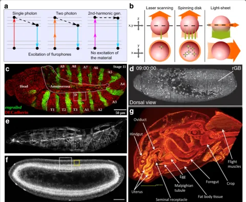
microscopy and micro-computed tomography (micro-CT), enable label-free imaging [86, 87] (Fig. 3a, b). In this section, we discuss variations of fluorescent imaging tech-niques and label-free imaging. We also cover the advan-tages and limitations of each imaging modality.
Confocal microscopy
Confocal microscopy uses a pinhole aperture to reject out-of-focus light to improve resolution and signal-to-noise ratio, compared to wide-field microscopy (Fig.3c) [88]. Confocal microscopes can achieve a penetration depth of up to around 100μm [89]. Confocal microscopy is divided into two main subcategories: laser scanning confocal microscopy and spinning disk confocal micros-copy [89]. In laser scanning confocal microscopy, a sin-gle illumination spot is rastered across the field of view. The image acquisition rate is relatively low because of the point-by-point scanning system, especially when ac-quiring 3D stacks with multiple fluorescent channels from a sample. Because of the small focal point, laser scanning confocal microscopy can cause significant photobleaching and the specimen’s long-term viability is compromised due to phototoxicity [89]. Continuous ef-forts have resulted in significant increase of scanning speeds to lessen this limitation [90]. Alternatively, a spin-ning disk that contains many focus pinholes provides a multipoint scanning strategy that significantly increases the collection rate. This reduces photobleaching and im-proves specimen viability. However, this comes at a cost of reduced 3D-sectioning capability and resolution.
Light-sheet fluorescent microscopy
In light-sheet microscopy, only a single plane of focus is illuminated (Fig. 3b). The camera detects fluorescence from a direction perpendicular to the light-sheet. The scanning speed of a light-sheet fluorescent microscopy is 100–1000 times faster than that of laser scanning con-focal microscope. These characteristics minimize both phototoxicity and photobleaching and enable long-term imaging experiments of 3D multicellular systems [84]. This advantage allows imaging of a beating heart of a zebrafish or imaging of whole Drosophilaembryos with fast rates of acquisition [91]. For example, Drosophila embryos can complete normal development even after being irradiated for 11,480 images by a light-sheet microscope [92]. The limited illumination of the speci-men also results in high signal-to-noise ratio.
can acquire 175 million voxels per second (Fig. 3d) [94, 95]. Chhetri et al. developed isotropic multiview light-sheet microscopy for long-term imaging with double the penetration depth and 500-fold larger temporal reso-lution than previous design of light-sheet microscopes [96]. Aided by image segmentation and computational tracking, researchers reconstructed the geometry of the entire tissue and measured morphogenic dynamics during embryo development [97]. Lattice light-sheet microscopy,
which results in an ultrathin light sheet, further increases the speed of image acquisition (scanning 200 to 1000 planes per second) with reduced phototoxicity [98].
Light-sheet microscopes can be constructed at rela-tively low cost, compared with other imaging technology setups. A great resource for building a customizable light-sheet microscope is an open hardware and software platform called OpenSPIM [99]. However, a significant challenge for light-sheet microscopes is how to process, Fig. 3Imaging technologies open doors to deeper insights ofDrosophila.aSingle-photon (confocal) microscopy and multi-photon microscopy visualize samples by exciting the fluorophore and detect the emitted fluorescence. Harmonic generation microscopy, however, does not involve excitation of target molecules for visualization. Second-harmonic generation involves the combination of two photons into one photon without loss of energy.bLaser scanning confocal and spinning disk confocal microscopes illuminate the whole sample and detects epifluorescence, while light-sheet only illuminates the focal plane and detects fluorescence from the perpendicular direction. Adapted with permission from [196].
store and move the very large datasets generated in single experiments.
Multi-photon fluorescence microscopy
Multi-photon fluorescence microscopy relies on the sim-ultaneous absorption of multiple photons to excite fluor-ophores (Fig. 3a). This process requires a high-energy laser concentrated at the laser focal point. Outside the focal point, the laser power is below the threshold re-quired for two-photon excitation. This allows multi-pho-ton microscopes to excite samples at a tiny volume around the focus point, thus reducing phototoxicity and extending the duration of in vivo imaging. The precise excitation at the focal point also improves the signal-to-noise ratio.
Multi-photon microscopes use near-infrared lasers with longer wavelengths (lower energy per photon) than lasers used in one-photon confocal microscopy. The near-infrared laser allows deeper penetration (2–3 times deeper for two-photon) into the sample, compared to confocal microscopy (Fig.3d) [85]. The laser, because of the longer wavelength, also scatters less. Therefore, multi-photon microscopy provides good 3D sectioning capability for thick specimens. Researchers were able to image calcium dynamics in Drosophila adult brain in vivo in behavioral studies and odor-activated neuron re-sponse due to the deep penetration capability of two-photon microscopy, which is the most commonly used multi-photon microscopy [100–102]. Besides two-photon, three-photon microscopy has received in-creasing popularity because of its increased penetration and signal-to-noise ratio. For example, scientists have successfully imaged through adult mouse skulls at > 500μm depth using three-photon microscopy [103].
However, multi-photon microscopy has low acquisi-tion rates due to the point scanning system and leads to accelerated photobleaching [104, 105]. Two-photon mi-croscopy also causes autofluorescence of some chromo-phores, such as NAD(P)H, which can cause significant noise for image acquisition [106]. The cost is also signifi-cantly higher because of the more sophisticated laser, optics, mechanics, and maintenance required. Neverthe-less, the improvement of functionality and the continu-ous reduction of costs will enable multi-photon laser scanning microscopy to be adopted by the wider research community. Multi-photon microscopy currently defines the upper limit of penetration depth in diffraction-limited microscopy [85].
Harmonic generation microscopy
The fluorescence microscopies discussed above have several innate shortcomings, such as photobleaching, phototoxicity, and the need to label the molecules [107]. Harmonic generation microscopy, on the other hand,
achieves label-free imaging. Harmonic generation refers to the nonlinear optics phenomenon where multiple photons reach a molecule and generate a new photon without the presence of a fluorophore. For example, during second-harmonic generation, two identical in-coming photons are combined to generate one outgoing photon with a wavelength of exactly half of the excita-tion beam (Fig.3a).
The biggest advantage of harmonic generation mi-croscopy is that it does not require labeling of the mol-ecules of interest. Harmonic generation microscopy also substantially reduces photobleaching and photo-toxicity because it does not rely on the excitation of fluorophores [108]. In addition, harmonic generation microscopy achieves deep penetration by using near-infrared wavelengths for the incident light. Harmonic generation microscopy has the ability to con-struct high-resolution three-dimensional images of several hundred microns of depth.
Harmonic generation provides additional structural in-formation on molecular or supra-molecular order not easily detectable with fluorescence strategies. Second-harmonic generation is caused by materials that are non-centrosymmetric [109]. These materials include collagen fibril/fiber structure (type I and II fibrillar collagen), myofilaments, fibers, polarized microtubule assemblies, and muscle myosin (Fig.3e) [87,110–112]. Second-har-monic generation microscopy has been used to image developing muscle structures and the trachea system in 2nd-instar larva, and the lipid bodies inDrosophilacells [112, 113]. Researchers used second-harmonic gener-ation microscopy to investigate the structure of Drosoph-ila sarcomeres and visualize myocyte activity to study rhythmic muscle contraction [114,115].
Third-harmonic generation occurs at structural inter-faces with local transitions of the refractive index [116]. Third-harmonic generation was used to image lipid in Drosophila and mouse embryos. When coupled with second harmonic generation microscopy and two-pho-ton imaging, one can explore the interactions between lipid, extracellular matrix and fluorescence-marked pro-teins (Fig. 3f ) [113, 117–119]. Researchers used third-harmonic generation to visualize rhodopsin in the eye [120], and to measure the morphogenetic movement in Drosophila embryos by visualizing lipid droplets around cell nuclei and the interfaces of yolk structures [121]. Together, second- and third-harmonic generation microscopy modalities serve as powerful label-free im-aging techniques.
Micro-CT
morphology of the specimen [122]. Micro-CT produces images with microscopic resolution and avoids artifacts due to processing of samples used for fluorescence imaging [123]. Because insects are made of only soft tissues, they are ideal for micro-CT. With very simple contrast staining, micro-CT can produce quantitative, high-resolution, high-contrast volume images of Dros-ophila,bumblebee, etc. [86, 124]. Micro-CT has become increasingly popular and is used to study morphological changes in a broad range ofDrosophilatissues (Fig.3g), including the female reproductive tract [125], neuronal structures [126], urolithiasis studies of calcium oxalate deposition [127], and wings for computational aero-dynamic analysis [128].
The combination of multiple imaging modalities opens new possibilities to utilize the strengths while avoiding the limitations of individual techniques. For example, Truong et al. combined two-photon microscopy with light-sheet microscopy to implement two-photon-scanned light-sheet microscopy for Drosophila embryos [129]. This combination achieved twice the penetration of one-photon light-sheet microscopy and is more than ten times faster than two-photon laser scanning copy. Researchers also combined multi-photon micros-copy with harmonic generation microsmicros-copy to construct a comprehensive picture of samples including both the fluorophore-labeled molecules and non-labeled struc-tural molecules [130]. However, a major challenge for systems bioengineers is to process large datasets gener-ated by these advanced imaging techniques. There is a critical need to automate the analysis of large datasets and to reduce high-dimensional data that includes infor-mation of molecular species and biophysical properties of cells through both space and time [131].
Trends of imaging technologies for multicellular systems Besides the introduction of new imaging principles, existing imaging technologies are often combined for multiplexing of functionalities that further increases in performance [93–96, 98]. There is also a trend of democratization of imaging technologies, from the OpenSPIM project supporting the construction of cus-tomized light-sheet microscopes to mobile phone-based microscopy [99, 132–134]. The increase in acquisition speed and resolution encourages the advance of image analysis methods to handle the ever-increasing amount of data generated from analysis of multi-cellular systems with Drosophila providing a versatile system for proof-of-concept studies.
Data-driven learning algorithms accelerate the quantitative analysis of multicellular systems The exponential increase in biological data acquisition rates challenges conventional analysis strategies [135].
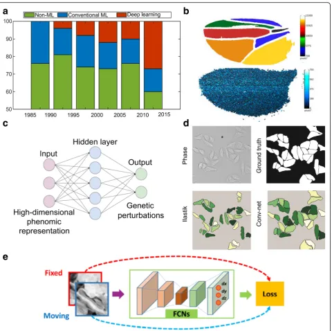
Integration of advanced algorithms for bio-image ana-lysis is thus highly desired. The result of a bio-image analysis pipeline can be as simple as quantification of fluctuations in cellular areas over time or as complex as a high-dimensional array of features of a Drosophila wing. In short, the goal of analysis is to convert images into arrays of numbers that are amenable to statistical evaluation. This helps create data-driven models or to validate predictions from phenomenological or mechan-istic models. In this section, we discuss how both conventional machine-learning and deep-learning algo-rithms play critical roles in the analysis of multicellular systems, using selected examples focused on the fruit fly. In particular, we show how deep learning is rapidly emerging as a solution to accelerate the analysis of bio-logical big data (Fig.4a).
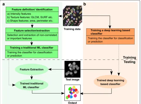
Machine-learning algorithms leverage training datasets to find features within the data to fulfill the task of either classification or prediction [136]. A feature is a measur-able property or characteristic of a phenomenon within the image. Feature extraction can either be manual or embedded within the algorithm’s architecture. Machine-learning algorithms are either supervised (requiring example input-output pairs to train the algorithm) or unsupervised (input data not annotated). Unsupervised learning algorithms, such as k-means clustering, perform poorly on noisy datasets and are frequently unsuited to bio-image analysis [137]. Therefore, supervised machine-learning algorithms are more commonly adopted for bio-image analysis (Fig.5).
One of the major challenges in cellular tracking is obtaining high-quality segmentation masks of cells and separating regions of interest from noisy images at each time points. Non-machine-learning techniques, such as Otsu’s method [138] and P-tile method [139], are very sensitive to noise and do not produce good quality seg-mentation masks. An alternative approach is using re-gion accumulation algorithms, such as watershed transformation [140] as implemented in EpiTools [141], where seed points are defined within the image and are iteratively grown to form the complete label [142]. How-ever, these algorithms result in over-segmentation and require further manual processing.
selection of features by a user (Fig. 4b). Incorporating too many features slows down the implementation of the algorithm and makes them unsuitable for real-time quantification. Manual feature selection and extraction also increase the processing time for each image and
hence make these algorithms unsuitable for big data processing.
To resolve these issues, researchers have started to use a class of machine learning algorithms called deep learning, which completely bypasses manual feature extraction. Fig. 4Data-driven learning accelerates the quantitative analysis in systems bioengineering.aThe literature on cell image analysis shows an exponentially increasing interest in cell segmentation and the emergence of new approaches for this purpose. In total, 250 journal papers describing cell segmentation methods were analyzed in [198].b) Upper panel shows automated extraction of trichrome densities forDrosophila
wings using an open source package, FijiWings. Lower panel shows heat map of intervein area and trichrome densities for the whole wing blade using the same software. Figure modified with permission from [199].cSchematic shows how the neural net architecture can be used for modelling many–one interactions between genetic perturbations and development. Figure modified with permission from [200].dA comparison of segmentation methods demonstrates that convolutional neural network performs better than Ilastik (based on random forest) for
Deep-learning techniques achieve higher accuracies than classical machine-learning methods. These algorithms rely on neural networks, where layers of neuron-like nodes mimic how human brains analyze information (Fig. 4c) [153]. Since deep learning is a relatively new concept in computer vision, its impact in the field of bio-image in-formatics is yet to be fully realized [154]. The architecture of neural networks automates the extraction of features, thus eliminating the need for feature selection (Fig. 5). Thus, deep-learning algorithms are suitable for processing large datasets as there is a significant reduction in compu-tational time achieved by avoiding a separate task of fea-ture extraction. Once trained, deep-learning algorithms can analyze data from new sources of bio-images.
Rapid development in processing capabilities and availability of packages, such as TensorFlow [155], Blocks and Fuel [156], Torch [157], Caffe [158] and
MATLAB, are making deep-learning techniques widely accessible to the systems biology and bioengineering communities. Deep-learning algorithms generate more accurate segmentation masks in less time, compared to conventional supervised learning algorithms.
full CNN called U-Net was created [160]. U-Net was used to segment cells inDrosophilafirst instar larva ven-tral nerve cord using only 30 training images, thus sig-nificantly reducing the size of training data required for conventional CNN. Duan et al. used CNN to identify and mark the heart region of Drosophila at different developmental stages [161]. The algorithm performs better than the conventional machine-learning algo-rithms (Fig.4d).
Additional applications of deep learning for analyzing multicellular systems in Drosophilainclude image regis-tration. For example, cultured samples often move dur-ing image acquisition. The movement, along with deformations within the tissue, makes spatial quantifica-tion of features a difficult task. Image registraquantifica-tion for biological samples is a two-step process: a) segmentation to identify regions to be registered, and (b) registration of the region of interest. Conventional machine-learning algorithms are not well-suited for this task as they often rely on manual identification of intensity-based features that vary over time. Liang et al. used deep learning to segment out the pouch from time-lapse movies of Drosophila wing discs that expresses GCaMP6, a genetically-encoded fluorescent sensor [162]. Segment-ing and registerSegment-ing the wSegment-ing disc is challengSegment-ing due to the highly dynamic and stochastic Ca2+ dynamics [162]. The full CNN architecture identifies high-level embed-ded patterns, which are sometimes impossible to identify and extract manually. Segmentation was followed by a modified traditional image registration approach for tracking the moving wing disc pouch. Similarly, a full CNN was also used with a novel non-rigid image registration algorithm to optimize and learn spatial transformations between pair of images to be registered (Fig.4e) [163].
Trends of data analysis techniques for multicellular systems
In summary, data-driven learning algorithms, such as machine learning and deep learning, serve as powerful new techniques for image processing of multicellular systems such as Drosophila. These algorithms can be used to tackle complicated problems and reveal struc-ture in data that is too big or too complex for the hu-man brain to comprehend. One of the biggest challenges in using these algorithms is that they require extremely large datasets that are well-annotated to train the algo-rithm. To circumvent this challenge, researchers have been working on ways to train models more efficiently with less data. Advancements in transfer learning enable the deep learning to apply classification capabilities ac-quired from one data type to another data type, thus in-creasing its robustness [164]. However, there are several challenges that need to be overcome to fully unleash the
power of deep learning in biological research. A signifi-cant challenge is to make these techniques accessible. Collaborations are required between computer vision re-searchers and biologists for developing general-use pack-ages. Support and proper documentation standards are needed for maintaining new computational packages to enable researchers to benefit and more quickly adopt new algorithm methodologies.
Concluding perspectives
Systematic approaches that integrate advanced micro-fluidic devices, imaging acquisition, and machine learn-ing are essential techniques for analyzing the development of multicellular systems. There is an emer-ging need and intensive focus toward accelerating the cycle of hypothesis generation and testing and interdis-ciplinary collaboration through the engineering of inte-grative experimental and computational pipelines (Fig. 1b). Significant progress is being made that combines device manufacturing, computer vision, statistical ana-lysis with mechanical automation of time-consuming biological experiments by multidisciplinary teams [165, 166].
From the traditional fluorescence-based imaging to X-ray-based micro-CT, we are seeing a range of new im-aging technologies being applied to multicellular systems, including genetic model systems such as Drosophila. Advances in traditional fluorescence-based imaging is also significantly increasing image-acquisition speed, penetration and signal-to-noise ratio [93, 95, 96, 102]. In the meantime, label-free imaging of structure and/or measurements of tissue mechanics is leading to broader applications [111, 167]. These imaging modal-ities further combine with other technologies to provide increasing imaging capabilities. An emerging bottleneck for automating multimodal imaging experiments is the need to develop capabilities for parallel imaging modules integrated with customizable multichannel microfluidic devices to image many biological samples at a time. This, in turn, will increase the need for data storage and management solutions for labs. The significant advances being made in acquisition speed and resolution also de-mands a paradigm shift of analysis methods to handle the gigabytes and terabytes of data that are generated per imaging session [94, 96]. These new trends are blur-ring the knowledge boundaries of different research disciplines and encouraging the collaboration of micro-fluidic device designers, imaging technicians and com-puter vision scientists.
learning accessible to cell and developmental biologists. Recently developed algorithms, based on the concept of transfer learning, has decreased the required sample sizes needed for training learning algorithms. For in-stance, U-Net required only 30 training images to analyze Drosophila larval neural cord, compared with hundreds of images needed for traditional CNN [160]. Algorithms that perform even faster than U-Net, such as context encoding networks, Mask R-CNN and Dee-plabv3+, have also been proposed recently [168–170]. However, a domain expert is required to implement these techniques, because they require fine-tuning of pa-rameters and hyperpapa-rameters within the network [171]. Currently, computer vision algorithms can handle a var-iety of tasks, including registration of dynamic imaging data, removal of obstructing elements in images, normalization of images, improvement of image quality, repair of data, and pattern discovery [172–174]. These algorithms will enable more robust and accurate quanti-fication of images of multicellular systems.
Finally, computational models are an additional tool for reverse-engineering multicellular systems. They are often required to generate new insights for explaining emergent phenomena. They also systematize the process of hypothesis generation to close the iterative loop in reverse-engineering multicellular systems (Fig. 1a). For example, the interplay between mechanical forces, bio-chemistry and genetics governs how cells organize them-selves into organs (as reviewed in [6]). These processes require computational models to integrate experimental data and reduce the complexity to identify underlying principles governing system behavior [175]. Historically, Drosophila provides an ideal playground for developing and testing computational models of many aspects of development including pattern formation [176–180], organ growth control [181] and morphogenesis [182].
Various methods have been used to model cell-based processes in Drosophila, with a significant focus on modelling cell mechanics during morphogenesis. These methods include cellular Potts models, vertex models, continuum models, viscoelastic models, subcellular element models and immersed boudary methods, to name a few. Interested readers are referred to several re-views that focus on computational model development and validation [46,47,183]. A key consideration in ana-lyzing multicellular systems is the need to account for heterogeneity (reviewed in [184]) and multiple length-scales (reviewed in [185, 186]). Another challenge is to develop multiscale models of physiological activities under different timescales, from milisecond to hours ([187], reviewed in [185, 188–190]). Finally, the inte-gration of inference tools that estimate the subcellular distribution of forces is enabling more direct compari-sons between model predictions and quantified
experimental image-based data (one such example in-cludes [191]). A couple of recent reviews on inference tools include [192–194].
A future goal for the reverse engineering of multicellu-lar system should be the integration of data acquisition and analysis as highlighted in this review with the devel-opment and validation of computational models to guide the analysis of multicellular systems into generalizable pipelines [46]. Because of the variability of the experi-mental data in biology, there is a need to integrate uncertainty into model development. A Bayesian prob-abilistic framework is one mathematical strategy that in-corporates uncertainty quantification into the optimization processes [195]. A Bayesian probabilistic framework can be used as a tool for estimating the pa-rameters required to run bioprocess simulations, using experimental data extracted from bio-image analysis. Using such frameworks for biological systems will help in the robust and accurate quantification of parameters involved in computational simulations. In conclusion, the integrative engineering analysis of multicellular sys-tems, often with Drosophila and other genetic model systems paving the way, is now reaching an exponential phase of synergistic growth.
Abbreviations
AdaBoost:Adaptive boosting; CNN: Convolutional neural network; FACS: Fluorescence-activated cell sorting; Micro-CT: Micro-computed tomography; SEM: Subcellular element model
Acknowledgements
The authors thank Cody Narciso, Pavel Brodskiy, Megan Levis, Dharsan Soundarrajan, Jamison Jangula, Ramezan Paravitorghabeh and an anonymous reviewer for feedback on earlier versions of the manuscript. The authors apologize for only being able to cite a subset of relevant examples and reviews due to space constraints.
Funding
The work in this manuscript was supported in part by NSF CBET-1553826 and NIH Grant R35GM124935 to JJZ.
Availability of data and materials
Not applicable.
Authors’contributions
QW and JJZ devised the structure of the manuscript. QW, NK, VV, JJZ designed the figures, and wrote the manuscript. All authors read and approved the final manuscript.
Ethics approval and consent to participate
Not applicable.
Consent for publication
Not applicable.
Competing interests
The authors declare that they have no competing interests.
Publisher’s Note
Received: 17 December 2018 Accepted: 7 April 2019
References
1. Nicholson JK, Holmes E, Lindon JC, Wilson ID. The challenges of modeling mammalian biocomplexity. Nat Biotechnol. 2004;22:1268–74.
2. Michener WK, Baerwald TJ, Firth P, Palmer MA, Rosenberger JL, Sandlin EA, et al. Defining and unraveling biocomplexity. BioScience. 2001;51:1018–23. 3. Kitano H. Systems Biology: A Brief Overview Science, vol. 295; 2002. p. 1662–4. 4. Janes KA, Chandran PL, Ford RM, Lazzara MJ, Papin JA, Peirce SM, et al. An
engineering design approach to systems biology. Integr Biol. 2017;9:574–83. 5. Narciso C, Zartman J. Reverse-engineering organogenesis through feedback
loops between model systems. Curr Opin Biotechnol. 2018;52:1–8. 6. Heisenberg C-P, Bellaïche Y. Forces in tissue morphogenesis and patterning.
Cell. 2013;153:948–62.
7. Oates AC, Gorfinkiel N, González-Gaitán M, Heisenberg C-P. Quantitative approaches in developmental biology. Nat Rev Genet. 2009;10:517–30. 8. Hariharan IK. Organ size control: lessons from Drosophila. Dev Cell. 2015;34:
255–65.
9. Edgar BA. How flies get their size: genetics meets physiology. Nat Rev Genet. 2006;7:907–16.
10. Bilen J, Bonini NM. Drosophila as a model for human neurodegenerative disease. Annu Rev Genet. 2005;39:153–71.
11. Pandey UB, Nichols CD. Human disease models in Drosophila melanogaster and the role of the Fly in therapeutic drug discovery. Pharmacol Rev. 2011; 63:411–36.
12. Matthews KA, Kaufman TC, Gelbart WM. Research resources forDrosophila: the expanding universe. Nat Rev Genet. 2005;6:179–93.
13. St Johnston D. The art and design of genetic screens: Drosophila melanogaster. Nat Rev Genet. 2002;3:176–88.
14. Adams MD, Sekelsky JJ. From sequence to phenotype: reverse genetics in
drosophila melanogaster. Nat Rev Genet. 2002;3:189–98.
15. Harding K, White K. Drosophila as a model for developmental biology: stem cell-fate decisions in the developing nervous system. J Dev Biol. 2018;6:25. 16. Hales KG, Korey CA, Larracuente AM, Roberts DM. Genetics on the Fly: a
primer on the Drosophila model system. Genetics. 2015;201:815–42. 17. Nüsslein-Volhard C, Wieschaus E. Mutations affecting segment number and
polarity in Drosophila. Nature. 1980;287:795–801.
18. Wharton KA, Johansen KM, Xu T, Artavanis-Tsakonas S. Nucleotide sequence from the neurogenic locus notch implies a gene product that shares homology with proteins containing EGF-like repeats. Cell. 1985;43:567–81. 19. Wodarz A, Nusse R. Mechanisms of Wnt signaling in development. Annu
Rev Cell Dev Biol. 1998;14:59–88.
20. Jennings BH. Drosophila–a versatile model in biology & medicine. Mater Today. 2011;14:190–5.
21. Das T, Cagan R. Drosophila as a novel therapeutic discovery tool for thyroid cancer. Thyroid. 2010;20:689–95.
22. Fukushiro-Lopes DF, Hegel AD, Rao V, Wyatt D, Baker A, Breuer E-K, et al. Preclinical study of a Kv11.1 potassium channel activator as antineoplastic approach for breast cancer. Oncotarget. 2017;9:3321–37.
23. Breuer E-K, Fukushiro-Lopes D, Dalheim A, Burnette M, Zartman J, Kaja S, et al. Potassium channel activity controls breast cancer metastasis by affecting
β-catenin signaling. Cell Death Dis. 2019;10:180.
24. Vidal M, Cagan RL. Drosophila models for cancer research. Curr Opin Genet Dev. 2006;16:10–6.
25. Gladstone M, Su TT. Chemical genetics and drug screening in Drosophila cancer models. J Genet Genomics. 2011;38:497–504.
26. Brumby AM, Richardson HE. UsingDrosophila melanogasterto map human cancer pathways. Nat Rev Cancer. 2005;5:626–39.
27. Pagliarini RA, Xu T. A genetic screen in Drosophila for metastatic behavior. Science. 2003;302:1227–31.
28. Miles WO, Dyson NJ, Walker JA. Modeling tumor invasion and metastasis in Drosophila. Dis Model Mech. 2011;4:753–61.
29. Moloney A, Sattelle DB, Lomas DA, Crowther DC. Alzheimer’s disease: insights from Drosophila melanogaster models. Trends Biochem Sci. 2010; 35:228–35.
30. Chan HYE, Bonini NM. Drosophila models of human neurodegenerative disease. Cell Death Differ. 2000;7:1075–80.
31. Whitworth AJ. 1-Drosophila Models of Parkinson’s Disease. Adv Genet. 2011: 1–50 Available from:http://www.sciencedirect.com/science/article/pii/ B978012380860800001X. [cited 12 Oct 2018].
32. Dionne MS, Schneider DS. Models of infectious diseases in the fruit fly Drosophila melanogaster. Dis Model Mech. 2008;1:43–9.
33. Palandri A, Martin E, Russi M, Rera M, Tricoire H, Monnier V. Identification of cardioprotective drugs by medium-scale in vivo pharmacological screening on a Drosophila cardiac model of Friedreich’s ataxia. Dis Model Mech. 2018; 11:dmm033811.
34. Owusu-Ansah E, Perrimon N. Modeling metabolic homeostasis and nutrient sensing in Drosophila: implications for aging and metabolic diseases. Dis Model Mech. 2014;7:343–50.
35. Bergantiños C, Vilana X, Corominas M, Serras F. Imaginal discs: renaissance of a model for regenerative biology. BioEssays. 2010;32:207–17.
36. Narciso C, Wu Q, Brodskiy P, Garston G, Baker R, Fletcher A, et al. Patterning of wound-induced intercellular ca 2+ flashes in a developing epithelium. Phys Biol. 2015;12:056005.
37. Matsubayashi Y, Millard TH. Analysis of the molecular mechanisms of Reepithelialization in Drosophila embryos. Adv Wound Care. 2014;5:243–50. 38. Brodskiy PA, Wu Q, Soundarrajan DK, Huizar FJ, Chen J, Liang P, et al.
Decoding calcium signaling dynamics during Drosophila wing disc development. Biophys J. 2019;116:725–40.
39. Chintapalli VR, Wang J, Dow JAT. Using FlyAtlas to identify betterDrosophila melanogastermodels of human disease. Nat Genet. 2007;39:715–20. 40. Fernández-Hernández I, Scheenaard E, Pollarolo G, Gonzalez C. The translational
relevance of Drosophila in drug discovery. EMBO Rep. 2016;17:471–2. 41. Kasai Y, Cagan R. Drosophila as a tool for personalized medicine: a primer.
Personalized Med. 2010;7:621–32.
42. Kamili F, Lu H. Recent advances and trends in microfluidic platforms for C. elegans biological assays. Annual Rev Anal Chem. 2018;11:245–64. 43. Hwang H, Lu H. Microfluidic tools for developmental studies of small model
organisms–nematodes, fruit flies, and zebrafish. Biotechnol J. 2013;8:192–205. 44. Yang F, Gao C, Wang P, Zhang G-J, Chen Z. Fish-on-a-chip: microfluidics for
zebrafish research. Lab Chip. 2016;16:1106–25.
45. Kim AA, Nekimken AL, Fechner S, O’Brien LE, Pruitt BL. Chapter 12 -Microfluidics for mechanobiology of model organisms. In: Doh J, Fletcher D, Piel M, editors. Methods in Cell Biology: Academic Press; 2018. p. 217–59. Available from:http://www.sciencedirect.com/science/article/pii/ S0091679X18300633. [cited 18 Nov 2018].
46. Fletcher AG, Cooper F, Baker RE. Mechanocellular models of epithelial morphogenesis. Philos Trans R Soc Lond B Biol Sci. 2017:372 Available from:https://www.ncbi.nlm.nih.gov/pmc/articles/PMC5379025/. [cited 14 Dec 2018].
47. Wyczalkowski MA, Chen Z, Filas BA, Varner VD, Taber LA. Computational models for mechanics of morphogenesis. Birth Defects Res C. 2012;96:132–52. 48. Whitesides GM. The origins and the future of microfluidics. Nature.
2006; Available from:http://www.nature.com/articles/nature05058. [cited 15 Oct 2018].
49. Ardeshiri R, Hosseini L, Amini N, Rezai P. Cardiac screening of intact Drosophila melanogaster larvae under exposure to aqueous and gaseous toxins in a microfluidic device. RSC Adv. 2016;6:65714–24.
50. Restrepo S, Basler K. Drosophila wing imaginal discs respond to mechanical injury via slow InsP3R-mediated intercellular calcium waves. Nat Commun. 2016;7:12450.
51. Mondal S, Ahlawat S, Rau K, Venkataraman V, Koushika SP. Imaging in vivo neuronal transport in genetic model organisms using microfluidic devices. Traffic. 2011;12:372–85.
52. Mondal S, Ahlawat S, Koushika SP. Simple Microfluidic Devices for in vivo Imaging ofC. elegans, Drosophila and Zebrafish. J Vis Exp. 2012:3780 Available from:https://www.ncbi.nlm.nih.gov/pmc/articles/PMC3490237/. [cited 2 Sep 2018].
53. Ghaemi R, Rezai P, Iyengar BG, Selvaganapathy PR. Microfluidic devices for imaging neurological response of Drosophila melanogaster larva to auditory stimulus. Lab Chip. 2015;15:1116–22.
54. Ghaemi R, Rezai P, Nejad FR, Selvaganapathy PR. Characterization of microfluidic clamps for immobilizing and imaging of Drosophila melanogaster larva’s central nervous system. Biomicrofluidics. 2017;11: 034113.
55. Heemskerk I, Lecuit T, LeGoff L. Dynamic clonal analysis based on chronic in vivo imaging allows multiscale quantification of growth in the Drosophila wing disc. Development. 2014;141:2339–48.
57. Chaudhury AR, Insolera R, Hwang R-D, Fridell Y-W, Collins C, Chronis N. On chip cryo-anesthesia of Drosophila larvae for high resolution in vivo imaging applications. Lab Chip. 2017;17:2303–22.
58. Ardeshiri R, Rezai P. Lab-on-chips for manipulation of small-scale organisms to facilitate imaging of neurons and organs, 2016 38th Annual International Conference of the IEEE Engineering in Medicine and Biology Society (EMBC); 2016. p. 5749–52.
59. Fan A, Tofangchi A, Venecia MD, Saif T. A simple microfluidic platform for the partial treatment of insuspendable tissue samples with orientation control. Lab Chip. 2018;18:735–42.
60. Delubac D, Highley CB, Witzberger-Krajcovic M, Ayoob JC, Furbee EC, Minden JS, et al. Microfluidic system with integrated microinjector for automated Drosophila embryo injection. Lab on a Chip. 2012;12:4911–9. 61. Ghaemi R, Arefi P, Stosic A, Acker M, Raza Q, Jacobs JR, et al. A microfluidic
microinjector for toxicological and developmental studies in Drosophila embryos. Lab Chip. 2017;17:3898–908.
62. Furlong EEM, Profitt D, Scott MP. Automated sorting of live transgenic embryos. Nat Biotechnol. 2001;19:153–6.
63. Chen CC, Zappe S, Sahin O, Zhang XJ, Fish M, Scott M, et al. Design and operation of a microfluidic sorter for Drosophila embryos. Sensors Actuators B Chem. 2004;102:59–66.
64. Chen CC, Wang JS, Solgaard O. Micromachined bubble-jet cell sorter with multiple operation modes. Sensors Actuators B Chem. 2006;117:523–9. 65. Bernstein RW, Zhang X, Zappe S, Fish M, Scott M, Solgaard O. Characterization
of fluidic microassembly for immobilization and positioning of Drosophila embryos in 2-D arrays. Sensors Actuators A Phys. 2004;114:191–6.
66. Bernstein RW, Scott M, Solgaard O. In: Ma Z, Jin G, Chen X, editors. BioMEMS for high-throughput handling and microinjection of embryos. Beijing; 2004. p. 67. Available from:http://proceedings.spiedigitallibrary.org/proceeding. aspx?doi=10.1117/12.584626. [cited 23 Oct 2018].
67. Chung K, Kim Y, Kanodia JS, Gong E, Shvartsman SY, Lu H. A microfluidic array for large-scale ordering and orientation of embryos. Nat Methods. 2011;8:171–6.
68. Levario TJ, Zhan M, Lim B, Shvartsman SY, Lu H. Microfluidic trap array for massively parallel imaging ofDrosophilaembryos. Nat Protoc. 2013;8:721–36. 69. Goyal Y, Levario TJ, Mattingly HH, Holmes S, Shvartsman SY, Lu H. Parallel
imaging of Drosophila embryos for quantitative analysis of genetic perturbations of the Ras pathway. Dis Model Mech. 2017;10:923–9. 70. Levario TJ, Zhao C, Rouse T, Shvartsman SY, Lu H. An integrated platform
for large-scale data collection and precise perturbation of liveDrosophila
embryos. Sci Rep. 2016;6:21366.
71. Lucchetta EM, Lee JH, Fu LA, Patel NH, Ismagilov RF. Dynamics ofDrosophila
embryonic patterning network perturbed in space and time using microfluidics. Nature. 2005;434:1134–8.
72. Lucchetta EM, Munson MS, Ismagilov RF. Characterization of the local temperature in space and time around a developing Drosophila embryo in a microfluidic device. Lab on a Chip. 2006;6:185–90.
73. Lucchetta EM, Carthew RW, Ismagilov RF. The Endo-siRNA pathway is essential for robust development of the Drosophila embryo. PLoS One. 2009;4:e7576.
74. Wang Z, Oppegard SC, Eddington DT, Cheng J. Effect of localized hypoxia on Drosophila embryo development. PLoS One. 2017;12:e0185267. 75. van Giesen L, Neagu-Maier GL, Kwon JY, Sprecher SG. A microfluidics-based
method for measuring neuronal activity inDrosophilachemosensory neurons. Nat Protoc. 2016;11:2389–400.
76. Zhang W, Sobolevski A, Li B, Rao Y, Liu X. An automated force-controlled robotic micromanipulation system for Mechanotransduction studies of Drosophila larvae. IEEE Trans Autom Sci Eng. 2016;13:789–97. 77. Narciso CE, Contento NM, Storey TJ, Hoelzle DJ, Zartman JJ. Release of
applied mechanical loading stimulates intercellular calcium waves in Drosophila wing discs. Biophys J. 2017;113:491–501.
78. Levis MK, Kumar N, Apakian E, Moreno C, Hernandez U, Olivares A, et al. Rapid fabrication of hybrid PETL-glass microfluidic devices for combined live imaging and multimodal perturbations of multicellular systems.
Biomicrofluidics. 2019. In press.
79. Shorr AZ, Mustafa Sonmez U, Minden JS, LeDuc P. High-throughput mechanotransduction in Drosophila embryos with mesofluidics. Lab on a Chip. 2019; Available from:https://pubs.rsc.org/en/content/articlelanding/ 2019/lc/c8lc01055b. [cited 18 Feb 2019].
80. Sonnen KF, Merten CA. Microfluidics as an emerging precision tool in developmental biology. Dev Cell. 2019;48:293–311.
81. Beauchamp MJ, Nordin GP, Woolley AT. Moving from millifluidic to truly microfluidic sub-100-μm cross-section 3D printed devices. Anal Bioanal Chem. 2017;409:4311–9.
82. Gong H, Woolley AT, Nordin GP. High density 3D printed microfluidic valves, pumps, and multiplexers. Lab Chip. 2016;16:2450–8.
83. Cosson S, Aeberli LG, Brandenberg N, Lutolf MP. Ultra-rapid prototyping of flexible, multi-layered microfluidic devices via razor writing. Lab on a Chip. 2015;15:72–6.
84. Power RM, Huisken J. A guide to light-sheet fluorescence microscopy for multiscale imaging. Nat Methods. 2017;14:360–73.
85. Ntziachristos V. Going deeper than microscopy: the optical imaging frontier in biology. Nat Methods. 2010;7:603–14.
86. Smith DB, Bernhardt G, Raine NE, Abel RL, Sykes D, Ahmed F, et al. Exploring miniature insect brains using micro-CT scanning techniques. Sci Rep. 2016;6:21768.
87. Campagnola PJ, Millard AC, Terasaki M, Hoppe PE, Malone CJ, Mohler WA. Three-dimensional high-resolution second-harmonic generation imaging of endogenous structural proteins in biological tissues. Biophys J. 2002;82:493–508. 88. Thorn K, Kellogg D. A quick guide to light microscopy in cell biology. MBoC.
2016;27:219–22.
89. Jonkman J, Brown CM. Any way you slice it—a comparison of confocal microscopy techniques. J Biomol Tech. 2015;26:54–65.
90. Choi S, Kim P, Boutilier R, Kim MY, Lee YJ, Lee H. Development of a high speed laser scanning confocal microscope with an acquisition rate up to 200 frames per second. Opt Express. 2013;21:23611.
91. Schmied C, Tomancak P. Sample preparation and mounting of Drosophila embryos for Multiview light sheet microscopy. In: Drosophila: methods and protocols. New York: Springer New York; 2016. p. 189–202. Available from:
https://doi.org/10.1007/978-1-4939-6371-3_10. [cited 4 Sep 2018]. 92. Huisken J, Swoger J, Bene FD, Wittbrodt J, Stelzer EHK. Optical sectioning
deep inside live embryos by selective plane illumination microscopy. Science. 2004;305:1007–9.
93. Greiss F, Deligiannaki M, Jung C, Gaul U, Braun D. Single-molecule imaging in living Drosophila embryos with reflected light-sheet microscopy. Biophys J. 2016;110:939–46.
94. Tomer R, Khairy K, Amat F, Keller PJ. Quantitative high-speed imaging of entire developing embryos with simultaneous multiview light-sheet microscopy. Nat Methods. 2012;9:755–63.
95. Krzic U, Gunther S, Saunders TE, Streichan SJ, Hufnagel L. Multiview light-sheet microscope for rapidin totoimaging. Nat Methods. 2012;9:730–3. 96. Chhetri RK, Amat F, Wan Y, Höckendorf B, Lemon WC, Keller PJ.
Whole-animal functional and developmental imaging with isotropic spatial resolution. Nat Methods. 2015;12:1171–8.
97. Khairy K, Lemon WC, Amat F, Keller PJ. Light sheet-based imaging and analysis of early embryogenesis in the fruit Fly. In: Tissue
morphogenesis: methods and protocols. New York: Springer New York; 2015. p. 79–97. Available from: https://doi.org/10.1007/978-1-4939-1164-6_6. [cited 4 Sep 2018].
98. Chen B-C, Legant WR, Wang K, Shao L, Milkie DE, Davidson MW, et al. Lattice light-sheet microscopy: imaging molecules to embryos at high spatiotemporal resolution. Science. 2014;346:1257998.
99. Pitrone PG, Schindelin J, Stuyvenberg L, Preibisch S, Weber M, Eliceiri KW, et al. OpenSPIM: an open-access light-sheet microscopy platform. Nat Methods. 2013;10:598–9.
100. Seelig JD, Chiappe ME, Lott GK, Dutta A, Osborne JE, Reiser MB, et al. Two-photon calcium imaging from head-fixedDrosophiladuring optomotor walking behavior. Nat Methods. 2010;7:535–40.
101. Wang JW, Wong AM, Flores J, Vosshall LB, Axel R. Two-photon calcium imaging reveals an odor-evoked map of activity in the Fly brain. Cell. 2003; 112:271–82.
102. Paoli M, Haase A. In vivo two-photon imaging of the olfactory system in insects. In: Olfactory receptors: methods and protocols. New York: springer New York; 2018. p. 179–219. Available from: https://doi.org/10.1007/978-1-4939-8609-5_15. [cited 3 Sep 2018].
103. Wang T, Ouzounov DG, Wu C, Horton NG, Zhang B, Wu C-H, et al. Three-photon imaging of mouse brain structure and function through the intact skull. Nat Methods. 2018;15:789.
104. Rebollo E, Karkali K, Mangione F, Martín-Blanco E. Live imaging in Drosophila: the optical and genetic toolkits. Methods. 2014;68:48–59. 105. Ustione A, Piston DW. A simple introduction to multiphoton microscopy. J
106. Huang S, Heikal AA, Webb WW. Two-photon fluorescence spectroscopy and microscopy of NAD (P) H and Flavoprotein. Biophys J. 2002;82:2811–25. 107. Friedl P, Wolf K, Harms G, von Andrian UH. Biological second and third
harmonic generation microscopy. Curr Protoc Cell Biol. 2007;34:4.15.1–4.15.21. 108. Campagnola P. Second harmonic generation imaging microscopy:
applications to diseases diagnostics. Anal Chem. 2011;83:3224–31. 109. Chu S-W, Chen I-H, Liu T-M, Sun C-K, Lee S-P, Lin B-L, et al. Nonlinear
bio-photonic crystal effects revealed with multimodal nonlinear microscopy. J Microsc. 2002;208:190–200.
110. Williams RM, Zipfel WR, Webb WW. Multiphoton microscopy in biological research. Curr Opin Chem Biol. 2001;5:603–8.
111. Chen X, Nadiarynkh O, Plotnikov S, Campagnola PJ. Second harmonic generation microscopy for quantitative analysis of collagen fibrillar structure. Nat Protoc. 2012;7:654–69.
112. Lin C-Y, Hovhannisyan VA, Wu J-T, Lin C-W, Chen J-H, Lin S-J, et al. Label-free imaging ofDrosophilalarva by multiphoton autofluorescence and second harmonic generation microscopy. J Biomed Opt. 2008;13:050502. 113. Débarre D, Supatto W, Pena A-M, Fabre A, Tordjmann T, Combettes L, et al.
Imaging lipid bodies in cells and tissues using third-harmonic generation microscopy. Nat Methods. 2006;3:47–53.
114. Greenhalgh C, Stewart B, Cisek R, Prent N, Major A, Barzda V. Dynamic investigation of Drosophila myocytes with second harmonic generation microscopy. Quebec City, 2006; 634308. Available from:http:// proceedings.spiedigitallibrary.org/proceeding.aspx?doi=10.1117/12.706559. [cited 7 Oct 2018]
115. Greenhalgh C, Prent N, Green C, Cisek R, Major A, Stewart B, et al. Influence of semicrystalline order on the second-harmonic generation efficiency in the anisotropic bands of myocytes. Appl Opt. 2007;46:1852–9. 116. Weigelin B, Bakker G-J, Friedl P. Third harmonic generation microscopy of
cells and tissue organization. J Cell Sci. 2016;129:245–55.
117. Débarre D, Supatto W, Farge E, Moulia B, Schanne-Klein M-C, Beaurepaire E. Velocimetric third-harmonic generation microscopy: micrometer-scale quantification of morphogenetic movements in unstained embryos. Opt Lett. 2004;29:2881–3.
118. Supatto W, Débarre D, Farge E, Beaurepaire E. Femtosecond pulse-induced microprocessing of live Drosophila embryos. Med Laser Appl. 2005;20:207–16. 119. Watanabe T, Thayil A, Jesacher A, Grieve K, Debarre D, Wilson T, et al.
Characterisation of the dynamic behaviour of lipid droplets in the early mouse embryo using adaptive harmonic generation microscopy. BMC Cell Biol. 2010;11:38.
120. Karunendiran A, Cisek R, Tokarz D, Barzda V, Stewart BA. Examination of Drosophila eye development with third harmonic generation microscopy. Biomed Opt Express. 2017;8:4504–13.
121. Supatto W, Débarre D, Moulia B, Brouzés E, Martin J-L, Farge E, et al. In vivo modulation of morphogenetic movements in Drosophila embryos with femtosecond laser pulses. PNAS. 2005;102:1047–52.
122. du Plessis A, Broeckhoven C, Guelpa A, le Roux SG. Laboratory x-ray micro-computed tomography: a user guideline for biological samples. Gigascience, vol. 6; 2017. Available from:https://academic.oup.com/ gigascience/article/6/6/gix027/3737665. [cited 19 Nov 2018]
123. Sombke A, Lipke E, Michalik P, Uhl G, Harzsch S. Potential and limitations of X-ray micro-computed tomography in arthropod neuroanatomy: a methodological and comparative survey. J Comp Neurol. 2015;523:1281–95. 124. Metscher BD. MicroCT for comparative morphology: simple staining
methods allow high-contrast 3D imaging of diverse non-mineralized animal tissues. BMC Physiol. 2009;9:11.
125. Mattei AL, Riccio ML, Avila FW, Wolfner MF. Integrated 3D view of postmating responses by the Drosophila melanogaster female reproductive tract, obtained by micro-computed tomography scanning. PNAS. 2015;112: 8475–80.
126. Mizutani R, Takeuchi A, Hara T, Uesugi K, Suzuki Y. Computed tomography imaging of the neuronal structure ofDrosophilabrain. J Synchrotron Radiat. 2007;14:282–7.
127. Chen W-C, Chen H-Y, Liao P-C, Wang S-J, Tsai M-Y, Chen Y-H, et al. Toward a new insight of calcium oxalate stones in Drosophila by micro-computerized tomography. Urolithiasis. 2018;46:149–55.
128. Brandt J, Doig G, Tsafnat N. Computational aerodynamic analysis of a micro-CT based bio-realistic fruit Fly wing. PLoS One. 2015;10:e0124824. 129. Truong TV, Supatto W, Koos DS, Choi JM, Fraser SE. Deep and fast live
imaging with two-photon scanned light-sheet microscopy. Nat Methods. 2011;8:757–60.
130. Zoumi A, Yeh A, Tromberg BJ. Imaging cells and extracellular matrix in vivo by using second-harmonic generation and two-photon excited
fluorescence. PNAS. 2002;99:11014–9.
131. Trisnadi N, Altinok A, Stathopoulos A, Reeves GT. Image analysis and empirical modeling of gene and protein expression. Methods. 2013;62:68–78. 132. Contreras-Naranjo JC, Wei Q, Ozcan A. Mobile phone-based microscopy,
sensing, and diagnostics. IEEE J Sel Topics Quantum Electron. 2016;22:1–14. 133. McLeod E, Wei Q, Ozcan A. Democratization of nanoscale imaging and
sensing tools using photonics. Anal Chem. 2015;87:6434–45. 134. Ozcan A. Mobile phones democratize and cultivate next-generation
imaging, diagnostics and measurement tools. Lab Chip. 2014;14:3187–94. 135. Camacho DM, Collins KM, Powers RK, Costello JC, Collins JJ. Next-generation
machine learning for biological networks. Cell. 2018;173:1581–92. 136. Angermueller C, Pärnamaa T, Parts L, Stegle O. Deep learning for
computational biology. Mol Syst Biol. 2016;12:878.
137. Raykov YP, Boukouvalas A, Baig F, Little MA. What to Do When K-Means Clustering Fails: A Simple yet Principled Alternative Algorithm. PLoS One. 2016;11:e0162259.
138. Xiong G, Zhou X, Ji L. Automated segmentation of Drosophila RNAi fluorescence cellular images using deformable models. IEEE Trans Circuits Sys I. 2006;53:2415–24.
139. Al-amri SS, Kalyankar NV. Image Segmentation by Using Thershod Techniques. 2010;2:4.
140. Ng HP, Ong SH, Foong KWC, Goh PS, Nowinski WL. Medical Image Segmentation Using K-Means Clustering and Improved Watershed Algorithm. 2006 IEEE Southwest Symposium on Image Analysis and Interpretation. Denver: IEEE; 2006. p. 61–5. Available from:http://ieeexplore. ieee.org/document/1633722/. [cited 14 Oct 2018]
141. Heller D, Hoppe A, Restrepo S, Gatti L, Tournier AL, Tapon N, et al. EpiTools: an open-source image analysis toolkit for quantifying epithelial growth dynamics. Dev Cell. 2016;36:103–16.
142. Mehnert A, Jackway P. An improved seeded region growing algorithm. Pattern Recogn Lett. 1997;18:1065–71.
143. Burges CJC. A tutorial on support vector machines for pattern recognition. Data Min Knowl Disc. 1998;2:121–67.
144. Viola P, Jones M. Rapid object detection using a boosted cascade of simple features. Proceedings of the 2001 IEEE Computer Society Conference on Computer Vision and Pattern Recognition CVPR 2001. Kauai: IEEE Comput Soc; 2001; I-511-I–518. Available from:http://ieeexplore.ieee.org/document/ 990517/. [cited 14 Oct 2018]
145. Liaw A. Wiener M. Classification and Regression by randomForest. 2002;2:6. 146. Carpenter AE, Jones TR, Lamprecht MR, Clarke C, Kang IH, Friman O, et al.
CellProfiler: image analysis software for identifying and quantifying cell phenotypes. Genome Biol. 2006;11:R100.
147. Sommer C, Straehle C, Kothe U, Hamprecht FA. Ilastik: Interactive learning and segmentation toolkit. 2011 IEEE International Symposium on Biomedical Imaging: From Nano to Macro. Chicago: IEEE; 2011. p. 230–3. Available from:http://ieeexplore.ieee.org/document/5872394/. [cited 14 Oct 2018]
148. Held M, Schmitz MHA, Fischer B, Walter T, Neumann B, Olma MH, et al. CellCognition: time-resolved phenotype annotation in high-throughput live cell imaging. Nat Methods. 2010;7:747–54.
149. Rajaram S, Pavie B, Wu LF, Altschuler SJ. PhenoRipper: software for rapidly profiling microscopy images. Nat Methods. 2012;9:635–7.
150. Shamir L, Orlov N, Eckley DM, Macura T, Johnston J, Goldberg IG. Wndchrm
–an open source utility for biological image analysis. Source Code Biol Med. 2008;3:13.
151. Schindelin J, Arganda-Carreras I, Frise E, Kaynig V, Longair M, Pietzsch T, et al. Fiji: an open-source platform for biological-image analysis. Nat Methods. 2012;9:676–82.
152. Pau G, Fuchs F, Sklyar O, Boutros M, Huber W. EBImage--an R package for image processing with applications to cellular phenotypes. Bioinformatics. 2010;26:979–81.
153. LeCun Y, Bengio Y, Hinton G. Deep learning. Nature. 2015;521:436–44. 154. Webb S. Deep learning for biology. Nature. 2018;554:555–7.
155. Abadi M, Barham P, Chen J, Chen Z, Davis A, Dean J, et al. TensorFlow: A System for Large-Scale Machine Learning; 2016. p. 265–83. Available from:
https://www.usenix.org/conference/osdi16/technical-sessions/presentation/ abadi. [cited 12 Dec 2018]